CHARACTERIZATION and MODIFICATION of ANTIOXIDANT PROTEINS from PLANT MATERIALS A Thesis Submitted to The Graduate School of Engineering and Sciences of zmir Institute of Technology in Partial Fulfillment of the Requirements for the Degree of MASTER OF SCIENCE in Biotechnology and Bioengineering by skender ARCAN October 2005 ZMR
Transcript
CHARACTERIZATION and MODIFICATION of ANTIOXIDANT PROTEINS from PLANT
MATERIALS
A Thesis Submitted to The Graduate School of Engineering and Sciences of
�zmir Institute of Technology in Partial Fulfillment of the Requirements for the Degree of
MASTER OF SCIENCE
in Biotechnology and Bioengineering
by �skender ARCAN
October 2005 �ZM�R
We approve the thesis of �skender ARCAN
Date of Signature
.......................................................................... 17.10.2005 Assoc. Prof. Dr. Ahmet YEMEN�C�O�LU Supervisor Department of Food Engineering �zmir Institute of Technology
.......................................................................... 17.10.2005 Assist. Prof. Dr. Sami DO�ANLAR Co-Supervisor Department of Biology �zmir Institute of Technology
.......................................................................... 17.10.2005 Prof. Dr. �ebnem HARSA Department of Food Engineering �zmir Institute of Technology
.......................................................................... 17.10.2005 Assist. Prof. Dr. O�uz BAYRAKTAR Department of Chemical Engineering �zmir Institute of Technology
.......................................................................... 17.10.2005 Assist. Prof. Dr. Figen TOKATLI Department of Food Engineering �zmir Institute of Technology
.......................................................................... 17.10.2005 Prof. Dr. Semra ÜLKÜ Head of Biotechnology and Bioengineering �zmir Institute of Technology
………………………..………………..
Assoc. Prof. Dr. Semahat ÖZDEM�R Head of the Graduate School
ACKNOWLEDGEMENT
This thesis marks a milestone in my academic career, so my sincere and hearty
gratitude needs to be expressed to a number of people who made this thesis possible. First I would like to thank my supervisor Assoc. Prof. Ahmet YEMEN�C�OGLU
whom has shared his experience and knowledge generously. As my supervisor, he has constantly motivated me to remain focused on achieving my goal in every situation.
I would also like to thank my family: my parents Azize and Mustafa ARCAN
for their support, encouragement, understanding, and love; and my brother Akın and my sister Alev for sharing everything they have when I need.
I would like to express my sincere thanks to my partner Pınar KAVCAR, for her patience when I spent most of my time for laboratory studies. Her love and support turned any fears of failure into desires to succeed. Thank you.
iv
ABSTRACT
In this study, the radical scavenging and iron chelating capacity of proteins from
heat treated (20 min at 90 oC) or thermally processed (20 min at 121 oC) chick-peas and
kidney-beans were compared. Lyophilized crude protein extracts from chick-peas
contained more protein (1.5-3 fold) and showed higher free radical scavenging (up to
2.3 fold) and iron binding capacity (up to 3 fold) than lyophilized crude protein extracts
form kidney-beans. The thermal processing of chick-peas did not cause a significant
change in the radical scavenging capacity of their lyophilized crude protein extracts, but
improved the iron chelating capacity of these proteins almost 80 %. However, the
thermal processing reduced both the radical scavenging and iron binding capacity of
crude lyophilized proteins form kidney beans by 20-40 % and 60 %, respectively.
Partial purification by ammonium sulfate precipitation or DEAE-cellulose
chromatography increased the antioxidant capacity of thermally processed chick-pea
proteins. The DEAE cellulose chromatography also showed the presence of 5 and 3
antioxidant protein fractions in heat treated and thermally processed chick-peas,
respectively. Hot acidic hydrolysis at 80 oC for 30 min in presence of 1.5 M HCl
increases the specific antioxidant activity of protein extracts, but causes the formation of
undesired Maillard reaction products. Hot extraction at 85 oC for 30 min at pH 2.5
extracts the antioxidant proteins selectively, whereas 85 oC for 30 min at pH 9.5 extracts
both antioxidant proteins and other proteins.
v
ÖZET
Bu çalı�mada ısı uygulaması (90 oC’de 20 dak) veya ısıl i�lem (121 oC’de 20
dak) uygulanmı� nohut ve kuru fasulye proteinlerinin serbest radikalleri inhibe etme ve
demir ba�lama kapasitleri kıyaslanmı�tır. Elde edilmi� sonuçlar, liyofilize edilmi� ham
fasulye protein ekstraktlarına kıyasla, liyofilize edilmi� ham nohut protein
ekstraktlarının protein içeri�inin 1.5-3 kat, antioksidant aktivitesinin 2.3 kat ve demir
ba�lama kapasitesinin 3 kat kadar daha yüksek olabilece�ini göstermi�tir. Isıl i�lem
uygulanması liyofilize ham nohut proteinlerinin serbest radikaller üzerindeki
aktivitesini etkilememekte, ancak demir ba�alma kapasitelerini yakla�ık % 80
artırmaktadır. Ancak, ısıl i�lem uygulaması fasulyelerden elde edilen liyofilize ham
protein ekstraktlarının serbest radikalleri inhibe etme ve demir ba�lama kapasitesini
sırasıyla % 20-40 ve % 60 oranında azaltmaktadır. Isıl i�lem görmü� nohut ham protein
ekstraktlarının amonyum sülfat veya DEAE-selüloz kolon kromatografisi ile kısmi
olarak safla�tırılması onların serbest radikalleri inhibisyon kapasitesinde artı�a neden
olmu�tur. DEAE-selüloz kromatografisi ayrıca, ısı uygulamı� nohut protein
ekstraktlarında 5, ısıl i�lem uygulanmı� nohut protein ekstraktlarında ise 3 antioksidant
protein fraksiyonu bulundu�unu göstermektedir. Protein ekstraktlarının spesifik
antioksidant aktivitesi 85 oC’de 30 dak 1.5 M HCl ile asidik hidrolizle artırılabilmekte,
ancak bu i�lem arzulanmayan Maillard reaksiyon ürünleri olu�turmaktadır. Di�er
yandan pH 2.5 ve 85 oC’de 30 dak yürütülen ekstraksiyon selektif olarak antioksidant
proteinlerin, pH 9.5 ve 85 oC’de 30 dak yürütülen ekstraksiyon ise antioksidant ve di�er
proteinlerin ekstraksiyonu amacıyla kullanılabilmektedir.
vi
TABLE OF CONTENTS
LIST OF FIGURES ....................................................................................................... xii
LIST OF TABLES........................................................................................................ xiv
6.1.1. Protein Content of Lyophilized Crude Protein Extracts of
Heat-Treated or Thermally Processed Chick-peas and Kidney-
beans ...............................................................................................50 6.1.2. Antioxidant Activity of Lyophilized Crude Protein Extracts of
Heat Treated or Thermally Processed Chick-peas and Kidney-
beans against ABTS Radical...........................................................51
ix
6.1.3. Fe+2 Chelating Capacity of Lyophilized Crude Protein
Extracts of Heat Treated or Thermally Processed Chick-peas
and Kidney-beans ...........................................................................58
6.1.4. Application of Ammonium Sulfate Precipitation and Dialysis
for Partial Purification of Crude Protein Extracts of Thermally
Processed Chick-peas and Kidney-beans .......................................60
6.1.4.1. Protein Content of Lyophilized Partially Purified Protein
Extracts Obtained from Thermally Processed Chick-peas
and Kidney-beans ....................................................................61
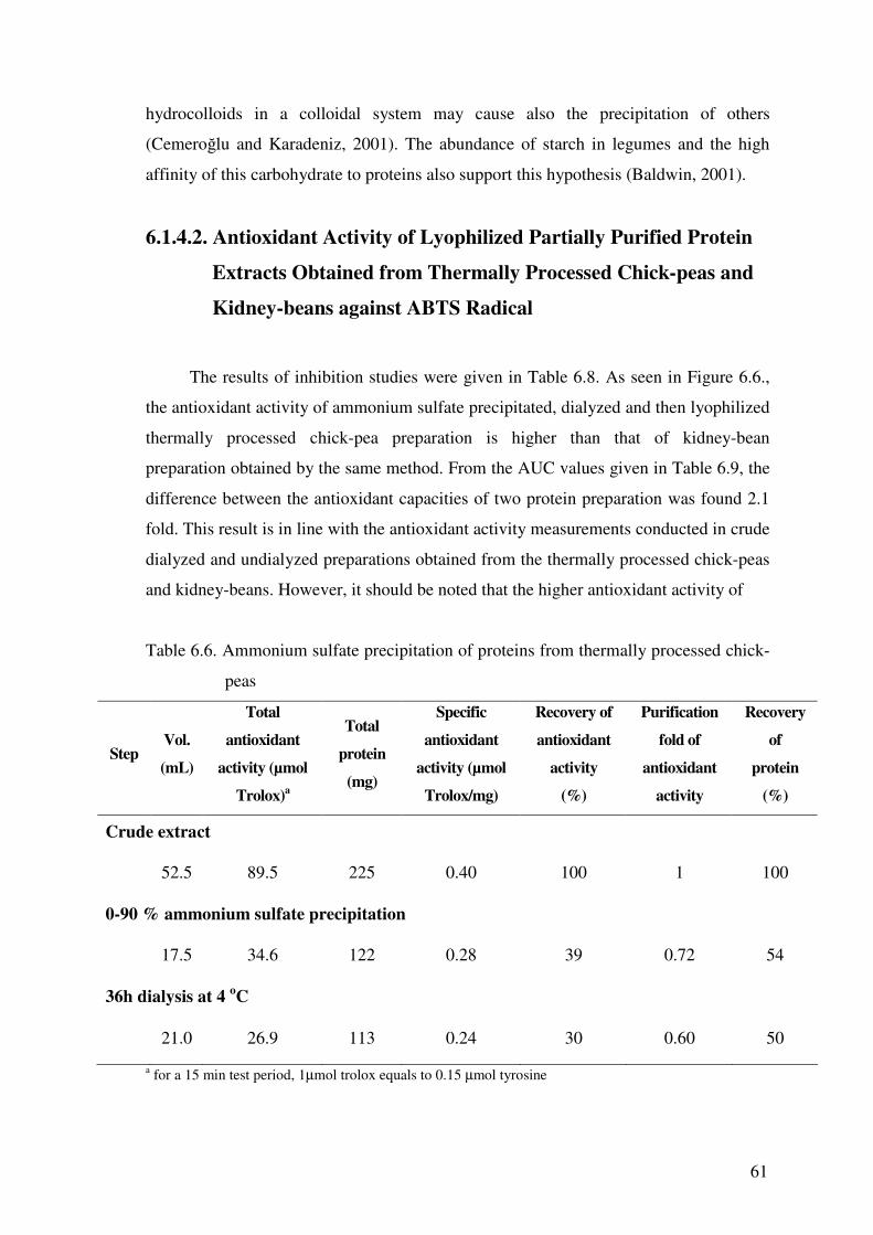
6.1.4.2. Antioxidant Activity of Lyophilized Partially Purified
Protein Extracts Obtained from Thermally Processed
Chick-peas and Kidney-beans against ABTS Radical.............61
6.1.5. Application of DEAE-cellulose Chromatography to Dialyzed
Crude Protein Extracts for Partial Purification of Heat
Treated or Thermally Processed Chick-pea Antioxidant
was purchased from Merck (Darmstadt). Trolox, Ferrous chloride tetrahydrate was
purchased from Fluka (Switzerland). Ferrozine (3-(2-Pyridyl)-5,6-diphenyl-1,2,4-
triazine-4’,4”-disulfonic acid Monosodium salt) was purchased from Fluka (USA).
Sericin (Silk Biochemical Co. Ltd) was kindly donated by Assistant Professor Dr. O�uz
Bayraktar from Izmir Institute of Technology.
5.2. Preparation of Samples
To prepare the heat treated samples, the legumes were first rehydrated in
distilled water at 12 h at room temperature. The samples were than heat treated at 90o C
for 20 minutes. On the other hand, for the preparation of thermally processed samples,
the legumes were put into flasks containing distilled water and thermally processed at
121o C for 20 minutes. The treated samples were processed immediately to acetone
powder.
5.3. Preparation of Acetone Powders
To remove phenolic compounds and lipids, acetone powders were used as source
of protein extracts. For the preparation of acetone powders, untreated, heat-treated or
44
thermally-processed chick-peas or kidney-beans (prepared by rehydration of 50 g dry
samples) were homogenized in a Waring blender for 3 min with 200 mL cold acetone.
The slurry obtained was filtered under vacuum from Buncher funnel containing a
Whatman No:1 filter paper and the solid residue remained on the filter paper was
collected. The homogenization with 200 mL cold acetone and filtration were then
repeated for two more times for the collected residue and the powder, left overnight to
evaporate the acetone, was stored at -18 oC until used for protein extraction.
5.4. Extraction and/or Modification Methods
5.4.1. Preparation of Crude Protein Extracts of Heat Treated or
Thermally Processed Chick-peas or Kidney-beans
The heat treatment of samples was applied for the inactivation of enzyme
lipoxygenase, whereas thermal processing was applied both for lipoxygenase
inactivation and modification of antioxidant activity of proteins. To prepare the crude
protein extracts from heat treated or thermally processed chick-peas or kidney-beans the
extraction method given by Genovese and Lajolo (1998) was applied by major
modifications. Briefly, 20 g acetone powder, 0.5 g insoluble PVPP and 180 ml distilled
water were mixed and extracted with a magnetic stirrer for 2 hours at room temperature.
The extract was then filtrated from a cheese-cloth (4 layers) to collect the filtrate and the
cake was discharged. The filtrate was then centrifuged for 30 min at 15000 g (4 oC) and
clarified. Half of the clarified supernatant was dialyzed for 72 h (48 h against 5 x 2 L
distilled water and 24 h against 3 x 2 L deionized water) at 4 oC, whereas the remaining
half was incubated for the same period at the same temperature without application of
dialysis. At the end of dialysis (or incubation without dialysis) the extracts were
clarified by centrifugation for 15 min at 4500 g (4 oC) and stored at -18 oC after they
were lyophilized. The lyophilization was conducted by using a freeze drier (Labconco,
FreeZone, 6 liter, Kansas City, MO, USA) working between -44 and -47 oC collector
temperature and 50 x 10-3 and 100 x 10-3 mBar vacuum. The sample container volume
was two to three times the sample volume.
45
5.4.2. Preparation of Crude Protein Extracts of Chick-peas by Hot
Acidic Hydrolysis
The hot acidic hydrolysis was applied mainly for the modification of antioxidant
activity of proteins. For this purpose, 4 g acetone powder from rehydrated chick-peas
was suspended in 65 ml deionzied water and the total volume of the suspension was
adjust to 130 ml with 3 N HCl. The extract was then heated to 85 oC and maintained at
this temperature for 10 or 30 minutes under continuous stirring. After heating, the
extract was cooled to room temperature in an ice water bath and its pH was brought to
neutrality by addition of 6 N NaOH. The final volume of this extract was then made up
155 mL, it was further stirred (30 or 50 min for 30 and 10 min heated samples,
respectively) at room temperature and clarified by centrifugation for 30 min at 15000 g
(4 oC). Half of the clarified supernatant was dialyzed for 24 h (against 3 x 2 L deionized
water) at 4 oC, whereas the remaining half was incubated for the same period at the
same temperature without application of dialysis. At the end of dialysis (or incubation
without dialysis) the extracts were clarified by centrifugation for 15 min at 5000 g (4 oC) and assayed for protein and antioxidant activity.
5.4.3. Preparation of Crude Protein Extracts of Chick-peas Obtained
by Hot Extraction Conducted at Different pH Values
5.4.3.1. Hot Extraction Conducted Close to Neutrality
The hot extractions conducted close to neutrality aimed mainly the inactivation of
enzyme lipoxygenase. On this purpose, 4 g acetone powder was suspended in 130 mL
deionzied water. The extract which pH was almost 6.5 was then heated to 85 oC and
maintained at this temperature for 30 or 60 minutes under continuous stirring. After
heating, the extract was cooled to room temperature in an ice water bath and its volume
was made up 150 mL. For extract heated for 30 min, an additional 30 min stirring was
applied at room temperature whereas the 60 min heated extract was used without further
stirring. The extracts stirred for total of 60 min were then clarified by centrifugation for
30 min at 15000 g (4 oC). Half of the clarified supernatant was dialyzed for 24 h
(against 3 x 2 L deionized water) at 4 oC, whereas the remaining half was incubated for
46
the same period at the same temperature without application of dialysis. At the end of
dialysis (or incubation without dialysis) the extracts were clarified by centrifugation for
15 min at 5000 g (4 oC) and assayed for protein and antioxidant activity.
5.4.3.2. Hot Extraction Conducted at Acidic or Basic pH Values
The hot extractions conducted at extreme pH values aimed both the modification
of proteins and increase of extraction yield of antioxidant proteins. In these extractions,
4 g acetone powder was suspended in 100 mL deionzied water. The pH of the extract
was then set to 2.5 (with 0.1 M HCl) or 9.5 (with 0.1 M NaOH) and it was heated to 85 oC and maintained at this temperature for 30 min under continuous stirring. The extract
was then cooled to room temperature in an ice water bath and its volume was made up
150 mL. After an additional 30 min stirring at room temperature, the extract was
clarified by centrifugation for 30 min at 15000 g and 4 oC. Half of the clarified
supernatant was then dialyzed for 24 h (against 3 x 2 L deionized water) at 4 oC,
whereas the remaining half was incubated for the same period at the same temperature
without application of dialysis. At the end of dialysis (or incubation without dialysis)
the extracts were clarified by centrifugation for 15 min at 5000 g (4 oC) and assayed for
protein and antioxidant activity.
5.5. Partial Purification of Crude Protein Extracts with Ammonium
Sulfate Precipitation and Dialysis
For partial purification, solid (NH4)2SO4 was added slowly to undialyzed crude
protein extracts at 4 oC up to 90 % saturation. The mixture was stirred slowly for 1.5 h
at 4 oC and the precipitate formed collected by 30 min (or 45 min) centrifugation at
15000 g (or 4500g) and 4 oC was dissolved in 20 ml distilled water. The extract was
then dialyzed for 24 h (or 36 h) at 4 oC (against 3 or 4 x 2L of distilled or deionized
water), clarified by centrifugation for 30 min at 15000 g (4 oC) and then lyophilized and
stored at -18 oC.
47
5.6. Partial Purification of Antioxidant Proteins by Dialysis and
DEAE-cellulose Column Chromatography
5.6.1. Purification of Antioxidant Proteins from Dialyzed Crude Protein
Extracts of Heat Treated or Thermally Processed Chick-peas
For the partial purification of heat treated or thermally processed chick-pea
proteins, crude protein extract was prepared by suspending 10 g acetone powder and
0.25 g PVPP in 90 ml distilled water. After 2 h stirring at room temperature, the mixture
was filtered from cheese cloth (4 layers), clarified by centrifugation for 30 min at 15000
g (4 oC) and dialyzed for 72 h (48 h against 5 x 2 L distilled water and 24 h against 3 x 2
L deionized water) at 4 oC. Following dialysis the extract was centrifuged for 15 min at
4500 g (4 oC) and loaded onto DEAE-cellulose column (2.4 cm diameter, 10 cm height)
previously equilibrated with 0.01 M pH 7.00 Na-phosphate buffer. The washing of the
column was conducted by 300 mL of equilibration buffer and the column was then
eluted with a continuous linear gradient of 0-1.5 M NaCl prepared in 0.01 M pH 7.00
Na-phosphate buffer. Fractions (5 mL) collected from the column were assayed for their
antioxidant activity against ABTS radical as described in section 5.7, and the inhibition
period of tests was shortened to 2 min to complete the measurements of all fractions as
soon as possible and prevent possible changes in the antioxidant properties of proteins.
The protein content of the fractions, on the other hand, was monitored by measuring
absorbance value at 280 nm.
5.6.2. Partial Purification of Antioxidant Proteins from Crude Protein
Extracts of Chick-peas Obtained by Hot Extraction
To purify antioxidant proteins obtained by hot extraction, 4 g acetone powder was
suspended in 130 mL deionzied water. The extract was then heated to 85 oC and
maintained at this temperature for 30 minutes under continuous stirring. After heating,
the extract was cooled to room temperature in an ice water bath, its volume was made
up 150 mL and it was further stirred for 30 min at room temperature. The extract was
then clarified by centrifugation for 30 min at 15000 g (4 oC), incubated for 24 h at 4 oC
and one more centrifuged for 15 min at 4500 g (4 oC). The crude protein extract was
48
then loaded onto a DEAE-cellulose column (2.4 cm diameter, 10.0 cm height)
previously equilibrated with 0.01 M pH 7.00 Na-phosphate buffer. The washing of the
column was conducted by x mL of equilibration buffer and the column was then eluted
with a continuous linear gradient of 0-1.5 M NaCl prepared in 0.01 M pH 7.00 Na-
phosphate buffer. Fractions (5 mL) collected from the column were assayed for their
antioxidant activity against ABTS radical and protein as described in section 5.6.1.
5.7. Determination of Antioxidant Activity against ABTS Radical
The antioxidant activity against ABTS radical was determined as described in Re
et al. (1999). The ABTS was dissolved in distilled water, oxidized by potassium
persulfate to form ABTS radical and then diluted with 5 mM pH 7.4 phosphate buffer
containing 150 mM NaCl (PBS). The reaction mixture for the measurements was
prepared by mixing 0.1 mL protein extract and 1.9 mL ABTS radical solution (initial
absorbance at 734 nm was almost 0.700). The discoloration of dark blue colored ABTS
radical by the antioxidant protein was monitored at 734 nm for 15 min. All
measurements were performed in triplicate The antioxidant capacities of lyophilized
protein preparations were determined by dividing the area of their % inhibition of
ABTS radical / concentration (�g/reaction mixture) ratio vs. period of inhibition test (in
1, 6 or 15 min) curves with that area of the same curve of the standard antioxidant
Trolox. The value determined by this calculation is called AUC (Area Under the Curve)
value and it represents the antioxidant capacity as µmol Trolox per mg of lyophilized
protein preparation. Bovine serum albumin and sericin were used as standard proteins
for comparison. During purification studies the antioxidant activity of proteins were
given as Trolox and Tyrosine equivalents (see standard curves in Appendixes A and B).
5.8. Determination of Fe+2 Chelating Capacity
The Fe+2 chelating capacity of protein extracts were determined as described in
Rajapakse et al. (2005). Briefly, 2 mL protein solution was mixed with 0.1 mL, 1mM
FeCl2.4H2O solution. After 30 min incubation at room temperature, 0.1 mL, 0.5 mM
ferrozine was added into mixture and its absorbance was read at 562 nm after 10 minute
incubation. The formation of blue color indicates weak Fe+2 chelating capacity whereas
49
the lack of any blue color development shows the strong Fe+2 binding. The percent Fe+2
chelating capacity of sample was determined by using the following formula; A1-A2/A3
x 100, where A1 is the final absorbance of the treated sample at 562 nm, A2 is the
original absorbance of the untreated sample at 562 nm and A3 is the absorbance of
blank obtained by treating deionized water in place of sample. The Fe+2 chelating
capacity of samples were given as EDTA equivalents (µmol) per mg of lyophilized
protein preparation (See standard curve in Appendix C). All measurements were
performed in triplicate.
5.9. Determination of Lipoxygenase Activity
In this study the enzyme lipoxygenase was used as an indicator for the
determination of suitable heat treatment periods of chick-peas and kidney-beans. The
presence of this enzyme in protein extracts intended to be used as antioxidant is
undesirable since the enzyme is responsible from lipid oxidation. During preliminaries
to determine a suitable heating condition for inactivation of enzyme in chick-peas or
kidney-beans the enzyme extract was prepared by homogenizing 10 g of heat treated or
unheated (control) sample with 50 mL distilled water in a Waring micro blender for 1
min. A sample taken from the extract was then clarified by centrifugation for 15 min at
15 000 g (4 oC) and used in test of enzyme activity. The activity of lipoxygenase was
determined spectrophotometrically by slightly modifying the method described in
Yemenicioglu (2002). The reaction mixture was formed by mixing 10 µL of clear
enzyme extract, 2.95 mL 0.05 M, Na-phosphate buffer (pH 7.0) and 0.05 mL linoleic
acid solution prepared with Tween 20 as described in Rackis et al (1972). The activity
of enzyme monitored at 234 nm and 30 oC constant temperature was determined from
the slope of the initial linear portion of absorbance vs. time curve.
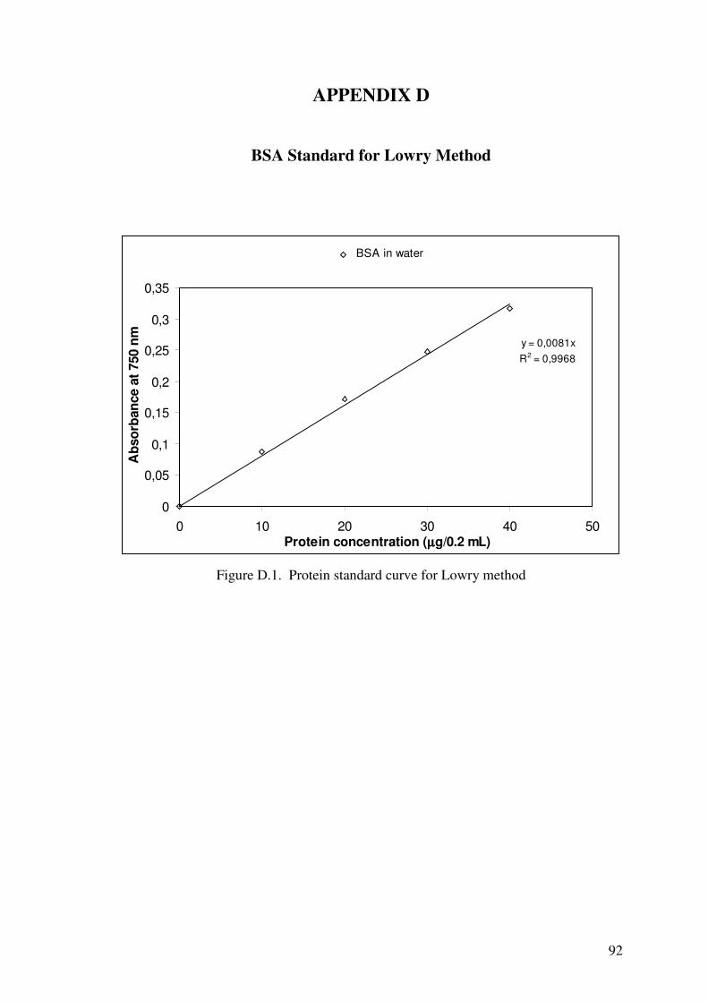
5.10. Determination of Protein Content
Protein was determined by the Lowry method by using bovine serum albumin
(BSA) as standard (see Appendices D) (Harris 1987).
50
CHAPTER 6
RESULTS AND DISCUSSIONS
6.1. Studies with Crude Protein Extracts of Heat Treated or Thermally
Processed Chick-peas or Kidney-beans Obtained by Water
Extraction
Since this work aimed studying the antioxidant activity of proteins, acetone
powders were used to obtain phenolic free preparations. In studies with heat-treated or
thermally processed chick-peas and kidney-beans, PVPP, an insoluble phenolic
scavenger, was also employed during extraction to ensure the complete elimination of
residual phenolics. On the other hand, the heat treatment (20 min at 90 oC) conditions
were optimized by using the enzyme lipoxygenase as an indicator. Since this enzyme is
responsible from the oxidation of lipids to hydroperoxides it should not exist in
preparations intended to be used as antioxidant. The thermal processing (20 min at 121 oC) was applied mainly to modify the antioxidant activity of proteins, but it also
inactivates the lipoxygenase enzyme.
6.1.1. Protein Content of Lyophilized Crude Protein Extracts of Heat-
Treated or Thermally Processed Chick-peas and Kidney-beans
Chick-peas and kidney-beans contain almost 27 % and 22 % protein, respectively
(WEB_1 2005, WEB_2 2005). In legumes, almost ~70 % the proteins consist of water
insoluble globulins whereas the remaining protein consist of water soluble albumins
(Genovese and Lajolo 1998, Vioque et al. 1999). Since extractions in this work were
conducted with water, the proteins discussed in this study are mainly water soluble
albumins.
As seen in Table 6.1, lyophilized crude chick-pea preparations contained
significantly higher protein than lyophilized crude kidney-bean preparations. The
thermal processing increased the protein content of undialyzed lyophilized chick-pea
extracts slightly. However, a slight reduction was observed in the protein content of
51
undialyzed lyophilized kidney-bean extracts by the thermal processing. The application
of dialysis caused an increase in protein/nonprotein substances ratio of lyophilized
chick-pea preparations. In fact, in these extracts almost half of the lyophilized
preparation was protein. In contrast, the dialysis did not affect the protein content of
lyophilized kidney-bean preparations significantly. During dialysis, low molecular
substances are removed from the extracts. The loss of some substances also occurred
due to insolubilization and consequent separation in centrifugation applied after
dialysis. For example, in heat treated chick-peas and kidney-beans, dialysis reduced the
amounts of lyophilized dry powders almost 49 and 79 %, respectively. However, the
increased protein content of dialyzed lyophilized chick-pea extracts indicates that the
separated substances in these extracts are mainly non-protein substances. The soluble
solids in the preparations other than the proteins may be polysaccharides such as soluble
fractions of starch and pectin and simple sugars. Also, it is possible that an important
part of the soluble solids exist as protein-polysaccharide complexes (Genovese and
Lajolo 1998, Baldwin 2001).
Table 6.1. Protein contents of lyophilized preparations obtained from crude protein
extracts of heat treated or thermally processed chick-peas and kidney-beans
Protein concentration in lyophilized preparation
(mg protein /mg lyophilized preparaiton)
Heat treated Thermally processed
Source of
Protein
undialyzed dialyzed undialyzed Dialyzed
Chick-peas
0.26 0.51 0.34 0.51
Kidney-beans
0.19 0.22 0.15 0.18
6.1.2 Antioxidant Activity of Lyophilized Crude Protein Extracts of
Heat Treated or Thermally Processed Chick-peas and Kidney-
beans against ABTS Radical
The results of inhibition tests for lyophilized crude protein extracts obtained from
heat treated or thermally processed chick-peas and kidney-beans showed the presence of
52
antioxidants in these sources (Table 6.2. and 6.3.). During tests, the inhibitions occurred
fast in the first minute. After that, the inhibition slowed down through the 6 min, but
continued slowly up to 15 min or more. Thus, the absorbance values of ABTS radical
solutions were monitored for 15 min and inhibition / concentration ratios of
preparations were evaluated for 1, 6 and 15 min separately.
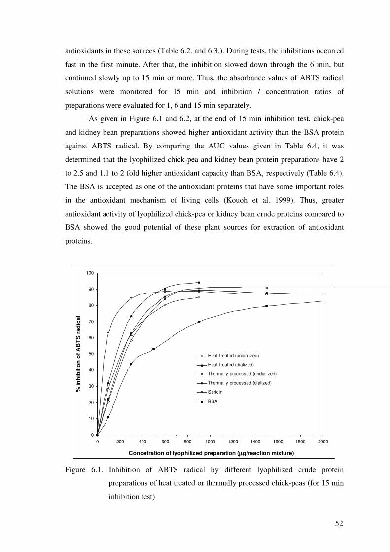
As given in Figure 6.1 and 6.2, at the end of 15 min inhibition test, chick-pea
and kidney bean preparations showed higher antioxidant activity than the BSA protein
against ABTS radical. By comparing the AUC values given in Table 6.4, it was
determined that the lyophilized chick-pea and kidney bean protein preparations have 2
to 2.5 and 1.1 to 2 fold higher antioxidant capacity than BSA, respectively (Table 6.4).
The BSA is accepted as one of the antioxidant proteins that have some important roles
in the antioxidant mechanism of living cells (Kouoh et al. 1999). Thus, greater
antioxidant activity of lyophilized chick-pea or kidney bean crude proteins compared to
BSA showed the good potential of these plant sources for extraction of antioxidant
proteins.
0
10
20
30
40
50
60
70
80
90
100
0 200 400 600 800 1000 1200 1400 1600 1800 2000
Concetration of lyophilized preparation (µµµµg/reaction mixture)
% in
hibi
tion
of A
BTS
radi
cal
Heat treated (undialized)
Heat treated (dialized)
Thermally processed (undialized)
Thermally processed (dialized)
Sericin
BSA
Figure 6.1. Inhibition of ABTS radical by different lyophilized crude protein
preparations of heat treated or thermally processed chick-peas (for 15 min
inhibition test)
53
Table 6.2. Inhibition of ABTS radical by lyophilized crude protein preparations of heat
treated or thermally processed chick-pea
Sample Concentration (µg/reaction mixture)
% inhibition of ABTS radical
1 min 6 min 15 min
Heat treated (undialyzed)
100 13.9 24.2 28.6
300 30.9 52.2 62.0
600 47.9 71.5 80.3
900 58.2 79.4 85.1
Heat treated (dialyzed)
100 22.0 29.4 32.4
300 49.1 66.0 73.6
600 67.7 84.7 90.5
900 77.9 91.6 94.5
Thermally processed (undialyzed)
100 12.8 18.7 20.9
300 34.0 51.7 58.3
600 53.4 75.4 83.9
900 68.2 86.7 90.6
1500 76.9 92.0 90.9
3000 85.6 92.7 90.9
Thermally processed (dialyzed)
100 15.3 21.0 22.3
300 40.2 56.2 62.9
600 61.6 80.3 85.3
900 73.0 87.2 89.4
1500 77.8 88.3 87.7
3000 83.6 87.1 85.2
54
Table 6.3. Inhibition of ABTS radical by lyophilized crude protein preparations of heat
treated or thermally processed kidney-beans
% inhibition of ABTS radical Sample Concentration
(µg/reaction mixture) 1 min 6 min 15 min
Heat treated (undialyzed)
20 0.71 1.7 2.3
100 7.8 12.3 13.4
300 24.5 36.9 42.2
600 43.7 62.1 71.5
900 57.7 76.2 82.9
Heat treated (dialyzed)
20 0.5 2.2 3.3
100 8.9 14.1 15.9
300 28.9 43.5 50.6
600 46.5 68.6 77.4
900 53.9 74.5 81.4
1500 64.5 82.1 86.6
3000 75.3 85.2 85.8
Thermally processed (undialyzed)
100 7.4 9.4 9.9
300 21.6 31.2 35.7
600 37.0 55.2 64.4
900 48.9 68.5 77.2
1500 58.1 74.0 78.4
3000 72.4 80.4 78.6
Thermally processed (dialyzed)
100 6.9 11.2 11.7
300 19.4 30.7 35.1
600 32.6 50.1 57.9
900 44.0 63.8 71.4
1500 61.2 82.3 85.8
3000 76.8 88.6 85.8
55
0
10
20
30
40
50
60
70
80
90
100
0 200 400 600 800 1000 1200 1400 1600 1800 2000
Concentration of lyophilized preparation (µµµµg/reaction mixture)
% in
hibi
tion
of A
BTS
radi
cal
Heat treated (undialized)
Heat treated (dialized)
Thermally processed (undialized)
Thermally processed (dialized)
Sericin
BSA
Figure 6.2. Inhibition of ABTS radical by different lyophilized crude protein
preparations of heat treated or thermally processed kidney-beans (for 15
min inhibition test)
On the other hand, both lyophilized crude chick-pea and kidney-bean
preparations showed lower antioxidant activity than sericin (Figure 6.3 and 6.4).
However, it should be noted that the standard proteins, BSA and sericin, used in this
study were highly pure, whereas lyophilized preparations’ protein content changed
between 15 to 51 %. These comparisons also clearly showed the greater antioxidant
activities of lyophilized crude chick-pea proteins than the lyophilized crude kidney-bean
proteins. The protein contents of chick-pea preparations were also higher. Thus, it
seems that the greater antioxidant activity is related with the higher protein content.
Thermally processed kidney-beans (dialyzed) 0.03 (0-1500) aThe numbers in the parenthesis indicate the range of data used in calculations (µg/reaction mixture)
0
10
20
30
40
50
60
70
80
90
100
0 500 1000 1500 2000 2500 3000 3500
Concentration of lyophilized preparation (µµµµg/reaction mixture)
B2 fraction of thermally processed chick-peas [AUC= 0.098 µµµµmol Trolox/mg
lyophilized protein]
0.129 (0-300) 0.174 (0-300) 0.190 (0-300) aThe numbers in the parenthesis indicate the range of data used in calculations (µg/reaction mixture)
72
0
0,05
0,1
0,15
0,2
0,25
0,3
0,35
0,4
0 2 4 6 8 10 12 14 16
Period of inhibition test (min)
% in
hibi
tion
of A
BTS
radi
cal/c
once
ntra
tion
( µµ µµg/
reac
tion
mix
ture
) rat
io
A2 from heat treated chick-peas
B2 from thermally processed chick-peas
sericinBSA
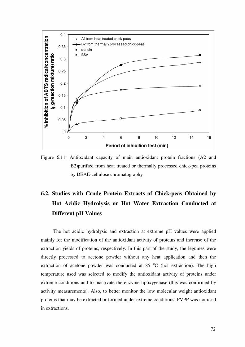
Figure 6.11. Antioxidant capacity of main antioxidant protein fractions (A2 and
B2)purified from heat treated or thermally processed chick-pea proteins
by DEAE-cellulose chromatography
6.2. Studies with Crude Protein Extracts of Chick-peas Obtained by
Hot Acidic Hydrolysis or Hot Water Extraction Conducted at
Different pH Values
The hot acidic hydrolysis and extraction at extreme pH values were applied
mainly for the modification of the antioxidant activity of proteins and increase of the
extraction yields of proteins, respectively. In this part of the study, the legumes were
directly processed to acetone powder without any heat application and then the
extraction of acetone powder was conducted at 85 oC (hot extraction). The high
temperature used was selected to modify the antioxidant activity of proteins under
extreme conditions and to inactivate the enzyme lipoxygenase (this was confirmed by
activity measurements). Also, to better monitor the low molecular weight antioxidant
proteins that may be extracted or formed under extreme conditions, PVPP was not used
in extractions.
73
6.2.1. Antioxidant Activity and Protein Content of Crude Protein
Extracts of Chick-peas Obtained by Hot Acidic Hydrolysis
In the literature the enzymatic hydrolysis with proteases has been mostly applied
to obtain protein hydrolysates (Amarowicz and Shahidi, 1997, Rival et al, 2001, Chen et
al, 1996). It is also well known that heating in highly acidic medium causes the
hydrolysis of proteins (Bull and Hahn, 1948, Greenberg and Burk, 1927). Thus, in this
study, limited acidic hydrolysis was applied to determine the effect of this treatment on
antioxidant activity and protein content of crude protein extracts of chick-peas. As seen
in Table 6.14, the application of 30 min heating in presence of 1.5 M HCl gave a crude
extract with high specific antioxidant activity. The application of dialysis reduced the
specific antioxidant activity of 30 min acid hydrolyzed samples moderately. Thus, the
increased antioxidant activity of hydrolyzed sample is not mainly due to low molecular
substances formed by acidic hydrolysis. The reduction of hot hydrolysis period to 10
min, on the other hand, reduced both the protein content and antioxidant activity, but
increased the specific antioxidant activity slightly.
The results showed that the 30 min acidic hydrolysis at 85 oC may be used to
increase the protein content and antioxidant activity of crude protein extracts. However,
it should be noted that the application of hot acidic hydrolysis caused also the formation
of Maillard reaction products. In hydrolyzed samples, the light brown color formed
during hydrolysis indicated the presence of Maillard reaction products in crude extracts.
The Maillard reaction products are formed by reaction between reducing sugars and
amino acids or proteins (Yoshimura et al, 1997). The antioxidant activity of Maillard
reaction products was reported by many different workers (Murakami et al, 2002,
Yoshimura et al, 1997, Duh et al, 2001). However, the use of preparations containing
Maillard reaction products needs some toxicological testing, since these products have
mutagenic activity (Murakami et al, 2002).
74
Table 6.14. Antioxidant activity and protein contents of crude protein extracts of chick-
peas obtained by hot acidic hydrolysis
Extraction conditions
of acetone powder
Protein
(mg/mL)
Antioxidant activity
(�mol trolox/mL)
Specific antioxidant
activity (�mol trolox/mg)
Suspension of acetone powder in 1.5 M HCl / continuous stirring at 85 oC for 30 min /
neutralization / continuous stirring at room temperature for 30 min / centrifugation
+ 24h incubation at 4 oC 4.19 2.34 0.56
+ 24h dialysis at 4 oC 2.95 1.26 0.43
Suspension of acetone powder in 1.5 M HCl / continuous stirring at 85 oC for 10 min /
neutralization / continuous stirring at room temperature for 50 min / centrifugation
+ 24h incubation at 4 oC 2.80 1.65 0.59
+ 24h dialysis at 4 oC 1.97 0.79 0.41
6.2.2. Antioxidant Activity and Protein Content of Crude Protein
Extracts of Chick-peas Obtained by Hot Water Extraction at
Different pH Values
The results of hot extractions conducted at different pH values were given in
Table 6.15. The application of hot extraction at pH 6.5 and 85 oC for 60 min increased
the specific antioxidant activity of extracts to almost 80 % of the specific antioxidant
activity of 30 min acid hydrolyzed sample without a considerable color change in the
protein extract. The application of dialysis, on the other hand, reduced the protein
content and antioxidant activity almost 60 and 75 %, respectively. The reduction of
heating period to 30 min at pH 6.5 and 85 oC reduced the specific antioxidant activity
and protein content. Also, 30 % reduction occurred in antioxidant activity by reduction
of heating period. Conducting hot extraction for 30 min at 85 oC by changing extraction
pH from 6.5 to 9.5 did not increase the specific antioxidant activity. However, this
increased the protein content of extracts almost 100 %, compared with the protein
contents of samples extracted at pH 6.5. On the other hand, conducting extraction for 30
min at 85 oC by changing extraction pH to 2.5 increased the specific antioxidant activity
to the highest level obtained in extraction studies. The protein content of the extract at
pH 2.5 reduced considerably. However, it seems that the conditions at this pH were very
75
suitable for the selective extraction of antioxidant proteins and/or other potential
antioxidants such as peptides, protein-phenolic or carbohydrate-phenolic associates. The
dialysis of the extract obtained at pH 2.5 reduced the specific antioxidant activity almost
40 %. Thus, it is clear that the important part of antioxidants in this extract is low
molecular weight compounds.
Table 6.15. Antioxidant activity and protein contents of crude protein extracts of chick-
peas obtained by hot extraction at different pH values
Extraction conditions
of acetone powder
Protein
(mg/mL)
Antioxidant activity
(�mol trolox/mL)
Specific antioxidant
activity (�mol trolox/mg)
Suspension of acetone powder in deionized water (pH was almost 6.5) / continuous stirring at 85
oC for 60 min / centrifugation
+ 24h incubation at 4 oC 3.70 1.70 0.46
+ 24h dialysis 4 oC 1.37 0.45 0.32
Suspension of acetone powder in deionized water (pH was almost 6.5) / Continuous stirring at 85
oC for 30 min + at room temperature for 30 min / centrifugation
+ 24h incubation at 4 oC 3.20 1.17 0.37
Suspension of acetone powder in deionized water / adjustment of pH to 9.5 / continuous stirring at
85 oC for 30 min + at room temperature for 30 min / adjustment of pH to 6.5 / centrifugation
+ 24h incubation at 4 oC 6.54 2.21 0.34
+ 24h dialysis at 4 oC 2.57 0.37 0.14
Suspension of acetone powder in deionized water / adjustment of pH to 2.5 / continuous stirring at
85 oC for 30 min + at room temperature for 30 min / adjustment of pH to 6.5 / centrifugation
+ 24h incubation at 4 oC 1.83 1.23 0.67
+ 24h dialysis at 4 oC 1.39 0.56 0.41
76
6.2.3. Application of Ammonium Sulfate Precipitation and Dialysis for
Partial Purification of Crude Protein Extracts of Chick-peas
Obtained by Hot Extraction Conducted at pH Values Close to
Neutrality
The summary of the ammonium sulfate precipitation of crude protein extracts
obtained by 30 min extraction of chick-pea acetone powder at pH 6.5 and 85 oC was
given in Table 6.16. For chick-pea crude proteins obtained by hot extraction, the
recovery of protein for ammonium sulfate precipitation was almost 15-20 % higher than
those of the previous ammonium sulfate precipitations conducted for crude proteins
extracted from thermally processed chick-peas and kidney-beans at room temperature
(see Table 6.6. and 6.7). However, similar to previous precipitations the recovery of
antioxidant activity by ammonium sulfate precipitation was low. In hot extracted chick-
pea crude proteins, the dialysis of ammonium sulfate precipitates further reduced the
recoveries of antioxidant activity and protein. Thus, it seems that the removal of low
molecular substances by dialysis and/or insolubilization during dialysis is very high in
ammonium sulfate precipitated crude protein extracts obtained by hot water extraction.
Table 6.16. Ammonium sulfate precipitation of crude chick-pea proteins obtained by
hot water extraction conducted at pH values close to neutrality
Step Vol.
(mL)
Total
antioxidant
activity (µmol
Trolox)a
Total
protein
(mg)
Specific
antioxidant
activity (µmol
Trolox/mg)
Recovery of
antioxidant
activity
(%)
Purification
fold of
antioxidant
activity
Recovery
of
protein
(%)
Crude extract
100 145.9 353 0.41 100 1 100
0-90 % ammonium sulfate precipitation
28 53.6 240 0.22 37 0.54 68
36h dialysis at 4 oC
40 21.6 76 0.28 15 0.69 22 a for a 15 min test period, 1 µmol trolox equals to 0,15 µmol tyrosine
77
6.2.4. Application of DEAE-cellulose Chromatography for Partial
Purification of Crude Chick-pea Antioxidant Proteins Obtained
by Hot Extraction Conducted at pH Values Close to Neutrality
For purification of antioxidant proteins from crude protein extracts obtained by 30
min extraction of chick-pea acetone powder at pH 6.5 and 85 oC, a crude extract was
applied to DEAE-cellulose column. To determine the antioxidant activity and protein
profiles of both low and high molecular weight fractions, the crude extract was not
dialyzed before chromatography (dialysis was applied before other chromatographic
separations given in Figures 6.8 and 6.9).
0
5
10
15
20
25
30
35
40
0 20 40 60 80 100 120 140Feaction number
% in
hibi
tion
of A
BTS
rad
ical
0
0.2
0.4
0.6
0.8
1
1.2
Pro
tein
(abs
at 2
80 n
m) o
r N
aCl c
once
ntra
tion
(M)
% inhibition of ABTS radical
Protein concentration
NaCl concentration
I C 2 I
I C 3 I
I C 4 I
IC 5I
I C 1 I
Figure 6.12. Purification of antioxidant proteins from chick-pea crude protein extracts
by DEAE-cellulose anion exchange chromatography (The crude extract
was obtained by 30 min extraction of acetone powder at pH 6.5 and 85 oC;
antioxidant activities were determined for 2 min inhibition test)
As seen in Figure 6.12, for the DEAE-cellulose chromatography of crude protein
extract, three main peaks, C1, C2 and C3, were obtained for the antioxidant activity.
From these peaks C3 peak contained two shoulders (C4 and C5), confirmed by the
78
protein peaks following the antioxidant activity peaks. The elution of significant amount
of antioxidant activity (followed by protein peaks) from the column by washing
suggests the presence protein fractions which can not bound by the DEAE-cellulose
anion exchange column. During previous DEAE-cellulose column chromatographic
studies of dialyzed crude extracts of thermally processed or heat treated chick-peas,
there was only little amounts of DEAE-cellulose unbound proteins (see section 6.1.5).
The crude protein extract obtained by hot extraction was not dialyzed. Thus, it is
possible that the unbound substances are low molecular weight proteins that contain
almost no or little negatively charged groups that contribute to anion exchange reactions
in DEAE-cellulose column. The rapid elution of C1 suggests the lack of negative
charges in these proteins whereas later elution of C2 suggests slight binding due to
limited number of negative charges. The lack of charges in these protein fractions may
also be due to the complex formation of these proteins with neutral polysaccharides and
masking of negative charges. Further studies are needed for full characterization of
antioxidant proteins in chick-peas.
The partial purification parameters for the antioxidant activity of the eluted
protein fractions were also given in Table 6.17. The results indicated that the highest
purification
Table 6.17. Purification of antioxidant proteins from chick-peasa
Step Vol.
(mL)
Total
antioxidant
activity(�mol
Trolox)b
Total
protein
(mg)
Specific
antioxidant
activity (µmol
Trolox/mg)
Recovery of
antioxidant
activity
(%)
Purification
fold of
antioxidant
activity
Recovery
of protein
(%)
Crude extract
50 58.5 160 0.37 100 1.00 100
DEAE cellulose anion exchange chromatography
C1 95 16.7 9.1 1.83 28.5 5.00 5.7
C2 150 16.8 6.1 2.76 28.8 7.53 3.8
C3 35 5.9 13.1 0.45 10.2 1.24 8.2
C4 40 5.6 22.4 0.25 9.6 0.68 14.0
C5 25 2.3 9,5 0.24 3.9 0.65 5.9
a extraction method: water extraction of acetone powder at 85o C, b for 15 min test period, 1 µmol trolox
equals to 0.15 µmol tyrosine
79
folds were obtained for the unbound fractions, C1 and C2. It is clear that the lack of
negative charges in these proteins capable to contribute anion exchange reactions was
responsible for the separation and resulting purification of these fractions. A slight
increase in purify of C3 was also observed, whereas other fractions’ purity declined due
to small amount of antioxidant activity but high protein content in these fractions.
On the other hand, the elution profile (a main peak and two shoulders) of
antioxidant activity by the initiation of NaCl gradient was quite similar with that of
thermally processed chick-peas (see Figure 6.9). Thus, it seems that the hot extraction
and thermal processing cause similar modifications in chick-pea proteins bind to
DEAE-cellulose. However, in chromatography of chick-pea crude proteins obtained by
hot extraction, the antioxidant activity loaded to column was higher than the antioxidant
activity eluted. Thus, it is clear that, unlike to thermal processing, hot extraction was not
effective in removal of prooxidants or antioxidant activity masking substances.
80
CHAPTER 7
CONCLUSIONS
• Kidney-beans and chick-peas contain antioxidant proteins with free radical
scavenging and metal chelating capacity.
• Lyophilized crude proteins from chick-peas contain more protein and higher free
radical scavenging activity than lyophilized crude proteins from kidney-beans.
• Free radical scavenging activity of lyophilized crude proteins from heat treated or
thermally processed kidney-beans and chick-peas is higher than the free radical
scavenging activity of bovine serum albumin. However, all lyophilized crude proteins
showed lower free radical scavenging activity than sericin.
• Lyophilized crude proteins from chick-peas and kidney-beans show greater iron
chelating capacity than bovine serum albumin and sericin.
• Thermal processing does not cause a significant change in free radical scavenging
capacity of lyophilized crude chick-pea proteins. However, it increases their iron
chelating capacity. In contrast, thermal processing reduces both free radical scavenging
and iron chelating capacity of lyophilized crude kidney-bean proteins.
• Dialysis increases the free radical scavenging capacity of lyophilized crude
thermally processed or heat treated chick-pea and heat treated kidney-bean proteins.
However, it does not cause a considerable change in the free radical scavenging
capacity of thermally processed kidney-bean proteins.
• Partial purification by ammonium sulfate precipitation or DEAE-cellulose anion
exchange chromatography removes the substances that mask the antioxidant activity or
prooxidants from crude protein extracts of thermally processed chick-peas and this
causes an increase in the free radical scavenging activity of these extracts.
81
• DEAE-cellulose chromatography showed the presence of five and three
antioxidant protein fractions in heat-treated and thermally processed chick-peas
respectively. The free radical scavenging activity of one of the purified antioxidant
protein fractions from heat treated chick-peas was very close to that of sericin.
• Hot acidic hydrolysis may be used to increase the protein content and specific
antioxidant activity of crude protein extracts. However, it causes also the formation of
undesirable light brown colored Maillard reaction products.
• Hot extraction at pH 2.5 can be used for the selective extraction of antioxidant
proteins, but this method is not very effective for the extraction of other proteins.
• Hot extraction at pH 9.5 is the most effective method for the extraction of
antioxidant proteins and other proteins.
82
FUTURE EXPECTATIONS
• The results of this study clearly showed the free radical scavenging activity and
iron chelating capacity of chick-pea proteins. However, further studies are needed to test
the antioxidant effects of these proteins in real food systems.
• The presence of antioxidant protein fractions in legumes shows the existence
of genes responsible for the synthesis of these proteins. Molecular biology may be used
as a tool to obtain legumes with high antioxidant protein content. A detailed scavenging
in different legumes cultivars is needed.
83
REFERENCES
Alaiz, M., Hidalgo, F.J., Zamoro, R., 1999. “Effect of pH and Temperature on Comparative Antioxidant Activity of Nonenzymatically Browned Proteins Produced by Reaction with Oxidized Lipids and Carbonhydrates”, ”, J. Agric. Food Chem., Vol. 47, pp. 748-752.
Amarowicz R., Shadidi F. 1997. “Antioxidant Activity of Peptide Fractions of Capelin Protein Hydrolysates”, Food Chemistry Vol. 58, No. 4, pp. 355-359.
Branden, C., Tooze, J., 1998. “Basic Structural Principles”, in Introduction to Protein
Structure (Grand Publishing Inc, New York), pp. 3-12. Baldwin, P.M., 2001. “Starch Granula-Associated Proteins and Polypeptides: a
Review”, Starch/Stärke, Vol. 53, pp. 475-503. Bull, H.B., Hahn, J.W., 1948. “The Acid Hydrolysis of Egg Albumin. I. Kinetic
Studies”, J. Am. Chem. Soc. , Vol. 70(6), pp. 2128-2131. Cemeroglu, B., Karadeniz, F., 2001. “Durultma”, in Meyve Suyu Üretim Teknolojisi
(Gıda Teknolojisi Derne�i Yayınları, Ankara), pp. 66-89. Chen, H., Muramoto, K., Yamauchi, F., Nokihara, K. 1996. “Antioxidant Activity of
Designed Peptides Based on the Antioxidative Peptide Isolated from Digests of a Soybean Protein”, J. Agric. Food Chem. Vol. 44, pp. 2619-2623.
Chen, H., Muramoto, K., Yamauchi, F. 1995. “Structural Analysis of Antioxidative
Peptides from Soybean �-Conglycinin”, J. Agric. Food Chem. Vol. 43, pp. 574-578.
Chen, H., Muramoto, K., Yamauchi, F., Fujimoto, K., Nokihara, K. 1998.
“Antioxidative Properties of Histidine-Containing Peptides Designed from Peptide Fragments Found in the Digests of a Soybean Protein”, J. Agric. Food Chem. Vol. 46, pp.49-53.
Clemente, A., Vioque, J., Vioque, R.S., Pedroche, J., Bautista, J., Milla F. 1999.
“Protein Quality of Chickpea (Cicer aritinum L.) Protein Hydrolysates”, Food Chemistry Vol. 67, pp. 269-274.
Damodaran S., 1996a. “Amino Acids, Peptides and Proteins”, in Food Chemistry,
edited by O. R. Fennema (Marcel Decker Inc, New York), pp. 321-429. Damodaran S., 1996b. “Functional Properties”, in Food Proteins Properties and
Characterization, edited by S. Nakai, H.W. Modler (Wiley-VHC Inc., New York), pp.167-234.
Davidek J., Velisek, J., Pokorny, J., 1990. “Fats, Oils, and Other Lipids”,in Chemical
Changes During Food Processing (Czechoslovak Medical Press, Czechoslovak), pp. 169-229.
84
Decker, E.A., 1998. “Antioxidant Mechanisms”, in Food Lipids, edited by C.C. Akoh,
and D.B. Min (Marcel Decker, New York), pp. 397-421. De Jong, G.A.H., Koppelman, S.J., 2002. “Transglutaminase Catalyzed Reactions:
Impact on Food Applications”, Journal of Food Science, Vol. 67, Nr. 8, p.2798. Duh, P., Yen, G., Yen, W., Chang, L., 2001. “ Antioxidant Effects of Water Extracts
from Barley (Hordeum vulgare L.) Prepared Under Different Roasting Temperatures”, J. Agric. Food. Chem. Vol. 49, pp. 1455-1463.
Erickson, M.C., 1998. “Lipid Oxidation of Muscle Foods”, in Food Lipids, edited by
C.C. Akoh and D.B. Min (Marcel Decker, New York), pp. 297-332. Friedman, M., 1996. “Nutrition”, in Food Proteins: Properties and Characterization,
editied by S. Nakai, H.W. Modler (Wiley-VCH, Inc, New York), pp. 281-332. Stryer, L., 1996. “Protein Structure and Function”, in Biochemistry (Freeman & Co.,
New York), pp. 17-45 Genovese, M.I. and Lajolo, F.M., 1998. “Influence of Natural Acid-soluble Proteins
from beans (Phaseolus vulgaris L.) on in Vitro Digestibility Determination”, Food Chemistry. Vol. 62, No. 3, pp. 315-323.
Greenberg, D.M., Burk, N.F., 1927. “The Rate of Hydrolysis of Solutions of Proteins in
Acids as Measured by the Formation of Amino Nitrogen”, J. Am. Chem. Soc., Vol. 49(1), pp. 275-286.
Harris, D. A., 1987 “Spectrophotometric assays”, in Spectrophotometry and
Spectrofluorometry, edited by D. A. Harris, C. L. Bashford, (Eds. ; IRL Press: Oxford, U.K.), pp 59_60.
“Proteins: Three-dimensional Structure and Function”, in Principles of Biochemistry (Prentice-Hall Inc., New Jersey), pp. 79-117
Hou, W., Lee, M., Chen, H., Liang, W., Han, C., Liu, Y., Lin, Y. 2001. “Antioxidant
Activities of Dioscorin, the Storage Protein of Yam (Dioscorea batatas Decne) Tuber”, J. Agric. Food Chem. Vol. 49, pp. 4956-4960.
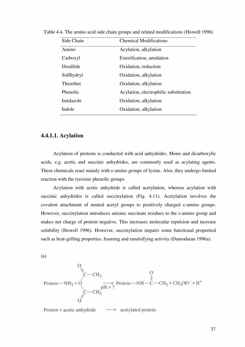
Howell, N.K., 1996. “Chemical and Enzymatic Modifications”, in Food Proteins:
Properties and Characterization, edited by S. Nakai, H.W. Modler (Wiley Inc., New York), pp.235-280.
85
Hu, M., McClements, D.J., Decker, E.A. 2003. “Lipid Oxidation in Corn Oil-in-Water Emulsions Stabilized by Casein, Whey Protein Isolate, and Soy Protein Isolate”, J. Agric. Food Chem. Vol. 51, pp. 1696-1700.
Hwang, J., Shue, Y., Chang, H. 2001. “Antioxidative activity of roasted and defatted
peanut kernels”, Food Research International. Vol. 34, pp. 639–647 Je, J., Park, P., Kim, S., 2004. “Antioxidant Activity of a Peptide Isolated from Alaska
Pollack (Theragra chalcogramma) Frame Protein Hydrolysate”, Food Research International, Vol. 52, Nr. 26, pp. 7842-7845.
Antioxidants”, in Food Antioxidants, edited by D.L. Madhavi, S.S. Deshpande, D.K. Salunlhe (Marcel Decker, New York), pp. 267-359.
86
Madhavi, D.L., Deshpande, S.S, Salunkhe, 1996c. “Summary, Copnclusions, and Future Research Needs”, in Food Antioxidants, edited by D.L. Madhavi, S.S. Deshpande, D.K. Salunkhe (Marcel Decker, New York), pp. 461-477.
Mathews, C.K., Van Holde, K.E., 1996. “Introduction to Proteins: The Primary Level of
Protein Structure”, in Biochemistry (The Benjamin/Cummings Publishing Company Inc., California), pp. 129-164.
Murakami, M., Shigeeda, A., Danjo, K., Yamaguchi, T., Takamura, H., Matoba, T.,
2002. “Radical-scavenging Activity and Brightly Colored Pigments in the Early Stage of the Maillard Reactions”, Journal of Food Science, Vol. 67., Nr. 1, p.93.
Nakamura, S., Ogawa, M., Nakai, S., Kato, A., Kitts, D.D., 1998. “Antioxidant Activity
of a Maillard-type Phosvitin-galactomannan Conjugate with Emulsifying Properties and Heat Stability”, J. Agric. Food Chem., Vol. 46, pp.3958-3963.
Nawar, W.W., 1996. “Lipids”, in Food Chemistry, edited by O.R. Fennema (Marcel
Decker Inc, New York), pp. 225-320. Nordberg, J., Arner, E.S.J., 2001. “Reactive Oxygen Species, Antioxidants, and the
Nicoli, M.C., Anese M., Parpinel M., 1999. “Influence of processing on the antioxidant
properties of fruit and vegetables”, Trends Food Sci. Tech., Vol. 10, pp. 94–100. Pena-Ramos, E.A., and Xiong, Y.L. 2002. “Antioxidant Activity of Soy Protein
Hydrolysates in a Liposomal System”, J. Food. Sci. Vol. 67. Okada, Y. and Okada, M. 1998. “Scavenging Affect of Water Soluble Proteins in Broad
Beans on Free Radicals and Active Oxygen Species”, Journal of Agricultural Food Chemistry Vol. 46, pp. 401-406.
O’Keefe, S.F., 1998. “Nomenclature and Classification of Lipids”, in Food Lipids,
edited by C.C. Akoh and D.B. Min (Marcel Decker, New York), pp. 1-36 . Rackis, J.J, Honig, D.H., Sessa, D.J., Moser, H.A., 1972. “Lipoxygenase and
Peroxidase Activities of Soybean as Related to the Flavor Profile During Maturation”, Cereal Chem., Vol. 49, pp. 586-597.
Rajalakshmi, D., Narasimhan, S., 1996. “Food Antioxidants: Source and Methods of
Evaluations”, in Food Antioxidants, edited by D.L. Madhavi, S.S. Deshpande, D.K. Salunlhe (Marcel Decker, New York), pp. 65-158
Rajapakse, N., Mendis, E., Jung, W., Je, J., Kim, S. 2005. “Purification of a Radical
Scavenging Peptide from Fermented Mussel Sauce and its Antioxidant Properties”, Food Research International Vol. 38, pp. 175–182.
87
Re, R., Pellegrini, N., Proteggente, A., Pannala, A., Yang, M., Rice-Evans, C., 1999. “Antioxidant Activity Applying an Improved ABTS Radical Cation Decolorization Assay”, Free Radical Biology & Medicine, Vol. 26, Nos. 9/10, pp. 1231-1237.
edited by C.C. Akoh and D.B. Min (Marcel Decker, New York), pp. 397-421 Rival, S.G, Boeriu, C.G., Wichers, H.J., 2001. “Caseins and Casein Hydrolysates. 2.
Antioxidative Properties and Relevance to Lipoxygenase Inhibition”, J. Agric. Food. Chem. Vol. 49, pp. 295-302.
Rocha, M.C.P., Genovese, M.I., Lajolo, F.M., 2002. “Albumins from the Bean
Phaseolus Vulgaris: Effect of Heat Treatment”, Journal of Food Biochemistry, Vol. 26, pp.191-208.
Saiga, A., Tanabe, S., Nishumura, T. 2003. “Antioxidant Activity of Peptides Obtained
from Porcine Myofibrillar Proteins by Protease Treatment”, J. Agric. Food Chem. Vol. 51, pp. 3661-3667.
Sakanaka, S., Tachibana, Y., Ishihara, N, Juneja, L.J. 2004. “Antioxidant Activity of
Egg-yolk Protein Hydrolysates in a Linoleic Acid Oxidation System”, Food Chemistry Vol. 86, pp. 99–103
Sapers, G.M., Ziolkowski, M.A., 1987. “Comparison of Erythorbic and Asacorbic
Acids as Inhibitors of Enzymatic Browning in Apple”, Journal of Food Science, Vol. 52, No. 6, p. 1732.
Segel, I.H., 1976. “Chemistry of Biological Molecules”, in Biochemical Calculations
(John Wiley & Sons Inc., New York), p. 107 Sikorski, Z.E., 1997. “Proteins”, in Chemical and Functional Properties of Food
Components, edited by Z.E. Sikorski (Technomic Publishing Co. Inc., Basel), pp. 119-160.
Shuler, M.L., Kargi, F., 2002. “An Overview of Biological Basics”, in Bioprocess
Engineering Basic Concept (Prentice-Hall Inc., New Jersey), pp. 11-56. Specchio, J.J., 1992. “Antioxidants”, in Encyclopedia of Food Science and Technology,
edited by Y.H. Hui (John Wiley and Sons. Inc., New York) Vol. 1, pp. 73-79. Voet, D., Voet J., 1995. “Amino Acids”, in Biochemistry (John Wiley Sons Inc.,
Toronto), pp.56-71. Voet, D., Voet J., 1995b. “Tree-dimensional Structures of Proteins”, in Biochemistry
(John Wiley Sons Inc., Toronto), pp. 141-196. Vioque, J., Sanchez-Vioque, R., Clemente, A., Pedroche, J., Bautista, J., Millan, F.,
1999. “Prufication and Partial Characterization of Chickpea 2S Albumin”, J. Agric. Food Chem., Vol. 47, pp. 1405-1409.