GROWTH RELATED DNA BINDING PROTEIN VOL. 17, NO. 6, 1978 983 Characterization of a DNA Binding Protein from Rat Liver Chromatin Which Decreases during Growth? Joseph J. Catino,*J Lynn C. Yeoman, Manley Mandel, and Harris Busch ABSTRACT: A nuclear nonhistone protein which decreases in chromatin during growth (Yeoman, L. C., et al. (1975) Cancer Res. 35, 1249) has been isolated in high purity from the chromatin of normal rat liver nuclei by gel electrophoresis and column chromatography. This protein, designated BA (Yeoman, L. C., et al. (1973) Biochem. Biophys. Res. Com- mun. 53, 1067), has a molecular weight of 31 000, an acidic to basic amino acid composition ratio of 0.9, and contains one tryptophan residue per molecule. Hydrazinolysis indicated M any studies have suggested that nonhistone chromosomal proteins regulate gene expression (Paul & Gilmour, 1968; Elgin & Weintraub, 1975; Busch et al., 1975). Specific control of a gene product by a highly purified protein, however, has not been demonstrated in eukaryotic systems. The isolation of individual nonhistone proteins is required to better evaluate nuclear protein functions and to relate them to the complex interactions that occur in vivo. The successful isolations of several nonhistone chromosomal proteins have already been reported (Goldknopf et al., 1975; Bliithmann, 1976; James et al., 1977; Yeoman et al., 1976b). The present study reports the isolation, purification, and partial characterization of protein BA which preferentially binds to eukaryotic DNA and is present in the chromatin of nongrowing tissues. This protein was absent or greatly reduced in Novikoff and Morris hepatomas and also in the Walker 256 carcinosarcoma (Yeoman et al., 1973, 1975a). A marked re- duction of protein BA was found in the chromatins of 18-h regenerating rat liver (Yeoman et al., 1975b) and PHA- stimulated’ human lymphocytes (Yeoman et al., 1976a). The nitrocellulose filter assay (Riggs & Bourgeois, 1968; Riggs et al., 1968) was used to evaluate the DNA binding properties of protein BA. Materials and Methods Preparation of Nuclei. Rat liver nuclei from normal male albino rats (Holtzman Co., Madison, Wis.) were prepared by the 0.5% citric acid method (Taylor et al., 1973). Phenyl- methanesulfonyl fluoride (Pierce Chemical Co., Rockford, Ill.) was added to all solutions in isopropyl alcohol to a final con- centration of 1 mM. Preparation of Chromatin Residual Proteins. Chromatin t From the Nuclear Protein Laboratory, Department of Pharmacology, Baylor College of Medicine, Houston, Texas 77030, and the Department of Biology, The University of Texas System Cancer Center, M. D. An- derson Hospital and Tumor Institute at Houston, Houston, Texas 77030. Received July 26, 1977. These studies were supported by the Cancer Program Project Grant CA-10893, P.6, awarded by the National Cancer Institute, Department of Health, Education and Welfare. * Predoctoral trainee supported by the Public Health Service Training Grant CA-5154-13 and 14, Department of Health, Education and Wel- fare. Abbreviations used: EDTA, ethylenediaminetetraacetic acid, disodium salt; PCA, perchloric acid; PHA, phytohemagglutinin; Tos-PheCH2C1, L-1-tosylamido-2-phenylethyl chloromethyl ketone. 0006-2960/78/04 17-0983$01 .OO/O protein BA has a lysine carboxyl terminus; however, the amino terminal is blocked as no reaction occurred with dansyl chlo- ride. Maps of tryptic peptides of protein BA contained 46 spots. Protein BA binding to various DNAs was examined by the nitrocellulose filter assay. Binding was slightly enhanced by 2 mM Mn2+ ion; Mg2+, however, decreased binding. Binding was optimal at neutral pH and an ionic strength of 0.2 M [NaCI] . Equilibrium competition binding studies indicated a binding preference of protein BA for dA-dT rich DNA. was prepared from isolated nuclei by the method of Marushige & Bonner (1966). Chromatin was extracted two times at 4 “C with 0.4 N H2S04 to remove acid-soluble proteins (Yeoman et al., 1973). The dehistonized chromatin was digested with DNase I (20 Fg/mg of DNA, Worthington Biochem. Corp., Freehold, N.J.) (Wilson & Spelsberg, 1973; Yeoman et al., 1973) and “chromatin fraction 11” was solubilized by the method of Gronow ( 1969). Polyacrylamide Gel Electrophoresis. Samples were di- alyzed to 10 M urea, 0.9 N acetic acid, 1% /3-mercaptoethanol (10 mg/mL) and loaded on 6% polyacrylamide, 4.5 M urea; 0.9 N acetic acid slab gels (10 X 7.5 X 0.3 cm) and electro- phoresed at 120 V for 2.5 h. The band corresponding to protein BA was identified by staining of side strips and isolated as previously described (Knecht & Busch, 197 1 ; Goldknopf et al., 1975). Analytical disc gels were stained with amido black and scanned at 540 nm on a Gilford Model 2000 gel scanner. Areas under peaks were measured by planimetry to determine the percentage of protein BA in the nuclear fraction. Chromatographic Purification. The concentrated protein was loaded on a Sephadex G-100 column, 0.7 X 42 cm, equi- librated with 0.9 N acetic acid. The elution profile was deter- mined by measuring the absorbance at 280 nm. Molecular Weight,Amino Acid Composition, and Terminal Amino Acid Determinations. Protein BA was hydrolyzed with 5.7 N HC1 at 110 OC for 22 h and analyzed on a Beckman Model 121-M amino acid analyzer. Tryptophan was deter- mined by hydrolysis with 3 N mercaptoethanesulfonic acid (Penke et al., 1974). Molecular weights were determined by the method of Shapiro et al. (1967). The method of Weiner et al. (1972) was used for amino-terminal determination. The carboxyl-terminal amino acid was determined after hydraz- inolysis at 85 OC, for 28 h (Akabori et al., 1952). Peptide Mapping. Protein BA was digested with two sepa- rate additions of l% (w/w) Tos-PheCH2CI2trypsin (Worth- ington Biochem. Corp., Freehold, N.J.) in 0.1 M N-ethyl- morpholine acetate (pH 8.0) at 37 OC. Peptides were coupled to dansyl chloride (Pierce Chemical Co., Rockford, Ill.) as previously described (Walker et al., 1976). Chromatography of the dansyl peptides was accomplished by a modification of the method of Tichy (1975). Solvent systems employed were 5% formic acid in the first dimension and benzene-acetic acid (9:2) in the second dimension. 0 1978 American Chemical Society
Transcript
G R O W T H R E L A T E D D N A B I N D I N G P R O T E I N V O L . 1 7 , N O . 6 , 1 9 7 8 983
Characterization of a DNA Binding Protein from Rat Liver Chromatin Which Decreases during Growth?
Joseph J. Catino,*J Lynn C. Yeoman, Manley Mandel, and Harris Busch
ABSTRACT: A nuclear nonhistone protein which decreases in chromatin during growth (Yeoman, L. C., et al. (1975) Cancer Res. 35, 1249) has been isolated in high purity from the chromatin of normal rat liver nuclei by gel electrophoresis and column chromatography. This protein, designated BA (Yeoman, L. C., et al. (1973) Biochem. Biophys. Res. Com- mun. 53, 1067), has a molecular weight of 31 000, an acidic to basic amino acid composition ratio of 0.9, and contains one tryptophan residue per molecule. Hydrazinolysis indicated
M any studies have suggested that nonhistone chromosomal proteins regulate gene expression (Paul & Gilmour, 1968; Elgin & Weintraub, 1975; Busch et al., 1975). Specific control of a gene product by a highly purified protein, however, has not been demonstrated in eukaryotic systems. The isolation of individual nonhistone proteins is required to better evaluate nuclear protein functions and to relate them to the complex interactions that occur in vivo. The successful isolations of several nonhistone chromosomal proteins have already been reported (Goldknopf et al., 1975; Bliithmann, 1976; James et al., 1977; Yeoman et al., 1976b).
The present study reports the isolation, purification, and partial characterization of protein BA which preferentially binds to eukaryotic DNA and is present in the chromatin of nongrowing tissues. This protein was absent or greatly reduced in Novikoff and Morris hepatomas and also in the Walker 256 carcinosarcoma (Yeoman et al., 1973, 1975a). A marked re- duction of protein BA was found in the chromatins of 18-h regenerating rat liver (Yeoman et al., 1975b) and PHA- stimulated’ human lymphocytes (Yeoman et al., 1976a). The nitrocellulose filter assay (Riggs & Bourgeois, 1968; Riggs et al., 1968) was used to evaluate the DNA binding properties of protein BA.
Materials and Methods Preparation of Nuclei. Rat liver nuclei from normal male
albino rats (Holtzman Co., Madison, Wis.) were prepared by the 0.5% citric acid method (Taylor et al., 1973). Phenyl- methanesulfonyl fluoride (Pierce Chemical Co., Rockford, Ill.) was added to all solutions in isopropyl alcohol to a final con- centration of 1 mM.
Preparation of Chromatin Residual Proteins. Chromatin t From the Nuclear Protein Laboratory, Department of Pharmacology,
Baylor College of Medicine, Houston, Texas 77030, and the Department of Biology, The University of Texas System Cancer Center, M. D. An- derson Hospital and Tumor Institute a t Houston, Houston, Texas 77030. Received July 26, 1977. These studies were supported by the Cancer Program Project Grant CA-10893, P.6, awarded by the National Cancer Institute, Department of Health, Education and Welfare. * Predoctoral trainee supported by the Public Health Service Training Grant CA-5154-13 and 14, Department of Health, Education and Wel- fare.
protein BA has a lysine carboxyl terminus; however, the amino terminal is blocked as no reaction occurred with dansyl chlo- ride. Maps of tryptic peptides of protein BA contained 46 spots. Protein BA binding to various DNAs was examined by the nitrocellulose filter assay. Binding was slightly enhanced by 2 mM Mn2+ ion; Mg2+, however, decreased binding. Binding was optimal at neutral pH and an ionic strength of 0.2 M [NaCI] . Equilibrium competition binding studies indicated a binding preference of protein BA for dA-dT rich DNA.
was prepared from isolated nuclei by the method of Marushige & Bonner (1966). Chromatin was extracted two times at 4 “ C with 0.4 N H2S04 to remove acid-soluble proteins (Yeoman et al., 1973). The dehistonized chromatin was digested with DNase I (20 Fg/mg of DNA, Worthington Biochem. Corp., Freehold, N.J.) (Wilson & Spelsberg, 1973; Yeoman et al., 1973) and “chromatin fraction 11” was solubilized by the method of Gronow ( 1969).
Polyacrylamide Gel Electrophoresis. Samples were di- alyzed to 10 M urea, 0.9 N acetic acid, 1% /3-mercaptoethanol (10 mg/mL) and loaded on 6% polyacrylamide, 4.5 M urea; 0.9 N acetic acid slab gels (10 X 7.5 X 0.3 cm) and electro- phoresed at 120 V for 2.5 h. The band corresponding to protein BA was identified by staining of side strips and isolated as previously described (Knecht & Busch, 197 1 ; Goldknopf et al., 1975). Analytical disc gels were stained with amido black and scanned at 540 nm on a Gilford Model 2000 gel scanner. Areas under peaks were measured by planimetry to determine the percentage of protein BA in the nuclear fraction.
Chromatographic Purification. The concentrated protein was loaded on a Sephadex G-100 column, 0.7 X 42 cm, equi- librated with 0.9 N acetic acid. The elution profile was deter- mined by measuring the absorbance at 280 nm.
Molecular Weight, Amino Acid Composition, and Terminal Amino Acid Determinations. Protein BA was hydrolyzed with 5.7 N HC1 at 110 OC for 22 h and analyzed on a Beckman Model 121-M amino acid analyzer. Tryptophan was deter- mined by hydrolysis with 3 N mercaptoethanesulfonic acid (Penke et al., 1974). Molecular weights were determined by the method of Shapiro et al. (1967). The method of Weiner et al. (1972) was used for amino-terminal determination. The carboxyl-terminal amino acid was determined after hydraz- inolysis at 85 OC, for 28 h (Akabori et al., 1952).
Peptide Mapping. Protein BA was digested with two sepa- rate additions of l% (w/w) Tos-PheCH2CI2 trypsin (Worth- ington Biochem. Corp., Freehold, N.J.) in 0.1 M N-ethyl- morpholine acetate (pH 8.0) at 37 OC. Peptides were coupled to dansyl chloride (Pierce Chemical Co., Rockford, Ill.) as previously described (Walker et al., 1976). Chromatography of the dansyl peptides was accomplished by a modification of the method of Tichy (1975). Solvent systems employed were 5% formic acid in the first dimension and benzene-acetic acid (9:2) in the second dimension.
0 1978 American Chemical Society
984 B I oc H E M I S T R Y C A T I N O E T A L .
FIGURE I : Two-dimensional polyacrylamide gel electrophoresis of 500 rg of liver chromatin residual proteins. Sample was prepared as described under Materials and Methods and electrophoresed as previously described (Ywman et al.. 1973). The first dimension of 6% polyacrylamide was run from right to left and the second dimension of 8% polyacrylamide from top to bottom. Protein spots w ~ r e stained with Coomassie Brilliant Blue R. The values on the right-hand margin indicate approximate molecular weights. Inset shows two-dimensional polyacrylamide gel electrophoresis of 20 wg of highly puriiied nucl~ar protein BA.
1 20 c
FIGURE 2 Sephadex G-IOOcolumnchromatographyofprotein BA iso- lated by preparative gel electrophoresis. The fractions included i n the shaded area of the graph were pooled for subsequent analyses. The second peak (fractions 12-19) contained urea and polyacrylamide contamination. The arrow denotes thc column void volume.
DNA Isolation. Normal rat liver nuclei were isolated as described above. DNA was prepared by the Marmur (1961) procedure as modified by Sitz et al. (1973). Electrophoretically pure proteinase K (50 &mL) (Merck) was substituted for Pronase. E. coli DNA was obtained from Worthington Bio- chemicals (Freehold, N.J.). Lactobacillus saliuarius DNA, Bacillus subtilis DNA, and Tetrahymena pyriformis DNA were isolated from logarithmically growing cells by a minor modification of the method of Marmur (1961) and the base composition calculated from the buoyant densities in CsCl (Mandel et al., 1968).
DNA Labeling. DNA was labeled with lzSI (New England Nuclear > 350 mCi/mL) by the Commerford (1971) proce- dure. Radioactive DNA was chromatographed on Sephadex G-50 in 0.05 M Tris-HCI buffer (pH 8.0). The DNA excluded from the column was resolved into single and double-stranded DNA on hydroxylapatite a t 60 OC (Miyazawa & Thomas, 1965). DNA was sheared by passage through a 20-gauge sy- ringe needle.
TABLE I: Distribution of Protein BA in Nuclear Extracts." % BA of
total Nuclear %nuclear nuclear fraction protein".* % BA protein
Saline-EDTA 14.0 10.4 1.5 Tris 11.5 0.4 N H 9 0 ~ 33.3 PCA supernatant 0.6 CF I I 40.0 3.7 1.5 CF I11 0.4
Whole nuclei 100.0 3.0
a Localization of protein BA in various nuclear extracts of normal rat liver nuclei. Six percent polyacrylamide gels of each fraction were stained and analyzed for content of protein BA as described under Materials and Methods. Yeoman et al., 1973.
DNA Binding Assay. Schleicher & Schuell nitrocellulose filters (25 mm diameter, 0.45 r m pore size) were pretrcated with 0.5 N KOH for 20 min a t room temperature (Lin & Riggs, 1970). For all binding experiments, 0.1 pg of [12511- DNA was incubated with purified protein BA in 0.01 M Tris-HCI buffer (pH 8). Metal ions, ionic strength, and pH are detailed in the figure legends. All samples were prepared in duplicate to a final volume of 225 r L . After room temperature incubation for 20 min, 200-pL aliquots were passed through the nitrocellulose filters. Filters were washed with 0.5 mL of 0.01 M Tris-HCI buffer (pH 8.0), dried, and counted in a Beckman Model LS-230 liquid-scintillation spectrometer (Herscowitz & McKillip, 1974).
In the competition experiments (Lin & Riggs, 1972), a fixed amount of protein BA and 1251-labeled normal liver DNA were incubated with increasing amounts of unlabeled DNA co- polymers (P-L Biochemicals) and unlabeled DNAs in 0.01 M Tris-HCI, 0.1 5 M NaCl (pH 7.6). Samples weie processed as described above.
Protein was determined by the method of Lowry et al. (1951); crystalline bovine serum albumin (Miles Research Labs.) was used as the standard. DNA was determined by the modified diphenylamine procedure (Richards, 1974).
Results Distribution of Protein BA. A two-dimensional gel of the
"chromatin fraction II", containing protein BA, is shown in Figure I . Protein BA was also found in the saline-EDTA (0.075 M NaCI, 0.025 M EDTA (pH 8.0)) extract of normal rat liver nuclei but not in other cell fractions. Quantitation of protein BA in nuclear cell fractions was achieved by gel scanning and planimetry (see Materials and Methods). Table 1 shows that approximately 3% of the total nuclear protein is accounted for by protein BA.
Isolation ofprotein BA. Preparative slab gel electrophoresis was performed as described in Materials and Methods (Knecht & Busch, 1971;Goldknopfet al., 1975).Theabsenceofother protein spots in Figure 1 (inset) shows that electrophoretically isolated protein BA is highly purified a t this point.
The protein isolated by electrophoresing the protein BA band from the preparative slabs into dialysis bags was sepa- rated from contaminating polyacrylamide and urea by cbro- matography on Sephadex G-100 (Figure 2). Contaminants extracted from polyacrylamide were eliminated in the void fraction of the column or remained on top of the column. Urea was eluted separate from protein BA in fractions 12-19.
Analysis ofprotein BA. The molecular weight of protein
G R O W T H R E L A T E D D N A B I N D I N G P R O T E I N V O L . 1 7 , N O . 6 , 1 9 7 8 985
TABLE 11: Amino Acid Composition of Protein BA.' Amino acid Mole percent
LYS His
9.7 4.7
Arg 8.3 ASP 8.3 Thr 3.8 Ser 4.8 GI" 11.0 Pro 6.5
Ala 5.2
Val 4.1 Met 2.5 Ile 3.9 Leu 9.1
Phe 5.9 Trp 0.5 Acidlbase 0.9 N terminal Blocked C terminal Lysine
GlY 7.3
CYS 0.0
T Y ~ 4.9
Amino acid composition of protein BA hydrolyzed with 5.7 N HCI at I10 "C for 22 h. Analyses were performed on a Beckman Model 121-M amino acid analyzer. Values are corrected for serine, threonine, and tyrosine destruction which occurs during hydrolysis. Tryptophan was analyzed using 3 N mercaptoethanesulfonic acid as previously described (Penke et al., 1974).
BA is 31 000 by comparison of its relative mobility to that of standard proteins of known molecular weight. Amino acid analyses (Table 11) show'glutamic acid and lysine to be the most plentiful amino acids. Protein EA contained 6.5 mol % of proline and has an acidic to basic amino acid ratio of 0.9. Unlike the histones, protein BA contains tryptophan and is not extractable in 0.4 N HzS04. Protein BA contains no detectable cysteine. The NH2 terminal of protein EA is blocked inasmuch as an NH2-terminal amino acid was not detected by the dansyl procedure (Weiner et al., 1972; Hartley, 1970), under condi- tions that provided positive results for myoglobin and lysozyme. Hydrazinolysis yielded a single C-terminal lysine.
Based on the approximate molecular weight of 3 I 000 and the lysine and arginine content of 9.7 and 8.3 mol %, respec- tively, it was calculated that approximately 45 peptides should be obtained from a tryptic digestion of the protein. Figure 3 shows the tryptic peptide map containing 46 resolved peptides, which is in good agreement with the theoretical value.
DNA Binding Characteristics. The nitrocellulose assay has been established as a sensitive method for detection of protein binding to double-stranded DNA (Riggs & Bourgeois, 1968; Riggs et al.. 1968).
To establish optimal conditions for protein EA binding to homologous DNA, constant DNA and limiting protein levels (approximately one-half DNA saturation) were used. The binding of protein EA to DNA was optimal in the pH range from 7 to 8. pH 8.0 was utilized for further investigations. Additionally, Mn2+ showed a slight enhancement of binding while Mg2+ produced a 33% reduction of protein binding. EDTA did not affect binding.
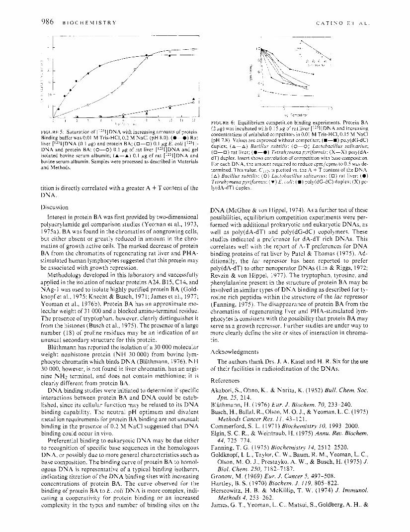
The ionic strength of the incubation buffer was critical for DNA binding (Figure 4). At concentrations greater than 0.2 M NaCI, the binding of protein BA to DNA rapidly dimin- ished. Figure 5 shows the saturation curves of nonhistone protein BA with double-stranded DNA from normal rat liver and E. coli. At pH 8.0 in 0.01 M Tris-HCI buffer containing
III;KKI, 3: I'cptidc map of I O iig 01 ~ p r m ~ n II,\ cht,mcd Ih digc4on s + i i h trypsin and d;m\yl;tlion wi th d;inryl chl imdc i ts dcrcrihed cinder bIa~criaIs and Methods. Thin-laycr chromatography is S K , fwmx acid. lcft to right. and benrenelacetic acid ( 9 2 ) bottom to top. Spots indicatcd by "X" wcrc found when the dansyl reaction was carried out in the irbsencc of protcin Bh.
i o ,
FIGURE 4: Effects of ionic strength on complex formation; 0. I wg of rat liver ['"I]DNA and 2 Wg of protein BA were incubated in 0.01 M Tris- HCI (pH 8.0) with increasing NaCl concentrations. Samples were pro- cessed as described in Materials and Methods.
0.2 M NaCI, saturation of eukaryotic DNA occurred much earlier than saturation of the prokaryotic DNA. For both DNAs the maximum retention on the filters was 60%. The background retention of radioactive DNA in the absence of protein has been subtracted. Approximately 2.5 times more protein was required to reach saturation with the prokaryotic DNA than with the eukaryotic DNA. Bovine serum albumin, purified by the same procedure as protein BA, did not bind DNA to 5 fig.
Equilibrium competition binding studies (Lin & Riggs, 1972) were used to compare binding affinities to ply(dA-dT) and poly(dC-dC) alternating copolymers as well as other DNAs. A fixed amount of labeled DNA and protein, in the linear portion of the saturation curve, was used with increasing amounts of unlabeled competitor. The competition for binding to normal liver DNA by poly(dA-dT), poly(dG-dC), and various DNAs is shown in Figure 6. Competition with poly(dA-dT) occurred a t a much lower concentration of competitor, as compared with poly(dC-dC) and natural DNAs. As can be seen in Figure 6 (insert), increased compe-
986 B I O C H E M I S T R Y
f'IGURE 5 : Saturation of [1251]DNA with increasing amounts of protein. Binding buffer was 0.01 M Tris-HCI, 0.2 M NaCl (pH 8.0). (0-0) Rat liver [1251]DNA (0.1 p g ) and protein BA; (0-0) 0.1 pg E . coli [1251]- DNA and protein BA; (0-0) 0.1 pg of rat liver [1251]DNA and gel isolated bovine serum albumin; (A-A) 0.1 p g of rat [1251]DNA and bovine serum albumin. Samples were processed as described in Materials and Methods.
tition is directly correlated with a greater A + T content of the DNA.
Discussion Interest in protein BA was first provided by two-dimensional
polyacrylamide gel comparison studies (Yeoman et al., 1973, 1975a). BA was found in the chromatins of nongrowing cells, but either absent or greatly reduced in amount in the chro- matins of growth active cells. The marked decrease of protein BA from the chromatins of regenerating rat liver and PHA- stimulated human lymphocytes suggested that this protein may be associated with growth repression.
Methodology developed in this laboratory and successfully applied in the isolation of nuclear proteins A24, B15, C14, and NAg-I was used to isolate highly purified protein BA (Gold- knopf et al.. 1975; Knecht & Busch, 197 1 ; James et al., 1977; Yeoman et al., 1976b). Protein BA has an approximate mo- lecular weight of 3 l 000 and a blocked amino-terminal residue. The presence of tryptophan, however, clearly distinguishes i t from the histones (Busch et al., 1975). The presence of a large number (1 8) of proline residues may be an indication of an unusual secondary structure for this protein.
Bluthmann has reported the isolation of a 30 000 molecular weight nonhistone protein ( N H 30 000) from bovine lym- phocyte chromatin which binds DNA (Bluthmann, 1976). N H 30 000, however, is not found in liver chromatin, has an argi- nine NH2 terminal, and does not contain methionine: it is clearly different from protein BA.
DNA binding studies were initiated to determine if specific interactions between protein BA and DNA could be estab- lished, since its cellular function may be related to its DNA binding capability. The neutral pH optimum and divalent metal ion requirements for protein BA binding are not unusual; binding in the presence of 0.2 M NaCl suggested that DNA binding could occur in vivo.
Preferential binding to eukaryotic DNA may be due either to recognition of specific base sequences in the homologous DNA, or possibly due to more general characteristics such as base composition. The binding curve of protein BA to homol- ogous DNA is representative of a typical binding isotherm, indicating titration of the DNA binding sites with increasing concentrations of protein BA. The curve observed for the binding of protein BA to E. coli DNA is more complex, indi- cating a cooperativity for protein binding or an increased complexity in the types and number of binding sites on the
~~~ L l--_-l 5 I O 15
pq Conpet l+or
FIGURE 6 : Equilibrium competition binding experiments. Protein BA (2 p g ) was incubated with 0.1 5 p g of rat liver [lZSI]DI\'A and increasing concentrations of unlabeled competitors in 0.01 M Tris-HCI, 0.15 M NaCl (pH 7.8). Values are expressed without competitor: (B-B) poly(dG-dC) duplex; (A--A) Bacillus subrilis: (0-0) Lactobacillus salicarius; (0-0) rat liver; (0 -0) Terrahynzena pyriformis; (X-X) poly(dA- dT) duplex. Insert show correlation of competition with base composition. For each DNA, the amount required to reduce cpm/cpmo to 0.5 was de- termined. This value, C I ~ ~ . is plotted vs. the A + T content of the DNA. ( A ) Bacillus subtilis; ( 0 ) Larrohacillus salirarius; (0) rat liver: ( 0 ) Tetrah~.menapl.ri formis: (v) E. c,oli: (B ) poly(dG-dC) duplex: ( X ) po- Iy(dA-dT) duplex.
DNA (McGhee & von Hippel, 1974). As a further test of these possibilities, equilibrium competition experiments were per- formed with additional prokaryotic and eukaryotic DNAs, as well as poly(dA-dT) and poly(dG-dC) copolymers. These studies indicated a preference for dA-dT rich DKAs. This correlates well with the report of A-T preferences for DNA binding proteins of rat liver by Patel & Thomas (1975). Ad- ditionally, the lac repressor has been reported to prefer poly(dA-dT) to other nonoperator DNAs (Lin & Riggs, 1972; Revzin & von Hippel, 1977). The tryptophan, tyrosine, and phenylalanine present in the structure of protein BA may be involved in similar types of DNA binding as described for ty- rosine rich peptides within the structure of the lac repressor (Fanning, 1975). The disappearance of protein BA from the chromatins of regenerating liver and PHA-stimulated lym- phocytes is consistent with the possibility that protein BA may serve as a growth repressor. Further studies are under way to more clearly define the site or sites of interaction in chroma- t in .
Acknowledgments
of their facilities in radioiodination of the DNAs.
References Akabori, S., Ohno, K., & Narita, K. (1952) Bull. Chem. SOC.
Bluthmann, H . (1976) Eur. J . Biochem. 70, 233-240. Busch, H., Ballal, R., Olson, M. 0. J., & Yeoman, L. C. (1975)
Commerford, S. L. (1971) Biochemistry I O , 1993-2000. Elgin, S. C. R., & Weintraub, M. (1975) Annu. Reu. Biochem.
Fanning, T. G . (1975) Biochemistry 14, 2512-2520. Goldknopf, I. L., Taylor, C. W., Baum, R. M., Yeoman, L. C.,
Olson, M. 0. J., Prestayko, A. W., & Busch, H. (1975) J . Biol. Chem. 250, 7 182--7 187.
The authors thank Drs. J. A. Kasel and H . R. Six for the use
Jpn. 25, 214.
Methods Cancer Res. 11, 43-121.
44, 725-714.
Gronow, M. (1969) Eur. J . Cancer 5, 497-508. Hartley, B. S. (1 970) Biochem. J . 119, 805-822. Herscowitz, H . B. & McKillip, T. W. (1974) J . Immunol.
James, G. T., Yeoman, L. C.. Matsui, S., Goldberg, A. H., & Methods 4 , 253-262.
F L U O R E S C E N C E S T U D I E S O N B A C T E R I A L L U C I F E R A S E V O L . 17, N O . 6, 1978 987
Busch, H. (1977) Biochemistry 16, 2384-2389.
1309.
1184.
Knecht, M. E., & Busch, H. (1971) Life Sci. 10, 1297-
Lin, S., & Riggs, A. D. (1970) Nature (London) 228,
Lin, S. , & Riggs, A. D. (1972) J . Mol. Biol. 72, 671-690. Lowry, 0. H., Rosebrough, N. J., Farr, A. L., & Randall, R.
Mandel, M., Schildkraut, C. L., & Marmur, J. (1968)
Marmur, J. (1961) J . Mol. Biol. 3, 208-218. Marushige, K., & Bonner, J. (1966) 1. Mol. Biol. 15, 160-
McGhee, J. D., & von Hippel, P. H. (1974) J . Mol. Biol. 86,
Miyazawa, Y. , & Thomas, Jr., C. D. (1 965) J. Mol. Biol. 1 I ,
Patel, G. L., & Thomas, T. L. (1975) in Chromosomal Pro- teins and Their Regulation of Gene Expression (Stein, G. S., & Kleinsmith, L. J., Eds.) pp 249-265, Academic Press, New York, N.Y.
Paul, J., & Gilmour, R. S. (1968) J. Mol. Biol. 34, 305- 316.
Penke, B., Ferenczi, R., & Kovaks, K. (1974) Anal. Biochem.
Revzin, A., & von Hippel, P. H. (1977) Biochemistry 16,
Richards, G. M. (1974) Anal. Biochem. 57, 369-376. Riggs, A. D., & Bourgeois, S. (1968) J . Mol. Biol. 34, 361-
J . (1951) J . Biol. Chem. 193, 265-275.
Methods Enzymol. 12, 184-195.
174.
469-489.
223-237.
60, 45-50.
4769-4776.
364.
J . Mol. Bioi. 34, 365-368.
Biophys. Res. Commun. 28; 8 15-820.
Cancer Res. 33, 3312-3318.
Exp. Cell Res. 82, 21 5-226.
Riggs, A. D., Bourgeois, S., Newby, R. F., & Cohn, M. (1968)
Shapiro, A. L., Venuela, E., & Maezel, J. V. (1967) Biochem.
Sitz, T. O., Nazar, R. N., Spohn, W. H., & Busch, H. (1 973)
Taylor, C. W., Yeoman, L. C., Daskal, I., & Busch, H. (1973)
Tichy, H. (1975) Anal. Biochem. 69, 552-557. Walker, J. M., Goodwin, G. H., & Johns, E. W. (1976) Eur.
Weiner, A. M., Platt, T., & Weber, K. (1972) J . Bioi. Chem.
Wilson, E. M., & Spelsberg, T. C. (1973) Fed. Proc., Fed. Am. SOC. Exp. Biol. 32, 608.
Yeoman, L. C., Taylor, C. W., Jordan, J. J., & Busch, H. (1973) Biochem. Biophys, Res. Commun. 53, 1067- 1076.
Yeoman, L. C., Taylor, C . W., Jordan, J. J., & Busch, H. (1975a) Exp. Cell Res. 91, 207-215.
Yeoman, L. C., Taylor, C. W., Jordan, J. J., & Busch, H. (1975b) Cancer Res. 35, 1249-1255.
Yeoman, L. C., Seeber, S., Taylor, C. W., Fernbach, D. J., Falletta, J. M., Jordan, J. J., & Busch, H. (1976a) Exp. Cell Res. 100, 47-55.
Yeoman, L. C., Jordan, J. J., Busch, R. K., Taylor, C. W., Savage, H. E., & Busch, H. (1976b) Proc. Natl. Acad. Sci.
J . Biochem. 62, 461-469.
247, 3242-325 1.
U.S.A. 73, 3258-3262.
Structural Studies on Bacterial Luciferase Using Energy Transfer and Emission Anisotropy?
Shiao-Chun Tu,$ Cheng-Wen Wu, and J . Woodland Hastings*
ABSTRACT: The distance between specific sites on bacterial luciferase was estimated by energy transfer. Luciferase was fluorescently labeled by reaction of an essential sulfhydryl group with N-( 1 -pyrene)maleimide and N - [p-(2-benzoxa- zolyl)phenyl]maleimide. Both of the modified enzymes bind 8-anilino- 1 -naphthalenesulfonate (Ans) with affinities similar to that exhibited by the native luciferase. Using each of the two fluorescent probes as a donor and the bound Ans as an accep- tor, the energy transfer efficiencies were determined by the resulting enhancement of fluorescence of the acceptor. The corresponding distance was calculated to be in the range of 21
B a c t e r i a l luciferase functions as a monooxygenase in cata- lyzing the bioluminescent mixed function oxidation of
t From the Biological Laboratories, Harvard University, Cambridge, Massachusetts 02138 (S.-C.T. and J .W.H.) , and the Department of Biophysics, Division of Biological Sciences, Albert Einstein College of Medicine, Bronx, New York 10461 (C.-W.W.). Received Augusr 10. 1977. This work was supported in part by National Institutes of Health Grants 5 F32 GM00348-02 and -03 to S.-C.T., National Science Foundation Grant BMS 74-23651 to J.W.H., and National Institutes of Health Grant GM-I9062 and American Cancer Society Grant BC-94 to C.-W.W.
t Present address: Department of Biophysical Sciences, University of Houston, Houston, Texas 77004.
0006-2960/78/04 17-0987$01 .OO/O
to 37 8. Energy-transfer studies were also carried out using fluorescence lifetime measurements of bound ANS, acting as a donor with bound FMN as an acceptor. The corresponding distance was calculated to be between 30 and 58 A. Using samples of 1uciferase:Ans complex and luciferase modified with N - ( 1 -pyrene)maleimide, the rotational correlation time of the enzyme-dye conjugate as a whole was found to be 47 f 2 ns. The observed rotational correlation time is much longer than that calculated for luciferase assuming a spherical structure, thus indicating an elongated form for the luciferase-dye con- jugate.
FMNHl ' and a long-chain aldehyde by molecular oxygen (Hastings & Gibson, 1963; Becvar & Hastings, 1975; Nealson & Hastings, 1972). The final reaction products are light,
I Abbreviations used are: FMNH2 and FMN, reduced and oxidized riboflavin 5'-phosphate; Ans, 8-anilino-l -naphthalenesulfonate: NBPM, N-[p-(2-benzoxazolyl)phenyl]maleimide; NPM, "V-( I-pyrene)maleimide; NBPS and NPS, the ,Y-[p-(2-benzoxazolyI)phenyl]succinimido group and the ,V-( I-pyrene)succinimido group, respectively (both are linked to the essential sulfhydryl group of luciferase): NBPS-E and NPS-E, the NBPS-luciferase adduct and the NPS-luciferase adduct, respectively; FMNb and AnSb, luciferase-bound FMN and Ans, respectively; q, quanta: Q, fluorescence quantum yield.