Copyright 2001 by the Genetics Society of America Characterization of the flamenco Region of the Drosophila melanogaster Genome Vale ´rie Robert, Nicole Prud’homme, Alexander Kim, 1 Alain Bucheton and Alain Pe ´lisson CGM/CNRS, 91198 Gif-sur-Yvette, France and IGH/CNRS, 34396 Montpellier, France Manuscript received August 10, 2000 Accepted for publication March 9, 2001 ABSTRACT The flamenco gene, located at 20A1–3 in the b-heterochromatin of the Drosophila X chromosome, is a major regulator of the gypsy/mdg4 endogenous retrovirus. As a first step to characterize this gene, z100 kb of genomic DNA flanking a P-element-induced mutation of flamenco was isolated. This DNA is located in a sequencing gap of the Celera Genomics project, i.e., one of those parts of the genome in which the “shotgun” sequence could not be assembled, probably because it contains long stretches of repetitive DNA, especially on the proximal side of the P insertion point. Deficiency mapping indicated that sequences required for the normal flamenco function are located .130 kb proximal to the insertion site. The distal part of the cloned DNA does, nevertheless, contain several unique sequences, including at least four different transcription units. Dip1, the closest one to the P-element insertion point, might be a good candidate for a gypsy regulator, since it putatively encodes a nuclear protein containing two double- stranded RNA-binding domains. However, transgenes containing dip1 genomic DNA were not able to rescue flamenco mutant flies. The possible nature of the missing flamenco sequences is discussed. G YPSY/MDG4 is an insect endogenous retrovirus rus able to spread both horizontally and vertically in spite of the fact that its expression and its regulation with infectious properties as shown by the integra- tion of new proviruses into the germline chromosomes by the flamenco gene occur only in the soma. The soma- to-germline transfer involved in the vertical spread of after horizontal transfer between laboratory strains (Kim et al. 1994; Song et al. 1994). As with any endogenous gypsy does not seem to be an infectious process, since a knock-out of the env gene, assumed to be reponsible retrovirus, gypsy is usually transmitted vertically like a Mendelian gene. However, in some genetic back- for the infectious properties of the virus (Teysset et al. 1998), does not affect its multiplication (Chalvet et grounds, vertically inherited gypsy proviruses can in- crease their copy number (Bucheton 1995). Compari- al. 1999). The respective importance of vertical and horizontal transmission for gypsy propagation, the mech- son of strains with low and high copy numbers led to the discovery of a gene called flamenco that controls anisms underlying both types of germline invasions by gypsy, and the mechanism by which flamenco represses gypsy proviral multiplication (Prud’homme et al. 1995). Deficiencies of flamenco, as well as permissive alleles, gypsy expression are still unclear. To address these issues, we set out to clone the flamenco gene. allow the multiplication of gypsy whereas restrictive al- leles do not. The latter are either dominant or semidom- The flamenco gene is located in 20A1–3, between the complementation groups extra organs (eo) and wings inant over permissive alleles (Pe ´lisson et al. 1997; Prud’- homme et al. 1995). Gypsy is allowed to amplify only in the apart (wap), a region contained in the b-heterochroma- tin of the X chromosome (Prud’homme et al. 1995). progeny of permissive females whatever the genotype of these progeny (Prud’homme et al. 1995). It is necessary Unlike a-heterochromatin, b-heterochromatin is nor- mally replicated in polytene chromosomes but, unlike and sufficient that somatic tissues of the mother have a permissive genotype for the functional gypsy proviruses euchromatin, it exhibits a poorly banded structure that forms the visible chromocenter (Heitz 1934; Yama- present in such tissues to amplify in the progeny (Chal- vet et al. 1999). Consistent with this observation is the moto et al. 1990). At the base of the polytene X chromo- some, the well-banded euchromatin is found distal to fact that the regulation of gypsy by flamenco is specific section 20 with respect to the centromere (Bridges to the somatic follicle cells that surround the female 1938). Heterochromatin is composed of repeated se- germline cells; the expression of a gypsy-lacZ reporter quences and is known to be a place for the accumulation transgene is totally abolished in restrictive follicle cells of defective transposable elements (Miklos et al. 1988; (Pe ´lisson et al. 1994). Thus, gypsy appears to be a retrovi- Vaury et al. 1989; Miklos and Cotsell 1990; Pimpi- nelli et al. 1995). On the X chromosome, only ribo- Corresponding author: Alain Pe ´lisson, Institut de Ge ´ne ´tique Humaine, somal genes have been localized in a-heterochromatin CNRS, 141 rue de la Cardonille, 34396 Montpellier Cedex 05, France. whereas complementation groups that could corre- E-mail: [email protected]spond to single copy genes appear to be located within 1 Present address: Department of Genetics, Lomonosov Moscow State University, 119899 Moscow, Russian Federation. the b-heterochromatin (Perrimon et al. 1989). A few Genetics 158: 701–713 ( June 2001)
Transcript
Copyright 2001 by the Genetics Society of America
Characterization of the flamenco Region of the Drosophila melanogaster Genome
Valerie Robert, Nicole Prud’homme, Alexander Kim,1 Alain Bucheton and Alain Pelisson
CGM/CNRS, 91198 Gif-sur-Yvette, France and IGH/CNRS, 34396 Montpellier, France
Manuscript received August 10, 2000Accepted for publication March 9, 2001
ABSTRACTThe flamenco gene, located at 20A1–3 in the b-heterochromatin of the Drosophila X chromosome, is a
major regulator of the gypsy/mdg4 endogenous retrovirus. As a first step to characterize this gene, z100kb of genomic DNA flanking a P-element-induced mutation of flamenco was isolated. This DNA is locatedin a sequencing gap of the Celera Genomics project, i.e., one of those parts of the genome in which the“shotgun” sequence could not be assembled, probably because it contains long stretches of repetitiveDNA, especially on the proximal side of the P insertion point. Deficiency mapping indicated that sequencesrequired for the normal flamenco function are located .130 kb proximal to the insertion site. The distalpart of the cloned DNA does, nevertheless, contain several unique sequences, including at least fourdifferent transcription units. Dip1, the closest one to the P-element insertion point, might be a goodcandidate for a gypsy regulator, since it putatively encodes a nuclear protein containing two double-stranded RNA-binding domains. However, transgenes containing dip1 genomic DNA were not able torescue flamenco mutant flies. The possible nature of the missing flamenco sequences is discussed.
GYPSY/MDG4 is an insect endogenous retrovirus rus able to spread both horizontally and vertically inspite of the fact that its expression and its regulationwith infectious properties as shown by the integra-
tion of new proviruses into the germline chromosomes by the flamenco gene occur only in the soma. The soma-to-germline transfer involved in the vertical spread ofafter horizontal transfer between laboratory strains (Kim
et al. 1994; Song et al. 1994). As with any endogenous gypsy does not seem to be an infectious process, sincea knock-out of the env gene, assumed to be reponsibleretrovirus, gypsy is usually transmitted vertically like a
Mendelian gene. However, in some genetic back- for the infectious properties of the virus (Teysset et al.1998), does not affect its multiplication (Chalvet etgrounds, vertically inherited gypsy proviruses can in-
crease their copy number (Bucheton 1995). Compari- al. 1999). The respective importance of vertical andhorizontal transmission for gypsy propagation, the mech-son of strains with low and high copy numbers led to
the discovery of a gene called flamenco that controls anisms underlying both types of germline invasions bygypsy, and the mechanism by which flamenco repressesgypsy proviral multiplication (Prud’homme et al. 1995).
Deficiencies of flamenco, as well as permissive alleles, gypsy expression are still unclear. To address these issues,we set out to clone the flamenco gene.allow the multiplication of gypsy whereas restrictive al-
leles do not. The latter are either dominant or semidom- The flamenco gene is located in 20A1–3, between thecomplementation groups extra organs (eo) and wingsinant over permissive alleles (Pelisson et al. 1997; Prud’-
homme et al. 1995). Gypsy is allowed to amplify only in the apart (wap), a region contained in the b-heterochroma-tin of the X chromosome (Prud’homme et al. 1995).progeny of permissive females whatever the genotype of
these progeny (Prud’homme et al. 1995). It is necessary Unlike a-heterochromatin, b-heterochromatin is nor-mally replicated in polytene chromosomes but, unlikeand sufficient that somatic tissues of the mother have
a permissive genotype for the functional gypsy proviruses euchromatin, it exhibits a poorly banded structure thatforms the visible chromocenter (Heitz 1934; Yama-present in such tissues to amplify in the progeny (Chal-
vet et al. 1999). Consistent with this observation is the moto et al. 1990). At the base of the polytene X chromo-some, the well-banded euchromatin is found distal tofact that the regulation of gypsy by flamenco is specificsection 20 with respect to the centromere (Bridgesto the somatic follicle cells that surround the female1938). Heterochromatin is composed of repeated se-germline cells; the expression of a gypsy-lacZ reporterquences and is known to be a place for the accumulationtransgene is totally abolished in restrictive follicle cellsof defective transposable elements (Miklos et al. 1988;(Pelisson et al. 1994). Thus, gypsy appears to be a retrovi-Vaury et al. 1989; Miklos and Cotsell 1990; Pimpi-nelli et al. 1995). On the X chromosome, only ribo-
Corresponding author: Alain Pelisson, Institut de Genetique Humaine, somal genes have been localized in a-heterochromatinCNRS, 141 rue de la Cardonille, 34396 Montpellier Cedex 05, France. whereas complementation groups that could corre-E-mail: [email protected]
spond to single copy genes appear to be located within1 Present address: Department of Genetics, Lomonosov Moscow StateUniversity, 119899 Moscow, Russian Federation. the b-heterochromatin (Perrimon et al. 1989). A few
Genetics 158: 701–713 ( June 2001)
702 V. Robert et al.
Females heterozygous for the X-linked deficiency and a bal-of these genes such as S6KII (Wassarman et al. 1994),ancer chromosome were crossed with males carrying the samefog (Costa et al. 1994), stn (Andrews et al. 1996), anddeficiency and a complementing Y-linked duplication (Prud’-
su(f), which is the most proximal (Mitchelson et al. homme et al. 1995). Embryos were collected for 18 hr at 238.1993), have been cloned. The fog gene was identified in After 1 more day at 238, the larvae that had nonlethal heterozy-
gous genotypes were manually discarded. The nonhatcheda 65-kb-long gene-poor region; the Drosophila genomeembryos, most of which were homozygous lethal, were dechori-consortium reported that “the gene density (. . .) dropsonated in 3% bleach before being frozen at 2808.abruptly to two genes in 400 kb around fog” (Adams et
Rescue experiments: Transgenic flies were recovered as col-al. 2000). The stn complementation group is a dicis- ored-eye w1 individuals after P-mediated transformationtronic gene that is found as a 12.5-kb island of unique (Spradling 1986) into the wOR(P) permissive stock. The
transgenes were then introduced into different flamenco con-DNA surrounded by repetitive sequences. On the distaltexts by two successive backcrosses with w2 females. Homozy-side of su(f), unique sequences are interspersed withgous transgenic females were crossed with males hemizygousrepeated sequences, whereas all the 30 kb isolated onfor a eo2, flam2, wap2 deficiency (l11) and homozygous for the
the proximal side of the gene (i.e., toward the centro- pgyp gypsy-LacZ transgene (Pelisson et al. 1994). Ovarian lacZmere) contain only repeated sequences (Tudor et al. expression was studied in the progeny of this cross. LacZ stain-
ing was performed as described previously (Pelisson et al.1996). light, an autosomal heterochromatic gene, has1994).also been characterized at the molecular level. It is 17
Libraries, screenings, and databases: A l phage library waskb long and its 3-kb RNA is expressed from single-copyconstructed with the DNA of flam py1(P) homozygous flies. This
DNA while its intronic and flanking regions consist of a DNA, partially digested by Sau3A, was ligated with the l Fixheterogenous array of middle-repetitive DNA sequences II vector arms (Stratagene, La Jolla, CA) following the protocol
provided by the manufacturer.(Devlin et al. 1990).The NotBamNot-CoSpeR (cosT) genomic cosmid libraryHere we report the P-element gene-tagging of flamenco
was kindly provided by J. Tamkun. It was constructed fromand the molecular characterization of a 100-kb walkthe restrictive y; cn bw sp strain. After transfer to Optitran BA-
around the P-element insertion. This walk mostly con- S85 reinforced nitrocellulose filters (Schleicher & Schuell,tains middle-repetitive DNA. Its distal part also contains Keene, NH), 3 3 104 clones were hybridized overnight at 428a few unique sequences including some transcription in 53 SSC, 53 Denhardt’s solution, 0.1% SDS, 50% for-
mamide with the U2 probe, i.e., the 3-kb BbsI-SalI fragmentunits. A detailed analysis of one of them (dip1) is pre-that flanks the flam py1(P) mutation, radiolabeled by randomsented here, although it has not yet been possible topriming (Megaprime; Amersham, Arlington Heights, IL). Outdemonstrate that this gene corresponds to flamenco. The of three positive clones, two were purified and their DNA was
possible nature of the missing flamenco sequences is dis- prepared with the plasmid Maxi kit (QIAGEN, Valencia, CA).cussed. RCPI-98 high-density hybridization filters containing the
Berkeley Drosophila Genome Project (BDGP) BAC librarywere purchased from the BACPAC Resource Center, as were19 clones from the library.MATERIALS AND METHODS
An ovary cDNA library was kindly provided by P. Tolias.The flamenco status of the Canton-S strain used to make thisDrosophila strains and fly care: Flies were maintained onlibrary is unknown. A total of 9 3 105 clones were screenedstandard Drosophila medium (Gans et al. 1975) at 258. Geneticas described above.symbols follow Lindsley and Zimm (1992). The strains of the
The databases constructed by BDGP were screened bylaboratory collection were previously described (Prud’hommeBLAST (Altschul et al. 1990). The BLAST queries were per-et al. 1995; Chalvet et al. 1999), including the sample offomed at the BDGP website with standard parameters. TheDf(1)lx deficiencies, which are wap2 X-ray-induced derivativescDNA clones were purchased from Research Genetics (Bir-of the 413(NP) chromosome, restrictive for gypsy expressionmingham, AL). The whole genome sequence assembly wasand mobilization (Pelisson et al. 1994). The y; cn bw sp restric-overviewed at the GenBank site (release 2) (http://www.ncbi.tive and the oreRw38 permissive stocks were the strains usednlm.nih.gov/PMGifs/Genomes/7227).to sequence the Drosophila genome by the American and
Restriction mapping of cosmid inserts: The protocol wasEuropean sequencing projects, respectively. The restrictiveadapted from Rackwitz et al. (1984). A total of 1 mg of cosmidRev(R) stock is referred to as RevI in Desset et al. (1999).DNA was digested by 5 units of l terminase (Epicentre, Madi-OvoD1 inactivation assay: The ovo gene is located on the Xson, WI) for 30 min at 258. After inactivation of the enzymechromosome at cytological position 4E2. It is a hot spot forat 658, 600 ng of this linearized DNA was preheated at 378insertion of gypsy (Mevel-Ninio et al. 1989). Crosses of permis-and then incubated with 0.1 unit of PstI (Biolabs). Aliquotssive females containing active gypsy proviruses with males car-of the digestion were sequentially stopped at 2, 8, 20, 45, andrying the ovoD1 dominant female sterile mutation produce
fertile daughters at high frequency. This abolition of the domi- 120 min by the addition of 10 mm EDTA and 125 mm NaCl.A total of 5 pmol of dephosphorylated oligonucleotides, eithernant female sterile phenotype results from insertions of gypsy
into the ovoD1 gene carried by the paternal chromosome. To cosL (AGGTCGCCGCCC) or cosR (GGGCGGCGACCT), was[g-32P]ATP-labeled with polynucleotide kinase (GIBCO BRL,estimate the frequency of these insertions, the daughters of
the test cross are scored for zero, one, or two ovaries. In most Gaithersburg, MD) as indicated by the manufacturer. One-half of each digested aliquot was incubated with 0.2 pmol ofcases fertile females have one ovary. The frequency of females
with two ovaries is about the square of that of females with either labeled oligonucleotide for 10 min at 658 and then 45min at 458. Samples were diluted 1/3 with loading bufferone ovary, suggesting that they result from two independent
events. Such females are therefore counted twice to calculate (13 TAE, 60 mm EDTA, 0.1% bromophenol blue and 50%glycerol), loaded on a 0.5% agarose gel, and fractionated atthe frequency of ovoD1 inactivation.
Collection of embryos homozygous for lethal deficiencies: 48 for 24 hr at 55V in 13 TAE buffer. Gels were vacuum dried
703Walk Around flam in D. melanogaster
on DE81 paper (Whatman) and exposed to X-Omat AR film seau and Bucheton 1999). Its mobilization can be in-(Kodak, Rochester, NY). duced by the P transposase in trans. It is located on
Subcloning nonrepetitive sequences: U2 was obtained bychromosome 2 of a strain that also contains an FM7csubcloning a 3-kb SalI-BbsI fragment from a clone of the lbalancer carrying a y2 allele and the restrictive allelephage library containing the 59 segment of the P element. U1
and d1 were obtained from the cos7a cosmid (Figure 3A). To flamFM7c(R). As shown in Figure 1A, females of this stockobtain U1, a HpaI-HindIII digestion was first performed to were crossed with males carrying the D2-3(99B) sourcesubclone a 0.9-kb fragment. U1 was then subcloned as a 0.2- of P transposase (Robertson et al. 1988) to inducekb HpaI-DraI fragment of this intermediate clone. d1 is a 1.4-
mobilization of P[lyB]. About 1000 FM7c chromosomeskb HindIII subclone of a 2.8-kb PstI-SalI intermediate clone.acquired a transposed copy of P[lyB], as judged by thePulsed-field gel electrophoresis: High molecular weight
DNA was prepared from adult flies essentially as described linkage of the y1 and B markers in G3. All but 1 ofpreviously (Birren and Lai 1993). About 250 frozen females these FM7c, P[lyB] chromosomes, when combined inwere ground to fine powder in liquid nitrogen. The powder G2 females with a chromosome carrying the permissivewas transferred to a 15-ml Dounce homogenizer (Kontes); 15
flamOR(P) allele, displayed the same ability as the parentalml of ice-cold NIB (10 mm Tris pH 8.5, 60 mm NaCl, 10 mmFM7c chromosome to prevent the transposition of gypsyEDTA, 0.15 mm spermine, 0.5% Triton X-100) was added to
the powder and the mix was homogenized with 5–10 strokes into the female sterile ovoD1 allele (0.1% G3 fertile fe-of pestle A. After transfer to an ice-cold Corex centrifuge tube, males). The exception was a mutant chromosome thatthe mix was spun 15 min at 580 3 g at 48 to pellet remaining allowed the inactivation of ovoD1 in five out of 23 G3large body parts. The supernatant was transferred to an ice-
females observed (22% ovoD1 inactivation). This chromo-cold Corex tube and spun 8 min at 5800 3 g at 48. The pelletsome contains a recessive permissive mutation that doeswas resuspended in 250 ml NIB and warmed at 378. An equal
volume of preheated (508) 1.2% InCert Agarose (FMC, Rock- not complement the known permissive alleles of fla-land, ME)/125 mm EDTA was added. Agarose blocks (100 menco and is therefore denoted flampy1(P).ml) were molded and hardened 5–10 min on ice. The resulting Mobilization of the P[lyB] element associated with thisplugs were incubated 20 hr at 508 in 250 ml of digestion buffer
permissive chromosome was then induced by crosses(50 mm EDTA, 1% SDS, 1 mg/ml proteinase K) and thenwith the D2-3(99B) transposase source (Figure 1B). Ex-dialyzed twice for 1 hr against 13 TE-40 mg/ml phenylmethyl-
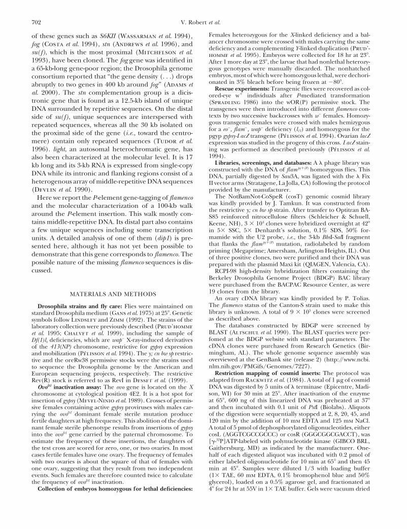
sulfonyl fluoride (PMSF) and four additional times each for cisions of P[lyB] from the FM7c, P[lyB] chromosome20 min in 13 TE. Samples were stored in 13 TE at 48. For were identified by looking for the loss of the y1 markerall tested enzymes, except for NotI (Promega, Madison, WI), in G1. As shown in Figure 2, out of 43 independent y2
digestion of DNA was performed in the liquid phase. A total ofderivatives of the FM7c, P[lyB] permissive chromosome,40 ml of melted plug was incubated overnight with restriction33 appeared to carry a restrictive revertant allele. Byenzyme buffer, BSA, and restriction enzyme in a final volume
of 80 ml. For NotI digestion, half plugs were incubated over- contrast, the 15 y1control derivatives all remained asnight in 200 ml of a mix of restriction endonuclease buffer, permissive as the parental chromosome. The frequentBSA, and enzyme. Before migration, a second proteinase K co-occurrence of excisions with reversions of thedigestion was performed either by adding 20 mg of enzyme
flampy1(P) allele strongly suggests that the P[lyB] insertionto the liquid samples or by incubating solid samples in 13is indeed responsible for this mutation.TAE-0.1 mg/ml proteinase K. Pulsed-field gel electrophoresis
(PFGE) was performed in 1% SeaKem LE agarose (FMC) gels Isolation of genomic DNA flanking the P insertionrun at 128, 160 V, on a 2015 Pulsaphor Plus (LKB, Piscataway, site: The 100-kb walk: Genomic DNA from flies homozy-NJ) apparatus, set at either 5- to 15-sec pulses for 20 hr (for gous for the flampy1(P) allele was used to construct a lrestriction mapping) or 25-sec pulses for 24 hr (for analysis
phage library. This library was screened with both endsof deficiencies). Gels were stained in 0.5 mg/ml ethidiumof the P element and 6 kb of genomic DNA spanning thebromide for 30 min and then photographed, blotted, and
hybridized as usual. P[lyB] insertion point was recovered. Three kilobases ofPoly(A)1 RNA extraction and Northern blot analysis: A this DNA consist of unique sequences (data not shown).
total of 100 mg of flies was homogenized in 2 ml of RNA Plus These sequences, in a fragment called U2, flank P[lyB]solution (Quantum Biotechnologies, Blaine, WA). Addition
(Figure 3B) and hybridize to the 20A region of theof 200 ml of chloroform allowed the extraction of total RNA.polytene X chromosome (data not shown). U2 was usedPoly(A)1 RNA was extracted from this preparation with the
polyATtract mRNA isolation system IV (Promega). Northern as an entry point to initiate a chromosome walk byblots were performed as previously described (Pelisson et al. screening a cosmid library (see materials and meth-1994) except that the running buffer was 23 3-(N-morpho- ods). Two positive clones were recovered (cos6a andlino) propanesulfonic acid (MOPS) instead of 13 MOPS.
cos7a, Figure 3A). A restriction map of these clones,PCR analysis: PCR was performed with genomic DNA essen-established by partial PstI digestion, shows that theytially as described previously (Chalvet et al. 1999). The se-
quences of the primers flanking the dip1 repetitive region encompass a total of 35 kb, 25 kb of which is commonwere 59-GCCTCTTCACTTTGACAG-39 and 59-CGGCACCAAT to both of them.TCACCTACAG-39. To look for additional unique sequences, Southern
blots of single and double digestions of the inserts in thetwo cosmid clones were probed by radiolabeled whole
RESULTSgenome DNA. Most of the fragments were readily la-beled, indicating that these clones were rich in repetitiveP-induced flamenco mutagenesis: P[lyB] is a nonauton-
omous P element carrying the y1 genetic marker (Bus- sequences. Two fragments that appeared weakly labeled
704 V. Robert et al.
Figure 2.—Excision of the y1 marker from the FM7c,flam py1(P) mutant chromosome is frequently associated withreversion of the permissive mutant phenotype. The numbersof independent excision events recovered in G1 females (de-noted FM7c, P[lyB]EX in Figure 1B) are plotted on the y-axisof the histogram. The x-axis indicates their respective levelsof permissiveness for gypsy mobilization as measured by thepercentage of fertile ovoD1 daughters of flamOR(P)/FM7c, P[lyB]EX
G2 mothers. The distribution of permissiveness levels of someFM7c, P[lyB] parental chromosomes is represented by the con-trol histogram. The three hatched squares correspond to re-vertants 679-5b, 679-14a, and 679-16a, the molecular structureof which was studied (see Figure 5).
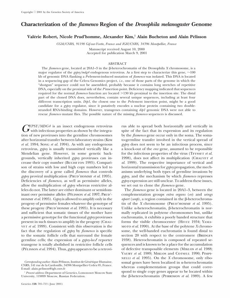
Figure 1.—Mating schemes used to select for transpositionsof the P[lyB] element into and out of the flamenco gene. Only
by the whole genome probe were further analyzed bythe relevant genetic markers are shown. (A) P-induced muta-genesis of the flamFM7c(R) allele on the FM7c restrictive chromo- several rounds of subcloning and hybridization to geno-some. FM7c, CyO, and TM6 are balancer chromosomes that mic blots until single-copy fragments could be identi-are transmitted through meiosis with little or no recombina- fied. Only one such sequence was found by this method,tion with the homologous chromosome. They carry, respec-
a 0.2-kb HpaI-DraI fragment that is hereafter referredtively, the B, Cy, and Ubx dominant markers, which, like Spto as U1 (see materials and methods and Figure 3).and Dr, can be recognized to select for the appropriate chro-
mosome combinations in heterozygous flies. G1 females car- The dissection process also revealed a 1.4-kb HindIIIrying the D2-3(99B) transposase source were crossed with fragment (denoted d1), which often labels more thanmales of the permissive gypsy-rich OR(520)/FM3 stock car- one fragment when used as a probe on genomic DNA.rying the y, v, and flamOR(P) markers. P[lyB] transpositions in
Further Southern blotting analyses indicated that d1 isthese G1 females were detected by the Cy y1 phenotype inpart of an z6-kb tandem duplication. Both copies oftheir daughters. G2 females that had inherited the y v FM7c
balancer, as judged by their v B phenotype, and that did the duplication, called D1 and D19, are contained in anot inherit the Dr D2-3(99B) chromosome 3 were individually 13-kb AvrII-SfiI fragment (Figure 3B). U1 and d1 arecrossed with ovoD1 males. The linkage of the y1 and B markers both located in the same part of the cos7a cosmid, whichindicated that P[lyB] had inserted into the FM7c balancer.
also contains the P[lyB] insertion point and U2. TheMutation of its flamFM7c(R) restrictive allele into a flam py1(P) per-missive allele was detected by the presence of fertile G3 females(see materials and methods for the rationale of the ovoD1
inactivation assay). (B) Reversion of the flam py1(P) mutation byexcision of the P[lyB] element from the FM7c, P[lyB] mutant B females. These females were individually crossed with males
of the permissive gypsy-rich OR(520)/FM3 stock. As a control,chromosome. G0 females containing the D2-3(99B) source oftransposase were crossed with males from a y v restrictive some of their y1 v B sisters were also studied. The ability of
both the parental and excision chromosomes to control gypsystrain. P[lyB]EX represents independent excision events oftheP[lyB]-linked y1 marker, which were selected in G1 as y v transposition was quantified by the ovoD1 inactivation assay.
705Walk Around flam in D. melanogaster
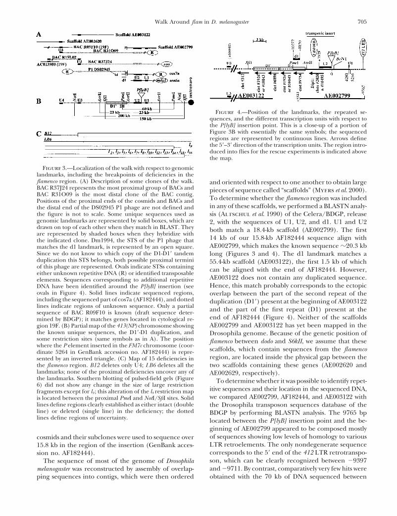
Figure 4.—Position of the landmarks, the repeated se-quences, and the different transcription units with respect tothe P[lyB] insertion point. This is a close-up of a portion ofFigure 3B with essentially the same symbols; the sequencedregions are represented by continuous lines. Arrows definethe 59–39 direction of the transcription units. The region intro-duced into flies for the rescue experiments is indicated abovethe map.
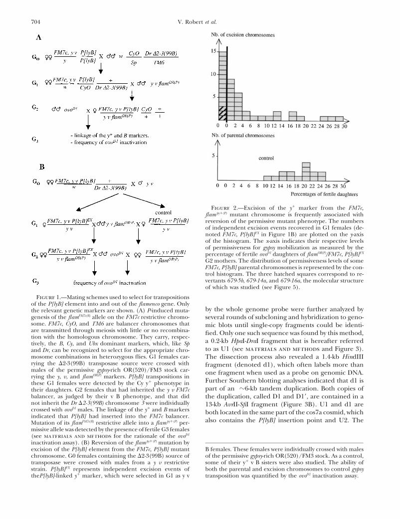
Figure 3.—Localization of the walk with respect to genomiclandmarks, including the breakpoints of deficiencies in theflamenco region. (A) Description of some clones of the walk. and oriented with respect to one another to obtain largeBAC R37J24 represents the most proximal group of BACs and pieces of sequence called “scaffolds” (Myers et al. 2000).BAC R31O09 is the most distal clone of the BAC contig.
To determine whether the flamenco region was includedPositions of the proximal ends of the cosmids and BACs andin any of these scaffolds, we performed a BLASTN analy-the distal end of the DS02945 P1 phage are not defined and
the figure is not to scale. Some unique sequences used as sis (Altschul et al. 1990) of the Celera/BDGP, releasegenomic landmarks are represented by solid boxes, which are 2, with the sequences of U1, U2, and d1. U1 and U2drawn on top of each other when they match in BLAST. They both match a 18.4-kb scaffold (AE002799). The firstare represented by shaded boxes when they hybridize with
14 kb of our 15.8-kb AF182444 sequence align withthe indicated clone. Dm1994, the STS of the P1 phage thatAE002799, which makes the known sequence z20.3 kbmatches the d1 landmark, is represented by an open square.
Since we do not know to which copy of the D1-D19 tandem long (Figures 3 and 4). The d1 landmark matches aduplication this STS belongs, both possible proximal termini 55.4-kb scaffold (AE003122), the first 1.5 kb of whichof this phage are represented. Ovals indicate STSs containing can be aligned with the end of AF182444. However,either unknown repetitive DNA (R) or identified transposable
AE003122 does not contain any duplicated sequence.elements. Sequences corresponding to additional repetitiveHence, this match probably corresponds to the ectopicDNA have been identified around the P[lyB] insertion (see
ovals in Figure 4). Solid lines indicate sequenced regions, overlap between the part of the second repeat of theincluding the sequenced part of cos7a (AF182444), and dotted duplication (D19) present at the beginning of AE003122lines indicate regions of unknown sequence. Only a partial and the part of the first repeat (D1) present at thesequence of BAC R09F10 is known (draft sequence deter-
end of AF182444 (Figure 4). Neither of the scaffoldsmined by BDGP); it matches genes located in cytological re-AE002799 and AE003122 has yet been mapped in thegion 19F. (B) Partial map of the 413(NP) chromosome showing
the known unique sequences, the D19-D1 duplication, and Drosophila genome. Because of the genetic position ofsome restriction sites (same symbols as in A). The position flamenco between dodo and S6kII, we assume that thesewhere the P element inserted in the FM7c chromosome (coor-
scaffolds, which contain sequences from the flamencodinate 5264 in GenBank accession no. AF182444) is repre-region, are located inside the physical gap between thesented by an inverted triangle. (C) Map of 15 deficiencies in
the flamenco region. B12 deletes only U4; LB6 deletes all the two scaffolds containing these genes (AE002620 andlandmarks; none of the proximal deficiencies uncover any of AE002629, respectively).the landmarks. Southern blotting of pulsed-field gels (Figure To determine whether it was possible to identify repet-6) did not show any change in the size of large restriction
itive sequences and their location in the sequenced DNA,fragments except for l9 ; this alteration of the l9 restriction mapwe compared AE002799, AF182444, and AE003122 withis located between the proximal PmeI and NotI/SfiI sites. Solid
lines define regions clearly established as either intact (double the Drosophila transposon sequences database of theline) or deleted (single line) in the deficiency; the dotted BDGP by performing BLASTN analysis. The 9765 bplines define regions of uncertainty. located between the P[lyB] insertion point and the be-
ginning of AE002799 appeared to be composed mostlyof sequences showing low levels of homology to variouscosmids and their subclones were used to sequence overLTR retroelements. The only nondegenerate sequence15.8 kb in the region of the insertion (GenBank acces-corresponds to the 59 end of the 412 LTR retrotranspo-sion no. AF182444).son, which can be clearly recognized between 29397The sequence of most of the genome of Drosophilaand 29711. By contrast, comparatively very few hits weremelanogaster was reconstructed by assembly of overlap-
ping sequences into contigs, which were then ordered obtained with the 70 kb of DNA sequenced between
706 V. Robert et al.
the P[lyB] insertion point and the end of AE003122. An distal position than U2 with respect to the centromere;(ii) U3 is also a more distal STS, since it matches theFB4/HB1 DNA transposon is located between positions
13597 and 14929 (Figure 4) and long stretches of complementary strand of 1 STS of the BAC R19L02,whose second STS matches GenBank entry AC013980the micropia LTR retrotransposon can be recognized
between coordinates 12,040 and 15,340 of AE003122 also originating from 19F. Moreover, this BLAST analy-sis of unique STSs suggested that they are all on the(data not shown). All but one of the six degenerate
retroelements found in these 70 kb are clustered in the distal side of the contig whereas most of the repetitiveones are proximal with respect to the centromere (datalast 1.5 kb of scaffold AE003122.
Additional clones were obtained from different librar- not shown). The sequence of the 43-kb most distal partof this contig is known, since U4 (but not U3) matchesies constructed by the Drosophila sequencing genome
projects after BLAST analysis of the BDGP/European the complementary strand of scaffold AE002620 be-tween positions 43,194 and 42,445.Drosophila Genome Project (EDGP) genomic clones
and the sequence-tagged site (STS) databases with the Orientation of the walk with respect to the centromere: Theorganization of the BAC contig was further confirmedU1, U2, and d1 sequences used as queries. The draft
sequence of the bacterial artificial chromosome (BAC) by using some PCR-amplified STSs to probe the blottedfingerprints. One of them, U3, enabled us to anchorclone R32M16 was found to contain U2 and d1. A more
detailed analysis reveals that it also contains unique se- the walk to the BAC contig and therefore to orient it withrespect to the centromere. U3 labels the P1 DS02945quences from cytological sections 15–16. We concluded
that this clone is rearranged. The STS Dm1994 of the but neither of the cosmids cos6a and cos7a, which aretherefore proximal to the P1 clone (Figure 3A). The factP1 phage DS02945 overlaps d1 in the same orientation.
The DS02945 insert is z70 kb long, which makes the that U3 is also complementary to the end of AE003122 isin agreement with the orientation of this scaffold towardwalk at least 100 kb long (Figure 3A). We could not
proceed further in this direction because the other STS the telomere.Molecular analysis of revertants: As shown in Figureof DS02945 is repeated. The same is true in the other
direction since the STSs of cos6a and cos7a match, re- 5A, the yellow gene, which was used as a marker to selectfor excisions, is located in the middle of the P[lyB]spectively, mdg1 and an unknown repetitive sequence.
The BAC contig: To get longer genomic DNA, U2 was element. To check for the possible retention of theflanks, BamHI-HincII and BamHI-HindIII DNA digestsused to probe the BDGP BAC library, which contains
inserts of 165 kb on average. A contig of 23 positive of three revertants were hybridized successively withboth ends of this element. In fact, the first probe wasclones was obtained. The inserts of 19 of them, as well
as those of the P1 and cosmid clones, were studied by a BamHI-EcoRI hybrid fragment containing 240 bp ofthe 39 end of P[lyB] flanked by 160 bp of genomic DNAthe fingerprint technique described by Marra et al.
(1997). The results of this analysis (not shown) can be (Figure 5A). This probe labeled the same fragments inrevertants 679-16a and 679-14a as in the flampy1(P) mutantsummarized as follows: (i) Four BACs have very little
overlap with the others. They were considered as re- flies, indicating that they result from imprecise excisionsleaving at least the right-hand side of the element inarranged and were ignored in this study. (ii) Among
the 15 others, two groups of 4 very similar BACs can be the footprints (Figure 5B). By contrast, revertant 679-5b showed the same restriction pattern as the parentalformed; one of them, represented by the clone R37J24,
seems to be located at one end of the contig (Figure FM7c chromosome, which, at this level of analysis, maybe interpreted as a precise excision event. These conclu-3A). (iii) The clone R31O09 seems to be located at the
other end of the contig. sions were supported by hybridization with a 274-bpprobe (coordinates 239–512 in the P-element sequence)The STSs of 16 of the 19 nonrearranged clones are
in the BDGP database: No clone has 2 unique STSs; 10 specific for the 59 end of P[lyB]. Only 679-16a and 679-14a hybridized to this probe (data not shown). In theclones, including R31O09 and R37J24, have 1 unique
STS (the other STS being made of repetitive elements); case of 679-16a, the sizes of the HincII-HincII andHindIII-BamHI fragments were different from those ofand 6 clones have 2 repetitive STSs. The unique STSs
of clones R31O09 and R37J24 were respectively named the corresponding fragments in flampy1(P) and 679-14a,indicating that only the HincII-HindIII 1-kb fragmentU4 and U3. The overall organization of the contig de-
duced from the fingerprint analysis was confirmed by flanking the P[lyB] element was unaffected by this im-precise excision.BLAST analysis performed with the 10 unique STSs.
Several clones are indeed very similar since they share Localization of the walk with respect to the breakpointsof deficiencies: We used the unique sequences previouslyat least pieces of STSs. This analysis also gave the orienta-
tion of some of the BAC clones with respect to each identified as landmarks to determine the localizationof the walk with respect to the breakpoints of the defi-other and to the centromere, as shown by the following
observations (Figure 3A): (i) U4 matches the draft se- ciencies affecting the flamenco region (Prud’homme etal. 1995). All these deficiencies are homozygous lethalquence of an additional BAC clone (R09F10) known to
be located in cytological section 19F, which is in a more because they uncover either the eo or the wap locus. To
707Walk Around flam in D. melanogaster
we took advantage of the observation that the LB6 defi-ciency uncovers all the unique sequences tested andcombined it with each deficiency studied. The resultsobtained by these four different strategies are summa-rized in Table 1 and Figure 3C. They show that noneof the flam1 deficiencies delete U1 or U2 but that theLB6 flam2 deficiency deletes both of these landmarks.This confirmed that the walk was indeed situated in theflamenco region. Moreover, U4 but not U3 is deleted bythe distal deficiency B12, which confirmed the orienta-tion of the walk with respect to the centromere andallowed the mapping of the B12 deficiency (and there-fore at least part of the eo sequences) distal to the U3landmark, which is .60 kb from the P[lyB] insertionpoint (Figures 3 and 4).
None of the unique sequences are uncovered by theproximal deficiencies tested. All the proximal deficien-cies denoted lx were derived from the same 413(NP)restrictive chromosome (Prud’homme et al. 1995). Therestriction map of this chromosome was established bydigesting its DNA with rare cutting enzymes and probingwith U2 and d1 (Figure 3B). We then asked whetherany of the restriction fragments with a long proximalextension would be affected by any of these proximaldeficiencies. l9 (Figure 3C) is the only deficiency forFigure 5.—Molecular structure of the P-induced flamencowhich modified fragments were observed; the 200-kbmutant and its revertant derivatives. (A) Partial restrictionNotI fragment (Figure 6) and the 150-kb SfiI fragmentmaps of the flamFM7c(R) and flam py1(P) parental and mutant alleles
(restriction sites not required for interpretation of the results (data not shown) present on the parental chromosomeare not shown). The box represents the P[lyB] insertion, brack- were replaced, respectively, by a 250-kb and a 200-kbeted by the P-element termini (in black) and flanked by FM7c fragment in l9. This was not the case for the 23-kb AvrIIgenomic DNA (thin line). (B) Comparison of the hybridiza-
and the 60-kb PmeI fragments. We can conclude that:tion patterns of three revertant alleles (679-16a, 679-14a, and(i) l9 is the closest rearrangement to U2, although its679-5b) with those of the flamFM7c(R) and flam py1(P) parental and
mutant alleles. The genomic DNA was digested by both distal breakpoint is located between the PmeI and NotI/BamHI, which cleaves the right-hand end of P[lyB], and by SfiI sites, that is, between 40 kb and 130 kb away fromeither HincII or HindIII, which cut in the DNA flanking the the P[lyB] insertion point; (ii) the breakpoints of theinsertion. The blot was hybridized with a 400-bp BamHI-EcoRI
two other flam2 proximal deficiencies are located proxi-fragment subcloned from the flam py1(P) genomic DNA. Thismal to the NotI/SfiI sites, that is, at .130 kb from theprobe includes the right-hand end of P[lyB] and 160 bp of
the flanking DNA. Revertant 679-5b has the same restriction P[lyB] insertion point. Two hypotheses can explain thesepattern as the parent flamFM7c(R) allele, which suggests that it results: Either flamenco is a gene larger than 130 kb orresults from the precise excision of the whole P element. The the region contains more than one gene involved intwo other revertants, which have the same BamHI-HincII and
the regulation of gypsy (see discussion).BamHI-HindIII junction fragments as the mutant flam py1(P),At least four different genes are present in the walk:retain the right-hand side of the insert.
With the aim of determining which gene(s) the P[lyB]insertion may affect, we looked for expressed sequencetags (ESTs) by BLASTN analysis. The location of thesolve the problem of lethality, several different strategies
were used to ask whether or not a unique sequence is corresponding cDNAs was confirmed by hybridizationto the genomic clones of the walk (Figure 4). At leastuncovered by a deficiency. First, we took advantage of
the existence of restriction site polymorphisms to com- seven distinct BDGP clots (nos. 312, 1926, 2020, 6898,9754, 10,043, and 10,495), and several individual cDNAspare various heterozygous combinations. Second, when
there was no polymorphism, we quantified the signals not classified in clots, were positive. They all map distalto the P[lyB] insertion point with respect to the centro-obtained on the Southern blots to determine whether
they resulted from the presence of one or two allelic mere. Four of the clots hybridize with the 6-kb duplica-tion described above. They correspond to two pairsdoses. Third, since most of the deficiencies to be studied
were embryonic lethal, we performed Southern blots (1926/10,043 and 6898/9754) of very similar clots. Bothclots of a pair appear to differ not only by alternativewith DNA extracted from homozygous embryos selected
on the basis of their lethal phenotype (see materials splicing but also by point mutations, which suggests thateach originates from a different copy of the duplication.and methods). Fourth, to resolve the final ambiguities,
708 V. Robert et al.
TABLE 1
Genetic and molecular analysis of deficiencies of the flamenco region
Genetic analysis Molecular analysis
eo flam wap U4 U3 U2 U1
Distal deficienciesB12 2 1e 1 2a 1b 1a NTLB6 2 2 1 NT NT 2a,c 2c
Proximal deficienciesR20 1 2 2 NT NT 1a,c 1c
R21 1 2 2 NT NT 1c 1c
16.2.13 1 2 2 NT NT 1a NTDCB1-35c 1 2 2 NT NT 1a NTl3, l9, l15 1 2 2 NT NT 1a,c 1c,d
l2, l 5, l6, l7, l8, l10, l12, l13, l14, l16 1 1 2 NT NT 1a,c 1a
R44 1 1 2 NT NT 1c NTl11 2 2 2 NT NT 2a,c NT
The genetic data are the results of complementation tests between the deficiencies and the three mutantgenes; for flam, mutant (2) and wild type (1) correspond, respectively, to the permissive and restrictivephenotypes. The 2 or 1 molecular data mean that the genomic landmark is or is not deleted by the deficiency.NT, the deficiency was not studied with this particular probe.
a,b,d The presence of the four genomic landmarks was investigated by Southern blot analysis of DNA fromheterozygous females (a,b, 1/deficiency or d, LB6/deficiency).
b The intensity of the band was quantified to compare the number of doses with that of the wild-type control.c Southern blot analysis was performed with DNA of embryos homozygous for the deficiency.e The chromosome bearing the B12 deficiency is permissive, as a result of a secondary mutation that could
be recombined away from the deficiency (Chalvet et al. 1999).
For instance, the consensus sequence of clot 9754 is common domains with the mammalian glucosidases II.The pair of clots 6898/9754 presents high similaritiesperfectly identical to the sequenced exonic part of the
D1 copy whereas up to seven differences are observed to Caenorhabditis elegans and Saccharomyces cerevisiae openreading frames (ORFs) and to murine and human ESTs,when this genomic sequence is compared with the 660
corresponding nucleotides of clot 6898. We infer that as well as lower similarities to prokaryotic genes involvedin nitrogen metabolism. The clots 2020 and 10,495 cor-the clones composing clot 9754 are encoded by the
proximal copy D1 and the clone of clot 6898 by the respond to two groups of cDNAs that differ only byalternative splicing. Three more cDNAs belonging todistal copy D19 (Figure 4).
The largest member of each clot or pair of clots was this class, dip1a (AF175713), UbxBP1 (AF218310), andklett (AJ250866), have been independently described,sequenced and analyzed by BLASTX. No hit was found
for the pair of clots 1926/10,043. Clot 312 shares some the products of which respectively interact with theDisco, Ultrabithorax, and SU(VAR)3-9 proteins. Puta-tive proteins encoded by members of this class have abipartite nuclear localization signal and two double-stranded RNA-binding domains (dsRBD). The dsRBDis present in many proteins (St. Johnston et al. 1992;Gibson and Thompson 1994), two of which are humanproteins involved in the control of virus propagation(Hovanessian 1989; Gatignol et al. 1991). Because ofthese two domains and because this transcription unit(hereafter called dip1) is the closest to the P[lyB] inser-tion point, we decided to characterize it and to test itsability to control gypsy mobilization.
Failure to rescue flamenco permissive flies with geno-mic dip1 transgenes: As shown in Table 2 and Figure
Figure 6.—Size of the NotI fragment in the proximal defi- 7A, at least 11 different types of dip1 transcripts wereciencies. High molecular weight DNA was prepared from fe-
obtained by using U2 to screen cDNA libraries andmales heterozygous for LB6 and the proximal deficiencies lx,EST databases. They vary by alternative splicing andanalyzed by PFGE, and hybridized with U2. 413(NP) is the
parental chromosome of the lx deficiencies. polyadenylation. All contain an ORF with the bipartite
709Walk Around flam in D. melanogaster
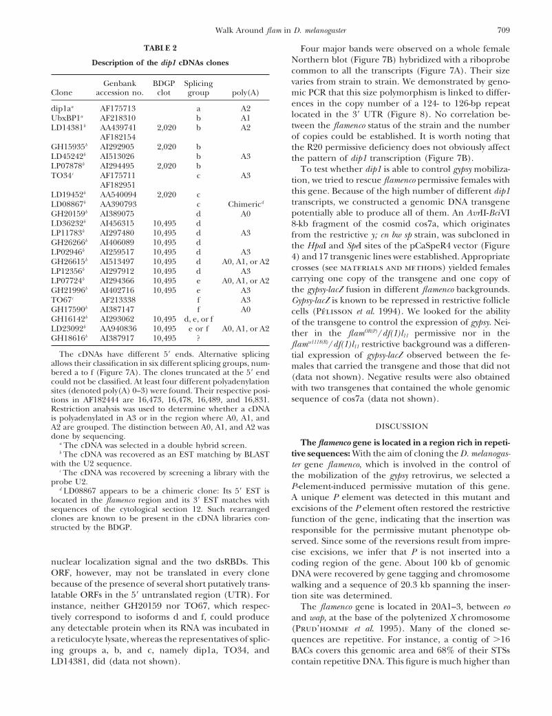
TABLE 2 Four major bands were observed on a whole femaleNorthern blot (Figure 7B) hybridized with a riboprobeDescription of the dip1 cDNAs clonescommon to all the transcripts (Figure 7A). Their sizevaries from strain to strain. We demonstrated by geno-Genbank BDGP Splicing
Clone accession no. clot group poly(A) mic PCR that this size polymorphism is linked to differ-ences in the copy number of a 124- to 126-bp repeatdip1aa AF175713 a A2located in the 39 UTR (Figure 8). No correlation be-UbxBP1a AF218310 b A1tween the flamenco status of the strain and the numberLD14381b AA439741 2,020 b A2of copies could be established. It is worth noting thatAF182154
GH15935b AI292905 2,020 b the R20 permissive deficiency does not obviously affectLD45242b AI513026 b A3 the pattern of dip1 transcription (Figure 7B).LP07878b AI294495 2,020 b To test whether dip1 is able to control gypsy mobiliza-TO34c AF175711 c A3 tion, we tried to rescue flamenco permissive females withAF182951
this gene. Because of the high number of different dip1LD19452b AA540094 2,020 ctranscripts, we constructed a genomic DNA transgeneLD08867b AA390793 c Chimericd
potentially able to produce all of them. An AvrII-BciVIGH20159b AI389075 d A0LD36232b AI456315 10,495 d 8-kb fragment of the cosmid cos7a, which originatesLP11783b AI297480 10,495 d A3 from the restrictive y; cn bw sp strain, was subcloned inGH26266b AI406089 10,495 d the HpaI and SpeI sites of the pCaSpeR4 vector (FigureLP02946b AI259517 10,495 d A3 4) and 17 transgenic lines were established. AppropriateGH26615b AI513497 10,495 d A0, A1, or A2
crosses (see materials and methods) yielded femalesLP12356b AI297912 10,495 d A3carrying one copy of the transgene and one copy ofLP07724b AI294366 10,495 e A0, A1, or A2the gypsy-lacZ fusion in different flamenco backgrounds.GH21996b AI402716 10,495 e A3
TO67c AF213338 f A3 Gypsy-lacZ is known to be repressed in restrictive follicleGH17590b AI387147 f A0 cells (Pelisson et al. 1994). We looked for the abilityGH16142b AI293062 10,495 d, e, or f of the transgene to control the expression of gypsy. Nei-LD23092b AA940836 10,495 e or f A0, A1, or A2 ther in the flamOR(P)/df(1)l11 permissive nor in theGH18616b AI387917 10,495 ?
flamw1118(R)/df(1)l11 restrictive background was a differen-The cDNAs have different 59 ends. Alternative splicing tial expression of gypsy-lacZ observed between the fe-
allows their classification in six different splicing groups, num- males that carried the transgene and those that did notbered a to f (Figure 7A). The clones truncated at the 59 end (data not shown). Negative results were also obtainedcould not be classified. At least four different polyadenylation
with two transgenes that contained the whole genomicsites (denoted poly(A) 0–3) were found. Their respective posi-sequence of cos7a (data not shown).tions in AF182444 are 16,473, 16,478, 16,489, and 16,831.
Restriction analysis was used to determine whether a cDNAis polyadenylated in A3 or in the region where A0, A1, and
DISCUSSIONA2 are grouped. The distinction between A0, A1, and A2 wasdone by sequencing.
The flamenco gene is located in a region rich in repeti-a The cDNA was selected in a double hybrid screen.tive sequences: With the aim of cloning the D. melanogas-b The cDNA was recovered as an EST matching by BLAST
with the U2 sequence. ter gene flamenco, which is involved in the control ofc The cDNA was recovered by screening a library with the the mobilization of the gypsy retrovirus, we selected a
probe U2. P-element-induced permissive mutation of this gene.d LD08867 appears to be a chimeric clone: Its 59 EST isA unique P element was detected in this mutant andlocated in the flamenco region and its 39 EST matches withexcisions of the P element often restored the restrictivesequences of the cytological section 12. Such rearranged
clones are known to be present in the cDNA libraries con- function of the gene, indicating that the insertion wasstructed by the BDGP. responsible for the permissive mutant phenotype ob-
served. Since some of the reversions result from impre-cise excisions, we infer that P is not inserted into a
nuclear localization signal and the two dsRBDs. This coding region of the gene. About 100 kb of genomicORF, however, may not be translated in every clone DNA were recovered by gene tagging and chromosomebecause of the presence of several short putatively trans- walking and a sequence of 20.3 kb spanning the inser-latable ORFs in the 59 untranslated region (UTR). For tion site was determined.instance, neither GH20159 nor TO67, which respec- The flamenco gene is located in 20A1–3, between eotively correspond to isoforms d and f, could produce and wap, at the base of the polytenized X chromosomeany detectable protein when its RNA was incubated in (Prud’homme et al. 1995). Many of the cloned se-a reticulocyte lysate, whereas the representatives of splic- quences are repetitive. For instance, a contig of .16ing groups a, b, and c, namely dip1a, TO34, and BACs covers this genomic area and 68% of their STSs
contain repetitive DNA. This figure is much higher thanLD14381, did (data not shown).
710 V. Robert et al.
Figure 7.—The dip1 transcription unit. (A) Dip1 encodes at least 11 different transcripts. Their structure was studied byrestriction mapping, PCR analysis, and partial sequencing. Solid boxes indicate the 59 (left) and the 39 (right) UTR. Hatchedboxes represent the putative translated regions. Open boxes indicate ORFs that are unlikely to be translated because they aredownstream of several putatively translatable small ORFs. Thin lines represent introns. The FB4/HB1 element and the 124- to126-bp tandem repeats are represented at the top. An antisense riboprobe was synthesized from a cDNA of class c; the sequencespresent in this probe and their orientation are indicated by two arrows. (B) Northern blot analysis of poly(A)1 RNA extractedfrom permissive (P) and restrictive (R) whole females and hybridized with the riboprobe shown in A. The RNA size polymorphismbetween strains is linked to the number of repetitions in the 39 UTR, which, in this sample of strains (see Figure 8), variesbetween five and seven. Since the probe is deleted by the distal deficiency LB6, the pattern observed with females heterozygousfor LB6 corresponds to the RNA produced by the homologous chromosome. When compared to the restrictive 413(NP) chromo-some, the R20 permissive proximal deficiency does not obviously affect the dip1 expression pattern.
the 8.74% of repetitive reads found by Myers et al. tion), including the follicle cells where flamenco down-regulates gypsy expression (Pelisson et al. 1994). (iv)(2000) during the sequencing of the Drosophila ge-
nome. Some of these repetitive STSs match known trans- Moreover, Dip1 also contains dsRBDs, which is reminis-cent of two dsRNA-binding proteins that behave as reg-posable elements such as roo, mdg1, or gypsy. Such a
wealth of repetitive DNA is characteristic of the b-het- ulators of viral proliferation. The first one is PKR, aserine/threonine kinase. During viral infection this pro-erochromatic region in which flamenco maps. This might
explain why the BAC contig falls into a gap in the Celera tein is activated by the presence of dsRNA to becomecompetent to block translation (Hovanessian 1989;sequencing project, since the shotgun sequencing of
clustered long repetitive units is difficult to assemble Clemens 1996). PKR can also be induced by interferontreatment and mediates the antiviral and antiprolifera-(Myers et al. 2000). Unique sequences and functional
transcriptional units were identified only in the distal tive effects of interferon (Dubois et al. 1989; Chong etal. 1992). The second dsRBD-containing protein is thepart of the cloned region.
One of the genes of the walk contains two double- TAR-binding protein (TRBP). It was first identified be-cause of its ability to bind the TAR stem-loop structurestranded RNA-binding domains also present in some
regulators of viral expression: The closest gene to the of human immunodeficiency virus (HIV; Gatignol etal. 1991). TRBP represses HTLV (Donzeau et al. 1997)insertion point of the P element was assumed to be the
best candidate for flamenco, because of the following and activates HIV by antagonizing the dsRNA-mediatedtranslational inhibition by the PKR (Park et al. 1994;circumstantial evidence: (i) As expected for a P-induced
mutation, the insertion occurred 59 of this gene. In this Benkirane et al. 1997).However, none of the attempts to show that dip1 iscase, P[lyB] is inserted z2 kb upstream of the beginning
of the coding region. (ii) This gene, which was discov- flamenco have succeeded. First, the proximal deficienciesaffecting flamenco do not show any obvious effect on theered independently (AF175713) in a double hybrid
screen for interactors of the Disco protein [hence its expression of dip1, as shown by Northern blots of wholefemales. Their effect, if any, must be subtle, for instance,name disco interacting protein 1 (dip1)], might be involved
in the tissue-specific autoregulation of the disco gene affecting dip1 expression only in a few tissues. Second,a genomic fragment cloned from a restrictive strain and(Lee et al. 1999). (iii) As expected for a transcription
factor, the Dip1 protein contains a putative nuclear encompassing the dip1 gene and z2.5 kb of upstreamDNA was unable to repress gypsy expression in an other-localization signal and accumulates in the nuclei of
many tissues (M. Mukhopadhyay, D. DeSousa, P. wise permissive background. Moreover, chromosomalrearrangements that knock out the flamenco functionPelka and A. Regina Campos, personal communica-
711Walk Around flam in D. melanogaster
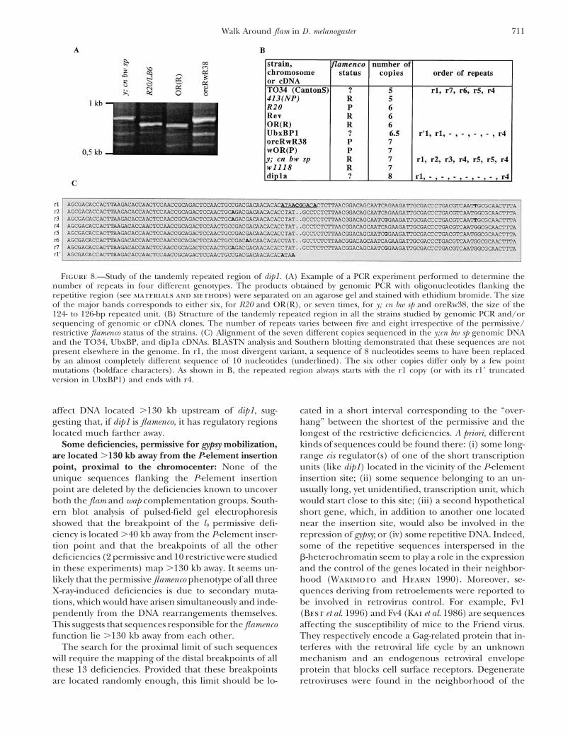
Figure 8.—Study of the tandemly repeated region of dip1. (A) Example of a PCR experiment performed to determine thenumber of repeats in four different genotypes. The products obtained by genomic PCR with oligonucleotides flanking therepetitive region (see materials and methods) were separated on an agarose gel and stained with ethidium bromide. The sizeof the major bands corresponds to either six, for R20 and OR(R), or seven times, for y; cn bw sp and oreRw38, the size of the124- to 126-bp repeated unit. (B) Structure of the tandemly repeated region in all the strains studied by genomic PCR and/orsequencing of genomic or cDNA clones. The number of repeats varies between five and eight irrespective of the permissive/restrictive flamenco status of the strains. (C) Alignment of the seven different copies sequenced in the y;cn bw sp genomic DNAand the TO34, UbxBP, and dip1a cDNAs. BLASTN analysis and Southern blotting demonstrated that these sequences are notpresent elsewhere in the genome. In r1, the most divergent variant, a sequence of 8 nucleotides seems to have been replacedby an almost completely different sequence of 10 nucleotides (underlined). The six other copies differ only by a few pointmutations (boldface characters). As shown in B, the repeated region always starts with the r1 copy (or with its r19 truncatedversion in UbxBP1) and ends with r4.
affect DNA located .130 kb upstream of dip1, sug- cated in a short interval corresponding to the “over-hang” between the shortest of the permissive and thegesting that, if dip1 is flamenco, it has regulatory regions
located much farther away. longest of the restrictive deficiencies. A priori, differentkinds of sequences could be found there: (i) some long-Some deficiencies, permissive for gypsy mobilization,
are located .130 kb away from the P-element insertion range cis regulator(s) of one of the short transcriptionunits (like dip1) located in the vicinity of the P-elementpoint, proximal to the chromocenter: None of the
unique sequences flanking the P-element insertion insertion site; (ii) some sequence belonging to an un-usually long, yet unidentified, transcription unit, whichpoint are deleted by the deficiencies known to uncover
both the flam and wap complementation groups. South- would start close to this site; (iii) a second hypotheticalshort gene, which, in addition to another one locatedern blot analysis of pulsed-field gel electrophoresis
showed that the breakpoint of the l9 permissive defi- near the insertion site, would also be involved in therepression of gypsy; or (iv) some repetitive DNA. Indeed,ciency is located .40 kb away from the P-element inser-
tion point and that the breakpoints of all the other some of the repetitive sequences interspersed in theb-heterochromatin seem to play a role in the expressiondeficiencies (2 permissive and 10 restrictive were studied
in these experiments) map .130 kb away. It seems un- and the control of the genes located in their neighbor-hood (Wakimoto and Hearn 1990). Moreover, se-likely that the permissive flamenco phenotype of all three
X-ray-induced deficiencies is due to secondary muta- quences deriving from retroelements were reported tobe involved in retrovirus control. For example, Fv1tions, which would have arisen simultaneously and inde-
pendently from the DNA rearrangements themselves. (Best et al. 1996) and Fv4 (Kai et al. 1986) are sequencesaffecting the susceptibility of mice to the Friend virus.This suggests that sequences responsible for the flamenco
function lie .130 kb away from each other. They respectively encode a Gag-related protein that in-terferes with the retroviral life cycle by an unknownThe search for the proximal limit of such sequences
will require the mapping of the distal breakpoints of all mechanism and an endogenous retroviral envelopeprotein that blocks cell surface receptors. Degeneratethese 13 deficiencies. Provided that these breakpoints
are located randomly enough, this limit should be lo- retroviruses were found in the neighborhood of the
712 V. Robert et al.
tion of sex-linked female-sterile mutants in Drosophila melanogaster.P-element insertion point. For instance, a 1.4-kb PstIGenetics 81: 683–704.
fragment located 20 kb proximal to this site was found Gatignol, A., A. Buckler-White, B. Berkhout and K. T. Jeang,1991 Characterization of a human TAR RNA-binding proteinto hybridize to the full-length gypsy probe (data notthat activates the HIV-1 LTR. Science 251: 1597–1600.shown). It might also be informative to investigate the
Gibson, T. J., and J. D. Thompson, 1994 Detection of dsRNA-bind-possible implication of such sequences in the regulation ing domains in RNA helicase A and Drosophila maleless: implica-
We thank Genevieve Payen-Groschene for her excellent technical Heitz, E., 1934 Uber a- and b-heterochromatin sowie konstanz undassistance and Leonie Ringrose for reading the manuscript. V.R. was bau der chromomeren bei Drosophila. Biologisches Zentralblattthe recipient of fellowships from the MESR and La Ligue Nationale 54: 588–609.
Hovanessian, A. G., 1989 The double stranded RNA-activated pro-Contre le Cancer. This work was supported by grants from the Associa-tein kinase induced by interferon: dsRNA-PK. J. Interferon Res.tion pour la Recherche sur le Cancer (ARC) and the FRM.9: 641–647.
Kai, K., H. Sato and T. Odaka, 1986 Relationship between thecellular resistance to Friend murine leukemia virus infection andthe expression of murine leukemia virus-gp70-related glycopro-LITERATURE CITEDtein on cell surface of BALB/c-Fv-4wr mice. Virology 150: 509–512.Adams, M. D., S. E. Celniker, R. A. Holt, C. A. Evans, J. D. Gocayne
et al., 2000 The genome sequence of Drosophila melanogaster. Kim, A., C. Terzian, P. Santamaria, A. Pelisson, N. Prud’hommeet al., 1994 Retroviruses in invertebrates: the gypsy retrotranspo-Science 287: 2185–2195.
Altschul, S. F., W. Gish, W. Miller, E. W. Myers and D. J. Lipman, son is apparently an infectious retrovirus of Drosophila melanogas-ter. Proc. Natl. Acad. Sci. USA 91: 1285–1289.1990 Basic local alignment search tool. J. Mol. Biol. 215: 403–
410. Lee, K. J., M. Mukhopadhyay, P. Pelka, A. R. Campos and H.Steller, 1999 Autoregulation of the Drosophila disconnectedAndrews, J., M. Smith, J. Merakovsky, M. Coulson, F. Hannan et
al., 1996 The stoned locus of Drosophila melanogaster produces gene in the developing visual system. Dev. Biol. 214: 385–398.Lindsley, D. L., and G. G. Zimm, 1992 The Genome of Drosophilaa dicistronic transcript and encodes two distinct polypeptides.
Genetics 143: 1699–1711. melanogaster. Academic Press, London.Marra, M. A., T. A. Kucaba, N. L. Dietrich, E. D. Green, B.Benkirane, M., C. Neuveut, R. F. Chun, S. M. Smith, C. E. Samuel
et al., 1997 Oncogenic potential of TAR RNA binding protein Brownstein et al., 1997 High throughput fingerprint analysisof large-insert clones. Genome Res. 7: 1072–1084.TRBP and its regulatory interaction with RNA-dependent protein
kinase PKR. EMBO J. 16: 611–624. Mevel-Ninio, M., M. C. Mariol and M. Gans, 1989 Mobilizationof the gypsy and copia retrotransposons in Drosophila melanogasterBest, S., P. Le Tissier, G. Towers and J. P. Stoye, 1996 Positional
cloning of the mouse retrovirus restriction gene Fv1. Nature 382: induces reversion of the ovoD dominant female-sterile mutations:molecular analysis of revertant alleles. EMBO J. 8: 1549–1558.826–829.
Birren, B., and E. Lai, 1993 Pulse-Field Gel Electrophoresis. A Practical Miklos, G. L., and J. N. Cotsell, 1990 Chromosome structure atinterfaces between major chromatin types: alpha- and beta-het-Guide. Academic Press, London.
Bridges, C. B., 1938 A revised map of the salivary gland X-chromo- erochromatin. Bioessays 12: 1–6.Miklos, G. L., M. T. Yamamoto, J. Davies and V. Pirrotta, 1988some of Drosophila melanogaster. J. Hered. 29: 11–13.
Bucheton, A., 1995 The relationship between the flamenco gene Microcloning reveals a high frequency of repetitive sequencescharacteristic of chromosome 4 and the beta-heterochromatin ofand gypsy in Drosophila: how to tame a retrovirus. Trends Genet.
11: 349–353. Drosophila melanogaster. Proc. Natl. Acad. Sci. USA 85: 2051–2055.Mitchelson, A., M. Simonelig, C. Williams and K. O’Hare, 1993Busseau, I., and A. Bucheton, 1999 A Drosophila enhancer detec-
tor transposon marked with the yellow gene. Dros. Inf. Serv. 82: Homology with Saccharomyces cerevisiae RNA14 suggests that phe-notypic suppression in Drosophila melanogaster by suppressor of forked94–95.
Chalvet, F., L. Teysset, C. Terzian, N. Prud’homme, P. Santama- occurs at the level of RNA stability. Genes Dev. 7: 241–249.Myers, E. W., G. G. Sutton, A. L. Delcher, I. M. Dew, D. P. Fasuloria et al., 1999 Proviral amplification of the gypsy endogenous
retrovirus of Drosophila melanogaster involves env-independent in- et al., 2000 A whole-genome assembly of Drosophila. Science287: 2196–2204.vasion of the female germline. EMBO J. 18: 2659–2669.
Chong, K. L., L. Feng, K. Schappert, E. Meurs, T. F. Donahue et Park, H., M. V. Davies, J. O. Langland, H. W. Chang, Y. S. Namet al., 1994 TAR RNA-binding protein is an inhibitor of theal., 1992 Human p68 kinase exhibits growth suppression in
yeast and homology to the translational regulator GCN2. EMBO interferon-induced protein kinase PKR. Proc. Natl. Acad. Sci.USA 91: 4713–4717.J. 11: 1553–1562.
Clemens, M. J., 1996 Protein kinases that phosphorylate eIF-2a and Pelisson, A., S. U. Song, N. Prud’homme, P. A. Smith, A. Buchetonet al., 1994 Gypsy transposition correlates with the productioneIF-2b and their role in eukaryotic cell translational control, pp.
139–172 in Translational Control, edited by M. J. Clemens. Cold of a retroviral envelope-like protein under the tissue-specific con-trol of the Drosophila flamenco gene. EMBO J. 13: 4401–4411.Spring Harbor Laboratory Press, Cold Spring Harbor, NY.
Costa, M., E. T. Wilson and E. Wieschaus, 1994 A putative cell Pelisson, A., L. Teysset, F. Chalvet, A. Kim, N. Prud’homme et al.,1997 About the origin of retroviruses and the co-evolution ofsignal encoded by the folded gastrulation gene coordinates cell
shape changes during Drosophila gastrulation. Cell 76: 1075– the gypsy retrovirus with the Drosophila flamenco host gene. Genet-ica 100: 29–37.1089.
Desset, S., C. Conte, P. Dimitri, V. Calco, B. Dastugue et al., 1999 Perrimon, N., D. Smouse and G. L. Miklos, 1989 Developmentalgenetics of loci at the base of the X chromosome of DrosophilaMobilization of two retroelements, ZAM and Idefix, in a novel
unstable line of Drosophila melanogaster. Mol. Biol. Evol. 16: 54–66. melanogaster. Genetics 121: 313–331.Pimpinelli, S., M. Berloco, L. Fanti, P. Dimitri, S. Bonaccorsi etDevlin, R. H., B. Bingham and B. T. Wakimoto, 1990 The organiza-
tion and expression of the light gene, a heterochromatic gene al., 1995 Transposable elements are stable structural compo-nents of Drosophila melanogaster heterochromatin. Proc. Natl.of Drosophila melanogaster. Genetics 125: 129–140.
Donzeau, M., E. L. Winnacker and M. Meisterernst, 1997 Spe- Acad. Sci. USA 92: 3804–3808.Prud’homme, N., M. Gans, M. Masson, C. Terzian and A. Bucheton,cific repression of Tax trans-activation by TAR RNA-binding pro-
tein TRBP. J. Virol. 71: 2628–2635. 1995 Flamenco, a gene controlling the gypsy retrovirus of Drosoph-ila melanogaster. Genetics 139: 697–711.Dubois, M. F., J. Galabru, P. Lebon, B. Safer and A. G. Hovanessian,
1989 Reduced activity of the interferon-induced double- Rackwitz, H. R., G. Zehetner, A. M. Frischauf and H. Lehrach,1984 Rapid restriction mapping of DNA cloned in lambdastranded RNA-dependent protein kinase during a heat shock
stress. J. Biol. Chem. 264: 12165–12171. phage vectors. Gene 30: 195–200.Robertson, H. M., C. R. Preston, R. W. Phillis, D. M. Johnson-Gans, M., C. Audit and M. Masson, 1975 Isolation and characteriza-
713Walk Around flam in D. melanogaster
Schlitz, W. K. Benz et al., 1988 A stable genomic source of heterochromatin boundary of the Drosophila melanogaster X chro-mosome. Genet. Res. 68: 191–202.P element transposase in Drosophila melanogaster. Genetics 118:
461–470. Vaury, C., A. Bucheton and A. Pelisson, 1989 The beta-hetero-chromatic sequences flanking the I elements are themselves de-Song, S. U., T. Gerasimova, M. Kurkulos, J. D. Boeke and V. G.
Corces, 1994 An env-like protein encoded by a Drosophila re- fective transposable elements. Chromosoma 98: 215–224.Wakimoto, B. T., and M. G. Hearn, 1990 The effects of chromo-troelement: evidence that gypsy is an infectious retrovirus. Genes
Dev. 8: 2046–2057. some rearrangements on the expression of heterochromaticgenes in chromosome 2L of Drosophila melanogaster. Genetics 125:Spradling, A. C., 1986 P element-mediated transformation, pp.
175–197 in Drosophila: A Practical Approach, edited by D. B. Rob- 141–154.erts. IRL Press, Oxford. Wassarman, D. A., N. M. Solomon and G. M. Rubin, 1994 The
St. Johnston, D., N. H. Brown, J. G. Gall and M. Jantsch, 1992 Drosophila melanogaster ribosomal S6 kinase II-encoding sequence.A conserved double-stranded RNA-binding domain. Proc. Natl. Gene 144: 309–310.Acad. Sci. USA 89: 10979–10983. Yamamoto, M. T., A. Mitchelson, M. Tudor, K. O’Hare, J. A. Davies
Teysset, L., J. C. Burns, H. Shike, B. L. Sullivan, A. Bucheton et al., 1990 Molecular and cytogenetic analysis of the hetero-et al., 1998 A Moloney murine leukemia virus-based retroviral chromatin-euchromatin junction region of the Drosophila melano-vector pseudotyped by the insect retroviral gypsy envelope can gaster X chromosome using cloned DNA sequences. Genetics 125:infect Drosophila cells. J. Virol. 72: 853–856. 821–832.
Tudor, M., A. Mitchelson and K. O’Hare, 1996 A 1.5 kb repeatsequence flanks the suppressor of forked gene at the euchromatin- Communicating editor: M. J. Simmons