antioxidants Article Chemical Profiling of Polyphenolics in Eucalyptus globulus and Evaluation of Its Hepato–Renal Protective Potential Against Cyclophosphamide Induced Toxicity in Mice Mosad A. Ghareeb 1, * , Mansour Sobeh 2,3 , Walaa H. El-Maadawy 4 , Hala Sh. Mohammed 5 , Heba Khalil 6 , Sanaa Botros 4 and Michael Wink 2, * 1 Medicinal Chemistry Department, Theodor Bilharz Research Institute, Kornaish El Nile, Warrak El-Hadar, Imbaba (P.O. 30), Giza 12411, Egypt 2 Institute of Pharmacy and Molecular Biotechnology, Heidelberg University, 44883-2462 Heidelberg, Germany; [email protected]3 AgroBioSciences Research Division, Mohammed VI Polytechnic University, Lot 660–Hay MoulayRachid, 43150 Ben-Guerir, Morocco 4 Pharmacology Department, Theodor Bilharz Research Institute, Kornaish El Nile, Warrak El-Hadar, Imbaba (P.O. 30), Giza 12411, Egypt; [email protected] (W.H.E.-M.); [email protected] (S.B.) 5 Department of Pharmacognosy, Faculty of Pharmacy (Girls), Al-Azhar University, Cairo 11311, Egypt; [email protected]6 Pathology Department, Theodor Bilharz Research Institute, Kornaish El Nile, Warrak El-Hadar, Imbaba (P.O. 30), Giza 12411, Egypt; [email protected]* Correspondence: [email protected] (M.A.G.); [email protected] (M.W.); Tel.: +20-2-010-1234-6834 (M.A.G.); +49-(0)6221-54-4880 (M.W.) Received: 3 July 2019; Accepted: 19 August 2019; Published: 19 September 2019 Abstract: Cyclophosphamide (CP) is a potent anti-neoplastic and immunosuppressive agent; however, it causes multi-organ toxicity. We elucidated the protective activities of Eucalyptus globulus (EG) leaf extract against CP-induced hepato–renal toxicity. Mice were treated with EG for 15 days plus CP on day 12 and 13 of the experiment. Using HPLC-DAD-ESI-MS/MS, 26 secondary metabolites were identified in EG leaf extract. Out of them, 4 polyphenolic compounds were isolated: (1) 4-(O-β-d-xylopyranosyloxy)-3,5-di-hydroxy-benzoic acid, (2) 4-(O-α-l- rhamnopyranosyloxy)-3,5-di-hydroxy-benzoic acid, (3) gallic acid, and (4) methyl gallate. Effects of EG extract on biochemical parameters, gene expression, and immune-histopathological changes were assessed in comparison to mesna positive control. Results showed that EG improved CP-increased serum ALT, AST, creatinine, and blood urea nitrogen levels. The hepatic and renal tissue levels of MDA, nitric oxide, protein carbonyl, TNF-α, IL-6, and immunohistochemical expression of nuclear factor kappa-B (NF-kB) and caspase-3 were reduced. Also, hepatic and renal GSH contents, and nuclear factor E2-related factor 2 (NRf2)/ hemoxygenase-1 (HO-1) signaling levels were increased. Histopathological findings supported our findings where hepatic and renal architecture were almost restored. Results revealed the protective effects of EG against CP-induced hepato–renal toxicity. These effects may be related to EG antioxidant, anti-inflammatory, and anti-apoptotic properties coupled with activation of Nrf2/HO-1 signaling. Keywords: Eucalyptus globulus; polyphenolics; cyclophosphamide; hepatotoxicity; nephrotoxicity; antioxidant; anti-inflammatory; Nrf2/HO-1 signaling Antioxidants 2019, 8, 415; doi:10.3390/antiox8090415 www.mdpi.com/journal/antioxidants
Transcript
antioxidants
Article
Chemical Profiling of Polyphenolics inEucalyptus globulus and Evaluation of ItsHepato–Renal Protective Potential AgainstCyclophosphamide Induced Toxicity in Mice
Mosad A. Ghareeb 1,* , Mansour Sobeh 2,3 , Walaa H. El-Maadawy 4, Hala Sh. Mohammed 5,Heba Khalil 6, Sanaa Botros 4 and Michael Wink 2,*
1 Medicinal Chemistry Department, Theodor Bilharz Research Institute, Kornaish El Nile, Warrak El-Hadar,Imbaba (P.O. 30), Giza 12411, Egypt
2 Institute of Pharmacy and Molecular Biotechnology, Heidelberg University, 44883-2462 Heidelberg,Germany; [email protected]
3 AgroBioSciences Research Division, Mohammed VI Polytechnic University, Lot 660–Hay MoulayRachid,43150 Ben-Guerir, Morocco
4 Pharmacology Department, Theodor Bilharz Research Institute, Kornaish El Nile, Warrak El-Hadar,Imbaba (P.O. 30), Giza 12411, Egypt; [email protected] (W.H.E.-M.); [email protected] (S.B.)
5 Department of Pharmacognosy, Faculty of Pharmacy (Girls), Al-Azhar University, Cairo 11311, Egypt;[email protected]
6 Pathology Department, Theodor Bilharz Research Institute, Kornaish El Nile, Warrak El-Hadar,Imbaba (P.O. 30), Giza 12411, Egypt; [email protected]
Received: 3 July 2019; Accepted: 19 August 2019; Published: 19 September 2019�����������������
Abstract: Cyclophosphamide (CP) is a potent anti-neoplastic and immunosuppressive agent;however, it causes multi-organ toxicity. We elucidated the protective activities of Eucalyptusglobulus (EG) leaf extract against CP-induced hepato–renal toxicity. Mice were treated withEG for 15 days plus CP on day 12 and 13 of the experiment. Using HPLC-DAD-ESI-MS/MS,26 secondary metabolites were identified in EG leaf extract. Out of them, 4 polyphenoliccompounds were isolated: (1) 4-(O-β-d-xylopyranosyloxy)-3,5-di-hydroxy-benzoic acid, (2) 4-(O-α-l-rhamnopyranosyloxy)-3,5-di-hydroxy-benzoic acid, (3) gallic acid, and (4) methyl gallate. Effects ofEG extract on biochemical parameters, gene expression, and immune-histopathological changes wereassessed in comparison to mesna positive control. Results showed that EG improved CP-increasedserum ALT, AST, creatinine, and blood urea nitrogen levels. The hepatic and renal tissue levels ofMDA, nitric oxide, protein carbonyl, TNF-α, IL-6, and immunohistochemical expression of nuclearfactor kappa-B (NF-kB) and caspase-3 were reduced. Also, hepatic and renal GSH contents, andnuclear factor E2-related factor 2 (NRf2)/ hemoxygenase-1 (HO-1) signaling levels were increased.Histopathological findings supported our findings where hepatic and renal architecture were almostrestored. Results revealed the protective effects of EG against CP-induced hepato–renal toxicity. Theseeffects may be related to EG antioxidant, anti-inflammatory, and anti-apoptotic properties coupledwith activation of Nrf2/HO-1 signaling.
Cyclophosphamide (CP) is prescribed in treating different types of neoplasms as well as animmunosuppressive drug in autoimmune diseases and organ transplantation [1,2]. Despite itschemotherapeutic efficacy and cost effectiveness, CP has a narrow therapeutic index and may beresponsible for severe toxicity of vital organs [3,4]. Toxicity to liver and kidney are considered the twomajor ones, being the key organs responsible for CP metabolism and excretion, respectively [5,6]. CP isa prodrug that is metabolized within hepatocytes to generate two reactive metabolites, phosphoramidemustard and acrolein [7]. Phosphoramide mustard possesses antineoplastic activity, while acrolein isreported to be responsible for the CP-induced cytotoxic effects [8,9]. Acrolein initiates oxidative stress viathe production of reactive oxygen species (ROS) causing the depletion of cellular defense mechanisms,induction of lipid peroxidation [10], nucleic acid damage and mutation [11]. The overproduction of ROSalso induces several signaling molecules including nuclear factor kappa-B (NF-kB), which regulatesthe activation of different pro-inflammatory cytokines including interleukin (IL)-6, IL-1β and tumornecrosis factor (TNF)-α [12]. Moreover, CP is reported to down- regulate the stress sensor transcriptionfactor nuclear factor erythroid 2-related factor 2 (Nrf2); one of the main defensive mechanisms againststress induced injuries [13,14].
CP is commonly administered with 2-mercaptoethane sulphonic acid (mesna); an adjuvantchemotherapeutic treatment regimen to counteract the prevalence of hemorrhagic cystitis and hematuriacaused by the toxic metabolite acrolein [15]. However, the recurrence of hemorrhagic cystitis wasreported with mesna [16] along with ineffectiveness in prevention of other reported CP-inducedadverse drug reactions [15].
Because no adjuvant regimen is reported to offer protection to the healthy organs and tissues againstCP toxicities without compromising its chemotherapeutic efficacy, enhancement of the antioxidantdefense system to avert and/or reduce the adverse effects of CP and its reactive metabolites has beensuggested [17]. Plant derived antioxidants preparations showed promising synergistic efficiencywhen co-administered with chemotherapeutic drugs mainly through their free radical scavenging andanti-inflammatory activities [18,19]. Natural antioxidants can enhance the current chemotherapeuticstrategies not only by guarding against the adverse effects of chemotherapy but stimulating the hostimmune status as well [20].
Eucalyptus globulus Labill. (Myrtaceae) (EG) is an evergreen tree that is cultivated worldwide [21].The leaf extract of EG exerts antimicrobial, antibacterial, anti-inflammatory, antioxidant, antihelmintic,and antiviral activities [22]. Moreover, previous studies reported that Eucalyptus extracts exhibitedpotent cytotoxic effects in various cell lines [23,24] and promising antitumor activity against Ehrlichascites carcinoma in mice [25,26]. These properties could be related to the abundance of phenoliccompounds in the extract such as caffeic acids, quinic, luteolin, dihydroxyphenylacetic, and hydrolysabletannins [27,28].
To the best of our knowledge, no previous study has investigated the hepatic and renal protectiveroles of EG against CP-intoxication. This study aims to identify and characterize the chemical profile ofE. globulus (EG) and also investigate its protective role against CP-induced hepatic and renal toxicitiesin comparison to the adjuvant drug "mesna" by investigating the related mechanisms of action.
2. Material and Methods
2.1. Drugs, Reagents and Instrumentations
CP (Endoxan®) and mesna (Urometixan®) were purchased from Baxter Oncology GMBH, Halle,Germany. Dimethyl sulfoxide (DMSO) and phosphate buffered saline solution (PBS) were purchasedfrom Sigma-Aldrich Chemical Co., MO, USA and Lonza Bio-products, Verviers, Belgium, respectively.All other solvents and reagents were of the highest grade commercially available. The 1H and 13C-NMRexperiments were carried out using a BRUKER 400 MHz NMR spectrometer and samples weredissolved in deuterated DMSO-d6.
Antioxidants 2019, 8, 415 3 of 19
2.2. Preparation, Extraction and Fractionation of EG Leaf Extract as well as Chromatographic Isolation
Fresh leaves of EG were collected from Giza Governorate in June 2016. The identification andauthentication of the collected plant was established by Dr. Tearse Labib, Botany Specialist, Departmentof Flora and Taxonomy, El-Orman Botanical Garden, Giza, Egypt.
Air dried leaves (1.5 kg) were extracted three times with methanol (4 L) at room temperature(25 ± 2 ◦C), the extract was concentrated via a rotatory evaporator to afford 220 g methanol extract.It was then defatted with 1.5 L petroleum ether (60–80 ◦C) to afford petroleum ether extract (25 g)and defatted methanol extract DME (175 g). DME (40 g) was subjected to polyamide (S6) columnchromatography (CC) using polyamide column eluted with H2O/EtOH mixtures up to pure EtOH. Byusing PC, UV light and spray reagents, similar fractions were collected together to obtain three mainfractions. Fraction (I) was purified via Sephadex LH-20/ EtOH & BIW (ethanol & butanol: isopropanol:water) to obtained compounds 1 and 2. Fraction II was subjected to successive CC on Sephadex LH-20/
EtOH & BIW to obtained compound 3. Fraction III was subjected to Sephadex LH-20 column andeluted via EtOH & BIW to yield compound 4.
2.3. HPLC-DAD-ESI-MS/MS Conditions
HPLC-DAD-ESI-MS/MS was employed to investigate the chemical constituents of the extract. TheLC system was Thermo Finnigan (Thermo electron Corporation, OK, USA), coupled with an LCQ Duoion trap mass spectrometer with an ESI source (ThermoQuest). A Silica gel C18 reversed-phase column(Zorbax Eclipse XDB-C18, Rapid resolution, 4.6 × 150 mm, 3.5 µm (Agilent, CA, USA) was used for theseparation process. Water with a gradient increase from 5% to 50% of acetonitrile (ACN) (with 1%formic acid each in the positive mode) was applied in 60 min, with a flow rate 1 mL/min, and thenincreased to 90% ACN in the next 30 min. The samples were injected automatically using auto samplersurveyor ThermoQuest. The instrument was controlled by Xcalibur software (Thermo Fisher ScientificInc., OK, USA). The MS operating conditions were applied in the negative ion mode, as previouslydescribed by us [29]. The ions were detected in a full scan mode and mass range of 50–2000 m/z.
2.4. Animals
Adult male Swiss albino mice weighing between 25 ± 5 g (purchased from the SchistosomeBiology Supply Center, Theodor Bilharz Research Institute, Giza, Egypt) were used. Mice were kept inthe animal house facility of the institute in standard polypropylene cages at 25 ± 2 ◦C temperature with50–60% relative humidity, 12 h light–dark cycles with free access to ad libitum food and water. Thestudy protocol was approved by the Research Ethics Committee of Theodor Bilharz Research Institute(PT: 19/2/467). All procedures were conducted in accordance with the guidelines of the NationalInstitutes of Health (NIH, 1996) and its amendments for the care and use of laboratory animals.
2.5. Experimental Design
Mice were randomly allocated into five groups each of six animals. Group 1 (control): Receivedvehicle for EG (0.5% DMSO in PBS solution) for 15 consecutive days; Group 2 (CP): Received CP in PBS(200 mg/kg, [30] on day 12 and 13 of the experiment, Groups 3 (CP + mesna): Received the positivecontrol mesna (40 mg/kg, [31] 1 h before and 4 h after each CP application, Groups 4 and 5 (CP + EG):Received EG in doses of 50 and 100 mg/kg [25,26], respectively once daily for 15 days along with CP(on day 12 and 13). All treatments were administered intraperitoneally (i.p.).
After the end of treatments mice were killed under anesthesia. Blood, kidney, and liver sampleswere collected. Sera were separated by centrifugation and stored at −80 ◦C until analysis. The liversand kidneys were washed in cold PBS; sections of livers and kidneys were fixed in 10% neutral bufferedformalin for histopathological and immunohistochemical examinations. The remaining tissue sampleswere homogenized (10% w/v) in cold PBS for biochemical analyses.
Antioxidants 2019, 8, 415 4 of 19
2.6. Determination of Liver and Kidney Toxicity Indices
Serum levels of alanine aminotransferase (ALT), aspartate aminotransferase (AST), creatinine,and blood urea nitrogen (BUN) were spectrophotometrically determined using commercially availablekits (Biodiagnostics, Cairo, Egypt).
2.7. Determination of Oxidative/Nitrosative Stress Markers and Protein Carbonyl in Liver and Kidney Tissues
The supernatants of homogenized liver and kidney samples were used for determination ofreduced glutathione (GSH) and lipid peroxidation (MDA) levels according to Ellman, 1957 [32] andOhkawa et al., 1979 [33], respectively. The protein carbonyl (PC) (OxiSelect Cell Biolabs, CA, USA) andnitric oxide (NO) levels (Biodiagnostics, Cairo, Egypt) were measured using commercially available kitsand the amount of total protein was determined by BCA protein assay kit (Thermo Fischer Scientific,IL, USA).
2.8. Determination of Nrf2/HO-1 Pathway Activation in Liver and Kidney Tissues
Gene expression levels of Nrf2 were determined using quantitative reverse transcriptase real timepolymerase chain reaction (qRT-PCR). Briefly, total RNA was isolated from hepatic and kidney samplesusing TRIzol (Invitrogen, CA, USA), and quantified using a nanodrop. RNA samples were used forDNA synthesis using cDNA Synthesis Kit. Amplification of the cDNA was carried out by SYBR Greenmaster mix (Applied biosystems, CA, USA). All values were normalized to the housekeeping β-actingene. Relative expression of studied genes was calculated using the comparative threshold cyclemethod (2−∆∆Ct method).
The sequences of PCR primer pairs used were:Nrf2 F: TTGTAGATGACCATGAGTCGC
R: CGCAGCTCAGTAACAGTCCGNext, the hepatic and renal levels of heme oxygenase-1 (HO-1) were measured using the commercial
ELISA kit according to the manufacturer’s instructions (OxiSelect Cell Biolabs, CA, USA).
2.9. Determination of Pro-inflammatory Markers and Caspase-3 in Liver and Kidney Tissues
The liver and kidney pro-inflammatory markers, TNF-α and IL-6, were measured using thecommercially available ELISA kits according to the manufacturer’s instructions (OxiSelect Cell Biolabs,CA, USA) and the amount of total protein was determined by a BCA protein assay kit.
Moreover, sections on charged slides (Superfrost charged slides, Thermo Scientific, Braunschweig,Germany) from liver and kidney tissues were immunohistochemically stained with anti-NF-κB andanti-caspase-3 (Santa Cruz Biotechnology, CA, USA, respectively) using an Ultra Benchmark machine(Roche, Tucson, USA) and an Optiview Detection kit with haematoxlin as the counterstain. The percentof positively stained brown nuclei and cytoplasm for NF-κB and caspase-3, respectively, were examinedin five microscopic fields (at x400 under Zeiss light microscopy, Jena, Germany).
2.10. Histopathological Examination
Liver and kidney tissues embedded in paraffin blocks were sectioned at 4 µm thickness. Sectionswere stained with hematoxylin/eosin (H&E) and blindly examined for the extent of liver and kidneydamage under bright field microscope (Olympus BX53F).
2.11. Statistical Analysis
Data are expressed as mean ± SEM. Statistical analysis was performed using one-way analysis ofvariance (ANOVA) followed by Tukey–Kramer as post hoc test for multiple comparisons (GraphPadSoftware, San Diego, CA, USA, version 5.03). p < 0.05 was considered statistically significant.
Antioxidants 2019, 8, 415 5 of 19
3. Results
3.1. HPLC-DAD-ESI-MS-MS Annotation and Chromatographic Isolation of Polyphenolic Compounds
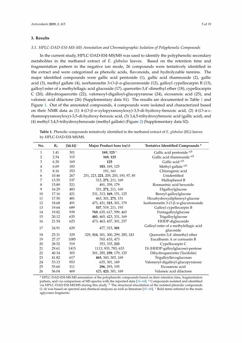
In the current study, HPLC-DAD-ESI-MS/MS was used to identify the polyphenolic secondarymetabolites in the methanol extract of E. globulus leaves. Based on the retention time andfragmentation pattern in the negative ion mode, 26 compounds were tentatively identified inthe extract and were categorized as phenolic acids, flavonoids, and hydrolyzable tannins. Themajor identified compounds were gallic acid pentoside (1), gallic acid rhamnoside (2), gallicacid (3), methyl gallate (4), isorhamnetin 3-O-β-d-glucuronoside (12), galloyl cypellocarpin B (13),galloyl ester of a methylellagic acid glucoside (17), quercetin-3,4’-dimethyl ether (18), cypellocarpinC (20), dihydroquercetin (22), valoneoyl-digalloyl-glucopyranose (24), eicosanoic acid (25), andvaloneic acid dilactone (26) (Supplementary data S1). The results are documented in Table 1 andFigure 1. Out of the annotated compounds, 4 compounds were isolated and characterized basedon their NMR data as (1) 4-(O-β-d-xylopyranosyloxy)-3,5-di-hydroxy-benzoic acid, (2) 4-(O-α-l-rhamnopyranosyloxy)-3,5-di-hydroxy-benzoic acid, (3) 3,4,5-trihydroxybenzoic acid (gallic acid), and(4) methyl 3,4,5-trihydroxybenzoate (methyl gallate) (Figure 2) (Supplementary data S2).
Table 1. Phenolic compounds tentatively identified in the methanol extract of E. globulus (EG) leavesby HPLC-DAD-ESI-MS/MS.
* HPLC-DAD-ESI-MS-MS annotation of the polyphenolic compounds based on their retention time, fragmentationpattern, and via comparison of MS spectra with the reported data [34–44]. a Compounds isolated and identifiedvia HPLC-DAD-ESI-MS/MS during this study. b The structural elucidation of the isolated phenolic compounds(1–4) was based on spectral and chemical analyses as well as literature [45–48]. c Bold items referred to the mainaglycones fragments.
Antioxidants 2019, 8, 415 6 of 19
Antioxidants 2019, 8, x FOR PEER REVIEW 6 of 23
fragmentation pattern, and via comparison of MS spectra with the reported data [34–44]. * The structural elucidation of the isolated phenolic compounds (1–4) was based on spectral and chemical analyses as well as literature [45–48]. * Bold items referred to the main aglycones fragments.
Figure 1. Negative HPLC-DAD-ESI-MS/MS profile of phenolic compounds from the methanol extract of E. globulus leaves. Peak numbers agree with those in Table 1. Figure 1. Negative HPLC-DAD-ESI-MS/MS profile of phenolic compounds from the methanol extractof E. globulus leaves. Peak numbers agree with those in Table 1.
3.2. EG Pretreatment Alleviated CP Induced Liver and Kidney Damage
CP caused a substantial increase in serum levels of ALT, AST, creatinine, and BUN (3, 3.6, 1.9, and3.4 fold, respectively) when compared to normal controls. Mesna modestly reduced their levels whencompared to CP treated animals. Pretreatment with EG at doses of 50 and 100 mg /kg resulted in asignificant dose dependent reduction in these parameters when compared to corresponding CP ormesna treated mice. EG when given in a dose of 100 mg/kg normalized the levels of ALT, AST, andcreatinine (Table 2).
Table 2. Protective effects of EG pretreatment on the serum markers against cyclophosphamide(CP)-induced hepato–renal toxicities in mice.
Data are represented as mean ± SEM (n = 6). * p < 0.05 vs. normal control, # p < 0.05 vs. CP, † p < 0.05 vs. CP+mesna,‡ p < 0.05 vs. CP+EG (50 mg/kg). Statistical analysis was done using one-way ANOVA followed by Tukey’s multiplecomparisons test. ALT: alanine aminotransferase, AST: aspartate aminotransferase, BUN: blood urea nitrogen, CP:cyclophosphamide, EG: E. globulus.
Antioxidants 2019, 8, 415 7 of 19
Antioxidants 2019, 8, x FOR PEER REVIEW 7 of 23
Figure 2. Chemical structures of phenolic compounds isolated from Eucalyptus globulus as well as some major annotated compounds by HPLC-DAD-ESI-MS/MS.
3.2. EG Pretreatment Alleviated CP Induced Liver and Kidney Damage
CP caused a substantial increase in serum levels of ALT, AST, creatinine, and BUN (3, 3.6, 1.9, and 3.4 fold, respectively) when compared to normal controls. Mesna modestly reduced their levels when compared to CP treated animals. Pretreatment with EG at doses of 50 and 100 mg /kg resulted in a significant dose dependent reduction in these parameters when compared to corresponding CP or mesna treated mice. EG when given in a dose of 100 mg/kg normalized the levels of ALT, AST, and creatinine (Table 2).
Figure 2. Chemical structures of phenolic compounds isolated from Eucalyptus globulus as well as somemajor annotated compounds by HPLC-DAD-ESI-MS/MS.
Hepatic tissues of CP treated mice revealed hydropic degeneration of hepatocytes, hyperemia,congestion, dilatation of sinusoids. Minor to moderate improvement in hepatic damage was recordedin mesna and EG (50 mg/kg) treated mice, respectively, whereas normal hepatic architecture wasrecorded upon pretreatment with 100 mg/kg EG (Figure 3A). Renal tissues of CP treated mice showeddegenerated renal tubules with hyaline casts, atrophy in the glomeruli with mesangeal proliferation,tubular degeneration and dilatation in bowman capsules. Pretreatment with EG in doses of 50 and100 mg/kg revealed a dose dependent improvement in the histological changes of the glomeruli andrenal tubules (Figure 3B).
Antioxidants 2019, 8, 415 8 of 19Antioxidants 2019, 8, x FOR PEER REVIEW 9 of 23
(A) Liver N
orm
al C
ontr
ol
CP
CP+
Mes
na
CP+
EG 5
0 m
g/kg
C
P+EG
100
mg/
kg
(B) Kidney
Figure 3. Photomicrographs showing the protective effects of EG pretreatment on CP–induced damage in hepatic (A) and renal (B) tissue sections stained with H&E (×400).
Figure 3. Photomicrographs showing the protective effects of EG pretreatment on CP–induced damagein hepatic (A) and renal (B) tissue sections stained with H&E (×400).
Antioxidants 2019, 8, 415 9 of 19
3.3. EG Pretreatment Mitigated CP-induced Oxidative/Nitosative Stress and Protein Carbonylation in Liverand Kidney Tissues
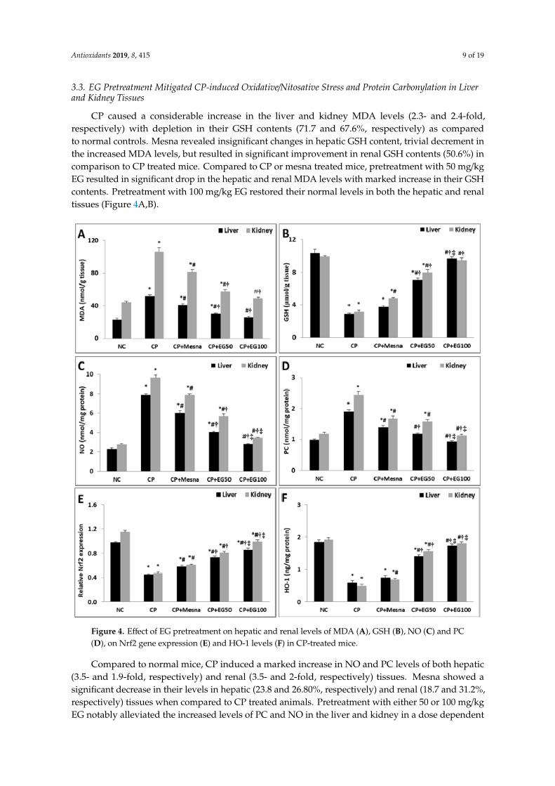
CP caused a considerable increase in the liver and kidney MDA levels (2.3- and 2.4-fold,respectively) with depletion in their GSH contents (71.7 and 67.6%, respectively) as comparedto normal controls. Mesna revealed insignificant changes in hepatic GSH content, trivial decrement inthe increased MDA levels, but resulted in significant improvement in renal GSH contents (50.6%) incomparison to CP treated mice. Compared to CP or mesna treated mice, pretreatment with 50 mg/kgEG resulted in significant drop in the hepatic and renal MDA levels with marked increase in their GSHcontents. Pretreatment with 100 mg/kg EG restored their normal levels in both the hepatic and renaltissues (Figure 4A,B).Antioxidants 2019, 8, x FOR PEER REVIEW 11 of 23
Figure 4. Effect of EG pretreatment on hepatic and renal levels of MDA (A), GSH (B), NO (C) and PC (D), on Nrf2 gene expression (E) and HO-1 levels (F) in CP-treated mice.
Data are represented as mean ± SEM (n = 6). * p < 0.05 vs. normal control, # p < 0.05 vs. CP, † p < 0.05 vs. CP + mesna, ‡ p < 0.05 vs CP + EG (50 mg/kg). Statistical analysis was done using one-way ANOVA followed by Tukey’s multiple comparisons test. MDA: malondialdehyde, GSH: reduced glutathione, NO: nitric oxide, PC: Protein carbonyl, Nrf2: nuclear factor erythroid 2-related factor 2, HO-1: heme oxygenase-1, NC: normal control, CP: cyclophosphamide, EG: E. globulus.
Figure 4. Effect of EG pretreatment on hepatic and renal levels of MDA (A), GSH (B), NO (C) and PC(D), on Nrf2 gene expression (E) and HO-1 levels (F) in CP-treated mice.
Compared to normal mice, CP induced a marked increase in NO and PC levels of both hepatic(3.5- and 1.9-fold, respectively) and renal (3.5- and 2-fold, respectively) tissues. Mesna showed asignificant decrease in their levels in hepatic (23.8 and 26.80%, respectively) and renal (18.7 and 31.2%,respectively) tissues when compared to CP treated animals. Pretreatment with either 50 or 100 mg/kgEG notably alleviated the increased levels of PC and NO in the liver and kidney in a dose dependent
Antioxidants 2019, 8, 415 10 of 19
way when compared to CP and mesna treated mice. Pretreatment with 100 mg/kg EG neutralized theincreased PC and NO levels in the liver and kidney of CP treated mice (Figure 4C,D).
Data are represented as mean ± SEM (n = 6). * p < 0.05 vs. normal control, # p < 0.05 vs. CP,† p < 0.05 vs. CP + mesna, ‡ p < 0.05 vs CP + EG (50 mg/kg). Statistical analysis was done usingone-way ANOVA followed by Tukey’s multiple comparisons test. MDA: malondialdehyde, GSH:reduced glutathione, NO: nitric oxide, PC: Protein carbonyl, Nrf2: nuclear factor erythroid 2-relatedfactor 2, HO-1: heme oxygenase-1, NC: normal control, CP: cyclophosphamide, EG: E. globulus.
3.4. EG Pretreatment Activated Nrf2/HO-1/Antioxidant Signaling in The Livers and Kidneys of CPTreated Mice
CP treated mice showed a substantial drop in hepatic and renal gene expression of Nrf2 (54 and59.1%, respectively) in comparison to normal controls. Mesna demonstrated a moderate decline inhepatic (28.9%) and renal (29.8%) expression of Nrf2 as compared to CP treated mice. Mice treated witheither 50 or 100 mg/kg EG exhibited a considerable rise in the hepatic (62.2 and 91.1%, respectively)and renal (72.3 and 110.6%, respectively) Nrf2 mRNA expression when compared with CP treatedanimals. Moreover, the enhancement in the hepatic (25.86 and 48.28%, respectively) and renal (32.79and 62.30%, respectively) Nrf2 expression was more prominent when compared to mesna treatedgroups (Figure 4E).
The cytoprotective isoenzyme HO-1 levels were significantly reduced in hepatic (68.7%) andrenal (74.5%) tissues of CP treated mice when compared to normal controls. Mesna did not reveal anysignificant changes in hepatic HO-1 levels whereas a significant rise of renal HO-1 levels (40.8%) wasrecorded in comparison with CP treated groups. Mice pretreated with EG in a dose of 50 and 100 mg/kgshowed a dose dependent increase in hepatic HO-1 levels when compared to either CP (2.4- and 3-fold)or mesna (1.9- and 2.3-fold) treated groups, respectively. Similarly, a dose dependent elevation inrenal HO-1 levels was detected in pretreated groups with EG (50 and 100 mg/kg) when compared toeither CP (3.2- and 3.7-fold) or mesna (2.3- and 2.6-fold) treated groups. Moreover, pretreatment with100 mg/kg EG normalized the reduced HO-1 levels in livers and kidneys of CP-treated mice (Figure 4F).
3.5. EG Pretreatment Down-regulated CP-induced Inflammation in Liver and Kidney Tissues
The levels of the pro-inflammatory cytokines, TNF-α and IL-6, were increased in the livers (92.1%and 129.9%) and kidneys (80.8% and 97.8%) of CP treated mice when compared to normal controls.Mesna caused a partial reduction in both hepatic and renal levels of TNF-α (23.6% and 17%) and IL-6(17.7% and 28.5%) when compared to CP treated groups. Pretreatment with EG resulted in a dosedependent reduction in their hepatic and renal levels in comparison to either CP or mesna treatedgroups. Additionally, EG pretreatment in a dose of 100 mg/kg neutralized the elevated hepatic andrenal levels of both cytokines (Figure 5A,B).
IHC expression of NF-κB revealed minimal basal levels in normal hepatic and renal tissues.However, a prominent up-regulation in its expression was detected in CP-treated liver (mainly inhepatocytes around the central vein) and kidney (identified in the renal tubules and glomeruli) tissuesby 52.2- and 60-fold, respectively, as compared to normal controls. Mesna showed a significantreduction by 16.7% and 25%, respectively, when compared to the CP-treated group. EG pretreatmentin doses of 50 and 100 mg/kg exhibited a dose dependent decline in hepatic expression of NF-κB incomparison to CP (54.8% and 83.4%, respectively) and mesna (45.7% and 80%, respectively) treatedgroups. A similar dose dependent down-regulation was observed in NF-κB renal expression incomparison to CP (55.6% and 77.8%, respectively) and mesna (40.7% and 70.4%, respectively) treatedgroups (Figure 5C,D).
Data are represented as mean ± SEM (n = 6). * p < 0.05 vs. normal control, # p < 0.05 vs. CP, † p < 0.05vs. CP + mesna, ‡ p < 0.05 vs. CP + EG (50 mg/kg). Statistical analysis was done using one-way ANOVAfollowed by Tukey’s multiple comparisons test. TNF-α: tumor necrosis-α, IL-6: interleukin-6, NF-κB:nuclear factor-kappa B, NC: normal control, CP: cyclophosphamide, EG: E. globulus.
Antioxidants 2019, 8, 415 11 of 19Antioxidants 2019, 8, x FOR PEER REVIEW 13 of 23
Live
r
Normal Control CP CP+Mesna CP+ EG 50 CP+ EG 100
Kid
ney
Figure 5. Effect of EG pretreatment on liver and kidney levels of pro-inflammatory markers (TNF-α (A), IL-6 (B)), IHC expression of NF-κB (×400, black arrows represent localization of positively stained brown nuclei with faint ignored background staining) (C) and semi-quantitative estimation of NF-κB positively stained nuclei in hepatic and renal tissues of CP-treated mice.
Figure 5. Effect of EG pretreatment on liver and kidney levels of pro-inflammatory markers (TNF-α(A), IL-6 (B)), IHC expression of NF-κB (×400, black arrows represent localization of positively stainedbrown nuclei with faint ignored background staining) (C) and semi-quantitative estimation of NF-κBpositively stained nuclei in hepatic and renal tissues of CP-treated mice.
3.6. EG Blocked CP-induced Apoptosis in the Liver and Kidney of Mice
IHC expression of caspase-3 was markedly up-regulated in CP treated hepatic (34.2-fold) and renaltissues (43.2-fold) when compared to untreated tissues. Caspase-3 positively stained cells were observedmainly in hepatocytes around the central vein and in the renal tubules and glomeruli. Administrationof mesna did not show significant changes in hepatic and renal caspase-3 expression when compared toCP-treated groups. EG pretreatment in doses of 50 and 100 mg/kg showed dose dependent reductionin hepatic expression of caspase-3 in comparison to CP (53.7% and 80.5%, respectively) and mesna
Antioxidants 2019, 8, 415 12 of 19
(48.7% and 78.4%, respectively) treated groups. Also, a dose dependent down-regulation in caspase-3renal expression was observed in comparison to CP (46.5% and 69.8%, respectively) and mesna (37.8%and 64.9%, respectively) treated groups (Figure 6A,B).Antioxidants 2019, 8, x FOR PEER REVIEW 15 of 23
Figure 6. Effect of EG pretreatment on IHC expression of caspase-3 (×400, black arrows represent localization of positively stained brown cytoplasm with ignored cross reaction in sinusoids as appeared in normal control section) (A) and semi-quantitative estimation of caspase-3 positively stained cells in hepatic and renal tissues of CP-treated mice.
Figure 6. Effect of EG pretreatment on IHC expression of caspase-3 (×400, black arrows representlocalization of positively stained brown cytoplasm with ignored cross reaction in sinusoids as appearedin normal control section) (A) and semi-quantitative estimation of caspase-3 positively stained cells inhepatic and renal tissues of CP-treated mice.
Antioxidants 2019, 8, 415 13 of 19
Data are represented as mean ± SEM (n = 6). * p < 0.05 vs. normal control, # p < 0.05 vs. CP,† p < 0.05 vs. CP + mesna, ‡ p < 0.05 vs. CP + EG (50 mg/kg). Statistical analysis was done using one-wayANOVA followed by Tukey’s multiple comparisons test. NC: normal control, CP: cyclophosphamide,EG: E. globulus.
4. Discussion
In this study, the hepato–renal protective activities of EG were examined in CP treated mice.Results were compared to the uroprotective thiol “mesna”, which is routinely prescribed as an adjuvanttreatment regimen to reduce the risk of hemorrhagic cystitis [15].
In this study, CP treated mice revealed a prominent increase in serum levels of hepatoxicitybiomarkers (ALT and AST) by approximately 3-fold, as previously described [17,49]. Hepatotoxicity isreported to be one of the major side effects of CP induced hepatic damage with increased permeability ofcell membrane and leakage of liver enzymes, specifically ALT and AST [50]. EG did not only normalizethe elevated levels of ALT and AST but also restored the normal hepatic architecture. In CP treatedmice, impairment in kidney functions was expressed as increased leakage of creatinine and urea intothe systemic circulation, glomerular degenerative changes, and atrophy in kidney tissues. The recordedsteep reduction in the levels of creatinine and BUN accompanied with amelioration of the kidneydegenerative changes in EG pretreated groups may point to a possible nephro-protective activity.
In the current study, the oxidative/antioxidative mechanisms were examined, since oxidative stressis well known to play a pivotal role in the pathogenesis of CP induced toxicity, which mainly resultsfrom its toxic metabolite acrolein [51]. Acrolein induces the generation of ROS leading to enhancedlipid peroxidation and reduction in the antioxidant defense system in liver and kidney tissues [14,52].In agreement with previous studies [17,49], our data showed an exacerbation in ROS productionas denoted by the significant increase in the final product of lipid peroxidation, MDA, in liver andkidney of CP treated mice. Moreover, a substantial depletion in hepatic and renal GSH contents (by72% and 68%, respectively) was observed. This could be attributed to the direct conjugation of CPand its metabolites (acrolein and phosphoramide mustard) to the free or protein-bound –SH groupsof GSH [53], denoting declined cellular defenses [54]. Pretreatment with EG restored the deficientthiol store in hepatic and renal cells by promoting the de novo synthesis of GSH. Previous studiesdocumented the free radical scavenging activities of EG in alloxan-induced oxidative stress [55] andacetaminophen-induced kidney damages in rats [56]. In this study HPLC-DAD-ESI-MS/MS analysis ofEG leaf extract revealed the presence of polyphenols with flavonoids (luteolin, kampferol and iridin)and tannins (gallotannins and ellagitannins) as major constituents. Flavonoids [29,57] and tannins [58]are reported to exhibit powerful antioxidant and free radical scavenging effects.
NO is known to play an essential role in regulating cellular stress [59]. NO directly result from theup-regulated expression of inducible NO synthase (iNOS) indicating impaired cellular viability [60].NO is apparently involved in CP induced toxicity [59,61], this is in coherence with our findings whereNO was significantly increased in both the hepatic and renal tissues of CP treated mice by almost3-fold. Moreover, the detected increase in NO was accompanied with elevated liver and kidney PCcontents in CP treated mice by almost 2-fold. PC content is a marker expressing the extent of proteindamage [62,63], and its enhanced levels is indicative of DNA damage and mutation resulting fromexcessive production of ROS in liver [64] and kidney [65] tissues. EG pretreatment led to normalizationof NO levels indicating hepato- and nephro-protection against CP induced oxidative stress. In addition,the reduced hepatic and renal PC contents may indicate significant recovery of DNA damage suggestingpossible assistant role for EG in tissue regeneration.
In addition, we investigated the impact of EG pretreatment on the up-regulation of theNrf2/HO-1/antioxidant signaling pathway, which is considered one of the key defense mechanismsagainst stress-associated injuries [66]. Under stress conditions, various stimuli as ROS andpro-inflammatory cytokines [67] adversely result in the down-regulation of Nrf2 expression inliver and kidney cells, thereby moderating the transcription of the cytoprotective isoenzyme heme
Antioxidants 2019, 8, 415 14 of 19
oxygenase-1 (HO-1) [14,19]. In the current work, CP resulted in a significant down-regulation of thehepatic and renal Nrf2 gene expressions along with marked reduction in their HO-1 levels, which couldbe linked to the excessive production of ROS, as previously reported [68,69]. Notably, pretreatmentwith EG demonstrated an activation of Nrf2 gene expression complemented with subsequent elevationin HO-levels in both hepatic and renal tissues implying that the recorded EG antioxidant effects can bepartly attributed to the up-regulation of the Nrf2/HO-1 antioxidant signaling pathway.
Additionally, the oxidative/nitrosative stress generated by CP is known to be associated with thesubsequent activation of inflammatory cascades [19,30]. NF-κB is a redox-sensitive transcription factorthat regulates the expression and activation of pro-inflammatory cytokines and other mediators ofinflammation including IL-6, TNF-α, and iNOS [70–72]. In the current study, CP-treatment elicited anincrease in NF-κB IHC expression with marked elevation in levels of TNF-α and IL-6 in hepatic andrenal tissues [14,60]. The increased NF-kB expression is known to trigger the expression of iNOS, whichin turn causes the overproduction of NO [73], as reported herein. The recorded decrease in the hepaticand renal levels of the pro-inflammatory cytokines (TNF-α and IL-6), NO along with subsequentdown-regulation in NF-κB expression in EG pretreated mice could be related to EG compensatorymechanism against the inflammatory milieu generated in CP treated mice. The anti-inflammatoryactivities of EG was previously demonstrated in inflammatory-mediated disorders [74].
CP induced oxidative/nitrosative stress and inflammatory responses are also known to triggerapoptotic cell death via both mitochondria-dependent and mitochondria-independent apoptoticpathways [75], leading to activation of the executioner apoptotic marker caspase-3 [76]. The IHCexpression of caspase-3 was up-regulated in both hepatic and renal tissues of CP treated mice.Pretreatment with EG extract counteracted the CP-induced apoptotic changes as evidenced by themarked regression in caspase-3 positively stained cells. This observed anti-apoptotic effect of EG couldbe directly linked to its antioxidant and anti-inflammatory properties.
In this study, mesna demonstrated minor to moderate hepatoprotective and nephroprotectiveactivities. EG showed better results in terms of dose dependent improvement in the assessedbiochemical and imunohistopathological parameters upon the use of EG, especially when the doseof 100 mg/kg was applied. These findings are in agreement with US FDA report (2009) [15] statingthe ineffectiveness of mesna in preventing or ameliorating other reported multi-organ toxicity elicitedby CP. Our findings may be explained in the view of the pharmacokinetic profile of mesna, which isknown to be distributed in the body in its biologically inactive disulfite form. It passes through thehepatic vasculature in an unchanged form [77] and undergoes reduction in the renal epithelial cells tothe pharmacologically active thiol form, mesna, which is then excreted in urine and combines withacrolein to form the stable uro-nontoxic compounds via its sulfhydryl group [78].
5. Conclusions
HPLC-MS/MS profiling of the tannins-rich leaf extract of E. globulus resulted in characterization of26 secondary metabolites including tannins, phenolic acids, and flavonoids. Moreover, our resultsrevealed that alleviation of CP-induced hepato/renal-toxicities can be related to the augmentationof antioxidant defenses at least partially through induction of Nrf2/HO-1 signaling with attenuationof excessive inflammatory responses as well as apoptosis in the hepatic and renal tissues. EG mayrepresent an effective and economic plant product that can protect against the risks of toxic CP activities.However, further investigations are required to examine the potential synergistic activity of EG on thechemotherapeutic efficacy of CP. Also, investigations are required in a clinical context to confirm itshepato- and nephro-protective activities.
Supplementary Materials: The following are available online at http://www.mdpi.com/2076-3921/8/9/415/s1.Annotation of the polyphenolic compounds using HPLC-DAD-ESI-MS/MS (S1) and structural elucidation of theisolated phenolic compounds (S2).
Author Contributions: M.A.G., performed the extraction, participated in the chemical characterization of theextract, participated in the structural elucidation of the isolated compounds, analyzed the data, wrote & revised
the paper, conceived and designed the project; M.S., participated in the chemical identification of the extractand wrote the paper; W.H.E.-M., shared in the design of work, conducted the animal study and biochemicalinvestigations, interpretation of obtained data, statistical analysis, shared in original draft preparation, graphicalabstract design as well as sharing in reviewing and editing of the submitted version of the manuscript; H.S.M.,performed the chromatographic isolation, participated in the identification of the compounds and wrote thepaper.; H.K., conducted the histopathological and immunohistochemical examinations; S.B., wrote & revised thepaper and conceived the study; M.W., revised the paper and conceived the study.
Funding: The authors received financial support from the Deutsche Forschungsgemeinschaft and Ruprecht-Karls-Universität Heidelberg within the funding program Open Access Publishing.
Acknowledgments: The authors would like to thank Dr. Tearse Labib, Botany Specialist, Department of Floraand Taxonomy, El-Orman Botanical Garden, Giza, Egypt, for her kind identification of the plant.
Conflicts of Interest: The authors declare no conflict of interest.
Abbreviations
HPLC-DAD-ESI-MS/MSHigh performance liquid chromatography coupled with diode array detection(DAD) and electrospray mass spectrometry (MS)
EG Eucalyptus globulusCP CyclophosphamideNF-κB Nuclear factor kappa-BPC Protein carbonylationNrf2 Nuclear factor E2-related factor 2HO-1 Hemoxygenase-1(IL)-6 Interleukin(TNF)-α Tumor necrosis factorROS Reactive oxygen speciesMesna (2-mercaptoethane sulphonic acid)DMSO Dimethyl sulfoxidePBS Phosphate buffered salineDME Defatted methanol extractPC Paper chromatographyUV Ultra-violetCC Column chromatographyBIW Butanol: isopropanol: wateri.p. IntraperitoneallyALT Alanine transaminaseAST Aspartate transaminaseBUN Blood urea nitrogenqRT-PCR Quantitative reverse transcriptase real time polymerase chain reactionH&E Hematoxylin/eosinMDA MalondialdehydeiNOS Inducible nitric oxide synthaseUS FDA United States Food and Drug AdministrationNO Nitric oxide
References
1. Rehman, M.U.; Tahir, M.; Ali, F.; Qamar, W.; Lateef, A.; Khan, R.; Quaiyoom, A.; Oday-O-Hamiza; Sultana, S.Cyclophosphamide-induced nephrotoxicity, genotoxicity, and damage in kidney genomic DNA of Swissalbino mice: The protective effect of ellagic acid. Mol. Cell. Biochem. 2012, 365, 119–127. [CrossRef] [PubMed]
2. Cuce, G.; Çetinkaya, S.; Koc, T.; Esen, H.H.; Limandal, C.; Balci, T.; Kalkan, S.; Akoz, M. Chemoprotectiveeffect of vitamin E in cyclophosphamide induced hepatotoxicity in rats. Chem. Biol. Interact. 2015, 232, 7–11.[CrossRef] [PubMed]
3. Basu, A.; Bhattacharjee, A.; Samanta, A.; Bhattacharya, S. Prevention of cyclophosphamide-inducedhepatotoxicity and genotoxicity: Effect of an l-cysteine based oxovanadium (IV) complex on oxidative stressand DNA damage. Environ. Toxicol. Pharm. 2015, 40, 747–757. [CrossRef] [PubMed]
4. Bhattacharjee, A.; Basu, A.; Biswas, J.; Bhattacharya, S. Nano-Se attenuates cyclophosphamide-inducedpulmonary injury through modulation of oxidative stress and DNA damage in Swiss albino mice. Mol. Cell.Biochem. 2015, 405, 243–256. [CrossRef] [PubMed]
5. Hamsa, T.P.; Kuttan, G. Protective role of Ipomoea obscura (L.) on cyclophosphamide-induced uro- andnephrotoxicities by modulating antioxidant status and pro-inflammatory cytokine levels. Inflammopharmacology2011, 19, 155–167. [CrossRef] [PubMed]
6. Jiang, W.; Liu, J.; Li, P.; Lu, Q.; Pei, X.; Sun, Y.; Wang, G.; Hao, K. Magnesium isoglycyrrhizinate showshepatoprotective effects in a cyclophosphamide-induced model of hepatic injury. Oncotarget 2017, 8,33252–33264. [CrossRef]
7. Zarei, M.; Shivanandappa, T. Amelioration of cyclophosphamide-induced hepatotoxicity by the root extractof Decalepis hamiltonii in mice. Food Chem. Toxicol. 2013, 57, 179–184. [CrossRef]
8. Olayinka, E.; Ore, A.; Ola, O.; Adeyemo, O.A. Ameliorative effect of gallic acid on cyclophosphamide-inducedoxidative injury and hepatic dysfunction in rats. Med. Sci. 2015, 3, 78–92. [CrossRef]
9. Gunes, S.; Ayhanci, A.; Sahinturk, V.; Altay, D.U.; Uyar, R. Carvacrol attenuates cyclophosphamide-inducedoxidative stress in rat kidney. Can. J. Physiol. Pharm. 2017, 95, 844–849. [CrossRef]
10. Shokrzadeh, M.; Ahmadi, A.; Naghshvar, F.; Chabra, A.; Jafarinejhad, M. Prophylactic efficacy of melatoninon cyclophosphamide-induced liver toxicity in mice. BioMed Res. Int. 2014, 2014, 470425. [CrossRef]
12. Kiuchi, H.; Takao, T.; Yamamoto, K.; Nakayama, J.; Miyagawa, Y.; Tsujimura, A.; Nonomura, N.; Okuyama,A. Sesquiterpene lactone parthenolide ameliorates bladder inflammation and bladder overactivity incyclophosphamide induced rat cystitis model by inhibiting nuclear factor-kappaB phosphorylation. J. Urol.2009, 181, 2339–2348. [CrossRef] [PubMed]
13. Lin, S.; Hao, G.; Long, M.; Lai, F.; Li, Q.; Xiong, Y.; Tian, Y.; Lai, D. Oyster (Ostrea plicatula Gmelin)polysaccharides intervention ameliorates cyclophosphamide-Induced genotoxicity and hepatotoxicity inmice via the Nrf2—ARE pathway. Biomed. Pharm. 2017, 95, 1067–1071. [CrossRef] [PubMed]
14. ALHaithloul, H.A.S.; Alotaibi, M.F.; Bin-Jumah, M.; Elgebaly, H.; Mahmoud, A.M. Olea europaea leafextract up-regulates Nrf2/ARE/HO-1 signaling and attenuates cyclophosphamide-induced oxidative stress,inflammation and apoptosis in rat kidney. Biomed. Pharm. 2019, 111, 676–685. [CrossRef] [PubMed]
15. US Food and Drug Administration (2009). Available online: https://www.accessdata.fda.gov/drugsatfda_docs/nda/2002/20-855_Mesnex_Prntlbl.pdf (accessed on 29 July 2017).
16. Yilmaz, N.; Emmungil, H.; Gucenmez, S.; Ozen, G.; Yildiz, F.; Balkarli, A.; Kimyon, G.; Coskun, B.N.;Dogan, I.; Pamuk, O.N.; et al. Incidence of cyclophosphamide-induced urotoxicity and protective effect ofMesna in rheumatic diseases. J. Rheumatol. 2015, 42, 1661–1666. [CrossRef] [PubMed]
17. Caglayan, C.; Temel, Y.; Kandemir, F.M.; Yildirim, S.; Kucukler, S. Naringin protects againstcyclophosphamide-induced hepatotoxicity and nephrotoxicity through modulation of oxidative stress,inflammation, apoptosis, autophagy, and DNA damage. Envrion. Sci. Pollut. Res. Int. 2018, 25, 20968–20984.[CrossRef] [PubMed]
18. Cerig, S.; Geyikoglu, F.; Bakir, M.; Colak, S.; Sonmez, M.; Koc, K. Hepatoprotective effect of oleuropeinagainst cisplatin-induced liver damage in rat. World Acad. Sci. Eng. Technol. 2016, 10, 260–267. [CrossRef]
19. Sherif, I.O. The effect of natural antioxidants in cyclophosphamide-induced hepatotoxicity: Role of Nrf2/HO-1pathway. Int. Immunopharmacol. 2018, 61, 29–36. [CrossRef]
20. Murali, V.P.; Kuttan, G. Enhancement of cancer chemotherapeutic efficacy of cyclophosphamide by Curculigoorchioides Gaertn and its ameliorative effects on cyclophosphamide-induced oxidative stress. Integr. CancerTher. 2015, 14, 172–183. [CrossRef]
21. White, D.A.; McGrath, J.F.; Ryan, M.G.; Battaglia, M.; Mendham, D.S.; Kinal, J.; Downes, G.M.; Crombie, D.S.;Hunt, M.E. Managing for water-use efficient wood production in Eucalyptus globulus plantations. For. Ecol.Manag. 2014, 331, 272–280. [CrossRef]
22. Nakhaee, A.; Bokaeian, M.; Saravani, M.; Farhangi, A.; Akbarzadeh, A. Attenuation of oxidative stressin streptozotocin-induced diabetic rats by Eucalyptus globulus. Indian J. Clin. Biochem. 2009, 24, 419–425.[CrossRef] [PubMed]
23. Al-Fatimi, M.; Friedrich, U.; Jenett-Siems, K. Cytotoxicity of plants used in traditional medicine in Yemen.Fitoterapia 2005, 76, 355–358. [CrossRef] [PubMed]
25. Islam, F.; Khatun, H.; Ghosh, S.; Ali, M.M.; Khanam, J.A. Bioassay of Eucalyptus extracts for anticanceractivity against Ehrlich ascites carcinoma (eac) cells in Swiss albino mice. Asian Pac. J. Trop. Biomed. 2012, 2,394–398. [CrossRef]
26. Islam, F.; Khatun, H.; Khatun, M.; Ali, S.M.; Khanam, J.A. Growth inhibition and apoptosis of Ehrlich ascitescarcinoma cells by the methanol extract of Eucalyptus camaldulensis. Pharm. Biol. 2014, 52, 281–290. [CrossRef][PubMed]
27. Boulekbache-Makhlouf, L.; Meudec, E.; Mazauric, J.P.; Madani, K.; Cheynier, V. Qualitative andsemi-quantitative analysis of phenolics in Eucalyptus globulus leaves by high-performance liquidchromatography coupled with diode array detection and electrospray ionisation mass spectrometry.Phytochem. Anal. 2013, 24, 162–170. [CrossRef]
28. Dezsi, S, .; Bădărău, A.S.; Bischin, C.; Vodnar, D.C.; Silaghi-Dumitrescu, R.; Gheldiu, A.M.; Mocan, A.; Vlase, L.Antimicrobial and antioxidant activities and phenolic profile of Eucalyptus globulus Labill. and Corymbiaficifolia (F. Muell.) K.D. Hill & L.A.S. Johnson leaves. Molecules 2015, 20, 4720–4734. [CrossRef] [PubMed]
29. Ghareeb, M.A.; Mohamed, T.; Saad, A.M.; Refahy, L.A.; Sobeh, M.; Wink, M. HPLC-DAD-ESI-MS/MS analysisof fruits from Firmiana simplex (L.) and evaluation of their antioxidant and antigenotoxic properties. J. Pharm.Pharm. 2018, 70, 133–142. [CrossRef] [PubMed]
30. Sharma, S.; Sharma, P.; Kulurkar, P.; Singh, D.; Kumar, D.; Patial, V. Iridoid glycosides fraction from Picrorhizakurroa attenuates cyclophosphamide-induced renal toxicity and peripheral neuropathy via PPAR-γ mediatedinhibition of inflammation and apoptosis. Phytomedicine 2017, 36, 108–117. [CrossRef]
31. Abdi, S.A.; Najmi, A.K.; Raisuddin, S. Cyclophosphamide-induced down-regulation of uroplakin II in themouse urinary bladder epithelium is prevented by S-allyl cysteine. Basic Clin. Pharm. Toxicol. 2016, 119,598–603. [CrossRef]
32. Ellman, G.L. Tissue sulfhydryl groups. Arch. Biochem. Biophys. 1959, 82, 70–77. [CrossRef]33. Ohkawa, H.; Ohishi, N.; Yagi, K. Assay for lipid peroxides in animal tissues by thiobarbituric acid reaction.
Anal. Biochem. 1979, 95, 351–358. [CrossRef]34. Wyrepkowski, C.C.; da Costa, D.M.G.; Sinhorin, A.P.; Vilegas, W.; De Grandis, R.A.; Resende, F.A.;
Varanda, E.A.; dos Santos, L.C. Characterization and quantification of the compounds of the ethanolic extractfrom Caesalpinia ferrea stem bark and evaluation of their mutagenic activity. Molecules 2014, 19, 16039–16057.[CrossRef] [PubMed]
35. Abu-Reidah, I.M.; Ali-Shtayeh, M.S.; Jamous, R.M.; Arráez-Román, D.; Segura-Carretero, A. HPLC–DAD–ESI-MS/MS screening of bioactive components from Rhus coriaria L. (Sumac) fruits. Food Chem. 2015, 166, 179–191.[CrossRef] [PubMed]
36. Dueñas, M.; Mingo-Chornet, H.; Pérez-Alonso, J.J.; Paola-Naranjo, R.D.; González-Paramás, A.M.;Santos-Buelga, C. Preparation of quercetin glucuronides and characterization by HPLC–DAD-ESI/MS.Eur. Food Res. Technol. 2008, 227, 1069–1076. [CrossRef]
37. Bhat, G.; Shawl, A.S.; Shah, Z.; Tantry, M. HPLC-DAD-ESI-MS/MS identification and characterization ofmajor constituents of Iris crocea, Iris germanica and Iris spuria growing in Kashmir Himalayas, India. J. Anal.Bioanal. Tech. 2014, 5, 223. [CrossRef]
38. Ghareeb, M.; Saad, A.; Ahmed, W.; Refahy, L.; Nasr, S. HPLCDAD-ESI-MS/MS characterization of bioactivesecondary metabolites from Strelitzia nicolai leaf extracts and their antioxidant and anticancer activitiesIn vitro. Phcog. Res. 2018, 10, 368–378. [CrossRef]
39. Gordon, A.; Jungfer, E.; da Silva, B.A.; Maia, J.G.S.; Marx, F. Phenolic constituents and antioxidant capacityof four underutilized fruits from the amazon region. J. Agric. Food Chem. 2011, 59, 7688–7699. [CrossRef]
40. Matsunami, K.; Takamor, I.; Shinzato, T. Radical-scavenging activities of new megastigmane glucosides fromMacaranga tanarius (L.) MULL.-ARG. Chem. Pharm. Bull. 2006, 54, 1403–1407. [CrossRef]
41. Boulekbache-Makhlouf, L.; Meudec, E.; Chibane, M.; Mazauric, J.P.; Cheynier, V.; Slimani, S.; Henry, M.;Madani, K. Analysis of phenolic compounds in fruit of Eucalyptus globulus cultivated in Algeria byhigh-performance liquid chromatography diode array detection mass spectrometry. J. Agric. Food Chem.2010, 58, 12615–12624. [CrossRef]
42. Singab, A.; Ayoub, N.; Al-Sayed, E.; Martiskainen, O.; Sinkkonen, J.; Pihlaja, K. Phenolic constituents ofEucalyptus camaldulensis Dehnh, with potential antioxidant and cytotoxic activities. Rec. Nat. Prod. 2011, 5,271–280.
43. Sandhu, A.K.; Gu, L. Antioxidant capacity, phenolic content, and profiling of phenolic compounds in theseeds, skin, and pulp of Vitis rotundifolia (muscadine grapes) as determined by HPLC-DAD-ESI-MSn. J. Agric.Food Chem. 2010, 58, 4681–4692. [CrossRef] [PubMed]
44. Chen, X.; Bergmeier, S. Compositions of glucose transport inhibitors as antitumor agents. International PCTPatent WO 2011119866 A1 20110929, 29 September 2011.
45. Schuster, B.; Winter, M.; Herrmann, K. 4-O-β-d-glucosides of hydroxybenzoic and hydroxycinnamicacids-their synthesis and determination in berry fruit and vegetable. Z. Naturforsch. 1986, 41, 511–520.[CrossRef]
46. Eldahshan, O.A. Isolation and structure elucidation of phenolic compounds of Carob leaves grown in Egypt.Curr. Res. J. Biol. Sci. 2011, 3, 52–55.
47. Ekaprasada, M.T.; Nurdin, H.; Ibrahim, S.; Hamidi, D. Antioxidant activity of methyl gallate isolated fromthe leaves of Toonasureni. Indones. J. Chem. 2009, 9, 457–460. [CrossRef]
48. Choi, J.G.; Mun, S.H.; Chahar, H.S.; Bharaj, P.; Kang, O.H.; Kim, S.G.; Shin, D.W.; Kwon, D.Y. Methyl Gallatefrom Galla rhois successfully controls clinical isolates of Salmonella infection in both in vitro and in vivosystems. PLoS ONE 2014, 9, e102697. [CrossRef]
49. Mansour, D.F.; Salama, A.A.A.; Hegazy, R.R.; Omara, E.A.; Nada, S.A. Whey protein isolate protects againstcyclophosphamide-induced acute liver and kidney damage in rats. J. Appl. Pharm. Sci. 2017, 7, 111–120.[CrossRef]
50. Kamel, E.M.; Mahmoud, A.M.; Ahmed, S.A.; Lamsabhi, A.M. A phytochemical and computational study onflavonoids isolated from Trifolium resupinatum L. and their novel hepatoprotective activity. Food Funct. 2016,7, 2094–2106. [CrossRef]
51. Zhu, H.; Long, M.H.; Wu, J.; Wang, M.M.; Li, X.Y.; Shen, H.; Xu, J.D.; Zhou, L.; Fang, Z.J.; Luo, Y.; et al.Ginseng alleviates cyclophosphamide-induced hepatotoxicity via reversing disordered homeostasis ofglutathione and bile acid. Sci. Rep. 2015, 5, 17536. [CrossRef]
52. Mahmoud, A.M.; Germoush, M.O.; Alotaibi, M.F.; Hussein, O.E. Possible involvement of Nrf2 and PPARγup-regulation in the protective effect of umbelliferone against cyclophosphamide-induced hepatotoxicity.Biomed. Pharm. 2017, 86, 297–306. [CrossRef]
54. Srivastava, A.; Shivanandappa, T. Hepatoprotective effect of the root extract of Decalepis hamiltonii againstcarbon tetrachloride-induced oxidative stress in rats. Food Chem. 2010, 118, 411–417. [CrossRef]
55. Ahlem, S.; Khaled, H.; Wafa, M.; Sofiane, B.; Mohamed, D.; Jean-Claude, M.; Abdelfattah, E.F. Oraladministration of Eucalyptus globulus extract reduces the alloxan-induced oxidative stress in rats. Chem. Biol.Interact. 2009, 181, 71–76. [CrossRef] [PubMed]
56. Dhibi, S.; Mbarki, S.; Elfeki, A.; Hfaiedh, N. Eucalyptus globulus extract protects upon acetaminophen-inducedkidney damages in male rat. Bosn. J. Basic Med. Sci. 2014, 14, 99–104. [CrossRef] [PubMed]
57. Ghareeb, M.A.; Sobeh, M.; Rezq, S.; El-Shazly, A.M.; Mahmoud, M.F.; Wink, M. HPLC-ESI-MS/MSprofiling of polyphenolics of a leaf extract from Alpinia zerumbet (Zingiberaceae) and its anti-inflammatory,anti-nociceptive, and antipyretic activities in vivo. Molecules 2018, 23, 3238. [CrossRef] [PubMed]
58. Sobeh, M.; Mahmoud, M.F.; Hasan, R.A.; Abdelfattah, M.A.O.; Sabry, O.M.; Ghareeb, M.A.; El-Shazly, A.M.;Wink, M. Tannin-rich extracts from Lannea stuhlmannii and Lannea humilis (Anacardiaceae) exhibithepatoprotective activities in vivo via enhancement of the anti-apoptotic protein Bcl-2. Sci. Rep. 2018,8, 9343. [CrossRef] [PubMed]
59. Goligorsky, M.S.; Brodsky, S.V.; Noiri, E. Nitric oxide in acute renal failure: NOS versus NOS. Kidney Int.2002, 61, 855–861. [CrossRef]
60. Mahmoud, A.M.; Al Dera, H.S. 18β-Glycyrrhetinic acid exerts protective effects against cyclophosphamide-induced hepatotoxicity: Potential role of PPARγ and Nrf2 upregulation. Genes Nutr. 2015, 10, 41. [CrossRef]
61. Andersson, M.C.; Tobin, G.; Giglio, D. Cholinergic nitric oxide release from the urinary bladder mucosa incyclophosphamide-induced cystitis of the anaesthetized rat. Br. J. Pharm. 2008, 153, 1438–1444. [CrossRef]
66. Panchal, S.K.; Poudyal, H.; Brown, L. Quercetin ameliorates cardiovascular, hepatic, and metabolic changesin diet-induced metabolic syndrome in rats. J. Nutr. 2012, 142, 1026–1032. [CrossRef] [PubMed]
67. Bryan, H.K.; Olayanju, A.; Goldring, C.E.; Park, B.K. The Nrf2 cell defence pathway: Keap1-dependent and-independent mechanisms of regulation. Biochem. Pharm. 2013, 85, 705–717. [CrossRef] [PubMed]
68. Abd El-Twab, S.M.; Hozayen, W.G.; Hussein, O.E.; Mahmoud, A.M. 18β-Glycyrrhetinic acid protectsagainst methotrexate-induced kidney injury by up-regulating the Nrf2/ARE/HO-1 pathway and endogenousantioxidants. Ren. Fail. 2016, 38, 1516–1527. [CrossRef] [PubMed]
69. Wu, T.; Li, J.; Li, Y.; Song, H. Antioxidant and hepatoprotective effect of swertiamarin on carbontetrachloride-induced hepatotoxicity via the Nrf2/HO-1 pathway. Cell. Physiol. Biochem. 2017, 41, 2242–2254.[CrossRef] [PubMed]
70. Farombi, E.O.; Shrotriya, S.; Surh, Y.J. Kolaviron inhibits dimethyl nitrosamine-induced liver injury bysuppressing COX-2 and iNOS expression via NF-κB and AP-1. Life Sci. 2009, 84, 149–155. [CrossRef]
71. Nafees, S.; Rashid, S.; Ali, N.; Hasan, S.K.; Sultana, S. Rutin ameliorates cyclophosphamide induced oxidativestress and inflammation in Wistar rats: Role of NFκB/MAPK pathway. Chem. Biol. Interact. 2015, 231, 98–107.[CrossRef]
72. Kandemir, F.M.; Kucukler, S.; Caglayan, C.; Gur, C.; Batil, A.A.; Gülçin, I. Therapeutic effects of silymarinand naringin on methotrexate-induced nephrotoxicity in rats: Biochemical evaluation of anti-inflammatory,antiapoptotic, and antiautophagic properties. J. Food Biochem. 2017, 41, e12398. [CrossRef]
74. Ji, Y.E.; Sun, X.; Kim, M.K.; Li, W.Y.; Lee, S.W.; Koppula, S.; Yu, S.H.; Kim, H.B.; Kang, T.B.; Lee, K.H. Eucalyptusglobulus inhibits inflammasome-activated pro-inflammatory responses and ameliorate monosodiumurate-induced peritonitis in murine experimental model. Am. J. Chin. Med. 2018, 46, 423–433. [CrossRef][PubMed]
75. Sinha, K.; Das, J.; Pal, P.B.; Sil, P.C. Oxidative stress: The mitochondria-dependent and mitochondria-independent pathways of apoptosis. Arch. Toxicol. 2013, 87, 1157–1180. [CrossRef] [PubMed]
76. Tsamandas, A.C.; Thomopoulos, K.; Zolota, V.; Kourelis, T.; Karatzas, T.; Ravazoula, P.; Tepetes, K.; Petsas, T.;Karavias, D.; Karatza, C.; et al. Potential role of bcl-2 and bax mRNA and protein expression in chronichepatitis type B and C: A clinicopathologic study. Mod. Pathol. 2003, 16, 1273–1288. [CrossRef] [PubMed]
77. Brock, N.; Pohl, J. Prevention of urotoxic side effects by regional detoxification with increased selectivity ofoxazaphosphorine cytostatics. IARC Sci. Publ. 1986, 78, 269–279.
78. Hensley, M.L.; Schuchter, L.M.; Lindley, C.; Meropol, N.J.; Cohen, G.I.; Broder, G.; Gradishar, W.J.; Green, D.M.;Langdon, R.J., Jr.; Mitchell, R.B.; et al. American Society of Clinical Oncology clinical practice guidelines forthe use of chemotherapy and radiotherapy protectants. J. Clin. Oncol. 1999, 17, 3333–3355. [CrossRef]