1 Jobling R, et al. J Med Genet 2018;0:1–6. doi:10.1136/jmedgenet-2017-105222 SHORT REPORT Chitayat-Hall and Schaaf-Yang syndromes: a common aetiology: expanding the phenotype of MAGEL2- related disorders Rebekah Jobling, 1,2 Dimitri James Stavropoulos, 1,3 Christian R Marshall, 1,4 Cheryl Cytrynbaum, 2 Michelle M Axford, 1 Vanessa Londero, 1 Sharon Moalem, 5 Jennifer Orr, 1 Francis Rossignol, 6,7 Fatima Daniela Lopes, 6,8,9 Julie Gauthier, 6 Nathalie Alos, 6,7 Rosemarie Rupps, 10 Margaret McKinnon, 10 Shelin Adam, 10 Malgorzata J M Nowaczyk, 11 Susan Walker, 4,12 Stephen W Scherer, 4,12,13 Christina Nassif, 6,7 Fadi F Hamdan, 6,7 Cheri L Deal, 6,7 Jean-François Soucy, 6,7 Rosanna Weksberg, 2 Patrick Macleod, 14 Jacques L Michaud, 6,7 David Chitayat 2,15 Phenotypes To cite: Jobling R, Stavropoulos DJ, Marshall CR, et al. J Med Genet Epub ahead of print: [please include Day Month Year]. doi:10.1136/ jmedgenet-2017-105222 ► Additional material is published online only. To view please visit the journal online (http://dx.doi.org/10.1136/ jmedgenet-2017-105222). For numbered affiliations see end of article. Correspondence to Dr David Chitayat, Division of Clinical Genetics and Metabolism, Department of Pediatrics, The Hospital for Sick Children, Toronto, ON M5G 1X8, Canada; David.Chitayat@ sinaihealthsystem.ca JLM and DC contributed equally. Received 16 December 2017 Revised 2 March 2018 Accepted 11 March 2018 ABSTRACT Background Chitayat-Hall syndrome, initially described in 1990, is a rare condition characterised by distal arthrogryposis, intellectual disability, dysmorphic features and hypopituitarism, in particular growth hormone deficiency. The genetic aetiology has not been identified. Methods and results We identified three unrelated families with a total of six affected patients with the clinical manifestations of Chitayat-Hall syndrome. Through whole exome or whole genome sequencing, pathogenic variants in the MAGEL2 gene were identified in all affected patients. All disease-causing sequence variants detected are predicted to result in a truncated protein, including one complex variant that comprised a deletion and inversion. Conclusions Chitayat-Hall syndrome is caused by pathogenic variants in MAGEL2 and shares a common aetiology with the recently described Schaaf-Yang syndrome. The phenotype of MAGEL2-related disorders is expanded to include growth hormone deficiency as an important and treatable complication. INTRODUCTION In 1990 Chitayat et al 1 reported siblings with distal arthrogryposis, hypopituitarism, intellec- tual disability and dysmorphisms. This condition is known as Chitayat-Hall syndrome or distal arthrogryposis with hypopituitarism including growth hormone (GH) deficiency, mental retar- dation and facial anomalies (OMIM #208080). A similar phenotype has been described in other patients, including one case with consanguineous parents. Autosomal recessive inheritance has been suggested based on the history of consanguinity and sibling recurrence. 1–3 Here we report six patients with Chitayat-Hall syndrome from four fami- lies, including updated information on the female proband originally reported by Chitayat et al. 1 All patients were found to have truncating sequence variants in the MAGEL2 gene, including the first reported disease-causing complex rearrangement involving MAGEL2. Patients with truncating variants in MAGEL2 have been described to have Schaaf-Yang syndrome (SHFYNG; OMIM #615547), a variable phenotype characterised by intellectual disability, early feeding difficulties followed by excessive weight gain in some patients, hypotonia, and contractures ranging in severity from distal arthrogryposis to severe arthrogryp- osis multiplex congenita. 4–6 We demonstrate that Chitayat-Hall syndrome has the same aetiology as SHFYNG, and that GH deficiency is an important feature of this condition. CLINICAL REPORTS The cohort was recruited from centres across Canada. All patients initially received a clin- ical diagnosis of Chitayat-Hall syndrome from a medical geneticist, with the exception of patient 4-I, who did not have a clinical diagnosis but was noted to have similar features. Clinical features are summarised in table 1. Pedigrees are shown in figure 1 and patient photographs in figure 2. Full phenotype reports are found in the online supple- mentary clinical information. Here we provide detailed information regarding GH deficiency in this cohort. Consent to publish clinical information was obtained from all families. Patient 1-I presented with poor growth velocity. She was treated with somatotropin until age 17. Her final height is on the 10th percentile. In addi- tion to GH deficiency, she has central hypothy- roidism and is treated with levothyroxine. 1 She has not been formally investigated for hypogonadism, but has amenorrhoea. At 4 months of age patient 2-I presented with rhythmic limb movements. At arrival to the emer- gency room, blood glucose was 2.2 mmol/L. She suffered recurrent hypoglycaemic episodes, with critical samples taken on three occasions and showing low GH levels: 1.04 μg/L, 0.8 μg/L and 2.45 μg/L with blood glucose concentrations of 1.3 mmol/L, 0.8 mmol/L and 2.3 mmol/L, respec- tively. She was started on somatotropin treat- ment at 0.17 mg/kg/week and the hypoglycaemic JMG Online First, published on March 29, 2018 as 10.1136/jmedgenet-2017-105222 Copyright Article author (or their employer) 2018. Produced by BMJ Publishing Group Ltd under licence. group.bmj.com on April 1, 2018 - Published by http://jmg.bmj.com/ Downloaded from
Transcript
1Jobling R, et al. J Med Genet 2018;0:1–6. doi:10.1136/jmedgenet-2017-105222
Short report
Chitayat-Hall and Schaaf-Yang syndromes: a common aetiology: expanding the phenotype of MAGEL2-related disordersrebekah Jobling,1,2 Dimitri James Stavropoulos,1,3 Christian r Marshall,1,4 Cheryl Cytrynbaum,2 Michelle M Axford,1 Vanessa Londero,1 Sharon Moalem,5 Jennifer orr,1 Francis rossignol,6,7 Fatima Daniela Lopes,6,8,9 Julie Gauthier,6 Nathalie Alos,6,7 rosemarie rupps,10 Margaret McKinnon,10 Shelin Adam,10 Malgorzata J M Nowaczyk,11 Susan Walker,4,12 Stephen W Scherer,4,12,13 Christina Nassif,6,7 Fadi F hamdan,6,7 Cheri L Deal,6,7 Jean-François Soucy,6,7 rosanna Weksberg,2 patrick Macleod,14 Jacques L Michaud,6,7 David Chitayat2,15
Phenotypes
To cite: Jobling r, Stavropoulos DJ, Marshall Cr, et al. J Med Genet epub ahead of print: [please include Day Month Year]. doi:10.1136/jmedgenet-2017-105222
► Additional material is published online only. to view please visit the journal online (http:// dx. doi. org/ 10. 1136/ jmedgenet- 2017- 105222).
For numbered affiliations see end of article.
Correspondence toDr David Chitayat, Division of Clinical Genetics and Metabolism, Department of pediatrics, the hospital for Sick Children, toronto, oN M5G 1X8, Canada; David. Chitayat@ sinaihealthsystem. ca
JLM and DC contributed equally.
received 16 December 2017revised 2 March 2018Accepted 11 March 2018
AbsTrACTbackground Chitayat-hall syndrome, initially described in 1990, is a rare condition characterised by distal arthrogryposis, intellectual disability, dysmorphic features and hypopituitarism, in particular growth hormone deficiency. the genetic aetiology has not been identified.Methods and results We identified three unrelated families with a total of six affected patients with the clinical manifestations of Chitayat-hall syndrome. through whole exome or whole genome sequencing, pathogenic variants in the MAGEL2 gene were identified in all affected patients. All disease-causing sequence variants detected are predicted to result in a truncated protein, including one complex variant that comprised a deletion and inversion.Conclusions Chitayat-hall syndrome is caused by pathogenic variants in MAGEL2 and shares a common aetiology with the recently described Schaaf-Yang syndrome. the phenotype of MAGEL2-related disorders is expanded to include growth hormone deficiency as an important and treatable complication.
InTroduCTIonIn 1990 Chitayat et al1 reported siblings with distal arthrogryposis, hypopituitarism, intellec-tual disability and dysmorphisms. This condition is known as Chitayat-Hall syndrome or distal arthrogryposis with hypopituitarism including growth hormone (GH) deficiency, mental retar-dation and facial anomalies (OMIM #208080). A similar phenotype has been described in other patients, including one case with consanguineous parents. Autosomal recessive inheritance has been suggested based on the history of consanguinity and sibling recurrence.1–3 Here we report six patients with Chitayat-Hall syndrome from four fami-lies, including updated information on the female proband originally reported by Chitayat et al.1 All patients were found to have truncating sequence variants in the MAGEL2 gene, including the first reported disease-causing complex rearrangement involving MAGEL2. Patients with truncating
variants in MAGEL2 have been described to have Schaaf-Yang syndrome (SHFYNG; OMIM #615547), a variable phenotype characterised by intellectual disability, early feeding difficulties followed by excessive weight gain in some patients, hypotonia, and contractures ranging in severity from distal arthrogryposis to severe arthrogryp-osis multiplex congenita.4–6 We demonstrate that Chitayat-Hall syndrome has the same aetiology as SHFYNG, and that GH deficiency is an important feature of this condition.
ClInICAl rePorTsThe cohort was recruited from centres across Canada. All patients initially received a clin-ical diagnosis of Chitayat-Hall syndrome from a medical geneticist, with the exception of patient 4-I, who did not have a clinical diagnosis but was noted to have similar features. Clinical features are summarised in table 1. Pedigrees are shown in figure 1 and patient photographs in figure 2. Full phenotype reports are found in the online supple-mentary clinical information. Here we provide detailed information regarding GH deficiency in this cohort. Consent to publish clinical information was obtained from all families.
Patient 1-I presented with poor growth velocity. She was treated with somatotropin until age 17. Her final height is on the 10th percentile. In addi-tion to GH deficiency, she has central hypothy-roidism and is treated with levothyroxine.1 She has not been formally investigated for hypogonadism, but has amenorrhoea.
At 4 months of age patient 2-I presented with rhythmic limb movements. At arrival to the emer-gency room, blood glucose was 2.2 mmol/L. She suffered recurrent hypoglycaemic episodes, with critical samples taken on three occasions and showing low GH levels: 1.04 μg/L, 0.8 μg/L and 2.45 μg/L with blood glucose concentrations of 1.3 mmol/L, 0.8 mmol/L and 2.3 mmol/L, respec-tively. She was started on somatotropin treat-ment at 0.17 mg/kg/week and the hypoglycaemic
JMG Online First, published on March 29, 2018 as 10.1136/jmedgenet-2017-105222
Copyright Article author (or their employer) 2018. Produced by BMJ Publishing Group Ltd under licence.
group.bmj.com on April 1, 2018 - Published by http://jmg.bmj.com/Downloaded from
2 Jobling R, et al. J Med Genet 2018;0:1–6. doi:10.1136/jmedgenet-2017-105222
Phenotypes
episodes decreased. She presented again at 11 months with a further episode of hypoglycaemia. Arginine stimulation testing confirmed GH deficiency with a peak GH level of 1.32 μg/L. Her somatotropin dose was adjusted to 0.18 mg/kg/week and her glycaemic control improved again. At age 2, a brain MRI revealed hypothalamic hypoplasia, with normal sella turcica. Her height increased from the 5th to the 25th percentile after treatment.
Her younger sister, patient 2-II, presented at 2 months with multiple hypoglycaemic episodes, including during an arginine stimulation test. GH was inappropriately low on multiple crit-ical samples: 4.8 μg/L, 4.6 μg/L and 3.3 μg/L with blood glucose concentrations of 2.2 mmol/L, 2.8 mmol/L and 1.8 mmol/L, respectively. The rest of the endocrine and metabolic work-up was normal. She was treated with somatotropin at 0.23 mg/kg/week, since her hypoglycaemia was more severe. Subsequently the dose was reduced to 0.185 mg/kg/week. After treatment her height increased from below the 3rd to the 25th percentile.
Brain MRI at age 6 showed hypersignal of the pituitary stalk and posterior pituitary.
Patient 3-II developed seizures at 12 months, thought to have been precipitated by hypoglycaemia (glucose 2.4 mmol/L). At 14 months of age her Insulin-like growth factor 1 (IGF-1) was 27 μg/L (reference value 49–342 μg/L). Arginine stimulation testing revealed GH deficiency (GH peak value 4.1 μg/L). Her blood glucose was monitored leading to a decision not to start somatotropin treatment. At 3 years of age she presented again with hypoglycaemic seizures. A critical sample showed an insulin level of 26 pmol/L, GH was low at 0.08 μg/L, beta-hy-droxybutyrate was 0.020 mmol/L (normal, 0.02–0.29 mmol/L) and free fatty acids was 263 µmol/L (normal, 100–900 µmol/L) for a glucose of 1.7 mmol/L. However, a second critical sample showed a fully suppressed insulin of <7 pmol/L and GH of 0.2 μg/L, for a glucose of 2.5 mmol/L. Her blood glucose was moni-tored regularly and the hypoglycaemic episodes have improved over time. She has not been treated with somatotropin.
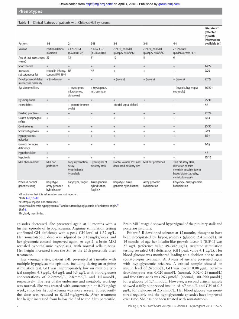
Table 1 Clinical features of patients with Chitayat-Hall syndrome
Patient 1-I 2-I 2-II 3-I 3-II 4-I
literature* (affected (n)/with information available (n))
MRI not performed Thin pituitary stalk, dilatation of third ventricle possibly due to hypothalamic atrophy, ventriculomegaly
Previous normal genetic testing
Karyotype, array genomic hybridisation
Karyotype, fragile X,
Array genomic hybridisation, fragile X
Karyotype, array genomic hybridisation
Array genomic hybridisation
Karyotype, array genomic hybridisation
NR indicates that this information was not reported.*Refs 4–6, 10–12.†Esotropia, myopia and strabismus.‡Hyperinsulinaemic hypoglycaemia10 and recurrent hypoglycaemia of unknown origin.11
§Ref 4.BMI, body mass index.
group.bmj.com on April 1, 2018 - Published by http://jmg.bmj.com/Downloaded from
3Jobling R, et al. J Med Genet 2018;0:1–6. doi:10.1136/jmedgenet-2017-105222
Phenotypes
Following the diagnosis of GH deficiency in her younger sister, patient 3-I was investigated. Her IGF-1 was low at <25 μg/L at 3 years and 10 months and 28 μg/L at 6 years 1 month (reference value 49–342 μg/L). Arginine stimulation testing revealed a peak value of 2.7 μg/L. Her blood glucose was monitored, but the decision was made not to start somatotropin treatment. Brain MRI at 4 years showed a small pituitary gland.
Patient 4-I had hypoglycaemic episodes requiring hospitalisa-tion at 6 months. GH deficiency was first suspected at 11 months and confirmed at 19 months. The GH measured during two hypoglycaemic episodes was low and a clonidine GH stimula-tion test showed a deficiency (GH peak value 4.42 μg/L). The arginine GH stimulation test was also abnormal (4.435 mg intra-venously ×1: GH peak value 3.53 μg/L). She was successfully treated with somatotropin. With treatment her height increased from below the 3rd to the 10th percentile. Brain MRI done at 3 months and repeated at 3 years and 7 months revealed a thin pituitary stalk and slight dilation of the third ventricle, possibly secondary to hypothalamic atrophy.
Detailed results of GH stimulation testing can be found in online supplementary clinical information, tables 1–3.
MeThodsFor all families, genetic analysis was performed by either whole genome sequencing (WGS) or whole exome sequencing (WES) with pathogenic variants confirmed by Sanger sequencing. For family 1, WGS of the proband and her father was performed. WES was performed on samples from affected patients in family 2, and the probands in families 3 and 4 (online supplementary methods).
Analysis of WES and WGS data prioritised variants based on allele frequency, presence in databases of medically relevant vari-ants including ClinVar7 and the Human Gene Mutation Data-base,8 predicted impact on coding sequence, phenotype in the OMIM database, zygosity, and mode of inheritance. In family 2, where both affected individuals were sequenced, shared variants were examined. In family 1 variants shared between the proband and her unaffected father were prioritised due to the paternal family history of similarly affected individuals (figure 1, online supplementary figure 5).
Since MAGEL2 is expressed exclusively from the paternal allele, only pathogenic variants located on the paternal allele will cause disease.9 To determine the parental origin of the c.2179_2180del variant identified in family 3, long-range PCR of MAGEL2 followed by Sanger sequencing was performed on genomic DNA after methylation-sensitive digestion, as described previously4 (online supplementary methods).
sequenCIng resulTsAll affected individuals were found to carry truncating variants in MAGEL2. Patient 1-I was found to have a complex rear-rangement interrupting the MAGEL2 gene, consisting of a 22 kb inversion and 3 kb deletion that removes the last 852 bp and the 3’ end of the gene (online supplementary figures 1–3). The variant was paternally inherited and segregation analysis for several additional family members was performed (figure 1). Siblings 2-I and 2-II have a nonsense variant (NM_019066; c.1762 C>T(p.Gln588Ter)) in MAGEL2. Parental samples were not available for testing. Patients 3-I and 3-II carry a frameshift variant (c.2179_2180del(p.Asp727Profs*6)) in MAGEL2. The
Figure 1 pedigrees and MAGEL2 variants identified in patients with Chitayat-hall syndrome. Filled black squares and circles indicate clinically affected individuals, black dots indicate carriers, V indicates that the familial variant was found in an individual, + indicates the reference sequence and Nt indicates that the individual was not tested.
group.bmj.com on April 1, 2018 - Published by http://jmg.bmj.com/Downloaded from
4 Jobling R, et al. J Med Genet 2018;0:1–6. doi:10.1136/jmedgenet-2017-105222
Phenotypes
variant was not present in parents or unaffected siblings, and was determined to be on the paternal allele (online supplementary figure 4). Patient 4-I has a previously reported frameshift inser-tion (c.1996dupC(p.Gln666Profs*47))6 in MAGEL2, inherited from her unaffected father.
dIsCussIonMultiple features first reported in Chitayat-Hall syndrome overlap with those described in the majority of individuals with SHFYNG, including contractures, hypotonia, developmental delay/intellectual disability, feeding difficulties, dysmorphisms, small hands and feet, and tapering fingers.1 4–6 10–12 Our cohort also has other features reported in a minority of individuals, including scoliosis, gastro-oesophageal reflux, increased subcu-taneous fat and prominent ridge over the metopic suture. While eye abnormalities are described, this is the first report of micro-cornea in patients with an MAGEL2-related disorder.6
The most common pathogenic sequence variant identified to date in MAGEL2, c.1996dupC(p.Gln666Profs*47), has been reported in 14 individuals from nine families diagnosed with SHFYNG.6 10 11 These individuals present with the features most commonly described in association with SHFYNG, including contractures, developmental delay/intellectual disability, dysmor-phism, hypotonia and feeding difficulties. Short stature was reported in 6/14 cases. Our patient 4-I, with the c.1996dupC
variant, has a very similar phenotype to the 14 reported patients, apart from her GH deficiency. Unfortunately, there is no infor-mation available regarding GH levels in these 14 individuals.
Deficiency of hormones produced by the anterior pituitary is a prominent feature of Chitayat-Hall syndrome. All patients reported here demonstrated biochemical abnormalities related to GH deficiency on more than one occasion, with either low IGF-1, low GH peak after arginine stimulation, low GH in the context of hypoglycaemia, or all of the above. One patient with SHFYNG has been previously reported to have GH deficiency, presenting with poor linear growth and treated from 2 years of age.4 However, short stature is common in these patients, and is likely caused by undiagnosed GH deficiency in some cases.4 6 11 Four patients in our study presented with hypoglycaemia, another manifestation of GH deficiency. Hypoglycaemic episodes have not been reported in the majority of patients with SHFYNG, although may go undiagnosed if not leading to convulsions or loss of consciousness.
The pathophysiology of GH deficiency in patients with MAGEL2 variants requires further investigation. MRI findings in our patients were not consistent, although it is notable that imaging for patients 2-I and 4-I demonstrated possible hypotha-lamic hypoplasia. Magel2 is expressed in both fetal and adult brain,9 13 and mouse studies have demonstrated robust expres-sion in the fetal hypothalamus. In adult mice Magel2 is mainly
Figure 2 Features of affected patients. (A) patient I-1—myopathic faces with droopy eyelids and open mouth. (B,C) patients 2-I and 2-II, respectively—both sisters had minor facial dysmorphism with a high forehead, a flat forehead in patient 2-I (B) and frontal bossing in patient 2-II (C) with a ridge over the metopic suture, deep set eyes, depressed nasal bridge with a broad nasal root and tip, and a ‘square’ chin with a horizontal groove over the chin. In patient 2-II (C) note the low-set ears with the right ear being lower than the left. (D) patient 3-I—high forehead with a ridge over the metopic suture, hypoplastic supraorbital ridges, deep set eyes, a broad nasal root and tip and a long philtrum, full cheeks, thin upper lip and retrognathia with a square chin and a horizontal groove over the chin. (e) hand of patient 3-II—‘puffy’ hand with proximally inserted thumb, tapering fingers and camptodactyly with absent distal flexion creases. (F) patient 3-II—deep set eyes, a broad nasal root and tip, long philtrum, thin upper lip, retrognathia, a ‘square’ chin with a horizontal groove over the chin and low-set ears. (G) patient 4-I—ridge over the metopic suture, deep set eyes, a broad nasal root and tip, thin upper lip, retrognathia with a square chin, a horizontal groove over the chin and low-set ears. (h) hand of patient 4-I—adducted thumb with the second and fifth fingers overlapping the third and fourth.
group.bmj.com on April 1, 2018 - Published by http://jmg.bmj.com/Downloaded from
5Jobling R, et al. J Med Genet 2018;0:1–6. doi:10.1136/jmedgenet-2017-105222
Phenotypes
expressed in the hypothalamus, including the arcuate nucleus where GH-releasing hormone (GHRH) is produced.14 15 There is evidence of GH deficiency related to hypothalamic dysfunc-tion in the Magel2-null mouse. Tennese and Wevrick16 found low levels of IGF-1 in female Magel2-null mice compared with controls. The mice demonstrated a blunted response to hypo-thalamic stimulation of the GH pathway with ghrelin compared with wild-type littermates, while their response to GHRH was equivalent, indicating a possible hypothalamic origin for the deficiency.16
Family 1 carries a complex rearrangement and partial deletion. To our knowledge this is the first report of such a change causing an MAGEL2-related disorder. The first 2.9 kb of the coding and the 5’ region are apparently intact, and it is possible that a truncated protein product is produced. It has been suggested that frameshift and nonsense variants in MAGEL2 escape nonsense-mediated decay and have a neomor-phic or dominant negative effect, explaining the milder pheno-type seen in full gene deletions.6 17 18 Functional studies are required to investigate this possibility, but are difficult to pursue given that the expression of MAGEL2 in adult tissues is very limited.9 13 This case illustrates the benefits of WGS as a diagnostic test, as this complex variant would not have been detected using exome, microarray or targeted sequencing methodologies.
In family 3 we demonstrated that the variant identified in the two affected sisters was on the paternal allele. It was not detectable in paternal blood by Sanger sequencing. This does not rule out the possibility of low level mosaicism in blood or other tissues. This is the third reported case of apparent mosa-icism in an unaffected father in MAGEL2-related disorder.10 11 In this situation, the recurrence risk could be up to 50%.
The phenotype of MAGEL2-related disorder continues to evolve, now including Chitayat-Hall syndrome. With the excep-tion of the endocrinological findings we describe, our patients’ phenotypes are very similar to those observed in patients with SHFYNG, and one of our patients carries the most common recurrent variant c.1996dupC reported in SHFYNG. This suggests that SHFYNG and Chitayat-Hall syndromes are likely the same disorder. A systematic investigation of endocrinolog-ical abnormalities in patients with MAGEL2-related disorder is needed and GH deficiency should always be considered in the context of poor growth and/or hypoglycaemia.
Author affiliations1Genome Diagnostics, Department of paediatric Laboratory Medicine, the hospital for Sick Children, toronto, ontario, Canada2Division of Clinical Genetics and Metabolism, Department of pediatrics, the hospital for Sick Children, toronto, ontario, Canada3Department of Laboratory Medicine and pathobiology, University of toronto, toronto, ontario, Canada4the Centre for Applied Genomics, the hospital for Sick Children, toronto, ontario, Canada5regenoron, New York City, New York, USA6ChU Sainte-Justine, Montréal, Quebec, Canada7Department of pediatrics, Université de Montréal, Montréal, Quebec, Canada8Life and health Sciences research Institute (ICVS), School of Medicine, University of Minho, Braga, portugal9ICVS/3B’s - pt Government Associate Laboratory, Guimarães, portugal10Department of Medical Genetics, University of British Columbia, Vancouver, British Columbia, Canada11Division of Clinical pathology, McMaster University, hamilton, ontario, Canada12program in Genetics and Genome Biology, the hospital for Sick Children, toronto, ontario, Canada13Department of Molecular Genetics and McLaughlin Centre, University of toronto, toronto, ontario, Canada14the Centre for Biomedical research, University of Victoria, Victoria, British Columbia, Canada
15the prenatal Diagnosis and Medical Genetics program, Department of obstetrics and Gynecology, Mount Sinai hospital, New York City, New York, USA
Acknowledgements We would like to thank the families who participated in this work. the authors also thank, the SickKids Centre for Genetic Medicine, the University of toronto McLaughlin Centre, the toronto Centre for Applied Genomics, and the GlaxoSmithKline-CIhr Chair in Genome Sciences at the hospital for Sick Children and the University of toronto (SWS), and the Fondation Jeanne et Jean-Louis Lévesque (JLM).
Contributors CC, SM, Fr, NA, rr, MM, SA, MJMN, CLD, rW, pM and DC performed clinical assessment and provided phenotypic information regarding the patients. Fr, FDL, JG, FFh, CN, J-FS, JLM, rJ, DJS, CrM, SWS, Jo and SW provided sequencing, data analysis, interpretation and validation of variants. rJ, VL and MMA performed phasing experiments for the variant in family 3. the manuscript was drafted by rJ, Fr, DC and JLM. All authors provided critical revision of the article.
Funding the McLaughlin Centre, University of toronto, toronto, Canada, and Fondation Jeanne et Jean- Louis Lévesque (JLM). the Centre for Genetic Medicine, the hospital for Sick Children, toronto, Canada. FDL has a fellowship funded by FCt - Fundação para a Ciência e a tecnologia (SFrh/BD/84650/2010).
reFerenCes 1 Chitayat D, hall JG, Couch rM, phang MS, Baldwin VJ. Syndrome of mental
retardation, facial anomalies, hypopituitarism, and distal arthrogryposis in sibs. Am J Med Genet 1990;37:65–70.
2 Smigiel r, Basiak A, Misiak B, pesz K. panhypopituitary insufficiency in a patient with clinical diagnosis of Chitayat-hall syndrome. Endokrynol Pol 2010;61:318–21.
3 rao V, el-Alem t, Aminu K, Mankad K, Cowan F, holder Se, Kinali M. Chitayat-hall syndrome: extending the clinical phenotype. Clin Dysmorphol 2013;22:156–60.
4 Schaaf Cp, Gonzalez-Garay ML, Xia F, potocki L, Gripp KW, Zhang B, peters BA, Mcelwain MA, Drmanac r, Beaudet AL, Caskey Ct, Yang Y. truncating mutations of MAGeL2 cause prader-Willi phenotypes and autism. Nat Genet 2013;45:1405–8.
5 Mejlachowicz D, Nolent F, Maluenda J, ranjatoelina-randrianaivo h, Giuliano F, Gut I, Sternberg D, Laquerrière A, Melki J. truncating Mutations of MAGeL2, a Gene within the prader-Willi Locus, Are responsible for Severe Arthrogryposis. Am J Hum Genet 2015;97:616–20.
6 Fountain MD, Aten e, Cho Mt, Juusola J, Walkiewicz MA, ray JW, Xia F, Yang Y, Graham Bh, Bacino CA, potocki L, van haeringen A, ruivenkamp CA, Mancias p, Northrup h, Kukolich MK, Weiss MM, van ravenswaaij-Arts CM, Mathijssen IB, Levesque S, Meeks N, rosenfeld JA, Lemke D, hamosh A, Lewis SK, race S, Stewart LL, hay B, Lewis AM, Guerreiro rL, Bras Jt, Martins Mp, Derksen-Lubsen G, peeters e, Stumpel C, Stegmann S, Bok LA, Santen GW, Schaaf Cp. the phenotypic spectrum of Schaaf-Yang syndrome: 18 new affected individuals from 14 families. Genet Med 2017;19:45–52.
7 Landrum MJ, Lee JM, Benson M, Brown G, Chao C, Chitipiralla S, Gu B, hart J, hoffman D, hoover J, Jang W, Katz K, ovetsky M, riley G, Sethi A, tully r, Villamarin-Salomon r, rubinstein W, Maglott Dr. ClinVar: public archive of interpretations of clinically relevant variants. Nucleic Acids Res 2016;44(D1):D862–8.
8 Stenson pD, Mort M, Ball eV, evans K, hayden M, heywood S, hussain M, phillips AD, Cooper DN. the human Gene Mutation Database: towards a comprehensive repository of inherited mutation data for medical research, genetic diagnosis and next-generation sequencing studies. Hum Genet 2017;136:665–77.
9 Boccaccio I, Glatt-Deeley h, Watrin F, roëckel N, Lalande M, Muscatelli F. the human MAGeL2 gene and its mouse homologue are paternally expressed and mapped to the prader-Willi region. Hum Mol Genet 1999;8:2497–505.
10 Soden Se, Saunders CJ, Willig LK, Farrow eG, Smith LD, petrikin Je, Lepichon JB, Miller NA, thiffault I, Dinwiddie DL, twist G, Noll A, heese BA, Zellmer L, Atherton AM, Abdelmoity At, Safina N, Nyp SS, Zuccarelli B, Larson IA, Modrcin A, herd S, Creed M, Ye Z, Yuan X, Brodsky rA, Kingsmore SF. effectiveness of exome and genome sequencing guided by acuity of illness for diagnosis of neurodevelopmental disorders. Sci Transl Med 2014;6:265ra168.
11 palomares-Bralo M, Vallespín e, Del pozo Á, Ibañez K, Silla JC, Galán e, Gordo G, Martínez-Glez V, Alba-Valdivia LI, heath Ke, García-Miñaúr S, Lapunzina p, Santos-Simarro F. pitfalls of trio-based exome sequencing: imprinted genes and parental mosaicism-MAGeL2 as an example. Genet Med 2017;19:1285–6.
group.bmj.com on April 1, 2018 - Published by http://jmg.bmj.com/Downloaded from
6 Jobling R, et al. J Med Genet 2018;0:1–6. doi:10.1136/jmedgenet-2017-105222
Phenotypes
12 Urreizti r, Cueto-Gonzalez AM, Franco-Valls h, Mort-Farre S, roca-Ayats N, ponomarenko J, Cozzuto L, Company C, Bosio M, ossowski S, Montfort M, hecht J, tizzano eF, Cormand B, Vilageliu L, opitz JM, Neri G, Grinberg D, Balcells S. A De Novo Nonsense Mutation in MAGeL2 in a patient Initially Diagnosed as opitz-C: Similarities Between Schaaf-Yang and opitz-C Syndromes. Sci Rep 2017;7:44138.
13 Lee S, Kozlov S, hernandez L, Chamberlain SJ, Brannan CI, Stewart CL, Wevrick r. expression and imprinting of MAGeL2 suggest a role in prader-willi syndrome and the homologous murine imprinting phenotype. Hum Mol Genet 2000;9:1813–9.
14 Kozlov SV, Bogenpohl JW, howell Mp, Wevrick r, panda S, hogenesch JB, Muglia LJ, Van Gelder rN, herzog eD, Stewart CL. the imprinted gene Magel2 regulates normal circadian output. Nat Genet 2007;39:1266–72.
15 Maillard J, park S, Croizier S, Vanacker C, Cook Jh, prevot V, tauber M, Bouret SG. Loss of Magel2 impairs the development of hypothalamic Anorexigenic circuits. Hum Mol Genet 2016;25:3208–15.
16 tennese AA, Wevrick r. Impaired hypothalamic regulation of endocrine function and delayed counterregulatory response to hypoglycemia in Magel2-null mice. Endocrinology 2011;152:967–78.
17 Buiting K, Di Donato N, Beygo J, Bens S, von der hagen M, hackmann K, horsthemke B. Clinical phenotypes of MAGeL2 mutations and deletions. Orphanet J Rare Dis 2014;9:40.
18 tacer KF, potts pr. Cellular and disease functions of the prader-Willi Syndrome geneMAGEL2. Biochem J 2017;474:2177–90.
group.bmj.com on April 1, 2018 - Published by http://jmg.bmj.com/Downloaded from
-related disordersMAGEL2of phenotypecommon aetiology: expanding the
Chitayat-Hall and Schaaf-Yang syndromes: a
ChitayatRosanna Weksberg, Patrick Macleod, Jacques L Michaud and DavidChristina Nassif, Fadi F Hamdan, Cheri L Deal, Jean-François Soucy, Adam, Malgorzata J M Nowaczyk, Susan Walker, Stephen W Scherer,Gauthier, Nathalie Alos, Rosemarie Rupps, Margaret McKinnon, Shelin Moalem, Jennifer Orr, Francis Rossignol, Fatima Daniela Lopes, JulieCheryl Cytrynbaum, Michelle M Axford, Vanessa Londero, Sharon Rebekah Jobling, Dimitri James Stavropoulos, Christian R Marshall,
published online March 29, 2018J Med Genet
http://jmg.bmj.com/content/early/2018/03/29/jmedgenet-2017-105222Updated information and services can be found at:
These include:
References
#ref-list-1http://jmg.bmj.com/content/early/2018/03/29/jmedgenet-2017-105222This article cites 18 articles, 2 of which you can access for free at:
serviceEmail alerting
box at the top right corner of the online article. Receive free email alerts when new articles cite this article. Sign up in the
Notes
http://group.bmj.com/group/rights-licensing/permissionsTo request permissions go to:
http://journals.bmj.com/cgi/reprintformTo order reprints go to:
http://group.bmj.com/subscribe/To subscribe to BMJ go to:
group.bmj.com on April 1, 2018 - Published by http://jmg.bmj.com/Downloaded from