Page 1
ABSTRACT
DE'IEMNATION OF PORPHYRIN RING ORIENTATION
IN SPINACH CHLOFDPLAST EXTRACT CHIOFDPHYLL
BLACK LIPID IVEMBRANES BY PHO'IUVOLTACE SPECTFDSGJPY
By
Herman G. Weller, Jr.
Photovoltage spectroscopy with polarized light was used to
investigate the structure of black lipid membranes formed from
spinach chloroplast extracts. The photovoltage action spectrum of
the chlorophyll black lipid membrane is similar to the absorption
spectrum of the membrane—forming solution, with a red and principal
blue peak. The maglitudes of these peaks were found to depend on
the direction of polarization of the exciting light. 'Ihis is
apparently a direct consequence of the dichroism of the mn‘brane.
The polarized light photovoltage data were used to obtain
information on the orientation of chloropmrll in the membrane.
The chlorophyll principal blue transition mment was
calculated to make an angle of“ 21 1 2° with the plane of the
membrane; the red transition moment, an angle of 38 1 2°. From
these angles, an angle (averaged over the chloroprwll a; and
chlorophyll g in the membrane) of 1&5 .+. 5° was calculated for that
Page 2
Herman G. Weller, Jr.
between the plane of the porphyrin ring and the plane of the
membrane .
Page 3
DE'IERMATION OF PORPHYRIN RING ORIENTATION
IN SPINACH CHIDROPLAST EXTRACT CHLOROPHYLL
BLACK LIPID MENBRANES BY PHOI‘OVOLTAGE SPECTHDSCOPY
By
Herman G. WEller, Jr.
A Thesis
Submitted to
Michigan State University V
in partial fulfillment of the requirements
for the degree of
Master of Science
Department of Biophysics
19714
Page 4
'Do Yamba Wiiga, Bob Foester,
Larry Morgan, and Abui Nbrgan
ii
Page 5
ACIQ‘IOWIEDGMEN’IS
The author wishes to express his gratitude to his major
advisor, Professor H. Ti Tien, in whose laboratory this research was
conducted. Interesting and informative discussion with the Master's
Thesis Committee members, Professor A. Haug, Professor S. Izawa, and
Dr. Tien, is also appreciated.
The financial support of this work was provided by a National
Institutes of Health Grant (GM—113971).
111
Page 6
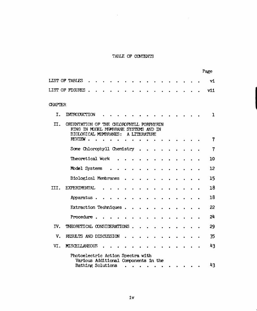
TABLE OF CONTENTS
LIST‘OF TABLES . . . . . . . . . . .
LIST'OF FIGURES . . . . . . . . .
CHAPTER
I. INTRODUCTION . . . . . . . . .
II. ORIENTATION OF THE CHLOROPHYII.PORPHYRIN
RDMG:DQP(HIH.NEWBRANE SYSTEMS AND IN
BIOLOGICAL MEMBRANES: A.LITERATURE
m 0 O O O O 0 0 O O O 0
Some Chlorophyll Chemistry . . . .
Theoretical Work . . . . . . .
.Model Systems . . . . . .
Biological Membranes . . . . . .
III. EXPERIMENTAL . . . . . . . .
Apparatus . . . . . . . .
Extraction Techniques . . . . . .
Procedure . . . . . . . . . .
IV. THEORETICAL CONSIDERATIONS . . . .
F RESULES AND DISCUSSION . . . . . .
VI. MISCELLANEOUS . . . . . . . . .
Photoelectric Action Spectra with
various Additional Components in.the
Bathing Solutions . . . . . .
iv
Page
V11
10
12
15
18
18
22
2h
29
35
A3
”3
Page 7
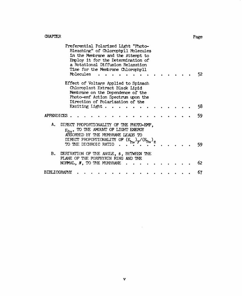
CHAPTER Page
Preferential Polarized Light "Photo-
Bleaching" of Chlorophyll Nblecules
in the Membrane and the Attempt to
Ehploy it for the Determination of
a Rotational Diffusion Relaxation
Time for the Membrane Chlorophyll
Molecules . . . . . . . . . . . . . . 52
Effect of Voltage Applied to Spinach
Chloroplast Extract Black lipid
Membrane on the Dependence of the
Photo-emf Action Spectrum upon the
Direction of Polarization of the
ExcitingLight......'....... 58
APPENDICES..................59
A. DIRECT PROPORTIONALITY OF THE PHOTO-EMF,
Em, TO THE AMOUNT OF LIGHT ENERGY
ABSORBED BY ms mam LEADS TO
DIRECT PROPORTIONALITY OF ( ) /( )
TO'IHEDICHROICRA'I'IO . .Ej‘Yy.E1.7".Z . . . . . 59
B. IZERIVA'I'ION OF THE ANGIE, a, BETWEEN ms
PLANE or THE PORPHYRIN RING AND ma
NomAL,m,To'IHEMEMBRANE . . . . . . . . . . 62
BIBLIOGRAPHY.................67
Page 8
LIST OF TABLES
Table Page
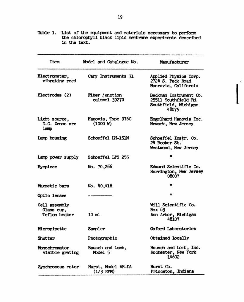
1. List of the Equipment and Materials Necessary
to perform the Chlorophyll Black Lipid
Membrane Experiments Described in the Text . . . . l9
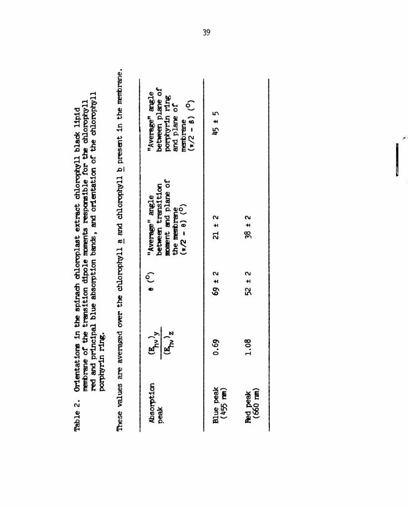
2. Orientations in the Spinach Chloroplast
Extract Chlorophyll Black Lipid Membrane of
the Transition Dipole Ivbments Responsible
for the Chlorophyll Red and Principal Blue
Absorption Bands, and Orientation of the
Chlorophyll Porphyrin Ring . . . . . . . . . 39
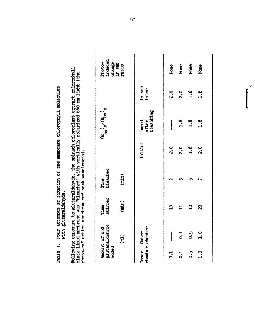
3. Four Attempts at Fixation of the Membrane
Chlorophyll Nblecules with Glutaraldehyde . . . . . 57
Page 9
LIST OF FIGURES
FigurePage
1. Schematic representation of the structure
ofchlorophyllaorg . . . . . . . . . . . 8
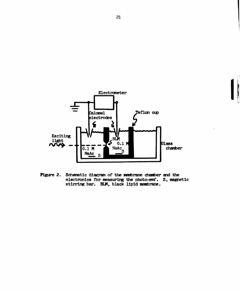
2. Schematic diagram of the membrane chamber
and the electronics for measuring the
phOto-enf O O O O O O O I O I O 0 O O 0 21
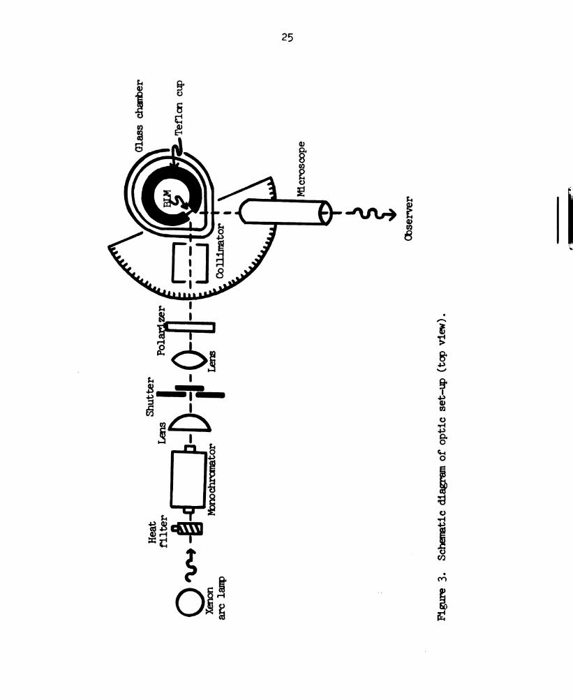
3. Schematic diagram of optic set-up (top view) . . . . 25
A. (A) Schematic diagram illustrating the
determination of the orientation of the
membrane relative to the direction of
propagation of the incident light,
(topview)... .........26
(B) Schematic diagram illustrating the
establishment of the direction of
polarization of the incident light with
the electric vector, E, vibrating parallel
to the plane of incidence (top view) . . . . . 26
5. Schematic diagram of the transition dipole
moment, M, of either the red or the principal
blue chlorophyll absorption band (see text) . . . . 30
6. Absorption spectrum and photo-emf action
spectra of the spinach chloroplast extract
blacklipidmembrane . . . . . . . . . . . 36
7. Photo-emf action spectra of the spinach
chloroplast extract black lipid membrane
with 1 mM FeCl3 in the outer chamber and
"water-soluble chlorophyll" in the imer
chamber................ All
8. Photo-emf action spectra of the spinach
chloroplast extract black lipid membrane
with 1 mM FeCl in the outer chamber and
o.1thhionin;intheimerchamber . . . . . . 1:6
vii
Page 10
Figure
9. Photo—emf action spectra of the spirech
chloroplast extract black lipid membrane
with 1 mM FeCl in the outer chamber and
0.3 m p—benz uirone in the inner
chamber...........
10. Photo—emf action spectra of the spinach
chloroplast extract black lipid membrane
with 1 mM FeCl in the outer chamber and
A mid L-asoorbi acid in the inner
chamber...........
11. Shape of a typical plot of the photo-emf
versustime..........
12. Schematic diagram of the relation between
the red transition moment vector, M , the
principal blue transition moment vector,
”8’ and the normal to the membrare, IV .
viii
Page 11
CHAPTERI
INTRODUCTION
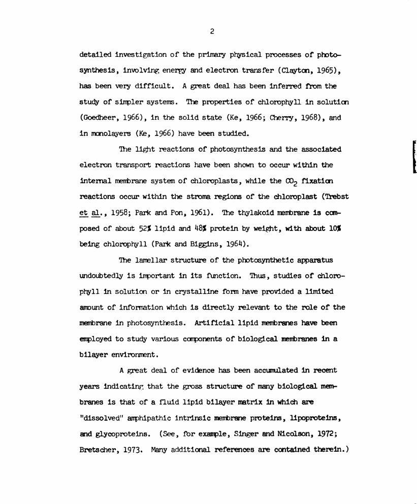
In green algae and higher plants, photosynthesis is the
process occurring in the chloroplasts in which (a) electromagnetic
energy from incident visible light activates the reduction of
nicotinamide adenine (NADP+) to NADPH and the oxidation of water,
and (b) carbon dioxide is reduced to {CHZO}n. (Lehninger, 1970)
The grana contain essentially all the photosynthetic
pigments of the chloroplast as well as the enzymes required for the
primary light—dependent reactions. The paired thylakoid membranes
are the sites of the light-trapping systems in the chloroplast
structure (Rabinowitch and Govindjee, 1969). The isolated chloro-
plast lamellae when illuminated perform electron transport from
water to ferredoxin, yielding oxygen gas and reduced ferredoxin.
Phosphorylation of ADP to ATP accompanies this electron transport
(Hill, 1937; Hill, 1965; Arnon _e_t_;a_l_., 19514).
The correspondence hemeen photochemical action spectra
and the light absorption spectra of various green algae and photo-
synthetic higher plants have led to the conclusion that chloroprwll
must be the primary light—trapping molecule in green cells (Clayton,
(1971).
Because of the complexity of the photosynthetic system
Page 12
detailed investigation of the primary physical processes of photo—
synthesis, involving energy and electron transfer (Clayton, 1965),
has been very difficult. A great deal has been inferred from the
stucw of simpler systems. The propertiesof chloroplwll in soluticn
(Goedheer, 1966), in the solid state (Ke, 1966; Cherry, 1968), and
in monolayers (Ke, 1966) have been studied.
The light reactions of photosynthesis and the associated
electron transport reactions have been shown to occur within the
internal membrane system of chloroplasts, while the (132 fixatim
reactions occur within the stroma regions of the chloroplast (Trebst
93 _a_l_. , 1958; Park and Pon, 1961). The thylakoid membrane is com-
posed of about 52% lipid and A81 protein by weight, with about 101
being chlorophyll (Park and Biggins, 196A).
The lamellar structure of’the photosynthetic apparatus
undoubtedly is important in its function. Thus, studies of chloro-
phyll in solution or in crystalline form have provided a limited
amount of infermation which is directly relevant to the role of’the
membrane in photosynthesis. Artificial lipid.membranes have been
employed to study various components of biological membranes in a
bilayer environment.
A great deal of evidence has been accumulated in recent
years indicating that the gross structure of many biological mem-
branes is that of a fluid lipid bilayer matrix in which are
"dissolved" amphipathic intrinsic membrane proteins, lipoproteins,
and glycoproteins. (See, for example, Singer and Nicolson, 1972;
Bretscher, 1973. Many additional references are contained therein.)
Page 13
Electron paramagnetic resonance studies with phospholipid bilayer-s
and rabbit sarooplasmic reticulum by McConnell and co-workers have
indicated that lipids may be very mobile in the plane of the membrane
(Kornberg and McConnell, 1971; Scandella _e_t_ gal. , 1972), but much less
mobile in a direction perpendicular to the plane of the membrane
(Kornberg and McConnell, 1971; McNamee and McConnell, 1973).
Studies by Frye and Edidin on intrinsic membrane proteins
complexed with fluorescent—labeled specific antibodies in the
envelopes of human cells and mouse cells caused to fuse under the
influence of Sendai virus, and by Nicolson and Singer on red blood
cell intrireic membrane proteins complexed with specific ferritin-
labeled antibodies, have shown that the proteins "dissolved" in the
plasma membrane may also be quite mobile laterally. (Frye and
Edidin, 1970; Nicolson and Singer, 1971a; Nicolson and Singer, 1971b;
Nicolson and Singer, 1971c)
The black lipid membrane has been introduced as a model
system for the study of biologcal membrane components in a bilayer
lipid matrix separating two aqueous soluticns by Mueller, Rudin,
Tien, and Wescott. (Mueller, Rudin, Tien, and Wescott, 1962) The
black lipid membrane exhibits many properties which are similar to
those of biological membranes, e.g., thickness, resistance,
capacitance, and interfacial tension (Tien, 1971).
The chlorophyll black lipid membrane separating two
aqueous phases has been proposed as a model system for the study of
the primary processes of photosynthesis of green plants. Various
properties of black lipid membranes in the dark have been measured,
Page 14
e.g. water permeability, bifacial tension, thickness, resistance,
and dielectric breakdown (Ting gt _a_l_. , 1968). Recently, light—
excitable properties such as fluorescence (Alamuti and Lauger, 1970) ,
absorbance (Steinemann gt__a_1_., 1971; Cherry _e_t_ 31;, 1971), and
photovoltage effects (Tien, 1968) have been investigated.
It has been found that with Fe3+ in one aqueous phase
visible light incident on the spinach chloroplast extract
chlorophyll-lipid bilayer induces a transmembrane voltage (Van and
Tien, 1970) . This was to be expected since a "photovoltaic" effect
in layers of chlorophyll a, t_)_, a+p_, and other pigrents applied to a
metallic electrode lowered into an electrolyte had been observed and
studied earlier by Yevstigneyev, Terenin, and co-workers
(Yevstigneyev and Terenin, 1951; Yevstigneyev, 1962; Termin and
Putseiko, 1961; Yevstigleyev and Savkina, 1963; Putseiko, 1963).
More recently, Getov and Jordanova have found that in
layers of chlorophyll a and :1 applied to a semi-transparent gold
electrode illumination causes a "photo-emf," the gold electrode
always being positive, and the spectral distribution of the photo—
emf upon illumination on the electrode almost coincides with the
optical absorption spectrum of chlorophyll (Getov and Jordanova,
1972).
Tre chlorophyll black lipid membrane photo-emf may be
comparable to the trans-thylakoid voltage calculated by Witt and
co—workers from absorbence changes at 515 nm of chlorophyll p_
during electron transport and protOphosprorylation in spinach
chloroplast preparations (Junge and Witt, 1968; Schliephake gt _a_l_.,
Page 15
1968 ; Witt, 1972). This calculation involved assumptims of
concomitant trans—thylakoid proton transfer, thickness and
dielectric constant of the membrane lipid layer, and the area of
thylakoid covered by one electron transport chain. Witt gt _a_l_.
arrived at values of about 50 mV for 1.5(10-5) sec of "saturating
intensity" excitation at 630 - 680 nm, about 200 mV for the maximum
voltage upon excitation of longer duration, and in permanent light
a steachl-state value of about 100 mV.
Tle light-induced emf of the artificial chlorophyll-lipid
membrane has been found to depend on the wavelength of the
illuminating light (Van and Tien, 1970). A "photo—emf action
spectrum" can be obtained by scammg the visible wavelengths.
The present work concerns tle finding that the magnitudes
of the peaks of the proto-emf action spectrum depend on the
direction of polarization of the exciting light. This appears to be
a direct consequence of the absorption properties of the chlorophyll
in the artificial membrane, and may be used to determine the
orientation of the chlorophyll porphyrin ring in the black lipid
membrane. (Weller and Tien, 1973)
The principal blue transition moment was calculated to
make an angle of 21 t 20 with the plane of the membrane; the red
transition moment, an angle of 38 1 2°. From these angles, an angle
of 145 r. 5° is calculated for that between the plane of the porphyrin
ring and the plane of the membrane. These values are averaged over
the chlorophyll _a and chlorophyll _b_ present in the membrane.
There exists a possibility that the acidic bathing
Page 16
solution employed in these experiments converts some or all of the
chlorophyll a and chlorophyll _b_ in the artificial membrane to
pheophytin a and preophytin b, respectively, by the removal of the
Mg atom from the center of each porphyrin ring. This
pheOprytinization reaction would depend on the degree of exposure of
the membrane chloropmll porphyrin rings to the aqueous phases. '
The porphyrin ring orientation angle determination in the
experiments described herein does not supply erough information to , f
ascertain the availability of the chelated chlorophyll Mg atom to N
the acidic bathing solutions. Thus there exists the possibility
that the porphyrin ring orientation angle obtained may be for
pheophytin and chlorophyll.
Page 17
CHAPTER II
ORIENTATION OF THE CHLOROPHYLL PORPIIYRIN
RDIG IN MODEL MEMBRANE SYSTEMS AND IN
BIOLOGICAL MMBRANFS: A LITERATURE REVIEW
Some Chlorgihyll Chemistry
Chlorophyll (Figure 1) is a molecule with an unusual
combination of electron donor-acceptor properties (Katz, 1973). The
ring V keto O=O group can function as donor, the central Mg atom as
acceptor. In the absence of extraneous nucleophiles, donor-acceptor
interactions form chlorophyll dimers, ((1112), and .oligomers, (Ch12)n.
With mmoiunctional electron donors, mmomeric chlorophyll species
form. BifUnctional donors may cross—link chloroplwlls through Mg
atoms to form large polynuclear adducts of colloidal dimensions.
Katz has examined the visible absorption spectra by com-
puter deconvolution techniques and found considerable similarity
between bulk or antenna chlorophyll in the plant and (0112)“.
(Katz, 1973) Electron spin resonance studies have suggested that
ESR photo—siglal I associated with the photosynthetic reaction
center of photosynthetic organisms could arise in a special pair of
chlorophyll molecules (Chl H20 Chl)+.
Katz pointed out that these structures can be combined to
give a structure that possesses both light—gathering properties and
photoactivity, (Chlz)n(Ch1 H20 Chl). The Junction is readily
Page 18
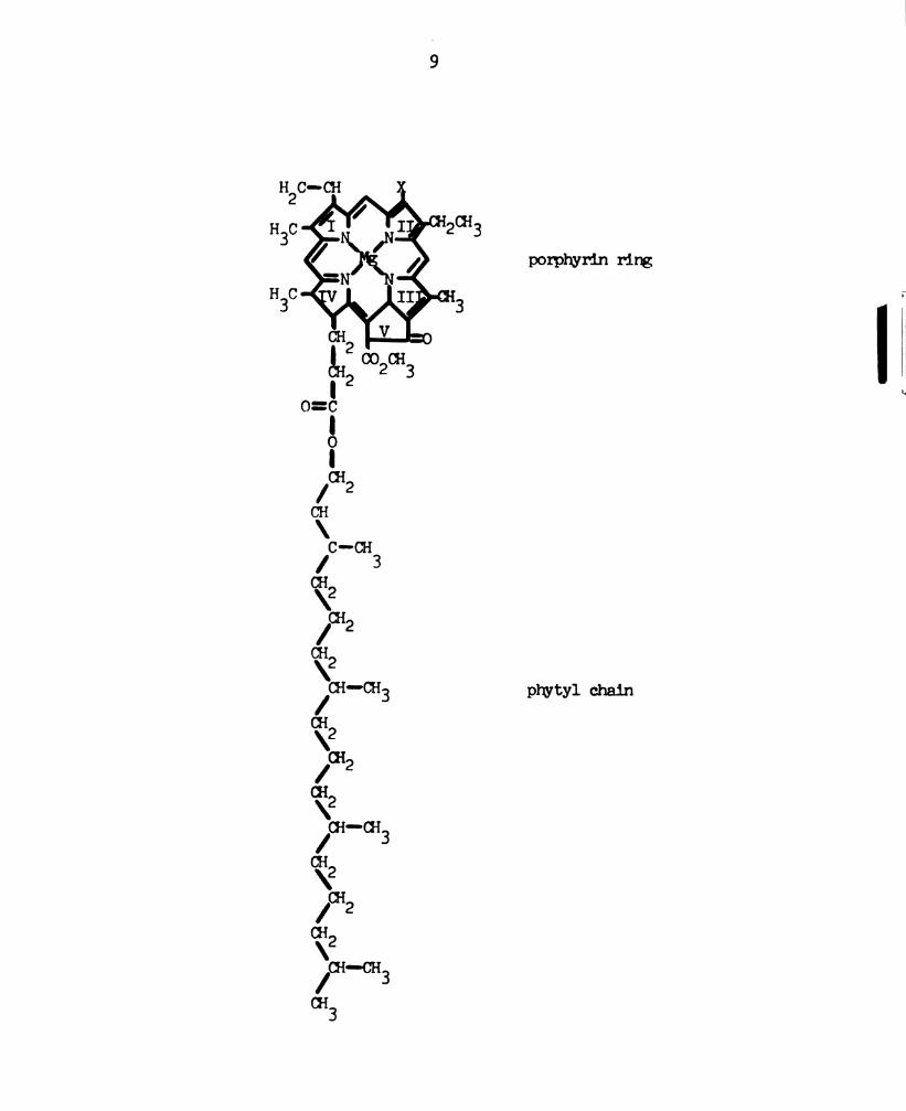
Figure 1. Schematic representation of the structure of chlorophyll _a_
or _b_. In chlorophyll a, X is -CH in chlorophyll b, X is3;
-CH0 (after Lehninger, 1970).
Page 19
m\m
mm
a...
gm3
E
m. m.
porphyrin ring
Page 20
10
effected by a keto 0-0—-Mg interaction between the terminal
chlorophyll molecule of (Ch12)n and the chloropmll of the special
pair that still l'es an Mg atom available for coordination. (Katz,
1973)
This model accounts for both optical and ESR properties of
plant chlorophyll. Such a structure can survive only if access of
water to it is strictly limited, otherwise the entire structure will
be converted to (Chl°H20)n. The electron-transport agents and the
enzymes required for the subsequent chemical reacticns of photo-
synthesis presumably would be in the membranes and hydrophilic
regions of the chloroplast‘that surround the chloroptmrll. (Katz,
1973)
Theoretical Work
Theoretical work based on experimental observations has
sugasted that chlorophyll molecules in photosynthetic systems are
oriented, and that energy transfer would be much facilitated by
suitable chromophore orientation.
For example , the finding that triplet excitation is
greatly enhanced, while the quantum yield of fluorescence is
diminished mam-fold, in chlorophyll aggregates over monaleric
chlorophyll suggested to Kasha that a suitable thylakoid
concentration of chloroptwll might allow absorbed energy to be
transferred in the chloroplast via excitation to a chlorophyll
"exciton band," followed by triplet excitation. (Kasha, 1959)
While discussirg energy reception and transfer in ploto-
synthesis , Calvin speculated about the orientation of chlorophyll in
Page 21
11
. the chloroplast lamella and suggested that the porphyrin rings lie
in a characteristic pattern, namely at an angle of about 145° to the
stacking axis. (Calvin, 1958; Calvin, 1959)
Seely calculated that emery transfer bemeen chlorophyll
molecules by a "slow mechanism" (colpatible with Forster's inductive
resonance theory) would be fastest when the chlorophyll transition
moment vectors are in a collinear arrangement and very small when the
vectors are parallel but an echelon by an angle of 60° (Seely, 1973a;
Seely, 1973b). This suggested that an expeditious use of orientation
would be to group as new chlorophylls as possible into collinear
files, stagered 600 from each other, so that transfer would be
rapid the length of the file but slow fiom one file to another. The
files would lead to the reaction center with as few changes in
orientation as possible.
On the basis of x-ray crystallographic studies, Kreutz has
postulated that the photosynthetic membrane is conposed of three
layers: protein, porphyrin ring, and lipid. (Kreutz, 1970; Kreutz,
1972) He felt that the chlorophyll molecules are anchored in the
protein layer by means of their phytol chains , and the contact
between protein and lipid is established by the 90mm?Em ring
which partially penetrate into the unsaturated fatty acid zones of
the lipid layer.
Based on the assumption that chloroplasts in the natural
state exhibit a dichroic ratio of D 8 l for both the chloropl'yll red
and principal blue absorption bands (with the exception of
chlorophyll-695, for which D > 1) , Kreutz calculated that the
porpmrin ring should make an angle of 5A.7° with the lamella plane.
Page 22
Model Systems
Chlorophyll is very hygroscopic (Ballschmniter and Katz,
1969) and water is necessary to form mnicrocrystalline chlorophyll
(Katz _e_t_ _a_l_. , 1968). Chlorophyll-water complexes have a similar
electron paramagnetic resonance spectrum to photosynthesizing
chloroplasts, wkereas anhydrone chlorophyll does not. (Katz gt _a_1_ . ,
1968)
An infrared absorption study of chlorophyll-water
aggregates has indicated that the water is hydrogen-bonded both to
the ring V ketone carbonyl and to the 0—2 ester carbonyl omen atoms
of the adjacent molecule (Ballschmiter and Katz, 1959).
From X—ray diffraction determination of the structure of
MgTPP'Hz) (T‘imkovitch and Tulinsky, 1969), Mch-H20°205H5N (Fischer
3}; §_1_., 1971), and methyl pheophorbide a_ (Fischer 913 a._l_., 1972),
Fischer and co—workers proposed a model of chlorophyll which has
dimensions similar to methyl pheophorbide a with the Mg atom 0.50 3
out of the plane and a water molecule 2.02 (A) above the 1% atom
(Fischer §_t_ _a_1_. , 1972) . Hydrogen bonds comect the water molecule
to the next chloromell. Repetition by simple translations would
lead to molecular crowding, but repetition by a 2 screw axis would1
permit a satisfactory fit.
Hanson reported that chlorophyllide can form a morolayer
consisting of close-packed porphyrin rings, and he assumed that the
porphyrin planes are tilted at a 55° angle with the plane as in
crystals. (Hanson, 1939)
From fluorescence polarization study on chlorophyll glipid
monolayers, at an air—water interface, Trosper and co—worlaers
Page 23
13
concluded that in pure chloropryll _a monolayers the pigrent
molecules are unordered, in chloroprwll _a_—monogalactolipid monolayers
the chlorophyll molecules are randomly dispersed, and in
chloropryll a_—"lipid" monolayers (with sulfolipid, oleyl alcolol, or
castor oil as "lipid") the chlorophyll molecules are partially
oriented (the porphyrin rings making an angle of from 0° to 50° with
the interface plane depending on the surface pressure). (Trosper,
1968) V
Brody investigated monolayers of chlorophyll g "complexed"
with various electron donors and acceptors at an air-water interface
(Brody, 1971). From the surface area/chloroptwll molecule in each
case, he calculated the angle between the porphyrin plane and the
water surface. He found, for example, angles of 39°, 37°, 189°, 116°,
and 119° for chlorqnhyll _a_ "complexed" with phenazine methosulfate
(P16), PMS + ascorbate, benzyl viologen (8V), ascorbate, and
delvdroascorbic acid, respectively.
Hoff incorporated chloropm'll _a_, chlorophyll b, and
bacteriochlorophyll g in an oriented phospholipid multilayer and
measured the orientation of the chlorophyll molecules by polarization
absorbsnce spectroscopy (Hoff, 197A). The multilayer contained
several Immndred monolayers , with one chloropmrll molecule per 200
phospholipid molecules. He found angles of 55A :t l.l°, 51.6 : 0.6°,
and 51.7 t o.2° between the porphyrin rirgs and the plane of the
multilayer for chlorophyll _a_, chloroplwll b, and bacteriochlorophyll
_a_, respectively.
From polarized absorption spectroscopy on an artificial
Page 24
ll}
chloropnyll black lipid membrane with a chlorophyll concentration up
to 2.5(1013) molecules/omz, Steiremann gt _a_l_. found values of
23 : 2°, 27 : 2°, 29 1 3°, and 29 a 2° for the angle between the
principal blue transition morent and the membrane plane for
chlorophyll a—phcsphaticwl etharolamine, chlorophyll a-dioleoyl—
phosphatidyl choline, chlorophyll _a-phosphaticbrl serirne, and
chlorophyll b—dioleoyl—prosphatidyl choline membranes, respectively
(Steinemnann _e_:c_ _a_l_. , 1972). They found angles of 35 1 1°, 3& 1 1°,
36 1 2°, and 28 : 2° for the red transition mnorents in the same
membranes (in the same order).
From these angles, they calculated values of 1m 3 3°,
“6 1 3°, 1&9 1 5°, and ‘42 .t 14° for the angle bemoan the porpryrin
ring and the plane of the membrane for the above membranes (in the
same order as above).
From polarized absorption spectroscopy on six chlorophyll-
egg lecithin bilayers in series separated by aqueous. phases, Cherry
and co-workers obtained orientation angles of 26° and 29.50 for the
chlorophyll a and chlorophyll t_> blue transition moments,
respectively (Cherry gt a_l_., 1972). 'Ihey found angles of 36.5° for
both the chlorophyll a and chloropmll 2 red transition mnorents.
From these angles, they calculated values of “8° for chlorophyll _a_
and 51° for chloropryll _b_ as the angle between the porpmrrin ring and
the plane of the membrane.
Hoff criticized the work of Cherry _et _a_l_. on the grounds
that (a) their technique is inherently much less semitive than his
multilayer technique, and permits only measurement of the dichroic
Page 25
15
ratio at one fixed angle, and (b) their values are calculated by
assuming that only one dipole moment contributes to the blue
absorption band. (Hoff,l9714)
Biological membranes
The first experimental results of Venice, Frey-Wyssling and
Steinmann, and Ruch with polarized light microscopy on unicellular
algae,Wand Closterium, were interpreted by these
researchers as an effect of the stacking of lamellae in the grena
(i.e., textural dichroism) rather than as an orientation of
pignents. (P'lenke, 1938; Menke, 1958; Frey-Wyssling and Steimnarm,
19148; Ruch, 1957)
Goedheer mode absorption measurements in polarized moro-
chrcmatic lignt onWand found a weak dichroism in light of
680 nm. (Goedheer, 1955) He concluded that there was a slight
orientation of chlorophyll a molecules.
Later, by means of linear dichroism and polarized
fluorescence measurements on the unicellular algae Moth and
Egglona, Olson and co-workers detected a form of chlorophyll with
maximum dichroism at about 705 mm and maximum polarized emission
near 716 nm. (Olson _e_t_:_ g. , 1961; Olson gt _a_l_., 1962; Olson _e_t_ g._1__. ,
1961411; Olson _e_t_ 11., 196%)
'Iromas gt 9;. found a distinct dichroism at about 680 nm
in spinach chloroplasts oriented at steel-water interfaces. They
interpreted this to be due to 2% of the chlorophyll a—680 being
oriented in the plane of the chloroplast lamellae. (Thomas 31; £1: ,
1967)
Page 26
16
Sauer and Calvin oriented spinach chloroplast f‘ragnents
by electric field (Sauer and Calvin, 1962) or by velocity gradient
(Sauer, 1965) and found a dichroic ratio significantly different from
unity only at 695 mm.
In the case of orientation in a hydrommamdc gradient, they
snowed that the long wavelength absorption oscillator lies parallel "j
to the streamlines of the sheer gradient, and assnmed this to be the
direction in which the planes of the chloroplast larellae are
oriented. They interpreted this dichroism at 695 mm as resulting
from 51 of the chlorophyll _a_ which is strongly oriented.
Morita and Miyazaki oriented the rod-shaped photosynthetic
bacterium Rhodopeudononas palustris cells in a flow-gradient and
lamellae in a thin film. (Morita and Miyazaki, 1971) They found
small dichroism at 590 mm and large dichroism at 800 on and 870 nm.
Geacintov gt _a_l. oriented inorella cells and spinach
chloroplasts in aqueous suspension by means of a static magnetic
field. (Geacintov _e_t_ a_}_. , 1971; Geacintov _e__t_ §_1__. , 1972;
Van Nostrand gt _a_l_. , 1973) They found significant dichroism in the
chlorophyll absorption band at 675 - 678 nm and polarized
fluorescence at about 685 nm, the chlorophyll _a_ fluorescence band.
They concluded that the bulk of the chlorophyll in _v_i_\_i_g is highly
oriented with its red transition moment preferentially parallel to
the plane of the lanellae.
Breton and fellow researchers oriented spinach chloroplasts
by application of a static mnagnetic field or by brushing then onto
an optically polisred quartz plate. (Breton gt _a_l_. , 1973) 'Ihey
oriented spinach chloroplast lamellae by brushing them onto a
Page 27
l7
polished quartz plate or by air—drying a drop of a suspension of
isolated lamellae on the plate. They measured the linear dichroism
spectrum of the oriented chloroplasts or lamellae with a spectro-
polarimeter and calculated the orientations of the dichroic
absorption bands' transition moments.
'Ihey found that the y-polarized transition moments of
chloropryll _a_-680 and longer wavelength forms of chlorophyll _a_ lie
at angles close to the lamellar plane (i.e. , at angles less than
25°-- 30° with the plane). Chlorophyll _a_-670 is less oriented or
oriented at an angle slightly less than 35° with the plane.
"Negative" dichroism in the Soret band of chlorophyll a implies that
the directions of x—polarized transitions are at angles of about 142°.
Chloropmll b—650 exhibited a low degree of order, making an angle
less than 350 with tre lamellar plane.
Page 28
CHAPTER III
EXPERIMENTAL
aratus
The equipment and parts required for the measurement of
the spinach chloroplast extract chlorophyll black lipid mnenbrane
photo-emf action spectrum and the chloropmll orientation in the
membrane are listed in Table l. lhe experimental set-up is
illustrated in Figure 2 (Fang, 1972; Van and Tien, 1970) and
Figure 3.
One requirement for the membrane chanber is that the
directions of the light beam incident on the membrane and the lignt
beam reflected from the mnenbrane be perpendicular to the plane of the
glass through which each passes. ‘Ihe other requirements are that
the mnenbrane chamber provide a stable support for the Teflon bealosr
and be of sufficient heignt to allow the outer aqueous medium to
extend above the hole in the beaker.
The mnenbrane chanber must be isolated from the vibrations
caused by the cooling fan for the arc lamp. 'Ihis was achieved by
placing styrofoam pads under the lamp housing and under the stand
supporting the membrane chalber. Good electrical irnsulation was
obtained by the use of non-metallic supports and coaxial cable.
The 2 mm diameter role for the membrane was bored in the
side of lemon beaker below the level to which the aqueous medium,
18
Page 29
19
Table 1. List of the equipment and materials necessary to perform
the chloropmll black lipid membrare experiments described
in the text.
Item rbdel and Catalogue No. Manufacturer
Electrometer,
vibrating reed
Electrodes (2)
Light source,
D.C. Xenon arc
lam
lamp rousing
Lamp power supply
Eyepiece
Phonetic bars
Optic lenses
Cell assembly
Glass cup ,
Teflon beaker
Micrcpipette
Shutter
Mitochromator
visible grating
Synchrorous motor
Cary Instruments 31
Fiber Junction
calomel 39270
Hanovia, Type 976C
(1000 W)
Schoeffel LH-151N
Selbeffel LPS 255
we 70,266
No. lno,u18
10ml
Sampler
Photographic
Bausch and Iomb,
Nbdel 5
Hurst, Model AR-DA
(1/3 RPM)
Applied Physics Corp.
272‘! S. Peck road
Monrovia, California
Beckman Instrument Co.
25511 Southfield Rd.
Southfield, Michigan
118075
Ehgelhard Hanovia Inc.
Newark, New Jersey
Schoeffel Instr. Co.
2” Booker St.
Westwood, New Jersey
Ednund Scientific Co.
Harrington, New Jersey
08007
Will Scientific Co.
Box 63
Ann Arbor, Michigan
11810?
Oxford Laboratories
Obtaired locally
Bausch and Iomb , Inc.
Rochester, New York
111602
Hurst Co.
Princeton , Indiana
Page 30
20
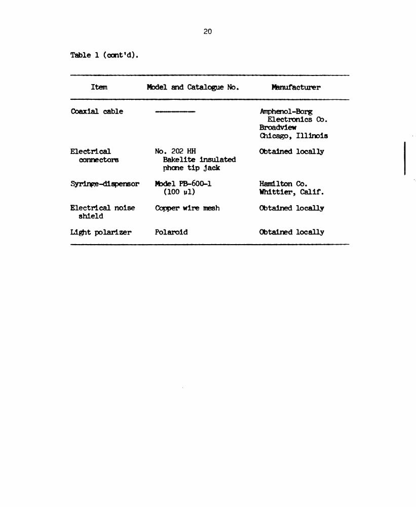
Table l (cont'd).
Item Model and Catalogue No . bonufacturer
Coaxial cable anhenol-Borg
Electronics Co.
Broadview
Chicago, Illirois
Electrical No. 202 HH Obtained locally
cornnectors Bakelite insulated
phone tip .1ack
Syringe-dispereor )bdel PB-600-l
(100 nil)
Electrical noise Copper wire mesh
shield
Light polarizer Polaroid
Hamilton Co.
Whittier, Calif.
Obtained locally
Obtaired locally
Page 31
21
Electrometer }
Figure 2. Schematic diagram of the membrane chamber and the
electronics for measuring the photo-emf. S, magnetic
stirring bar. 8114, black lipid membrane.
Page 32
22
0.1 M potassium acetate, was added. The membrane was formed by
applying a small amount of the membrae-forming solution with a
micro—syringe. 'Ib facilitate application, a 2 - in mm piece of
0.038" polyetmlere tubing was placed on the end of the micro-
Miriam
The positions of the lenses were adjusted until the
exciting light was focused upon the menbrane. 'Iie visible wave-
lengths were scamed by rotating the monochromator can with a smell
motor (Hurst Nbdel AR DA) at approximately 2.5 rum/sec. The lignt-
induced membrane emf was monitored with a calonel electrode in each
aqueous prose and a Cary 31 electrometer. A convenient recorder
speed was 2 in/min.
Extraction Technicmes
'Ihe membrane-forming solution was prepared by isolating
chloroplasts from commercial spinach and then extracting chlorophyll
arnd lipoid materials . All glassware employed was rinsed with acetone
and hot distilled water before use in order to avoid contamination
by soaps. ‘Ihe specific steps in the extraction are given below (Tien
and Howard, 1969) .
l. 'Ihe ribs and stalks were removed from 10 oz fresh
spinach. The leaves were washed thorongnly, then dried.
2. The leaves were added to a 300 mnl solution of 0.5 M
3 buffer (pH 7.5) in a Waring
blender at low speed. When all the leaves were added,
sucrose and 0.05 M KHCO
high speed was used for 30 sec. 'li'e mixture was then
Page 33
5.
23
filtered through for layers of cheesecloth; tre
residue was discarded. I
The filtrate was centrimged in approximately 110 mnl
quantities at 8700 g for 5 minutes; the supernatant
was discarded.
The chloroplasts were re-suspended in the buffered
sucrose solution (approximately 15 ml per test tube)
using the Vortex mixer, then centrimged at 8700 g for
5 minutes , discarding the supernatant.
The chloroplasts were broken by adding 25 m1 of glass-
distilled water to each test tube, mixed with the
Vortex mixer, tren allowed to stand for 5 mninutes. The
mixture was then centrimged at 9700 g for 10 minutes;
the supernatant was discarded.
The residue was extracted with 90 mnl of 2:1 petroleum
ether: methanol (by volune) in the Waring blender at
medium speed for l mninute, then centrimged at 5100 g
for 10 mninutes.
The top layer was pipetted off into a my flask and
evaporated to dryress. It was then re-suspended in
5 mnl of 1:1 n-butanol: dodecane (by volume), and stored
inthedarkatO-lnoc.
Page 34
21!
Procedure
The membrane was formed in 0.1 M acetate buffer, pH 5,
across a circular aperture of 2 mm diareter in a Teflon beaker set
inside a glass cup. After the menbrane had reached the black stage,
Fe013 was added to the inner chanber to bring the 1=ne3+ ion
concentration to 1 mM. 'ne open-circuit potential difference across
the membrare was monitored by a Cary 31 electrometer via a calomnel
electrode in tie aqueous phase on each side of tie membrane (Tien
and Howard, 1969) .
The black lipid membrane was excited with lig'nt from a
1000 w D.C. Xeon arc lamp (Hanovia, Type 976 C) which was passed
througn a teat filter, a visible grating mornochronator (Bausch and
Lomb, Model 5), a plano-convex lens, a shutter, a converging lens, a
polarizer, and a collimator (Figure 3). During formation, the
membrane was observed with dim green lignt(~525 hm).
After application of the membrane-forming solution across
the aperture, the membrane thinned first to a thickress of less than
1 um. At this stage, if the mnenbrare is observed at an angle with
the normal equal to the angle of incidence of illuminating lignt,
interference frings are seen (Figure 11a). In this way, the
orientation of the mnenbrane relative to the direction of propagation
of tie incident lignt was determined.
What of the lignt reflected norm the membrane is polarized
with the direction of vibration of the electric vector parallel to
the plane of the membrane, i.e., perpendicular to the plane of
incidence (Figure lb). The direction of polarization of the
Page 35
Figure
3.
Schereticdiagramofoptic
set-up
(top
view).
Microscope
(Inserver
I
I g!
‘l
25
Page 36
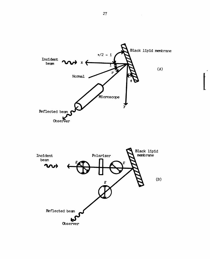
Figure ll.
26
(A) Schemnatic diagram illustrating the determination of
the orientation of the membrane relative to the direction
of propagation of the incident light, -'x (top view).
1, angle of incidence; r, angle of reflection. i = r = 4:.
(B) Schematic diagram illustrating the establishment of
the direction of polarization of the incident light with
the electric vector, 15’, vibrating parallel to the plane
of incidence (top view).
Page 37
27
Incident
beam W x6
(A)
y
Reflected beamn
Observer
Incident \ membrane
beam
Mr)
Reflected beam
Observer
Page 38
28
illuminating light was varied, by rotating the polarizer about the
direction of propagation, until the direction for which tie
interference fringes were observed to have a minimum intensity. This
established the direction of polarization.of the incidbnt light with
the electric vector vibrating parallel to the plane of incidence.
When the thickress of the membrane has fallen much below
1000 X, destructive interference gives rise to the optically "black"
appearance (Tien and Howard, 1969). When the "black" menbrane is
illuminated with exciting light, an electromotive force (open-circuit
voltage) is generated across it, with the side in contact with Fe3+
icn.becoming:more negative than the other'sideu The magnitude of
this "photo—emf" is dependent upon the wavelength of the exciting
light, and a "photo-emf’action spectrum" can be obtained by scanning
the visible wavelengtre (Van and Tien, 1970).
Page 39
CHAPTER IV
'IHEDRE‘I‘ICAL CCNSIIERA'IICNS
Before presenting the results and discussing their
significance, a consideration of sore aspects of theoretical back-
ground upon which the present interpretation is based is in order.
First, it is assumed that the chloropmrll black lipid membrane is a
lyotropic liquid crystalline system with smectic structure. In a
smectic structure tie molecules are arranged in layers, with their
long axes parallel to each other and approximately normal to the
plane of the layers. Tie molecules can move in two directions in the
plane and can rotate about one axis (Brown, 1967). In the
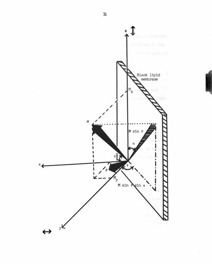
chlorcpmrll black lipid membrane, each transition moment is probably
restricted to an direction which lies on a conical surface making
an angle of o with the rormel, N, as depicted in Figure 5.
The orientation of each transition mnonent, M, is given by
the angle between M and the normal, N, to the membrane. The
components of M along the two directions, y and z, of polarization of
the incident light are 1% and M2, where
Pk-Msinesindcosoi-Mcosesinc (l)
Mz-Msinecosc (2)
29
Page 40
Figure 5.
30
Schematic diagram of the transition dipole moment, M, of
either the red or the principal blue chlorophyll
absorption band (see text). -x, the direction of
propagation of the exciting light.
Page 41
a?
Black lipid
xef
Page 42
32
The dichroic ratio, D, which is defined as the ratio of the
absorbence for morochromnatic light polarized in the y-direction (i.e.
torizontally polarized) to that for the light polarized in the
z-direction (i.e., vertically polarized) (Setlow and Pollard, 1962) ,
is given by
D-fi— , (3)
A2
If the absorption band for lignt polarized in the
y-direction has the same shape as the absorption band for lignt
polarized in the z-direction, then
I
Z - 1y (1)
Z
where I3, and 12 are the integrated intensities for the bands (Orchin
and Jaffé, 1971). Since the integrated intensity of an absorption
band is proportional to the square of the transition dipole moment,
_‘fx. . a . i (5)A2 12 w:
Dichroism of chloropl'yll black lipid membranes has been
attributed to orientation of chloropmll molecules in the membrane
(Crerry _e_t_a_1_., 1971; Qnerry gt gl_., 1972; Steinemenn 935931., 1972).
The (kpendence of the magnitude of the blue and red peaks in the
photo-emf action spectrum on the direction of polarization of the
exciting lignt is apparently a direct consequence of the dichroism
of the membrane. If so, then ratios of emf magnitudes of each peak
Page 43
33
for the stated two directions of polarization of incident lignt
should allow calculation of the orientations in the membrane of the
transition dipole mmnents of the blue and red absorption bands. For
each peak the ratio of photo-emfs, (Em )y/(Ehv)z varied less then
81 with a fourfold increase in light intensity.
If the photo-emf, Em, is proportional to the amount of
light energy absorbed by the mnenbrane, then the ratio of the emf
magnitudes for horizontally polarized light to vertically polarized
light for the red peak or the principal blue peak gives the ratio of
the absorbances for these directions of polarization (see
Appendix A), and
WWW)" - "‘2' (6)
<sz 7?
Since each transition moment is probabe restricted to any
direction which lies on a conical surface mnaldrng on angle of a with
the normal, My and 142 must be integrated over all a,
-1(F‘hv)y (2') f3. ’6 do
(am),5 (2:)‘1 I? u: do
(7)
( )
Msm2¢ + 2ctn26sin2¢ (8)
(gnu)!
Since the orientation, c, of the membrane is knom, the
last equation can be used to calculate the direction, a, of each
Page 44
3H
transitionnmnment.fromnthe1magnitudes of the polarized light
induced.photo-emf peaks. Since there are both chlorophyll a and
chlorophyll g in the spinach chloroplast (Park and Biggins, 1961:),
the value fer 0 thus obtained is actually an average over'both types
of’chlorophyll present in the membrane.
Polarized absorption and fluorescence measurements with
chlorophyll have shown that the two transitionnmnmenms responsible
for the red and principal blue absorption bands are perpendicular to
each other and lie in the plane of the porpl-wrin ring (Rabimwitch,
1956; Goedneer, 1966). 'Ihe orientations, “R and GB, of the red and
principal blue dipole mnomnents with respect to the normal, N, to the
membrane then supply enough infbrmation.that the orientation of’the
chlorophyll porptvrin ring can be calculated. With the assmptions
that the red and principal blue dipole'moments are mutually
perpendicular and lie in the plane of the porpmrin ring, it may be
shown (see Appendix B) that the angle, 8, between the plane of’the
porphyrin.ring and.the normal to the>membrane is given by the
expression
2, 2 2cosB cos 0R+coseB (9)
Page 45
(HAP'ERV
E30138 AND DISCLBSICN
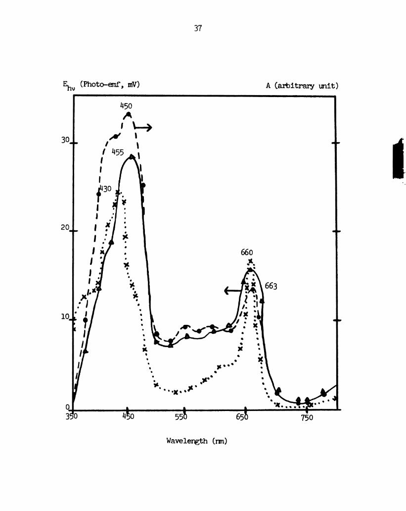
A typical photo-emf action spectrum of a chloroplast
extract black lipid membrane, obtained by the method outlined in the
experimental section, is shown in Figure 6. . 'Ihe photo-emf action
spectrum shows a slight dependence on the direction of wavelenan
scan, with peaks shifting about 5 mm and each peak magnitude
changing by a factoer of about 1.2. The peaks in the action spectrum
are shifted to the red from the peaks for the bulk solution
absorption spectrum. Otherwise, the photo-emf spectrum bears a
strong resemblance to the absorption spectrum for chloroptmrll a, with
peaks at 1130 arnd 660 on. Other researchers have observed a red
shift in the red and blue peaks from the chlorophyll absorption
spectrum in bulk to the absorption spectrum in chlorophyll black
lipid membranes (Steinemam 9331;, 1971; Cherry 33gb, 1971). It
is likely that this is responsible for the red shift in the pinto-
emf action spectrum.
The photo-emf blue peak is of greater magnitude for the
incident light polarized perpendicular to the plane of incidence
than for it polarized parallel to it. The situation is opposite for
the red peak. From the photo-emf peak values for horizontally and
vertically polarized light, the orientation 0 of each transition
35
Page 46
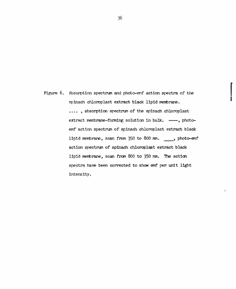
Figure 6 .
36
Absorption spectrum and photo-emf action spectra of the
spinach chloroplast extract black lipid membrane.
.... , absorption spectrum of the spinach chloroplast
extract membrane-ferming solution in bulk. -—-,‘photo—
emf action spectrum of spinach chloroplast extract black
lipid membrane, scan from 350 to 800 rmn. _, photo-emf
action spectrum of spinach chloroplast extract black
lipid membrane, scan from 800 to 350 nm. The action
spectra have been corrected to show emf per unit light
intensity.
Page 47
37
Em (Photo-emf, mV) A (arbitrary unit)
Wavelength (nm)
Page 48
38
moment can be calculated with the aid of the equations deve10ped
above. 'Ihe results are summarized in Table 2. ‘Ihe principal blue
transition moment was calculated to make an angle of 21 i: 2° with the
plane of the membrane; the red transition moment, an angle of
38 1 2°. From these angles, an angle (averaged over the
chlorophyll a and chlorophyll b in the membrane) of 1&5 1 5° is
calculated for that between the plane of the porphyrin ring and the
plane of the membrane (Weller and Tien, 1973).
These results can be compared with values for chlorophyll
porphyrin ring orientation obtained from polarized absorption
spectroscopy on artificial chloroprwll membranes by other
researchers. For chlorophyll-egg lecithin black lipid membranes,
Cherry gt _a;l_. found angles of 188° for chlorophyll a_ and 51° for
chlorophyll 2 (Cherry _e_t_ a1. , 1972). Steinemann and co-workers
found angles of M r 3°, 1:6 i: 3°, and 1&9 1 5° for chlorophyll a-
phosphatich'l ethanolamirne , chlorophyll _a_-dioleoyl-phosphatich'l
choline, and chlorophyll _a—phosphatidyl serine membranes,
respectively. They found ”2 i '40 for chlorophyll b__-dioleoy1—
phosphatidyl cholire membranes.
'Ihere exists the possibility that the acidic bathing
solution employed in these experiments converts some or all of the
chlorophyll a and chlorophyll t_n in the artificial membrane to
pheoprwtin a and pheophytin _b_, respectively, by the renoval of the
Mg atom from the center of each porphyrin ring. The central Mg atom
of chlorophylls is readily displaced by strong and weak acids
(Willstatter and Hocheder, 1907) .
Page 49
Table
2.
Orientatiore
inthespinach
chloroplastextract
chlorophyllblack
lipid
membraneof
the
transition
dipolemoments
responsible
for
the
chlorophyll
redandprincipalblueabsorption
bancb
,andorientationofthe
chloropl'wll
porphyrin
ring.
These
values
are
averagedoverthe
chlorophylla
and
chlorophyll
p_present
in
themembrane.
(mm)y
o(°)
(am)z
Absorption
peak
"Average"
angle
betweentransition
moment
andplaneof
themembrane
(I/2
-6)
(0)
"Average"
angle
betweenplaneof
porphyrin
ring
andplaneof
merbrane
(m/Z
-8)
(o)
Bluepeak
0.59
69
i:2
(1455on)
Redpeak
1.08
52
i2
(660mm)
2122
38:2
11515
39
Page 50
1&0
In aqueous acetone, the rate of pheopmtinization is first
order in acid concentration (Joslyn and Mackinney, 1938) and in
chlorophyll concentration (Mackinney and Joslyn, 19110). The rate
constant for Mg displacement in 20% aqueous acetone is S - 6 times
larger for chlorophyll _a_ than for chloropl'yll _b (Schanderl _e_t_ _a_l_. ,
1962). Activation energy for chlorophyll a was about 11 kcal.
'Ihe plneophytinization of chloropmn in the lipid bilayer
would depend on the degree of exposure of the membrane ctnloroptwll
porphyrin rings to the acid. Loss of m from chlorophyll has been
found to be 13 times as fast in a chlorophyll monomolecular layer at
an air-water interface (pH in) as in acetone (Rosoff and Aron, 1965).
The rate in the monolayer is sensitive to pressure and the presence
H
ofO 0a , andrg".2’
The rate constant for the mnonolayer pheophytinization
mereased with increasing pressure. For example, the rate coretant
1 "-1was 1.36(103) min' at an initial pressure of about 6 (hues/om
(i.e. , a molecular area of 120 :2 per chlorqnmll molecule) and
l.‘43(102) min-'1".-1 at 16 dynes/om. These results suggested that the
onange in orientation of the chlorophyll molecules in the monomer
is responsible for the availability of the porpmrrin ring m for
reaction.
However, the chloroplwll embedded in a lipid bilayer
matrix may not be as exposed to the acidic bathing solution as
chloroplwll in solution or in a monolayer. The porpm'rin ring
orientation angle determdnation in the experiments described herein
does not supply enougn intonation to ascertain the availability of
Page 51
ill
the chelated chloropmrll It atom to the acid.
Comparison of the photo—emf action spectrum and the
absorption spectra of chloroptwll in solution and in a lipid bilayer
with the absorption spectra of pheoprvtin in solution seems to
indicate that a very small amount of chlorophyll in the chloroplast
extract chlorophyll-lipid bilayer is pheopmrtinized.
The absorption maxima for chlorophyll _a_ and chlorophyll _b
in ether have been reported as 1630 nm, 662 rm and 1453 mm, 6142 nmn,
respectively. The maxima for pheophytin a and pheoplwtin b in ether
are 1408 mu, 667 mm and “314 mm, 655 nm, respectively (Goedheer, 1966).
Pheophytinization shifts the principal blue peak about 20 mm to a
lower wavelength and the red peak 5 - 13 nm to a higher wavelength.
waever, Cherry gt; _a_l_. found maximna at 1039 nm, 672 nm and
1466 rm, 653 nm for chloropmll a and chlorophyll b_, respectively, in
the chlorophyll-egg lecithin bilayer (Cherry _e_t_ a_1_. , 1972). These are
red shifts from the bulk spectrum of 9 - 13 nm for the principal blue
peaks and about 11 nm for the red peaks. Steirnemann and co-workers
found maxima at 1&3? mm and 672 nm for chlorophyll a in the
chlorophyll-dioleoyllecithin bilayer (Steinemam _e_t_ _a_l_. , 1971).
‘Ihese are red shifts of about 5 mm for the principal blue peak and
10 nm for the red peak from the bulk spectrum.
‘Ii'e photo-emf actiwn spectrum peaks (Figure 6) for the
chloroplast extract chlorophyll black lipid membrane are red-shifted
20 - 25 nmn from the bulk absorption spectrum for the principal blue
peak and about 3 nm for the red peak.
Pheophytinization of chlorophyll, or even subsequent
Page 52
112
oomplexing of a Fe atom in the center of the porptwrin ring, would
change only slightly the directions of the red and principal blue
transition mnonents relative to the symmetry axes of the porphyrin
ring. For example, the direction of the chlorophyll red transition
moment vector is shifted about llo by replacement of the chelated
1% atom by a Fe atom (Platt, 1956).
The calculation of the angle of tilt, B, of the porphyrin
ring is not unduly sensitive to the assurption of a right angle
between the red and principal blue transition moments. For example,
if this angle is perturbed up to 10° fromn the value of 90°, the
resultant uncertainty in a is still only 2 5° (Cherry 9}; _a_l_. , 1972).
The polarized light-induced emf in black chloropm'll-lipid
mnenbranes is apparently a sensitive technique for investigating the
orientation of chlorophyll in a bilayer lipid membrane. It requires
only one membrane and a simple electronic set-up. Furthermore, the
existence of the phenomenon of light-induced emf in artificial
chlorophyll membranes suggests that a similar phenomenon may occur
_ip_ :13 in the chloroplast thylakoid membrane during the process of
transduction of lignt energy into chemical energy.
Page 53
OiAP'IERVI
MISGZILANEOUS
Photoelectric Action Spectra with Various Additional
Caponents in the Bathing Solutiens
The dependence of the photo—emf action spectrum of the
spinach chloroplast extract chlorophyll black lipid membrane upon the
direction of polarizatian of the excitirg light was measured for the
following four sets of bathing solution components (Figures 7 - 10).
The exciting light was incident initially upon the solutien shown at
the left side of the membrane.
l.
Fe3+ (lmfl) Black Lipid Water-soluble"
Membrane chloropmrll
HAc (pH 5)
HAc (pH 5)
2. 3... .1;
Fe (1mm Black Lipid Thionine (10 a)
humane
HAc (pH 5) HM (pH 5)
3. 3+ _u
Fe (lmfl) Black Lipid p—Benzoquinme (3)(1o nun)
Membrane
HAc (EH 5) HM: (PH 5)
“3
Page 54
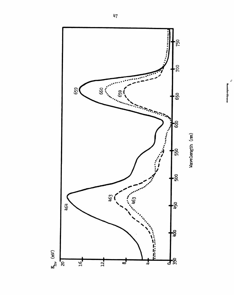
Figure 7.
Nil
Photo—emf action spectra of the spinach chloroplast
extract black lipid membrane with l mnM FeCl3 in the
outer chamnber and "water-soluble chloropl'wll" in the
inner chamber. 0.1 ml of "water-soluble chloroprwll"
4L-
from KIRK Laboratories, Inc. , Plainview, New York (no
concentration value was supplied by them) was added.
_, action spectrum with unpolarized exciting light.
---, action spectrum with the exciting light polarized
perpendicular to the plane of incidence. . , action
spectrum with the exciting light polarized parallel to
the plane of incidence.
Page 55
Ehv 6..
““7
633
Sun-
583
u:b
5&1
“5
21-
....\
..
.0
I0..\
..O
'..-°“°°‘°$"-\
/‘.-'
.°
I.
..'0
1!-
’..
v11
IL
l
350
400
uBo
560
séo
Wavelength(m)
Page 56
Figure 8.
1:6
Photo-emf action spectra of the spinach chloroplast
extract black lipid membrane with 1 mid FeCl3 in the outer
chamber and 0.1 m thionine in the imer chamber.
_, action spectrum with unpolarized exciting light.
---, action spectrum with the exciting light polarized
perpendicular to the plane of incidence. . .. . , action
spectrum with the exciting light polarized parallel to
the plane of incidence.
Page 57
E's.
<mV>
20
16
11614
Wavelength
(rm)
559
750
"7
Page 58
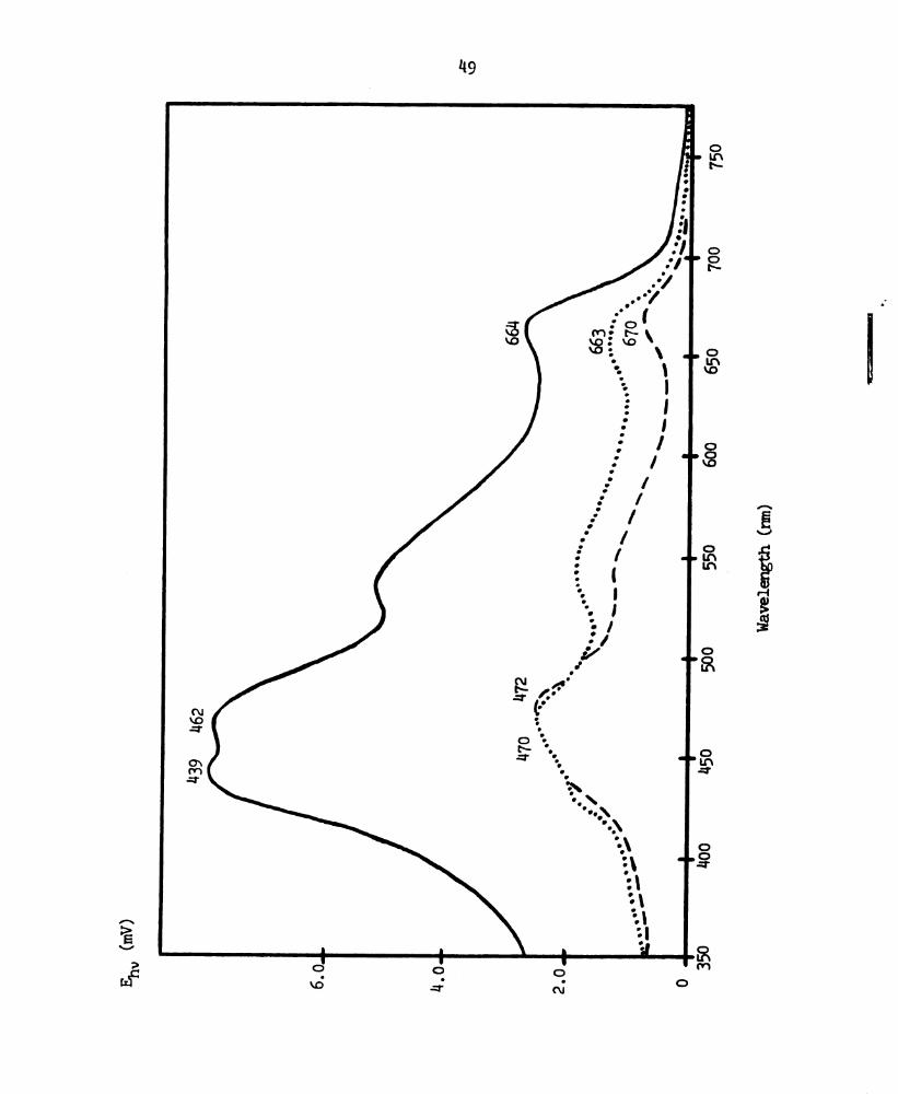
Figure 9.
148
Photo-emf action spectra of the spinach chloroplast
extract black lipid menbrane with 1 mid FeCl3 in the
outer chamber and 0.3 mM p-benzoquinone in the inner
chamber. ______, action spectrum with urnpolarized
exciting light. -—-, action spectrum with the exciting
light polarized perpendicular to the plane of incidence.
. . . . , action spectrum with the exciting light polarized
parallel to the plane of incidence.
Page 59
Q9
(mfl)1xfiheneaen
09L
00L
059
009
095‘
O
In
:1-
O
O
:1-
8m
.nllll
ouoeloo..fi.>.r‘
V:
IL
::
l0
ux
-—~-
0I
\
o.
0L9
.........
\\
’.:.'..00000
...000000...
...'.o
\~_~\
I...
0....
\/.0
£99
...°o.
00.......\
’3.
'00.
.0.
.lo
099
1-o°n
29w
6£n
(am)
“”3
Page 60
Figure 10.
50
Photo-emf action spectra of the spinach chloroplast
extract black lipid membrane with 1 mM FeCl3 in the
outer chamber and 14 mM L-ascor'bic acid in tlne inner
chamber. _, action spectrum with unpolarized
exciting light. --—-, action spectrum with the
exciting light polarized perpendicular to the plane
of incidence. . . . . , action spectrum with the
exciting light polarized parallel to the plane of
incidence.
Page 61
Eh.(m
-.
30
‘459
51
Page 62
52
Fey-(1m?!) Black Lipid L-Ascorbic d
Mme (In) (10" ) H
mm (PH 5)
HAG (DH 5)
All of these spectra exhibit a dependence of the red and
principal blue peaks upon the direction of polarization of the
exciting light which is similar to that exhibited by the membrane
with only Fie3+ in one bathing solution. However, the addition of
another species of dlromcphore ("water-solnble" chloropmll or
thionine) and/or the alteration of the photochemistry probably
rembr incorrect an interpretation of charges in the ratio
(Envy/(Em), as reflecting only changes in the orientation of the
membrane chlorophyll ring.
Preferential "Polarized Lijnt [Iihoto-Bleachirg" of
Gnlorepm'll Molecules in the Membrane and the
Attenpg to Engloy it for the Determination of a
Rotational Diffusion Relaxation Time for the
Membrane Chlorophyll Molecules
Under continuous illumination of the spinach chloroplast
extract chlorophyll black lipid nembrane with light of either the red
or the principal blue photo-emf peak wavelength, the photo-emf first
(a) increases linearly (Figure 11), then (b) the rate of increase of
F1” decreases urntil (c) Em remains constant or decreases. 'nne
existence of this saturation of the capacity of the meninrane to
transduce light energy into energy stored in an electric field
suggests a possible method for the determrmatien of the diffusional
\
A
Page 63
53
an, (mV)
A
3 ..
2 P I’M
1 .-
a
4 s 1 1
o i 15 30 “5 60 >Tile (sec)
Lignt
on
Figure 11. Shape of a typical plot of the photo-emf versus time.
Excitation of the spinach chloroplast extract chloropwll
black lipid snenbrane is at either the principal blue peak
or the red peak wavelength. The athirg solutions are
0.1 M acetic acid (pH 5) with 10' M FeCl3 in the inner
chamber. See text for further details.
Page 64
514
rotation frequency of the chlorophyll molecules in the membrane.
If the membrane chlorophyll molecules with their red (or
principal blue) transition moments more aligned in a certain directien
—- e.g. , more vertically oriented than horizontally —- could be
preferentially "photo-bleached" with plane-polarized light, then there
would be a snbsequent amnonmt of time before rotational diffusion would
again randomize the azimuthal orientations of the "bleached"
chlorophyll molecules.
During this randomizing interval, excitirg lignt plane-
polarized normal to the polarization direction of the previously
enployed "bleaching" lignt would give rise to a photo-emf of nearly
"urnbleached" magnitude, whereas light polarized in the "bleaching"
plane would result in a photo-emf of smaller magnitude than that when
"unbleached." I.e., the ratio (Ehv)y/(Ehv)2 will have a value
different than when the merbrane is "unbleached." The time required
to return to the "nmbleached" value of (Ehv)y/(Ehv)z is then related
to the rotational diffusion relaxatien time for the membrane
chlorophyll molecules .
In order for an experimental determination of the
rotational diffusion frequency to be performed using preferential
"bleaching" of membrane chloropmll molecules with plane-polarized
lignt, it would be desirable first to slow that the preferential
"bleaching" can occur. Le. , it must be shown that the mnentnrane
chloroplwll molecules can be "fixed" (e.g. , with glutar'aldehyde,
mo“, or each) so that preferential "pl'otobleaching" can be observed
over a period of time of the order of a few seccnm or more.
Page 65
This technique for the determination of the rotational
diffusion relaxation time for the chloropmrll molecules in the ‘
membrane is in some aspects similar to the techniques of Brown and
Gone who established first that (a) dichroism of frog retinal rod
outer segment viewed end-en can be photo-induced by partially
bleaching r'hocbpsin with plane—polarized lignt following fixation with
glutamldehyde (Brown, 1972), and then that (b) the rapid decay of the
dichroism induced by a flash of plane-polarized light provides a
direct measure of the relaxation time —- 20 usec --- of rhodopsin in
the receptor menbrane (Gene, 1972).
Isolated rhodopsin is highly dichroic, absorbing light most
strongly when the electric vector is parallel to the leg conjugated
chain of ll-cis retinal (Clayton, 1971). Rodopsin consists of the
chromphore ll-cis retinal attached by a Schiff's base linkage to the
lipoprotein cpsin. Several chemical and structural properties of
rhodopsin suggest that rotations of the chromqnhore accurately reflect
rotations of the entire molecule .
Photoisanerizatien of retinal from ll-cis to all-trans
initiates a bleaching process which cornsists of a series of
configuration transitians in opsin, and leads eventually to the
release of retinal:
hv
modopsin -—) Preluni —-) Lumi —)nete I
(“98 nm) (5"3 nm) ('497 nm) (8 mm)
~lvRetinal + (rosin (—— Para L—Meta II
(387 mm) (“65 nun) (330 ml)
Page 66
56
line absorption maxima are shown below the configurations .
Retinal in rhodopsin and lumirl'odopsin has an absorption maximum at
about 500 nm, whereas in prelumirrodopsin the maxim shifts to 5113 mm.
At the meta II stage, the spectrum shifts for to the blue, becoming
similar to that of free retinal.
Attempts were made to "fix" the chloronlwll molecules in the
spinach chloroplast chlorophyll black lipid mnanbrane with
glutaralchmrde, and subsequently to induce a change in the ratio
(h'.h\,))/(n~:m)Z with plane-polarized light at the photo-emf red peak
(Table 3). It was ibund that the presence of glutaraldemrde has no
effect , or perhaps only a small effect , on the membrane photo-emf
action spectrum. However, no change in the ratio (Shay/(E1102 upon
excitation with plane-polarized light was observed. Simnilar
concentrations of men also proved ineffective.
Some consents m be in order on possible reasons for the
ineffectiveness of glutaraldemrde in "fixing" the membrane chlorophvll
molecules sufficiently to enable pinto-induction of dichroism.
Glutaralderwde has been used by researchers principally to stabilize
proteins by cross-lirnldrg them. It may only react with lipids con-
tainirg free ammo MB (e.g. , phosphatidyl ethanolamine)
(Jolmston and Roots, 1972). The reaction of an amends with a
primary amine is expected on the basis of classical organic reactions
(i.e. , Schiff base formation).
Althougn Brown observed very strorg plotoinduced dichroism
with a 2 . 51 glutaraldemde concentration fixation of rl'ocbpsin in the
disk membranes (Brown, 1972) , there are important differences between
A-
Page 67
Table
3.
Four
attempts
at
fixationof
themembranechloropmllmolecules
withglutamldehvde.
Fbllowingexposure
toglutamldehvde,
the
spinachchloroplast
extract
chlorophyll
black
lipidmembranewas
"bleached"with
verticallypolarized
660nm
light
(the
photo-emf
action
spectrumredpeakwavelength).
Amountof
251
Time
Time
()
()
Photo-
glutaraldehyde
stirred
bleached
Eh”
Y/Rh"
2induced
added
change
inemf
(ml)
(min)
(min)
ratio
57
Inner
Orter
Initial
Immed.
15
sec
chamberchamber
after
later
bleaching
0.1
---
10
2.0
----
2.0
None
0.1
0.1
11
2.0
1.8
2.0
None
0.5
0.5
10
1.8
1.8
1.6
None
Nmmfi
1.0
1.0
20
2.0
1.8
1.8
None
Page 68
58
his system and an artificial marbrane. 'lhe retinal rod outer segment
is a tissue and rhodopsin is lat-31y protein. 'ihe black lipid mnem-
brane contaim only chloroptwll and lipids .
It must be noted that Vasquez and co-workers (vanquez _e_t a}, ,
1971) employed a 23 glutamldemrde concentration in fimtion of black
lipid membranes for electron microscopy. The black lipid membrane
types which they fixed were (a) lipidic -- total phospholipids of the
cerebral cortex and cholesterol, (b) lipidic-proteolipidic --- with
small ononmnts of proteolipid from Electrophoms electrqalax, and
(c) proteolipidic -- fromn Electrophorus electroplax.
Effect of Voltage _Applied to Spinach animust
Extract Black Lipid Mennbrane on the Dependence of
the Photo-emf Action Spectrum upon the Direction
of Polarization of the Exciting Lignt
Externally applied voltages across the membrane up to 35 mV
had to observable effect on the dependence of the photo-emf action
spectrum upon the direction of polarization of the exciting lignt.
Page 70
APPENDIX A
DIRECT Pmpom'IonAm'rr 0F rue mono-Em, Em,
mmmorumammomsim
mm IEADS TO DIRECT Paommovm or
(RM/(31192. TO THE DICHHDIC RATIO
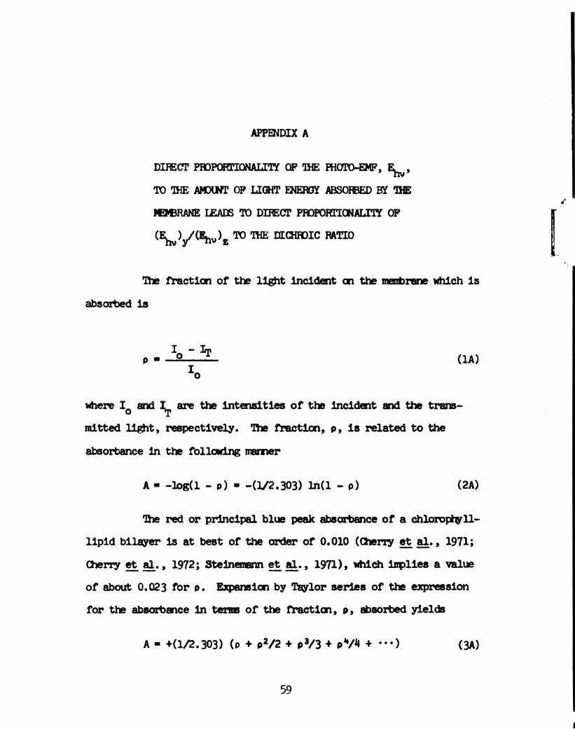
The fraction of the light incident on the membrane which is
absorbed is
(1A)
where Io and IT are the intensities of the incident and the trans-
mitted lignt, respectively. The fraction, 9, is related to the
absorbance in the following mnamer
A - -Joa(1 - o) - -(l/2.303) 1m(1 - a) (2A)
The red or principal blue peak absorbance of a chlorophyll-
lipid bilayer is at best of the order of 0.010 (Cherry e_t_ 9:1. , 1971;
men-y 93 a_1_., 1972; Steinennann 2331;, 1971), which implies a value
of about 0.023 for 9. Expansion by Taylor series of the expression
for the absorbance in term of the fraction, 9 , absorbed yields
A - +(l/2.303) (p + 92/2 + 03/3 + 0V“ + "') (3A)
59
Page 71
60
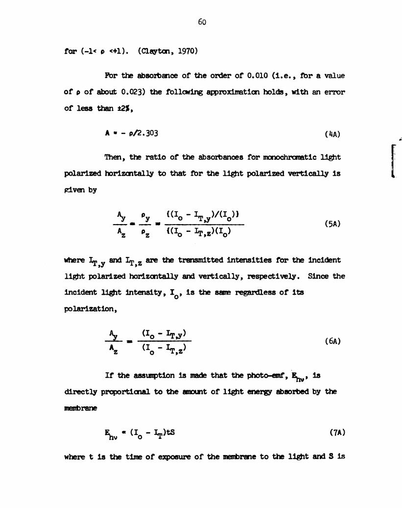
for (-1< o <+l). (Clayton, 1970)
For the absorbance of the order of 0.010 (i.e. , for a value
of p of about 0.023) the following approxinnnation holds, with an error
of less than :21,
A - - p/2.303 (HA)
Then, the ratio of the absorbances for monochromatic lignt
polarized torizontally to that for the light polarized vertically is
given by
A}, 9y {(Io - IT y)/(IG)}
--— - —— - ’ (5A)A 9 (IO " Lr’z)(Io)
where IT y and IT,z are the transmitted intensities for the incident
’
light polarized horizontally and vertically, respectively. Since the
incident lignt intensity, 10, is the same regardless of its
polarization,
(I - )i- o ITJ (6A)
Az (Io - IT,Z)
If the assnmnption is made that the photo-emf, Em, is
directly proportional to the amount of light enery absorbed by the
membrane
an . (Io ~LI.)tS <7A)
wheretisthetimeofeiqnosureofthemenbrenetotheligntandSis
Page 72
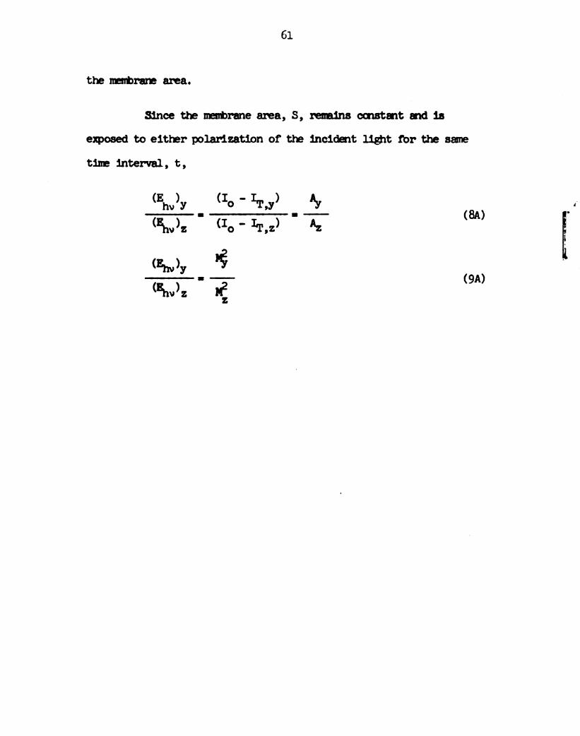
61
the manbrare area.
Since the mnenbrane area, 8, remains constant and is
exposed to either polarization of the incident lignt for the same
time interval, t,
(Ehv)y (IO ' ITJ) .. ‘3' (8A)
‘2(Elnv)z - (Io " I‘I',z)
(Em)y @
an”). ' “2
(9A)
Page 73
APPBWDIX B
WATIWGF'IHEANGIE, 8,m
MMOF'IHEPOWRJNGAND
MNOML,N,'ID'IIEW
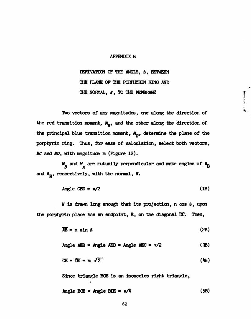
'No vectors of am magnitudes, one along the direction of
the red trareition moment, MB, and the other along the direction of
the principal blue transition mnonent , NR,
porphyrin ring. ‘lhus, for ease of calculation, select both vectors,
determine the plane of the
BC and 80, with magnitude nm (Figure 12).
”B and MR are mutually perpendicular and make angles of GB
and respectively, with the normal, 10.OR,
Arngle can - v2 (18)
N is dram long enough that its projection, n cos 8, upon
the porprwrin plane has an endpoint, E, on the diagonal E. Then,
m-nsina (23)
AngleAE-MgleAED-Angleflc-w/Z (38)
55-53an (‘8)
Since triangle so: is an isosceles rignt triangle,
0
MgleBm-AngleBIE-w/H (SB)
62
Page 74
53
—>~a
Figure 12. Schematic diagram of the relation between the red
transition moment vector, , tie principal blue
transition moment vector, , and the normal to the
membrane, I. See text for mrther details.
Page 75
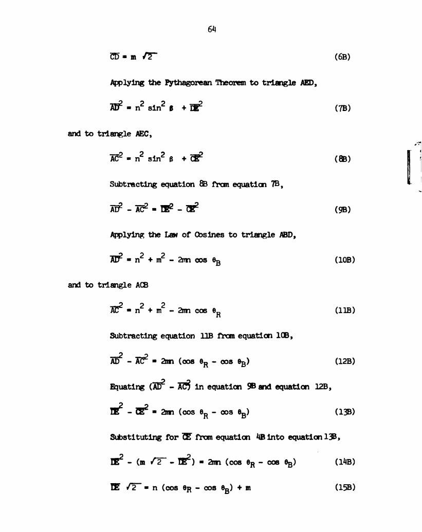
614
CD— 8 rm ’2'" (68)
Applying the Pythagorean Theorem to triangle AED,
KHz-n2 sinZB +E2 (7s)
and to triangle AEC,
Inez-n2 sinZB +fi‘? (m)
Subtracting equation 88 from equation 73 ,
I132 - 2T6?- - m?- - :22 (98)
Applying the Lav of Oosines to triangle ABD,
ADE-n2+m2-2nvncos 913 (103)
and to triangle ACB
Ez-nzi-mz-ancosoR (118)
Subtracting equation 118 from equation 1(8,
fi-Ké-amwoseR-cosoB) (128)
Ehuating (1'52 - R; in equation 95“ equation 128,
mz—Ez-Zmnwos eR-cos GB) (138)
Substituting for E from equation l|Binto equation 19.
mz-(m/T-m2)-am(eoseR-coee3) (nus)
El'é—Inn(cosoR-ooseB)+mn (158)
Page 76
65
Squaring both sides of equation 19,
2 -n(cosoR-cos03)+2mn(cosoR-cos05)+m
(168)
Substituting for m2 from equation 168 into equation 78,
2
B52-n2sin284-n/2 (cos OR-cos GB)2
+mrn(cos eR-coseB)+nm2/2 (178)
Tin—DQ-nZ-mnz/Z-nz ooszsi-nZ/Z (008 93-008 93)2
+mn (cos OR" cos 88) (1%)
Substituting for (In? - n2) from equation 1CD into
equation 188,
2
m2-2mncos OBI-Irma/Z-n2 cosza+n2/2 (cos oR-cos GB)
+ mn (cos OR - cos GB) (198)
m2/2 -mrn (cos 08 + cos OR) II n2/2 (cos OR - cos 98)2
- n2 0082 a (2m)
Substituting the trigonometric identity (sinza - 1 - coszs)
in equation 78,
TAI_)2a-n2(1-ccs28)+fi-.=2-n2--n2cornea +E2 (218)
nacosze - m2 I n2 - A152 (228)
Applying the Law of (bsines to triangle 813,
nzccszs - m2 + E2 - 2m (rs) cos “5° (23)
Page 77
66
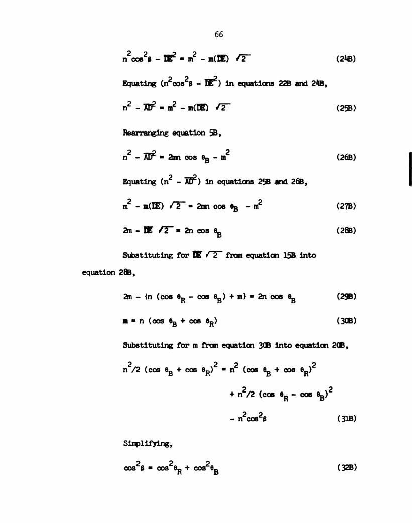
nzcoszs - m2 - m2 - mm) ’2— (2'48)
Equating (nzooszs - E2) in equations 228 and 2118,
nZ-Zfiz-nz-Mm IT (258)
88mins equation 58.
nZ-Ez-amcoseB-nmz (26s)
Equating (n2 - H52) in equations 258 and 268,
mz-lm(fi) fir-anncosea -nm2 (278)
am-E/E—IchoseB (288)
Substituting for m I 2 from equation 158 into
equation 2m,
2nm-{n(cosoRucosnnB)-n-nm}--32rncos(lB (2Q)
In-n(ooseB+cos OR) (3%)
Substitutirg for m from equation 3m into equation 2m,
2 2 _ 2 2
n/2(coseB+coseR) n (costar-omen)
2 2+n/2 (cos OR-cos OB)
- nzcoszs (318)
Siflplimns,
cosB-cos26R+coseB (323)
Page 79
L9
“[2‘stsau.°§"i(Inst)'8‘Suau
'62‘92:‘Rtomnewwith‘(zL6U'v'a‘ano
'00noon
1W..‘II‘aatwaw3mmwewin.‘(IL6I)'nn'8‘uoimm
'003908
Ina—mason..‘I‘Jaszniw3mmpunam‘(OLGU°x'H‘uonmto
140xMan"00mmnqndHavens
“weanlmonauurcommanumerals.‘(5961)'3‘uowato
'219‘L92mov
'EW'urmoors‘(ZL6'[)'cn‘ua-Iduowe“)1‘nenn“r“H‘K-uao
’ISE‘Eu'qumwas
'Bfildommeteors‘(IL6'0'0‘WMwe")1‘mn"r'3‘Mano
.091:CE0°08om.m.66(896I)or.3(map
‘Lnt‘128°thmeowwas‘(656Uin‘mm
'091:‘(92-0)
2151:!!th‘Ktonmocmremnantuampnooas‘(9961)in‘mm
“SE‘952morenanmm‘(zL6t)'nn‘d‘umas
'0‘!‘on;755mm‘(L96U'H'0‘wma
'nnEt‘agz'Emmmowsnam°z‘(u6t)'8's‘M
'239‘IBIestates‘(ELGU'8'wManama-Ia
'zn‘nnIEmov'smdora
'Imnoom‘(ELGU'9‘unottteaare“u‘mm-taqoun‘’f‘uoiaaa
1992‘I6"503"was'WT‘(696t)T'r‘zzvnnweon‘ammems
__.__'an9‘91."00%
wasw'r‘(nnSGU'a'N‘uattvpm“a'1‘KBIQP‘JM“Io‘W
Page 80
68
Fiscler, n. s., Tenpleton, D. 11., Zalncin, A., and Calvin, M. (1971),
J. in. den. §_o_c_. 93, 2622.
Fischer, M. S., Tenpleton, D. 8., Zalkin, A., and Calvin, M. (1972),
J. 55. (hem. _S_oc_. 918, 3613.
Frey-Uysslfing, A. and Steinmnn, E. (19118), Biochlm. Biopms. Acta
2, 25 .
Frye, C. D. and Edidin, M. (1970), _J_. Cell _S_ci. 7, 313.
Geacintov. N. 8., Van Nostrand, F., Pope, 14., and 'I‘inkel, J. 8.
(1971), Biochim. 83m. Acta 226, 65.
Geacintov, N. 3., Van Nostrand, F., Becker, J. F., and Tinkel, J. B.
(1972), Biochim. Biopmrs. Acta 267, 65.
Getov, G. K. and Jordanova, S. T. (1972), Biofizika 17, 782.
Goedheer, J. c. (1955), Biochim. sigpnm. Acta 16, 1:71.
Goedreer, J. C. (1966), "The Chloroprwlls," Vernon, L. P. and
Seely, G. R., eds., Academic Press, New York, 1“.
Hanson, E. A. (1939), flecueil Trav. Q11. Neerl. 36, 183.
Hill, R. (1937), Nature, 881.
Kill, R. (1965) 'Bsws in Biodnemistry," Cowbell, P. N. and
Gmune, G. D. ’ a. , 1.
Hoff, A. J. (19718), Photochem. and Ptotobiol. 19, 51.
Johnston, P. V. and Ibots, 8. I. (1972), "Nerve Menbranes," Pergamon
Press, New York, 116.
Joslyn, M. A. and Ahclflnney, G. (1938), 9;. _Ag. Chem. _Sg_c_. 60, 1132.
Junge, N. and Witt, H. T. (1968), _a_. Naturforsch. 23, 1571.
Kasha, n. (1959). 533. Mod. m. 31, 162.
Katz, J. J., Ballschmiter, K., Garcia-Abrin, 14., Strain, H. 8., and
Uphaus, R. A. (1968). Proc. Nat. Acad. Sci., (BA 60, 100.
Katz, J. J. (1973), Natm'wissenschaften 60, 32.
Re, 8. (1966), "line Gnloropl'ylls," Vernon, L. P. and Seely, G. 8.,
ecb., Acadennic Press, New York, 253.
Kornberg, R. D. and McConnell, H. N. (1971a), Proc. Nat. Acad. Sci.,
EA 68, 25614.
Page 81
69
Kornberg, R. D. and McConnell, H. M. (19718), Biochemistry 10, 1111.
Kreutz, w. (1970), 591. 2233.- 33. 3, 53.
Kreutz, w. (1972), Angewandte m, _1_n_t_. 11, 551.
Lelrninger, A. L. (1970), "Bioctemistry," Worth Publishers, Inc.,
New York.
ibcldnney, G. and Joslyn, M. A. (19110), J. A_m_. _Qn_e_m_. &- 62, 231.
McNamee, M. G. and McConnell, H. M. (1973), Biochemistry 12,2951.
Menke, N. (1938), 50113195. 85, 256.
Menke, W. (1958), _Z_. E2: 1&6, 26.
Morita, s. and Miyazald, T. (1971), m. m. 5333 21:5, 151.
Mleller, P., Rudin, D. 0., Tien, H. T., and Nescott, N. C. (1962),
Nature 19“, 979.
Nicolgcé'n, g. L. and Singer, 3. J. (1971a) Proc. 1595. Acad. §_o_i__., Lg;
. 9 2.
Nicolson, G. L., Masouredis S. P., and Singer, S. J. (1971b), Proc.
Nat. Acad. Sci., gs; 68, lu16. “"'
Nicolson, G. L., Hyman, R., annd Singer, S. J. (1971c), ._I_. Cell Biol.
50, 905.
Olson, R. A., Butler H. L., and Jenning, I. H. (1961), Biochim.
alarms. Acta 51), 615.
Olson, R. A., Butler, W. L., and Jennings, H. H. (1962) Biochimn.
Bigms. Acta
Olson, R. A. , Jennings, W. H., and Butler, W. L. (196%), Biochimn.
Biophys. Acta 88, 318.
Olson, R. A., Jennings, N. 8., and Butler, 1!. L. (196%), Biochimn.
913m. Acta 88, 331.
Orchin, M. and Jaffe, H. H. (1971), "Symmetry, Orbitals, and Spectra,"
Johnn Niley & Sons, Inc., New York, 2013.
Park, a. s. and Porn, N. a. (1961), J. _nnp_l_. Biol. 3, 1.
Park, a. a. and Higgins, J. (1961:), Science 11m, 1009.
Page 82
70
Platt, J. R. (1956), "Radiation Biology," Hollaender, A., ed.,
I’chraw-Hill Book 06. , New York, 71.
Putaeinoo, Ye. x. (1963). Dokl. Akad. Nauk. 'SSSR 150, 393.
beinowitch, E. I. (1956), "Photosynthesis end blated Processes,"
Interscience Publishers, Inc., New York, vol. II, pt. 2, 1793.
Rabinovitch, E. and GovindJee (1969), "Protosynthesis," John Wiley
A Sons, Inc., New York, 95.
Rnsoff, N. and Aron, c. (1965), _J_. m. _cn-ml. 69, 21.
Ruch, F. (1957), QB. _ce_l_1_aes. Suppl. 1:, 58.
Sauer, x. (1965), Biomical ._I_c_>_gr_na_1 5, 337.
Sauer, K. and Calvin, N. (1962), g. _nn_gl__. 8101. In, 951.
Scandella, C. J., Devaux, P., and McConnell, H. M. (1972), P_r_'_oc_.
Nat. §c__ad_. _S__c__i. IB__A_ 69, 2056.
Sctenderl, s. n. Gnichester, 0.0., endlsrsh, 0. L. (1962), J. 313.
cm. 27, 3665.
Schliepmke, N., Junge, W. , and Witt, H. '1‘. (1968), g. Naturforsch
23b, 1571.
Seely, G. R. (19738), ~_I_. 'Iheor. Biol. #0, 173.
Seely, G. R. (1973b), J. Mar. Bio___l_. '10, 189.
Setlow, R. 8. and Pollard, E. C. (1962), "M olecular Biophysics,"
Addison-Wesley Oonpany, Inc., Beading, Ahssachusetts, 21h.
Sinyr, S. J. and Nicolson, G. L. (1972), Science 175, 720.
Steinenem, A., A13nmrti,N., Broomann, N., Abracall, 0., and
Laupr, P. (1971), J. hbmbrane Bi__o_l. 13,2814.
Steinemem, A., Stark, G., and Laser, P. (1972), g. lubrane Biol.
9. 177.
Terenin, A. N. and Putseiko, Ye. x. (1961), Proc. fllntern. Biochem.
935. Akad. Nauk. ‘sssa.
Thomas, J. 8., VanLierop, J. IL, and filer Ken, I. (1967), Biochim.
m. Acta lll3, 2014.
Tien, H. T. (1968), ._I_. Phys. Germ. 72, 11512.
Page 83
71
Tien, H. T. (1971), "line dentistry of Biosurfaces," Hair, H. L., ed.,
M. Deldner, Inc., New York, 239.
Tien, H. '1'. and Howard, R. s. (1969), 'aecnniques of Moe and
colloid menstry and Physics," Good, n. J. gt _a_l_., eds.,
H. Deldcer, Inc., New York, 109.
Timkovitch, a. and‘I‘ulinsky, A. (1969), J. £13. om. _ng. 91, M30.
Ting, H. P., menoeller, W. A., Lalitha, 8., Diana, A. L., and
Tien, H. T. (1968), Biochim. m. Acta 163, A39.
Trebst, A. V., Tsujimoto, H. Y., and Arron, D. I. (1958), Nature 182,
351.
Trosper, T., Park, H. 8., and Sauer, K. (1968), Ptotochemn. and
Photobiol. 7, 1851.
Van, N. 'r. and Tien, H. 'r. (1970), J. m. Gnemn. 7a. 3559.
Van Nostrand, P., Geacintov, N. E., and Becker, J. F. (1973),
BigMical Society Abstracts RPM-lilo .
Vdsquez, 0., Parisi, N., and neaobertis, E. (1971), J. Menorane
8101. 69 3530
Weller, H.uG., Jr. and Tien, H. T. (1973), Biochim. m. Acta
325. 33.
Willstatter, R. and Hocheder, F. (1907), 5111. men. 359, 205.
Witt, H. T. (1972), Bioenergetics 3, 117.
Yevstigneyev, V. 8. and Terenin, A. N. (1951), lbkl. Akad. Nauk.
$813 81, 223.
Yevstigneyev, V. B. 2291. (1962), Biofizika 7, 298.
Yevstigneyev, V. B. and Savkina, I. G. (1963), Biofizika 8, 181.
Page 84
17
RIII'.
(Inn.III!I(all.
(II(I.IE!
1293 03
mum:3
INIHHHIN