An Official American Thoracic Society Workshop Report: Use ofAnimal Models for the Preclinical Assessment of Potential Therapiesfor Pulmonary FibrosisR. Gisli Jenkins, Bethany B. Moore, Rachel C. Chambers, Oliver Eickelberg, Melanie Konigshoff, Martin Kolb,Geoffrey J. Laurent, Carmel B. Nanthakumar, Mitchell A. Olman, Annie Pardo, Moises Selman, Dean Sheppard,Patricia J. Sime, Andrew M. Tager, Amanda L. Tatler, Victor J. Thannickal, and Eric S. White; on behalf of the ATSAssembly on Respiratory Cell and Molecular Biology
THIS OFFICIAL WORKSHOP REPORT OF THE AMERICAN THORACIC SOCIETY WAS APPROVED MARCH 2017
Numerous compounds have shown efficacy in limitingdevelopment of pulmonary fibrosis using animal models, yet fewof these compounds have replicated these beneficial effects inclinical trials. Given the challenges associated with performingclinical trials in patients with idiopathic pulmonary fibrosis(IPF), it is imperative that preclinical data packages be robust intheir analyses and interpretations to have the best chance ofselecting promising drug candidates to advance to clinical trials.The American Thoracic Society has convened a group of expertsin lung fibrosis to discuss and formalize recommendations forpreclinical assessment of antifibrotic compounds. The panelconsidered three major themes (choice of animal, practicalconsiderations of fibrosis modeling, and fibrotic endpoints forevaluation). Recognizing the need for practical considerations,we have taken a pragmatic approach. The consensus view is that
use of the murine intratracheal bleomycin model in animalsof both genders, using hydroxyproline measurements forcollagen accumulation along with histologic assessments, is thebest-characterized animal model available for preclinicaltesting. Testing of antifibrotic compounds in this model isrecommended to occur after the acute inflammatory phase hassubsided (generally after Day 7). Robust analyses may alsoinclude confirmatory studies in human IPF specimens andvalidation of results in a second system using in vivo orin vitro approaches. The panel also strongly encourages thepublication of negative results to inform the lung fibrosiscommunity. These recommendations are for preclinicaltherapeutic evaluation only and are not intended to dissuadedevelopment of emerging technologies to better understandIPF pathogenesis.
ContentsMaterials and MethodsAnimal Use in Fibrosis Models
Species ConsiderationsAge ConsiderationsSex ConsiderationsGenetically Modified Animals
Practical Aspects of Fibrosis ModelsIdentify the A Priori Goal of EachIn Vivo Experiment
Route of Delivery in Lung FibrosisModels
Kinetics of Lung Fibrosis ModelsReproducibility and Statistical PowerEndpoints for PreclinicalAssessments
Correspondence and requests for reprints should be addressed to Bethany B. Moore, Ph.D., University of Michigan, 4053 BSRB, 109 Zina Pitcher Place, AnnArbor, MI 48109-2200. E-mail: [email protected]
Am J Respir Cell Mol Biol Vol 56, Iss 5, pp 667–679, May 2017
Many compounds show efficacy in limitingfibroblast/myofibroblast activation in vitro,or beneficial effects in animal models oflung fibrosis. However, very few showingefficacy in animal models have translatedinto successful therapies for idiopathicpulmonary fibrosis (IPF). Possibleexplanations may relate to limitations ofanimal models and/or flaws in trial design.There is urgent need to increase thepotential for successful translation ofpreclinical models to improved patient care,because unsuccessful clinical trials wastevaluable patient and financial resources,undermine confidence, and, ultimately,reduce the chance for future successfultrials. Thus, preclinical data packages thatrobustly predict successful clinical trials areneeded to help trialists and pharmaceuticalcompanies decide which drug(s) to developtherapeutically.
IPF is a complex, heterogeneous, andprogressive disease of unknown etiology.Animal models don’t fully recapitulatephysiologic findings of IPF (1, 2) orhistopathologic pattern of usual interstitialpneumonia. However, they do enablemechanistic investigations relevant tofibrogenesis. Our goal was to definethe minimum standard practiceguidelines for preclinical therapeuticassessment, with a goal of enhancingpreclinical data assessment goingforward. Overall, committee participantsbelieve that judicious use of bleomycin, andother models, can be effective in determiningthe utility of potential new therapies.
Materials and Methods
To identify standards for use of animalmodels in preclinical drug design, theAmerican Thoracic Society convened anexpert panel to define optimal experimentalprotocols to ensure that in vivo animalmodeling studies have the highest chance ofdiscriminating between potentially effectiveand ineffective antifibrotic compounds. U.S.and international experts on animal modelsof lung fibrosis participated. Members ofthe writing committee submitted conflict ofinterest statements before the workshop.No important conflicts were identified orbecame apparent during the workshop.
The panel considered three majorthemes (choice of animal, practicalconsiderations of fibrosis modeling, andfibrotic endpoints for evaluation) as
outlined below. After viewing expertpresentations, participants discussed keyquestions and needs. Participantswere encouraged to express opinionsand recommendations. Additionalrecommendations were formulated duringteleconferences among writing committeemembers after the workshop. Disagreementwas resolved by discussion and consensus.All workshop attendees reviewed andrevised the manuscript before submission.
Recommendations were also informedby the Animal Research: Reporting ofIn Vivo Experiments guidelines (online athttps://www.nc3rs.org.uk/arrive-guidelines [3])with the aim of minimizing animalexperimentation while improvingreproducibility and repeatability withinscientific research (4, 5).
Animal Use in FibrosisModels
Species ConsiderationsA single-model system may never fullyrecapitulate all aspects of human IPFbiology. Prominent IPF features include itsprogressive and irreversible nature and sexpredilection for older males. Similarly,murine models don’t fully recapitulateclassical IPF histopathology (6, 7), likelyexplained by anatomic differences betweenmurine and human lungs (8), temporalhomogeneity of animal models, andpotentially distinctive pathobiologicmechanisms operating in human disease.Furthermore, there’s considerable strainvariation in response to insults used toinduce fibrosis (9). However, alternativeanimal models may not offer betterdiscrimination for pharmacologicalassessment. Rats may have histopathologythat is more reminiscent of IPF, althoughdirect comparisons between rats and micesuggest similar responses to lung injury.Comparative anatomy of the domesticatedpig and ferret more closely resemblehumans than do mice (10, 11), and bothhave been used to model cystic fibrosis(12–14), but neither to study IPF.Australian sheep develop fibrosis inresponse to bleomycin (15), whereas otheranimals develop spontaneous lung fibrosis,including horses (16, 17), donkeys (18),cats (19), and West Highland whiteterriers (20). Horses develop fibrosis afterexperimental gherpesvirus infection (21),but none of the other animals have been
proven as tractable models of experimentalfibrosis. Furthermore, no therapies havebeen proven to alter the course of fibrosis inthese animals, and the cost of purchaseand housing of these species makes themdifficult for preclinical studies. However,given the potential advantages associatedwith the comparative anatomy andspontaneous fibrosis in some of theseanimals, we would encourage furtherevaluation of these models.
Currently, the panel recommends thatmice be considered the first line animalmodel for preclinical testing, with rats usedsubsequently if a second species is required,or practical considerations make miceunsuitable.
Age ConsiderationsIPF is a disease of advanced age; however,most biomedical research is performed inmice 6–8 weeks old. Estimates have beenmade to correlate the relative age of mice tohuman age equivalents (Table 1), but fewfibrosis studies have taken advantage ofaged mice. Studies assessing bleomycin inolder mice revealed more exuberantfibrosis, but this remained associated withenhanced inflammatory responses (22, 23).Some studies have demonstratedmechanisms more reminiscent of IPF(6, 22, 24, 25). Hecker and colleagues (26)found that aged mice suffer from impairedresolution or regeneration after injury,consistent with studies showing that scarformation is reduced in fetal wounds(27–29). In addition, three studies haveshown that infectious insult with murinegherpesvirus can induce worse fibrosis inmice aged 15–24 months compared withyounger mice (30–32).
Taken together, aged mice may moreclosely reflect human pulmonary fibrosis.While this is an understudied area, there is
Table 1. Comparative Ages of Mice andHumans*
Mouse AgeHuman AgeEquivalent
8–12 wk z20 yr10–12 mo 38–47 yr old18–24 mo 56–69 yr old (typical IPF
diagnosis time)
Definition of abbreviation: IPF, idiopathicpulmonary fibrosis.*Based on Refs. 106, 107.
AMERICAN THORACIC SOCIETY DOCUMENTS
668 American Journal of Respiratory Cell and Molecular Biology Volume 56 Number 5 | May 2017
currently no evidence that the pathwaysleading to fibrosis are different in youngcompared with old mice, although oldermice may resolve injury more slowly (26).Furthermore, histological changesassociated with aging, such as increasedairway thickness (33), may complicatehistological assessments necessary toconfirm biochemical endpoints of matrixdeposition (e.g., hydroxyproline assays).
Given the significant practical hurdlesassociated with generation of aged mice,(e.g., time and cost), the panel found nocurrently compelling reason to recommendeither standard or prioritized use of agedmice for pharmacological testing, unlessthere is a particular age-related target(e.g., epithelial stress responses or redoximbalance) that warrants suchinvestigations.
Sex ConsiderationsAlthough IPF is more common in menthan women, the explanation for this is notfully understood. It may reflect the knowneffect of estrogens on collagen metabolismprotecting premenopausal women, orcertain X-linked genes (34) preventingdevelopment of lung fibrosis. Similarly astudy has highlighted that male mice havea greater response to bleomycin thanfemale mice, regardless of age (22).However, the precise mechanism ofprotection remains unknown, women dodevelop pulmonary fibrosis, and theNational Institutes of Health currentlyrecommends use of both male and femaleanimals in all research studies.
We would recommend that initialstudies be performed in male animals;however, it may be appropriate to confirmkey findings in female mice (possibly usinghigher doses of bleomycin) before progressinglead compounds.
Genetically Modified AnimalsGenetically modified mice have generatedgreat insights into the pathogenesis ofpulmonary fibrosis (34–38). Sometransgenic models have been proposed asbetter models of progressive fibrosis due tothe spontaneous development of diseaseover time (39), although replication andindependent validation are needed toconfirm these findings. Furthermore,caution must be used in interpreting datafrom genetically modified mice. Transgenicoverexpression of a protein allows crudeassessment of that protein’s function, but
not interpretations of how physiology maychange when the protein is expressed atnormal levels. Constitutive “knockout”animals often have genetic compensationfor missing genes/proteins that may skewoutcomes of pharmacological studies.Similarly, doxycycline and tamoxifen, usedto induce transgenic expression, may haveindirect effects on inflammatory andfibrotic responses that confound otherpharmacological agents. Therefore, thesemodels are better for discovery researchthan preclinical testing.
Within IPF research, recent studiescreated transgenic mice harboring genemutations associated with familial formsof IPF. Examples include increasedsusceptibility of mice expressing mutantsurfactant protein C (40) or genetic deletionof the shelterin component, Trf1, oftelomerase from epithelial cells (36) that aremore susceptible to bleomycin-inducedfibrosis, and, recently, prolonged deletion ofTRF1fl/fl in surfactant protein C-Crerecombinase–expressing cells by exposure totamoxifen lead to spontaneous fibrosis (41).
The other area in which geneticallymodified animals are useful is providingmore quantitative analysis of drug action oras lineage reporters. For example, fibroblastsfrom mice expressing luciferase undercontrol of the collagen promoter show agreater dynamic range than is measureableby hydroxyproline assay (42). Cells fromthese mice may allow analysis of drugaction on collagen expression at earliertime points before protein cross-linking.Performing studies on fibroblasts fromthese mice ex vivo provides an opportunityto test drug actions on a sensitive anddynamic variable, such as collagengene expression. This could allow fordetermination of half maximal inhibitoryconcentration (IC50) doses in vitro for latertesting in vivo.
When these observations are takentogether, the panel recommends thatgenetically modified mice may be moresuited to discovery research than drugtesting, unless the overexpressed or mutantprotein is the candidate drug target.However, certain mutant mice may offeropportunities to purify lineage-traced cellpopulations or provide greater dynamicrange for analysis of collagen gene regulation(42) that could be helpful in conjunctionwith other fibrotic endpoints andapproaches.
Practical Aspects of FibrosisModels
Identify the A Priori Goal of EachIn Vivo ExperimentBefore performing in vivo modelingexperiments, there should be clear exvivo/in vitro rationale from relevant cellularexperiments. Identification of relevanttargets in annotated human tissuespecimens, or use of human or animalorgan and tissue cultures, can allowthe building of a portfolio to supportpreclinical advancement. Such studiesprovide proof of concept and buildconfidence in the likely success, orotherwise, of in vivo studies.
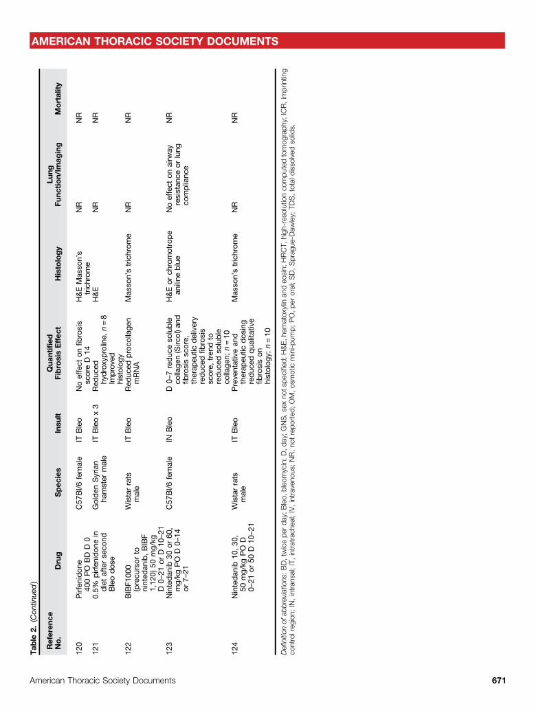
Animal models can roughly be dividedinto those that have a strong inflammatorycomponent andmodels of less inflammatoryinjury. Use of bleomycin, radiation,silica, asbestos, FITC, and many cytokineoverexpression systems lead to fibrogenesisafter a robust inflammatory response,whereas transgenic delivery oftransforming growth factor (TGF)-b,TGF-a, targeted depletion of epithelial cells,and models of IPF fibroblasts delivered toimmunodeficient mice are less dependenton inflammation (1). Use of models fromeach category can help determine whetherthe drug effect is really independent ofinflammation, a crucial consideration whenassessing potential antifibrotic molecules.Determining the precise goal ofexperiments before deciding which in vivoplatform to use is important. For example,experiments aiming to establish potentialtherapeutic efficacy, pharmacokinetic andpharmacodynamic relationships, toxicitystudies, or studies calculating therapeuticwindows may require assessmentsat different time points. Similarly,understanding whether a drug has engagedits biological mechanism of action is afundamental aspect of evaluating theoutcome of clinical trials, and animalmodels are particularly useful fordeveloping such pharmacodynamicbiomarkers for use in early-phase clinicaltrials. Thus, care should be taken indetermining which models may bestprovide a platform at different stages ofthe preclinical development pipeline foreach unique drug target. Interestingly,both pirfenidone and nintedanib showedefficacy, albeit marginal, in more than oneanimal model (43) (Table 2).
AMERICAN THORACIC SOCIETY DOCUMENTS
American Thoracic Society Documents 669
Tab
le2.
Preclinical
Studieswith
Pirfen
idon
ean
dNintedan
ibin
Rod
ents
Referen
ceNo.
Drug
Spec
ies
Insu
ltQua
ntified
FibrosisEffec
tHistology
Lung
Func
tion/Im
aging
Mortality
108
Aeros
olized
pirfen
idon
eD
8C57
Bl/6
mice
GNS
ITBleo
Inhibite
dSirius
red
bioch
emistryan
dAsh
croftsc
oreD
22,n=4
H&EMas
son’s
trichrom
ePirfen
idon
eincrea
sed
dyn
amic
complianc
e
NR
109
Pirfen
idon
e10
mg/kg
PO,D
6C57
Bl/6
Jfemale
ITBleo
Inhibite
dsircol
D14
Noeffect
onAsh
croftsc
oren=6
Mas
son’strichrom
eNR
NR
110
Pirfen
idon
e50
0mg/kg
PO,
D21
SD
rats
male
ITBleo
Noeffect
onCol1
mRNAD
28,n=8
H&EMas
son’s
trichrom
eNR
NR
111
Pirfen
idon
e30
0mg/kg
PO,D
0C57
Bl/6
female
OM
Bleo
Red
uced
collage
nSirc
olan
dfibrosis
scoreD
14,n=6
Mas
son’strichrom
eNR
NR
112
Pirfen
idon
e40
0mg/kg
PO,
D10
C75
Bl/6
male
ITBleo
Noeffect
onhy
droxy
proline,
n=10
Sirius
redH&E
NR
40%
Mortality
with
Pirf/Bleo.
No
sign
ifica
ntdifferen
cefrom
control,
n=40
113
Pirfen
idon
e10
0mg/kg
BD,
D3–
23
Swiss-albino
micemale
ITBleo
Pos
sible
reduc
tion
hydroxy
prolineD
21,n=3,
reduc
edfibrosissc
ore,
n=5
H&EMas
son’s
trichrom
eHRCT
Improve
dmortalityin
pirfen
idon
eap
prox.
50%
versus
60%
114
Pirfen
idon
e10
mg/D
Aeros
olized
D10
C57
Bl/6
male
ITBleo
Red
uced
hydroxy
proline,
n=4,
timepoint
notclea
r
Mas
son’strichrom
eNR
NR
115
Pirfen
idon
e50
0mg/kg
,PO,D
21
ICR
micemale
IVBleo
Red
uced
Ash
croft
score,
D28
,n=8
Mas
son’strichrom
eNR
NR
116
Pirfen
idon
e40
0mg/kg
,PO
D0,
PO
D7,
PO
D14
MiceGNS
ITBleo
Red
uced
hydrox
yprolinen=7,
D0–14
redu
ced
hydrox
yproline,
n=12
,D
7–14
H&EMas
son’s
trichrom
eRed
uced
elas
tanc
e,n=11
,D
0–14
,reduc
edelas
tanc
e,n=24
,D
7–14
NR
117
Pirfen
idon
e10
0mg/kg
TDS
ICR
micemale
IVBleo
Red
uced
hydroxy
proline
n=10
,D
10an
dD
28.reduc
edfibrosis
score,
n=10
H&EMas
son’s
trichrom
eNR
NR
118
Pirfen
idon
e40
0mg/kg
/dyD
14ICR
micemale
IVBleo
Red
uced
fibrosis
scoren=3an
dhy
droxy
proline(n=
3–5)
D35
andD
49.
H&E
NR
NR
119
0.5%
Pirfen
idon
ePO
D22
Golden
Syrian
hamster
male
ITBleo
Significa
ntRed
uctio
nin
hydroxy
proline
D14
&D
21,n=4
NR
NR
NR
(Con
tinue
d)
AMERICAN THORACIC SOCIETY DOCUMENTS
670 American Journal of Respiratory Cell and Molecular Biology Volume 56 Number 5 | May 2017
The panel’s recommendations are foran integrated approach to drug developmentusing both relevant animal models andappropriate ex vivo/in vitro approaches toprovide robust validation of pharmacologicalmechanisms of action and a dosingframework (Figure 1).
Route of Delivery in Lung FibrosisModelsWhen considering bleomycin, mostinvestigators use a single intratrachealadministration (1), but repeatedintratracheal (44) or repeatedintraperitoneal (45) administrations mayoffer more robust and nonresolving fibroticpathology. Repetitive intraperitonealinjections may result in profound epithelialcell hyperplasia, although the evidence isinsufficient to make firm recommendations.
When considering a singleadministration of bleomycin, theoropharyngeal route leads to pulmonaryfibrosis persisting for up to 6 months (46),while requiring shorter-duration animalsedation, eliminating a surgical procedureand associated postsurgical analgesia, andallowing more rapid post-procedurerecovery than the intratracheal route of
administration. The fibrotic response thatdevelops after both methods appears similar;however, direct comparisons between thesetwo routes of administration have not beenformally assessed. Two different models mayprovide more robust data for preclinicalassessment. Reasonable choices wouldinclude adenoviral-driven TGF-boverexpression, FITC, asbestosis, or silica, alladministered into the lung, or radiationdelivered locally to the thorax (reviewed inRefs. 1, 9).
The panel recommends use of a singleoropharyngeal administration of bleomycinand that a second model be considered forpreclinical testing. In addition, the U.S. Foodand Drug Administration will require atleast two species for toxicology, but notefficacy studies.
Kinetics of Lung Fibrosis ModelsThe time course for fibrotic outcomesshould be well characterized, andstandardized within laboratories. This isimportant when considering timings ofintervention with antifibrotic compounds.The bleomycin model is characterized byperiods of acute lung injury (Days 0–7),fibroproliferation (Days 3–14), and
established fibrosis (generally Days 14–28)that generally resolves over a variable timeperiod (47).
The consensus of the panel is that it isinsufficient to only test an antifibroticcompound before evidence of histologicalfibrosis (e.g., giving the drug before Day 7 inthe acute bleomycin model). Rather, it ismore meaningful to deliver the drugtherapeutically after histological evidence offibrosis (e.g., no sooner than Days 7–10 inthe acute bleomycin model) postinstillationafter peak of the acute inflammatory phaseof the lung injury response.
When considering therapeutic dosingin other models (e.g., adenoviral–TGF-b),results are less likely to be confoundedby antiinflammatory effects. However, itremains important to ensure that therapiesare only administered after the resolution ofany inflammation, and the precise timing inother models of fibrosis is an importantarea for future clarification.
It is unclear whether recurrentbleomycin administration offers anyadvantage over the acute bleomycin modelfor preclinical assessment of antifibrotictherapies. The major advantage of thismodel is that recurrent injury and fibrosismay better replicate the progressive natureof human disease, although the mechanisticassumption remains the same, namely, thatprogressive fibrosis is caused by an externalinsult rather than internal genetic/epigeneticresponse to injury. However, we wouldsupport further investigation to determinewhether these models lead to epigeneticchanges, which promote fibrosis.
Reproducibility andStatistical Power
A key aspect of improving translatabilityof preclinical assessment is choosingappropriate, quantitative endpoints (discussedsubsequently here) and undertaking a prioripower calculations using a predefined primaryoutcome measure. All other endpoints shouldbe considered secondary, exploratoryendpoints requiring further assessment forvalidation. Care needs to be taken in dealingwith missing data, especially in animals thatmay not complete the study due to death, orbreaching animal welfare limits. Although notcurrently routine, consideration should begiven to undertaking sensitivity analysis incircumstances of high levels of missingdata (4). Another practice recommendation
Figure 1. Integrating human disease data with preclinical animal studies. Preclinical assessment ofdrug candidates should make judicious use of animal models, such as the bleomycin model in mice,with strong biochemical and histologic assessments of collagen deposition. In addition, animal modelsand culture systems can be used for efficacy, safety, and imaging studies. Target validation shouldinvolve annotated human specimens. An integrated approach that tests effects of compounds onestablished fibrosis should help move promising targets to clinical trials. iPSCs, inducible pluripotentstem cells; PD, pharmacodynamics; PK, pharmacokinetics. Figure adapted from a slide shown byDr. Shelia Violette, who presented this at the American Thoracic Society Conference in May 2015.
AMERICAN THORACIC SOCIETY DOCUMENTS
672 American Journal of Respiratory Cell and Molecular Biology Volume 56 Number 5 | May 2017
would be to test potential drug candidates in asecond laboratory working in the same model,or even a different model.
The panel highly recommends thatnegative data should be published, andhopes that journal editors will valuepublishing high-quality negative results toavoid unnecessary duplication of effortand to reduce unnecessary animalexperimentation.
Endpoints for PreclinicalAssessments
The most accurate analysis for lung fibrosisinvolves several endpoints, includingcombinations of biochemical quantificationof collagen content, histologic assessment offibrotic distribution, and other optionalparameters, such as gene expression, lungfunction, and radiographic imaging. Here,we consider common methodologies usedfor fibrosis endpoint assessments andconsider emerging endpoints that may havevalue in the future.
Biochemical Assessment of CollagenContentHydroxyproline content in total lungsamples is a surrogate for collagen content(1 mg hydroxyproline = 6.94 mg collagen),and is expressed as micrograms per lung(right, left, or both) (48). Commonly,fibrosis is patchy after introduction ofthe stimulus by inhalation, often withconsiderable variation between lungs.Therefore, processing both lungs to powderin liquid nitrogen or homogenized togetherbefore analysis of hydroxyproline, or otherbiochemical assessments, should beconsidered. If the fibrotic stimulus isdeposited with excellent reproducibility, thesame lung should be consistently used forthis assay to allow comparison betweenmice and groups. Although this mayincrease the standard deviation of the dataobtained, it permits use of one lung formorphometric analysis, which, overall,could reduce the number of animalsrequired in the experiment. Sirius red isoften used as a colorimetric estimate offibrosis in tissue homogenates; however,the values seem to overestimate absolutevalues obtained by the gold standardhydroxyproline assay (48, 49). In addition,collagen content can be determined usingSircol collagen assays kits. However, it is
important to be aware that this methodonly allows analysis of “newly formed”acid and pepsin-soluble collagens, and,therefore, is not useful for measuringinsoluble collagens integrated into “mature”scar tissue, and Sircol measurementsaccount for only a small fraction of totallung collagen determined by HPLC (46). Inaddition, care must be taken to use amodified version of the assay that is notadversely impacted by serum proteins presentin the samples due to vascular leak. Thus,although this method is suitable for collagenproduced in cell culture systems, the panelbelieves that this assay does not best reflectthe effect of an intervention on lung collagenaccumulation and fibrosis in vivo.
Given these considerations, the panelrecommends hydroxyproline measurement asthe optimal primary endpoint for preclinicalassessment of novel therapeutic agents.
Gene ExpressionAnother common practice is reportingextracellular matrix levels by measuringlevels of gene expression, generally byquantitative PCR. Although thesemeasurements can be supportive to theoverall assessment, they are difficult tointerpret if done in isolation. For example,collagen mRNA is extremely stable (50), sosmall changes in gene expression can correlatewith large differences in protein levels.
The panel currently recommends thatgene expression studies always beaccompanied by biochemical parameters.
Morphological Approach forHistological AssessmentMasson’s trichrome staining is usedto assess fibrosis histologically, and,usually, the severity of lung fibrosis issemiquantified in stained tissue sectionsthrough an Ashcroft scoring system (51),where the grade of fibrosis is scored from0 (normal lung) to 8 (total fibrousobliteration of fields) by examiningrandomly chosen sections. Morerecently, digital scoring systems usingsemiautomated image analysis havebeen used (52, 53) or a combination ofsemiquantitative scoring by blinded readersand digital analysis (54). The Sirius red stain(55) consists of a dye that binds to the Gly-x-ytriple-helix structure found in all collagenfibers. Fibrillar collagens are visualized asbirefringent structures under polarized light,but tissues can also be examined bybrightfield microscopy and semiquantified
by morphometric (visual) scoring systems(56–59). Recently, a digital imaging additionand subtraction method was proposedto determine the relative area offibrillar collagens and cell content in asemiautomated process using standardsoftware (60). For morphological analysis,the panel recommends that lungs should befixed at a constant hydrostatic pressure up toa maximum of 30 cm H2O based on normallung (61, 62). Incomplete perfusion provokescollapse, whereas excessive pressure mayprovoke alveolar wall rupture, leadingto erroneous interpretation. With regard tomeasures of a-smooth muscle actin toidentify myofibroblasts as a surrogate forfibrosis, recent studies suggest that collagenand a-smooth muscle actin do not alwayscorrelate with each other (63, 64).
The panel recommends that due to theinherent bias associated with morphologicalscoring systems, exacerbated by the sporadicnature of histologic assessment, histologyshould focus on the morphological andmolecular characteristics of fibrosis but mustnever be used to quantify fibrosis withoutbiochemical assessment of lung collagen.
Lung FunctionLung function assessment is helpful incorroborating fibrotic changes and/ortherapeutic response of interventions, butpresents considerable technical challenges(52, 54, 65). Lung function measures showgood correlation with morphological changesat peak of fibrosis, but recover when the lesionextent decreases, even when there is still clearevidence of histologic fibrosis (52). Somefunctional parameters can be measuredthrough noninvasive methods with whole-body plethysmography, where the animal isconscious and unrestrained; however, thistechnique evaluates few parameters (e.g.,respiratory frequency, tidal volume), and thesemeasures show marginal or no differencesbetween injured and normal lungs (66).
Invasive methods are performed withforced ventilator maneuvers in anesthetized,tracheostomized animals; thus, adisadvantage is that these measures mayonly be used as single-timepoint outcomes (66).There are two invasive methods, Buxco-force pulmonary maneuvers, and FlexiVent,which allow the measurement of severalfunctional tests of clinical relevance for lungfibrosis, such as pressure–volume curves (66).With forced oscillation techniques, lungimpedance can be examined and, through it,several variables, such as lung tissue elastance
AMERICAN THORACIC SOCIETY DOCUMENTS
American Thoracic Society Documents 673
and tissue damping (54, 66, 67). Some studiessuggest that values obtained by forcedoscillation techniques correlate better withextent of fibrosis than do pressure–volumecurves (47, 54). In larger animals (e.g., rats),gas transfer can also be measured. Importantly,lung mechanics measurements with theseinvasive methods before killing experimentalmice should not alter results obtained byhistology or collagen determinationsperformed on the same lungs (54).
In general terms, pulmonary functiontests can evaluate response to drugs orantifibrotic mediators (68, 69). Certainly, animportant goal in the field is to developmethods that allow monitoring the samemouse over time. As such, repetitive, invasivemeasurements of lung function may beperformed in intubated instead oftracheostomized mice with similar results(70). However, studies so far are limited, andthe effect of repeated invasive testson morphology and collagen content isunknown. Alternatively, the measurement ofarterial blood oxygen saturation with pulseoximetry is noninvasive, and can be tested onconscious mice (71).
Currently, the panel does notrecommend pulmonary function as aroutine part of the preclinical assessment ofdrugs. However, it does recommend furtherevaluation of the potential for use of pulseoximetry as a repeatable, noninvasivemeasure of the development of lung injuryand fibrosis over time, and recognizesthat lung function measurements in
experienced hands can be part of acomposite endpoint if supported bybiochemical measures.
ImagingMultiple techniques have been developedto acquire functional, molecular, and/oranatomical images of the body, includingbioluminescence, fluorescence, magneticresonance imaging, planar X-ray, X-raycomputed tomography (CT), nuclearimaging with positron emission tomography,and single-photon emission CT (SPECT) (67).However, experience with most ofthese methods in animal models of lungfibrosis is scant. An early study used Luciferyellow carbohydrazide fluorescent dye toselectively visualize connective tissue matrixmacromolecules, enabling quantitativeassessment of lung fibrosis (72).
In the last decade, multiple groups havetaken advantage of noninvasive imagingtechniques to give longitudinal informationon individual animals in pulmonary fibrosismodels, predominantly using in vivo micro-CT of the lungs (73–76). Micro-CT isfeasible and allows dynamic visualization ofdisease progression and response to therapy.However, the degree of resolution isconstrained by respiratory and cardiacmotion; therefore, implementation ofgating for small-animal CT imaging isrecommended (77). Abnormal imagesrelevant to interstitial lung diseases, such asground glass attenuation, reticular opacities,consolidation, traction bronchiectasis, and
honeycombing, can be semiquantified byradiologists (blinded to the pathologic results)scoring, for example, on a scale from0 (absent) to 5 depending on the involvedarea (78). Micro-CT data can also be digitallyprocessed and the aerated and total lungvolumes, lung tissue (including lesions), andmean lung density can be more preciselyquantified (73). Importantly, several studiesshowed that repeated high-resolution lungmicro-CT scans for long-term imagingprotocols don’t provoke radiotoxicity-induced lung damage (79, 80).
High-resolution micro-CT analysiswith spatial resolution of approximately20 mm has also been employed inmurine lungs ex vivo. Fibrotic changesrevealed by micro-CT fully matchedequivalent histologic sections, and thisapproach allows evaluation oftherapeutic dosing once fibrosis isalready established (46).
The panel didn’t feel there wassufficient evidence to recommend routineuse of CT scanning for preclinicalassessment of antifibrotic therapy.However, the potential advantagesassociated with CT scanning, especiallywhen measuring evolution of fibrosis overtime, merit further investigation.
InflammationThe role of chronic inflammation in IPFremains controversial (81, 82). However,it may be useful to quantify changes,as inflammation does not completely
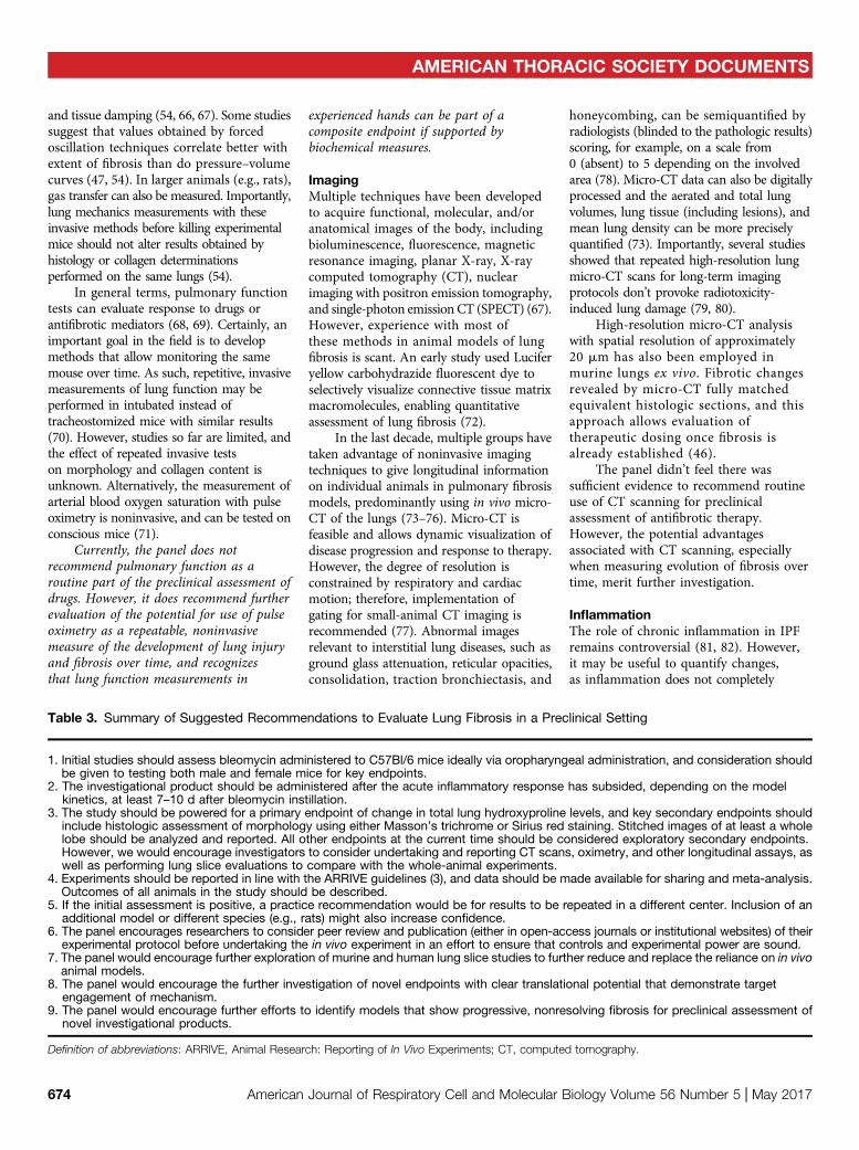
Table 3. Summary of Suggested Recommendations to Evaluate Lung Fibrosis in a Preclinical Setting
1. Initial studies should assess bleomycin administered to C57Bl/6 mice ideally via oropharyngeal administration, and consideration shouldbe given to testing both male and female mice for key endpoints.
2. The investigational product should be administered after the acute inflammatory response has subsided, depending on the modelkinetics, at least 7–10 d after bleomycin instillation.
3. The study should be powered for a primary endpoint of change in total lung hydroxyproline levels, and key secondary endpoints shouldinclude histologic assessment of morphology using either Masson’s trichrome or Sirius red staining. Stitched images of at least a wholelobe should be analyzed and reported. All other endpoints at the current time should be considered exploratory secondary endpoints.However, we would encourage investigators to consider undertaking and reporting CT scans, oximetry, and other longitudinal assays, aswell as performing lung slice evaluations to compare with the whole-animal experiments.
4. Experiments should be reported in line with the ARRIVE guidelines (3), and data should be made available for sharing and meta-analysis.Outcomes of all animals in the study should be described.
5. If the initial assessment is positive, a practice recommendation would be for results to be repeated in a different center. Inclusion of anadditional model or different species (e.g., rats) might also increase confidence.
6. The panel encourages researchers to consider peer review and publication (either in open-access journals or institutional websites) of theirexperimental protocol before undertaking the in vivo experiment in an effort to ensure that controls and experimental power are sound.
7. The panel would encourage further exploration of murine and human lung slice studies to further reduce and replace the reliance on in vivoanimal models.
8. The panel would encourage the further investigation of novel endpoints with clear translational potential that demonstrate targetengagement of mechanism.
9. The panel would encourage further efforts to identify models that show progressive, nonresolving fibrosis for preclinical assessment ofnovel investigational products.
Definition of abbreviations: ARRIVE, Animal Research: Reporting of In Vivo Experiments; CT, computed tomography.
AMERICAN THORACIC SOCIETY DOCUMENTS
674 American Journal of Respiratory Cell and Molecular Biology Volume 56 Number 5 | May 2017
resolve during the evolution of thefibrotic response, and might confoundinterpretations, especially with agents thatimpact inflammatory response. Inflammationis usually evaluated in bronchoalveolar lavage(BAL) or lung digest specimens. In thebleomycin model, BAL to obtain lungleukocytes is typically done in the first weekpostinstillation. Inflammatory cell profiles,including total cell numbers, percentages,and even cell subsets, such as M1/M2macrophages and CD4/CD8 lymphocytes,can be determined. In addition, cytokines areeasily explored in BAL fluids or lunghomogenates. The extent of inflammatorylesions may be semiquantified in histologysections from lungs stained with hematoxylinand eosin. However, the panel does notrecommend routine assessment ofinflammatory cell profiles for the preclinicalassessment of antifibrotic drugs, except to apriori confirm presence or absence of effects oninflammatory pathways for a given molecule.
Emerging Endpoints
Novel Imaging StudiesImaging modalities are widely used to assessfibrosis; although many are in earlydevelopment for in vivo modeling, afew warrant commentary as potentialpreclinical surrogates. Molecular imagingtechniques use tracer molecules labeledwith contrast reagents for visualizationto track cellular events and molecularprocesses in their native environments inintact living subjects (83). These techniquesallow assessment of important moleculartargets not shed into extracellular fluidsthat cannot be sampled in peripheral blood.Molecular imaging probes visualizingrecently synthesized collagen, or theTGF-b–activating integrin avb6, havealready been demonstrated to noninvasivelytrack development of pulmonary fibrosis inthe bleomycin mouse model by magneticresonance imaging or SPECT, respectively(84, 85). High levels of avb6 integrins,detected by immunohistochemistry,correlate with poor outcome in IPF (86).Using SPECT scanning, it is possible toidentify up-regulated avb6 integrins inbleomycin-induced lung fibrosis, predictthe development of fibrosis, anddemonstrate target engagement usinganti–avb6 integrin blocking antibodies(85). Use of SPECT and other noninvasiveimaging techniques combined with
molecular probes for use in murinemodels of lung fibrosis has recently beenreviewed (87). Second harmonic generationmicroscopy is an exciting, emerging, “label-free” technique for assessing collagen invarious tissues, and has been used to scoreliver fibrosis (88) and to assess collagenstructure in IPF (89) and bleomycin-induced lung fibrosis (90). However, itsgreatest potential is for real-time collagenassessments in vivo, in both animalmodels and human disease, throughmicroendoscopic second harmonicgeneration microscopy (91).
Novel Peripheral Blood BiomarkersSeveral prognostic/diagnostic biomarkershave been identified in IPF usingtranscriptional or protein profiling (92–96).Another emerging technology ismeasurement of matrix neo-epitopes createdduring fibrotic remodeling. Examples includespecific fragments of type IV collagen a1(C4M12a1) and a3 (C4M12a3) chains inserum as indicators of fibrosis when measuredby ELISA (97). Recent human IPF studieshave also shown that neo-antigens generatedby matrix metalloproteinase cleavage areuseful biomarkers in IPF (98). Thus, onepotential advantage of these techniques is thattheir use in animal models may be translatedto human studies. One note of caution isthat neo-epitope panels currently in usefor human studies have not been fullycharacterized for murine tissues. There areseveral potential reasons: first, cleavage sites onproteins in mice and humans differ; and,second, quantities of cleaved proteins differsignificantly at baseline in the two species.
When taken together, the panel isexcited about these emerging imaging andbiomarker technologies, but does not believethat there is currently sufficient experienceto recommend this approach for generalpreclinical drug testing. The panelrecommends that investigators continue todevelop specific imaging probes and bloodbiomarkers to predict disease progression,and to predict responses to new treatments.The panel is also enthusiastic about the highpotential translatability of these approachesinto clinical studies.
Ex Vivo Lung Slices for PreclinicalTestingEx vivo precision-cut human lung slices forpreclinical testing are generally obtainedfrom lung cadaver, surgical resections, orexplanted lungs, which are filled with low
melting point agarose to allow precision slicesof 300–1,000 mm in thickness for use inculture (99, 100). Such lung slices are viablein culture for 5–7 days and preserve three-dimensional architecture (99). By Day 7, suchcultures generally show prominent apoptosis,but movement of media over the slices mayincrease viability. These slices can also beobtained from the lungs of patients with IPFand used to validate findings from in vitroand in vivo studies in human tissue, as well asa dosing framework of novel therapeuticagents (101).
Preliminary studies treating slices withbleomycin, or influenza, have not yieldedchanges ex vivo that are observed in vivo(29, 73), although encouraging results usingTGF-b have been found (102). It is possibleto generate lung slices from mice that havebeen treated with bleomycin in vivo, andthese have increased levels of extracellularmatrix (103–105); there are encouragingresults using nintedanib and pirfenidone inthese slices as well (102). An importantfeature of murine or human lung slicecultures is that they can be used to assesseffects of drugs directly on structuralcells in the absence of vasculature orinflammatory cell influx. Furthermore,a single mouse lung can generateapproximately 30 slices for analysis, makingthis an emerging alternative to some whole-animal experiments, potentially reducingoverall animal numbers used for research.
In conclusion, the panel is excited aboutthe promise that lung slice cultures holdfor identification of novel targets, kineticsstudies, and ability to compare biologybetween human andmurine systems, and theinvestigation of slices from IPF lungs. Thepanel encourages further investigationcomparing pharmacological manipulation offibrotic murine and human lung slices tofurther develop and improve these assays,and accumulate evidence to use this approachfor future preclinical drug testing.
Conclusions
The panel recommendations (Table 3)are for comprehensive, preclinicalevaluation of potential antifibrotictherapies based on currently availabledata. They are not to be regarded as aminimum dataset for publication, or asrecommendations applicable to discoverybiology, although some principlesare clearly transferable. n
AMERICAN THORACIC SOCIETY DOCUMENTS
American Thoracic Society Documents 675
This official workshop report was prepared by an ad hoc committee of the Assembly on Respiratory Cell and Molecular Biology.
Members of the subcommittee are asfollows:
R. GISLI JENKINS, M.D., PH.D. (Co-Chair)BETHANY B. MOORE, PH.D. (Co-Chair)RACHEL C. CHAMBERS, PH.D., FRSBOLIVER EICKELBERG, M.D.MARTIN KOLB, M.D., PH.D.MELANIE KONIGSHOFF, M.D., PH.D.GEOFFREY J. LAURENT, PH.D., F.MED.SCI.CARMEL B. NANTHAKUMAR, PH.D.MITCHELL A. OLMAN, M.D., M.A.ANNIE PARDO, PH.D.MOISES SELMAN, M.D.DEAN SHEPPARD, M.D.PATRICIA J. SIME, M.D.ANDREW M. TAGER, M.D.AMANDA L. TATLER, PH.D.VICTOR J. THANNICKAL, M.D.ERIC S. WHITE, M.D., M.S.
Author disclosures: R.G.J. received researchsupport from Biogen, Galecto, GlaxoSmithKline,and Novartis Pharma; served on an advisorycommittee for Biogen, Boehringer Ingelheim,GlaxoSmithKline, InterMune, PharmAkea, andRoche; served as a consultant and as a speakerfor Roche; served as a speaker for InterMune;served as a consultant for Nuformix andPulmatrix; and served on a data and safetymonitoring board for Boehringer Ingelheim.B.B.M. received research support from SyntrixBiosystems. R.C.C. received research supportfrom GlaxoSmithKline; her spouse is anemployee of and has stocks, stock options, orother ownership interests in GlaxoSmithKline.M. Kolb received research support from ActelionPharmaceuticals, Boehringer Ingelheim, GileadSciences, Janssen Pharmaceuticals, andPrometic; served as a consultant for BoehringerIngelheim, F. Hoffmann-La Roche, GileadSciences, GlaxoSmithKline, JanssenPharmaceuticals, and Prometic; served as a
speaker for Boehringer Ingelheim andF. Hoffmann-La Roche; served on anadvisory committee for AstraZeneca; andserved on a data safety and monitoring board forF. Hoffmann-La Roche. D.S. served as aconsultant, had commercialized intellectualproperty, and had stocks, stock options, orother ownership interests with PliantTherapeutics. P.J.S. served on an advisorycommittee for GlaxoSmithKline and UCBBiosciences; served as a consultant forBoehringer Ingelheim and Third Rock Ventures;and served on a data and safety monitoring boardfor InterMune. A.M.T. received research supportfrom Biogen Idec, InterMune, and BoehringerIngelheim; served on an advisory committee andhas stocks, stock options, or other ownershipinterests with PharmAkea Therapeutics and PliantTherapeutics; and served as a consultant forBlade Therapeutics. O.E., M. Konigshoff, G.J.L.,C.B.N., M.A.O., A.P., M.S., A.L.T., V.J.T., E.S.W.report no relevant commercial interests.
References
1. Moore BB, Lawson WE, Oury TD, Sisson TH, Raghavendran K,Hogaboam CM. Animal models of fibrotic lung disease. Am J RespirCell Mol Biol 2013;49:167–179.
2. Chua F, Gauldie J, Laurent GJ. Pulmonary fibrosis: searching for modelanswers. Am J Respir Cell Mol Biol 2005;33:9–13.
3. Kilkenny C, Browne WJ, Cuthi I, Emerson M, Altman DG.Improving bioscience research reporting: the ARRIVE guidelinesfor reporting animal research. Vet Clin Pathol 2012;41:27–31.
4. Numbers matter. Nature 2015;520:263–264.5. Cressey D. Surge in support for animal-research guidelines. Nature
2016;530: doi:10.1038/nature.2016.19274.6. Rabeyrin M, Thivolet F, Ferretti GR, Chalabreysse L, Jankowski A, Cottin
V, Pison C, Cordier JF, Lantuejoul S. Usual interstitial pneumonia end-stage features from explants with radiologic and pathologicalcorrelations. Ann Diagn Pathol 2015;19:269–276.
7. Raghu G, Collard HR, Egan JJ, Martinez FJ, Behr J, Brown KK, Colby TV,Cordier JF, Flaherty KR, Lasky JA, et al.; ATS/ERS/JRS/ALATCommittee on Idiopathic Pulmonary Fibrosis. An officialATS/ERS/JRS/ALAT statement: idiopathic pulmonary fibrosis:evidence-based guidelines for diagnosis and management. Am JRespir Crit Care Med 2011;183:788–824.
8. Rackley CR, Stripp BR. Building and maintaining the epithelium of thelung. J Clin Invest 2012;122:2724–2730.
9. Moore BB, Hogaboam CM. Murine models of pulmonary fibrosis. Am JPhysiol Lung Cell Mol Physiol 2008;294:L152–L160.
10. Judge EP, Hughes JM, Egan JJ, Maguire M, Molloy EL, O’Dea S.Anatomy and bronchoscopy of the porcine lung: a model for translationalrespiratory medicine. Am J Respir Cell Mol Biol 2014;51:334–343.
11. Leigh MW, Gambling TM, Carson JL, Collier AM, Wood RE, Boat TF.Postnatal development of tracheal surface epithelium andsubmucosal glands in the ferret. Exp Lung Res 1986;10:153–169.
12. Sun X, Sui H, Fisher JT, Yan Z, Liu X, Cho HJ, Joo NS, Zhang Y, Zhou W,Yi Y, et al. Disease phenotype of a ferret CFTR-knockout model ofcystic fibrosis. J Clin Invest 2010;120:3149–3160.
13. Sun X, Yan Z, Yi Y, Li Z, Lei D, Rogers CS, Chen J, Zhang Y, Welsh MJ,Leno GH, et al. Adeno-associated virus-targeted disruption of theCFTR gene in cloned ferrets. J Clin Invest 2008;118:1578–1583.
14. Rogers CS, Stoltz DA, Meyerholz DK, Ostedgaard LS, Rokhlina T, Taft PJ,Rogan MP, Pezzulo AA, Karp PH, Itani OA, et al. Disruption of theCFTR gene produces a model of cystic fibrosis in newborn pigs.Science 2008;321:1837–1841.
15. Organ L, Bacci B, Koumoundouros E, Barcham G, Milne M, Kimpton W,Samuel C, Snibson K. Structural and functional correlations in a largeanimal model of bleomycin-induced pulmonary fibrosis. BMC PulmMed 2015;15:81.
16. Wong DM, Belgrave RL, Williams KJ, Del Piero F, Alcott CJ, Bolin SR,Marr CM, Nolen-Walston R, Myers RK, Wilkins PA. Multinodularpulmonary fibrosis in five horses. J Am Vet Med Assoc 2008;232:898–905.
17. Williams KJ, Maes R, Del Piero F, Lim A, Wise A, Bolin DC, Caswell J,Jackson C, Robinson NE, Derksen F, et al. Equine multinodularpulmonary fibrosis: a newly recognized herpesvirus-associatedfibrotic lung disease. Vet Pathol 2007;44:849–862.
18. Miele A, Dhaliwal K, Du Toit N, Murchison JT, Dhaliwal C, Brooks H,Smith SH, Hirani N, Schwarz T, Haslett C, et al. Chronicpleuropulmonary fibrosis and elastosis of aged donkeys: similarities tohuman pleuroparenchymal fibroelastosis. Chest 2014;145:1325–1332.
19. Williams K, Malarkey D, Cohn L, Patrick D, Dye J, Toews G.Identification of spontaneous feline idiopathic pulmonary fibrosis:morphology and ultrastructural evidence for a type II pneumocytedefect. Chest 2004;125:2278–2288.
20. Corcoran BM, Cobb M, Martin MW, Dukes-McEwan J, French A,Fuentes VL, Boswood A, Rhind S. Chronic pulmonarydisease in West Highland white terriers. Vet Rec 1999;144:611–616.
21. Williams KJ, Robinson NE, Lim A, Brandenberger C, Maes R, Behan A,Bolin SR. Experimental induction of pulmonary fibrosis in horses withthe gammaherpesvirus equine herpesvirus 5. PLoS One 2013;8:e77754.
22. Redente EF, Jacobsen KM, Solomon JJ, Lara AR, Faubel S, Keith RC,Henson PM, Downey GP, Riches DW. Age and sex dimorphismscontribute to the severity of bleomycin-induced lung injury andfibrosis. Am J Physiol Lung Cell Mol Physiol 2011;301:L510–L518.
24. Sueblinvong V, Neujahr DC, Mills ST, Roser-Page S, Ritzenthaler JD,Guidot D, Rojas M, Roman J. Predisposition for disrepair in the agedlung. Am J Med Sci 2012;344:41–51.
25. Sueblinvong V, Neveu WA, Neujahr DC, Mills ST, Rojas M, Roman J,Guidot DM. Aging promotes pro-fibrotic matrix production andincreases fibrocyte recruitment during acute lung injury. Adv BiosciBiotechnol 2014;5:19–30.
AMERICAN THORACIC SOCIETY DOCUMENTS
676 American Journal of Respiratory Cell and Molecular Biology Volume 56 Number 5 | May 2017
26. Hecker L, Logsdon NJ, Kurundkar D, Kurundkar A, Bernard K, Hock T,Meldrum E, Sanders YY, Thannickal VJ. Reversal of persistentfibrosis in aging by targeting Nox4–Nrf2 redox imbalance. Sci TranslMed 2014;6:231ra47.
27. Thannickal VJ, Zhou Y, Gaggar A, Duncan SR. Fibrosis: ultimate andproximate causes. J Clin Invest 2014;124:4673–4677.
28. Yates CC, Hebda P, Wells A. Skin wound healing and scarring: fetalwounds and regenerative restitution. Birth Defects Res C EmbryoToday 2012;96:325–333.
29. Walmsley GG, Maan ZN, Wong VW, Duscher D, Hu MS, Zielins ER,Wearda T, Muhonen E, McArdle A, Tevlin R, et al. Scarless woundhealing: chasing the holy grail. Plast Reconstr Surg 2015;135:907–917.
30. Bueno M, Lai YC, Romero Y, Brands J, St Croix CM, Kamga C, Corey C,Herazo-Maya JD, Sembrat J, Lee JS, et al. PINK1 deficiency impairsmitochondrial homeostasis and promotes lung fibrosis. J Clin Invest2015;125:521–538.
31. Naik PN, Horowitz JC, Moore TA, Wilke CA, Toews GB, Moore BB.Pulmonary fibrosis induced by g-herpesvirus in aged miceis associated with increased fibroblast responsiveness totransforming growth factor-b. J Gerontol A Biol Sci Med Sci2012;67:714–725.
32. Torres-Gonzalez E, Bueno M, Tanaka A, Krug LT, Cheng DS,Polosukhin VV, Sorescu D, Lawson WE, Blackwell TS, Rojas M, et al.Role of endoplasmic reticulum stress in age-related susceptibility tolung fibrosis. Am J Respir Cell Mol Biol 2012;46:748–756.
33. Matsuoka S, Uchiyama K, Shima H, Ueno N, Oish S, Nojiri Y.Bronchoarterial ratio and bronchial wall thickness on high-resolutionCT in asymptomatic subjects: correlation with age and smoking. AJRAm J Roentgenol 2003;180:513–518.
34. Tatler AL, Habgood A, Porte J, John AE, Stavrou A, Hodge E, Kerama-Likoko C, Violette SM, Weinreb PH, Knox AJ, et al. Reduced Etsdomain-containing protein ELK1 promotes pulmonary fibrosis viaincreased integrin avb6 expression. J Biol Chem 2016;291:9540–9553.
35. Munger JS, Huang X, Kawakatsu H, Griffiths MJ, Dalton SL, Wu J,Pittet JF, Kaminski N, Garat C, Matthay MA, et al. The integrin avb6binds and activates latent TGFb 1: a mechanism for regulatingpulmonary inflammation and fibrosis. Cell 1999;96:319–328.
37. Baron RM, Choi AJ, Owen CA, Choi AM. Genetically manipulatedmouse models of lung disease: potential and pitfalls. Am J PhysiolLung Cell Mol Physiol 2012;302:L485–L497.
38. Lee CG, Homer RJ, Zhu Z, Lanone S, Wang X, Koteliansky V, Shipley JM,Gotwals P, Noble P, Chen Q, et al. Interleukin-13 induces tissuefibrosis by selectively stimulating and activating transforming growthfactor beta(1). J Exp Med 2001;194:809–821.
39. Park DS, Cohen AW, Frank PG, Razani B, Lee H,Williams TM, Chandra M,Shirani J, De Souza AP, Tang B, et al. Caveolin-1 null (2/2) miceshow dramatic reductions in life span. Biochemistry 2003;42:15124–15131.
40. Lawson WE, Cheng DS, Degryse AL, Tanjore H, Polosukhin VV, Xu XC,Newcomb DC, Jones BR, Roldan J, Lane KB, et al. Endoplasmicreticulum stress enhances fibrotic remodeling in the lungs. Proc NatlAcad Sci USA 2011;108:10562–10567.
41. Naikawadi RP, Disayabutr S, Mallavia B, Donne ML, Green G, La JL,Rock JR, Looney MR, Wolters PJ. Telomere dysfunction in alveolarepithelial cells causes lung remodeling and fibrosis. JCI Insight 2016;1:e86704.
42. Horan GS, Wood S, Ona V, Li DJ, Lukashev ME, Weinreb PH, Simon KJ,Hahm K, Allaire NE, Rinaldi NJ, et al. Partial inhibition of integrin avb6prevents pulmonary fibrosis without exacerbating inflammation. Am JRespir Crit Care Med 2008;177:56–65.
43. Myllarniemi M, Kaarteenaho R. Pharmacological treatment ofidiopathic pulmonary fibrosis - preclinical and clinical studies ofpirfenidone, nintedanib, and n-acetylcysteine. Eur Clin Respir J2015;2:26385.
44. Degryse AL, Tanjore H, Xu XC, Polosukhin VV, Jones BR, McMahonFB, Gleaves LA, Blackwell TS, Lawson WE. Repetitive intratrachealbleomycin models several features of idiopathic pulmonary fibrosis. AmJ Physiol Lung Cell Mol Physiol 2010;299:L442–L452.
45. Collins SL, Chan-Li Y, Oh M, Vigeland CL, Limjunyawong N, Mitzner W,Powell JD, Horton MR. Vaccinia vaccine–based immunotherapyarrests and reverses established pulmonary fibrosis. JCI Insight2016;1:e83116.
46. Scotton CJ, Hayes B, Alexander R, Datta A, Forty EJ, Mercer PF,Blanchard A, Chambers RC. Ex vivo micro-computed tomographyanalysis of bleomycin-induced lung fibrosis for preclinical drugevaluation. Eur Respir J 2013;42:1633–1645.
47. Schiller HB, Fernandez IE, Burgstaller G, Schaab C, Scheltema RA,Schwarzmayr T, Strom TM, Eickelberg O, Mann M. Time- andcompartment-resolved proteome profiling of the extracellular nichein lung injury and repair. Mol Syst Biol 2015;11:819.
48. Campa JS, McAnulty RJ, Laurent GJ. Application of high-pressureliquid chromatography to studies of collagen production by isolatedcells in culture. Anal Biochem 1990;186:257–263.
49. Kliment CR, Englert JM, Crum LP, Oury TD. A novel methodfor accurate collagen and biochemical assessment of pulmonary tissueutilizing one animal. Int J Clin Exp Pathol 2011;4:349–355.
50. Maatta A, Ekholm E, Penttinen RP. Effect of the 39-untranslated regionon the expression levels and mRNA stability of alpha 1(I) collagengene. Biochim Biophys Acta 1995;1260:294–300.
51. Ashcroft T, Simpson JM, Timbrell V. Simple method of estimatingseverity of pulmonary fibrosis on a numerical scale. J Clin Pathol1988;41:467–470.
52. Fernandez IE, Amarie OV, Mutze K, Konigshoff M, Yildirim AO,Eickelberg O. Systematic phenotyping and correlation of biomarkerswith lung function and histology in lung fibrosis. Am J Physiol LungCell Mol Physiol 2016;310:L919–L927.
53. Chung EJ, McKay-Corkum G, Chung S, White A, Scroggins BT,Mitchell JB, Mulligan-Kehoe MJ, Citrin D. Truncated plasminogenactivator inhibitor-1 protein protects from pulmonary fibrosismediated by irradiation in a murine model. Int J Radiat Oncol BiolPhys 2016;94:1163–1172.
54. Manali ED, Moschos C, Triantafillidou C, Kotanidou A, Psallidas I,Karabela SP, Roussos C, Papiris S, Armaganidis A, Stathopoulos GT,et al. Static and dynamic mechanics of the murine lung afterintratracheal bleomycin. BMC Pulm Med 2011;11:33.
55. Junqueira LC, Bignolas G, Brentani RR. Picrosirius staining pluspolarization microscopy, a specific method for collagen detection intissue sections. Histochem J 1979;11:447–455.
56. Cai Y, Kimura S. Secretoglobin 3A2 exhibits anti-fibrotic activity inbleomycin-induced pulmonary fibrosis model mice. PLoS One 2015;10:e0142497.
57. Zhao H, Qin HY, Cao LF, Chen YH, Tan ZX, Zhang C, Xu DX.Phenylbutyric acid inhibits epithelial-mesenchymal transition duringbleomycin-induced lung fibrosis. Toxicol Lett 2015;232:213–220.
58. Mercer RR, Scabilloni JF, Hubbs AF, Battelli LA, McKinney W, Friend S,Wolfarth MG, Andrew M, Castranova V, Porter DW. Distribution andfibrotic response following inhalation exposure to multi-walledcarbon nanotubes. Part Fibre Toxicol 2013;10:33.
59. Rydell-Tormanen K, Andreasson K, Hesselstrand R, Risteli J,Heinegard D, Saxne T, Westergren-Thorsson G. Extracellular matrixalterations and acute inflammation; developing in parallel duringearly induction of pulmonary fibrosis. Lab Invest 2012;92:917–925.
60. Vogel B, Siebert H, Hofmann U, Frantz S. Determination of collagencontent within picrosirius red stained paraffin-embedded tissuesections using fluorescence microscopy. MethodsX 2015;2:124–134.
61. Soutiere SE, Mitzner W. On defining total lung capacity in the mouse.J Appl Physiol 2004;96:1658–1664.
62. Vasilescu DM, Knudsen L, Ochs M, Weibel ER, Hoffman EA. Optimizedmurine lung preparation for detailed structural evaluation via micro-computed tomography. J Appl Physiol 2012;112:159–166.
63. Sun KH, Chang Y, Reed NI, Sheppard D. a-Smooth muscle actin is aninconsistent marker of fibroblasts responsible for force-dependentTGFb activation or collagen production across multiple models oforgan fibrosis. Am J Physiol Lung Cell Mol Physiol 2016;310:L824–L836.
64. Rock JR, Barkauskas CE, Cronce MJ, Xue Y, Harris JR, Liang J, Noble PW,Hogan BL. Multiple stromal populations contribute to pulmonary fibrosiswithout evidence for epithelial to mesenchymal transition. Proc NatlAcad Sci USA 2011;108:E1475–E1483.
AMERICAN THORACIC SOCIETY DOCUMENTS
American Thoracic Society Documents 677
65. Irvin CG, Bates JH. Measuring the lung function in the mouse: thechallenge of size. Respir Res 2003;4:4.
66. Vanoirbeek JA, Rinaldi M, De Vooght V, Haenen S, Bobic S, Gayan-Ramirez G, Hoet PH, Verbeken E, Decramer M, Nemery B, et al.Noninvasive and invasive pulmonary function in mouse models ofobstructive and restrictive respiratory diseases. Am J Respir Cell MolBiol 2010;42:96–104.
67. Gammon ST, Foje N, Brewer EM, Owers E, Downs CA, Budde MD,Leevy WM, Helms MN. Preclinical anatomical, molecular, andfunctional imaging of the lung with multiple modalities. Am J PhysiolLung Cell Mol Physiol 2014;306:L897–L914.
68. Shimbori, C, Bellaye PS, Xia J, Gauldie J, Ask K, Ramos C, Becerril C,Pardo A, Selman M, Kolb M, et al. Fibroblast growth factor-1 attenuatesTGF-b1–induced lung fibrosis. J Pathol 2016;240:197–210.
69. Jarman ER, Khambata VS, Yun Ye L, Cheung K, Thomas M,Duggan N, Jarai G. A translational preclinical model ofinterstitial pulmonary fibrosis and pulmonary hypertension:mechanistic pathways driving disease pathophysiology. PhysiolRep 2014;2:e12133.
70. De Vleeschauwer SI, Rinaldi M, De Vooght V, Vanoirbeek JA,Vanaudenaerde BM, Verbeken EK, Decramer M, Gayan-Ramirez GN,Verleden GM, Janssens W. Repeated invasive lung functionmeasurements in intubated mice: an approach for longitudinal lungresearch. Lab Anim 2011;45:81–89.
71. Early MA, Lishnevsky M, Gilchrist JM, Higgins DM, Orme IM, Muller WA,Gonzalez-Juarerro M, Schenkel AR. Non-invasive diagnosis of earlypulmonary disease in PECAM-deficient mice using infrared pulseoximetry. Exp Mol Pathol 2009;87:152–158.
72. Antonini JM, Hemenway DR, Davis GS. Quantitative image analysis oflung connective tissue in murine silicosis. Exp Lung Res 2000;26:71–88.
73. Vande Velde G, Poelmans J, De Langhe E, Hillen A, Vanoirbeek J,Himmelreich U, Lories RJ. Longitudinal micro-CT providesbiomarkers of lung disease that can be used to assess the effect oftherapy in preclinical mouse models, and reveal compensatorychanges in lung volume. Dis Model Mech 2016;9:91–98.
74. De Langhe E, Vande Velde G, Hostens J, Himmelreich U, Nemery B,Luyten FP, Vanoirbeek J, Lories RJ. Quantification of lung fibrosisand emphysema in mice using automated micro-computedtomography. PLoS One 2012;7:e43123.
75. Nagatani Y, Nitta N, Otani H, Mukaisho K, Sonoda A, Nitta-Seko A,Takahashi M, Murata K. Quantitative measurement of bleomycin-induced lung fibrosis in rabbits using sequential in vivo regionalanalysis and high-resolution computed tomography: correlation withpathologic findings. Acad Radiol 2011;18:672–681.
76. Rodt T, von Falck C, Dettmer S, Halter R, Maus R, Ask K, Kolb M, Gauldie J,Langer F, Hoy L, et al. Micro-computed tomography of pulmonaryfibrosis in mice induced by adenoviral gene transfer of biologicallyactive transforming growth factor-b1. Respir Res 2010;11:181.
77. Bartling SH, Kuntz J, Semmler W. Gating in small-animal cardio-thoracic CT. Methods 2010;50:42–49.
78. Jin GY, Bok SM, Han YM, Chung MJ, Yoon KH, Kim SR, Lee YC.Effectiveness of rosiglitazone on bleomycin-induced lung fibrosis:assessed by micro-computed tomography and pathologic scores.Eur J Radiol 2012;81:1901–1906.
79. Vande Velde G, De Langhe E, Poelmans J, Bruyndonckx P, d’Agostino E,Verbeken E, Bogaerts R, Lories R, Himmelreich U. Longitudinal in vivomicrocomputed tomography of mouse lungs: no evidence forradiotoxicity. Am J Physiol Lung Cell Mol Physiol 2015;309:L271–L279.
80. Detombe SA, Dunmore-Buyze J, Petrov IE, Drangova M.X-ray dosedelivered during a longitudinal micro-CT study has no adverse effecton cardiac and pulmonary tissue in C57bl/6 mice. Acta Radiol 2013;54:435–441.
81. Selman M, King TE, Pardo A; American Thoracic Society; EuropeanRespiratory Society; American College of Chest Physicians.Idiopathic pulmonary fibrosis: prevailing and evolving hypothesesabout its pathogenesis and implications for therapy. Ann Intern Med2001;134:136–151.
82. King TE Jr, Pardo A, Selman M. Idiopathic pulmonary fibrosis. Lancet2011;378:1949–1961.
83. Sosnovik DE, Weissleder R. Emerging concepts in molecular MRI. CurrOpin Biotechnol 2007;18:4–10.
84. Caravan P, Yang Y, Zachariah R, Schmitt A, Mino-Kenudson M, Chen HH,Sosnovik DE, Dai G, Fuchs BC, Lanuti M. Molecular magneticresonance imaging of pulmonary fibrosis in mice. Am J Respir Cell MolBiol 2013;49:1120–1126.
85. John AE, Luckett JC, Tatler AL, Awais RO, Desai A, Habgood A,Ludbrook S, Blanchard AD, Perkins AC, Jenkins RG, et al.Preclinical SPECT/CT imaging of avb6 integrins for molecularstratification of idiopathic pulmonary fibrosis. J Nucl Med 2013;54:2146–2152.
86. Saini G, Porte J, Weinreb PH, Violette SM, Wallace WA, McKeever TM,Jenkins G. avb6 integrin may be a potential prognostic biomarker ininterstitial lung disease. Eur Respir J 2015;46:486–494.
87. Zhou Y, Chen H, Ambalavanan N, Liu G, Antony VB, Ding Q, Nath H,Eary JF, Thannickal VJ. Noninvasive imaging of experimental lungfibrosis. Am J Respir Cell Mol Biol 2015;53:8–13.
88. Gailhouste L, Le Grand Y, Odin C, Guyader D, Turlin B, Ezan F, Desille Y,Guilbert T, Bessard A, Fremin C, et al. Fibrillar collagen scoring bysecond harmonic microscopy: a new tool in the assessment of liverfibrosis. J Hepatol 2010;52:398–406.
89. Kottmann RM, Sharp J, Owens K, Salzman P, Xiao GQ, Phipps RP,Sime PJ, Brown EB, Perry SW. Second harmonic generationmicroscopy reveals altered collagen microstructure in usual interstitialpneumonia versus healthy lung. Respir Res 2015;16:61.
90. Pena AM, Fabre A, Debarre D, Marchal-Somme J, Crestani B, Martin JL,Beaurepaire E, Schanne-Klein MC. Three-dimensional investigationand scoring of extracellular matrix remodeling during lung fibrosis usingmultiphoton microscopy. Microsc Res Tech 2007;70:162–170.
91. Ducourthial G, Leclerc P, Mansuryan T, Fabert M, Brevier J, Habert R,Braud F, Batrin R, Vever-Bizet C, Bourg-Heckly G, et al.Development of a real-time flexible multiphoton microendoscope forlabel-free imaging in a live animal. Sci Rep 2015;5:18303.
92. Sokai A, Handa T, Tanizawa K, Oga T, Uno K, Tsuruyama T, Kubo T,Ikezoe K, Nakatsuka Y, Tanimura K, et al. Matrix metalloproteinase-10: a novel biomarker for idiopathic pulmonary fibrosis. Respir Res2015;16:120.
93. Rosas IO, Richards TJ, Konishi K, Zhang Y, Gibson K, Lokshin AE,Lindell KO, Cisneros J, Macdonald SD, Pardo A, et al. MMP1 andMMP7 as potential peripheral blood biomarkers in idiopathicpulmonary fibrosis. PLoS Med 2008;5:e93.
94. Herazo-Maya JD, Noth I, Duncan SR, Kim S, Ma SF, Tseng GC,Feingold E, Juan-Guardela BM, Richards TJ, Lussier Y, et al.Peripheral blood mononuclear cell gene expression profiles predictpoor outcome in idiopathic pulmonary fibrosis. Sci Transl Med 2013;5:205ra136.
95. Ashley SL, Xia M, Murray S, O’Dwyer DN, Grant E, White ES, Flaherty KR,Martinez FJ, Moore BB. Six-somamer index relating to immune,protease and angiogenic functions predicts progression in IPF. PLoSOne 2016;11:e0159878.
96. White ES, Xia M, Murray S, Dyal R, Flaherty CM, Flaherty KR,Moore BB, Cheng L, Doyle TJ, Villalba J, et al. Plasma surfactant protein-D, matrix metalloproteinase-7, and osteopontin index distinguishesidiopathic pulmonary fibrosis from other idiopathic interstitialpneumonias. Am J Respir Crit Care Med 2016;194:1242–1251.
97. Sand JM, Larsen L, Hogaboam C, Martinez F, Han M, Røssel Larsen M,Nawrocki A, Zheng Q, Karsdal MA, Leeming DJ. MMP mediateddegradation of type IV collagen alpha 1 and alpha 3 chains reflectsbasement membrane remodeling in experimental and clinical fibrosis—validation of two novel biomarker assays. PLoS One 2013;8:e84934.
98. Jenkins RG, Simpson JK, Saini G, Bentley JH, Russell AM,Braybrooke R, Molyneaux PL, McKeever TM, Wells AU, Flynn A, et al.Longitudinal change in collagen degradation biomarkers in idiopathicpulmonary fibrosis: an analysis from the prospective, multicentrePROFILE study. Lancet Respir Med 2015;3:462–472.
99. Uhl FE, Vierkotten S, Wagner DE, Burgstaller G, Costa R, Koch I,Lindner M, Meiners S, Eickelberg O, Konigshoff M. Preclinicalvalidation and imaging of Wnt-induced repair in human 3D lungtissue cultures. Eur Respir J 2015;46:1150–1166.
100. Wohlsen A, Martin C, Vollmer E, Branscheid D, Magnussen H,Becker WM, Lepp U, Uhlig S. The early allergic response insmall airways of human precision-cut lung slices. Eur Respir J2003;21:1024–1032.
AMERICAN THORACIC SOCIETY DOCUMENTS
678 American Journal of Respiratory Cell and Molecular Biology Volume 56 Number 5 | May 2017
101. Mercer PF, Woodcock HV, Eley JD, Plate M, Sulikowski MG,Durrenberger PF, Franklin L, Nanthakumar CB, Man Y,Genovese F, et al. Exploration of a potent PI3 kinase/mTORinhibitor as a novel anti-fibrotic agent in IPF. Thorax 2016;71:701–711.
102. Lehmann M, Buhl L, Wagner D, Behr J, Lindner M, Konigshoff M.Late-breaking abstract: anti-fibrotic effects of nintedanib andpirfenidone in 2D versus 3D lung cultures. Eur Respir J 2016;48(Suppl 60):OA478.
103. Tatler AL, Barnes J, Habgood A, Goodwin A, McAnulty RJ,Jenkins G. Caffeine inhibits TGFb activation in epithelial cells,interrupts fibroblast responses to TGFb, and reducesestablished fibrosis in ex vivo precision-cut lung slices. Thorax2016;71:565–567.
104. Burgstaller G, Vierkotten S, Lindner M, Konigshoff M, Eickelberg O.Multidimensional immunolabeling and 4D time-lapse imaging ofvital ex vivo lung tissue. Am J Physiol Lung Cell Mol Physiol 2015;309:L323–L332.
105. Hansen NU, Karsdal MA, Brockbank S, Cruwys S, Rønnow S,Leeming DJ. Tissue turnover of collagen type I, III and elastin iselevated in the PCLS model of IPF and can be restored back to vehiclelevels using a phosphodiesterase inhibitor. Respir Res 2016;17:76.
106. Geifman N, Rubin E. The mouse age phenome knowledgebase anddisease-specific inter-species age mapping. PLoS One 2013;8:e81114.
107. Flurkey K, Currer JM, Harrison DE. The mouse in aging research. In:Fox JG, editor. The mouse in biomedical research, 2nd ed.Burlington, MA: American College Laboratory Animal Medicine;2007. pp. 637–672.
108. Xie Y, Jiang H, Zhang Q, Mehrotra S, Abel PW, Toews ML, Wolff DW,Rennard S, Panettieri RA Jr, Casale TB, et al. Upregulation ofRGS2: a new mechanism for pirfenidone amelioration of pulmonaryfibrosis. Respir Res 2016;17:103.
109. Boehme SA, Franz-Bacon K, DiTirro DN, Ly TW, Bacon KB. MAP3K19is a novel regulator of TGF-beta signaling that impacts bleomycin-induced lung injury and pulmonary fibrosis. PLoS One 2016;11:e0154874.
110. Wu YH, Li XW, Li WQ, Li XH, Li YJ, Hu GY, Liu ZQ, Li D.Fluorofenidone attenuates bleomycin-induced pulmonary fibrosisby inhibiting eukaryotic translation initiation factor 3a (eIF3a) in rats.Eur J Pharmacol 2016;773:42–50.
111. Inomata M, Kamio K, Azuma A, Matsuda K, Kokuho N, Miura Y,Hayashi H, Nei T, Fujita K, Saito Y, et al. Pirfenidoneinhibits fibrocyte accumulation in the lungs in bleomycin-inducedmurine pulmonary fibrosis. Respir Res 2014;15:16.
112. Lv XX, Wang XX, Li K, Wang ZY, Li Z, Lv Q, Fu XM, Hu ZW.Rupatadine protects against pulmonary fibrosis by attenuatingPAF-mediated senescence in rodents. PLoS One 2013;8:e68631.
113. Niu CH, Wang Y, Liu JD, Wang JL, Xiao JH. Protective effects ofneferine on amiodarone-induced pulmonary fibrosis in mice. Eur JPharmacol 2013;714:112–119.
114. Trivedi R, Redente EF, Thakur A, Riches DW, Kompella UB. Localdelivery of biodegradable pirfenidone nanoparticles amelioratesbleomycin-induced pulmonary fibrosis in mice. Nanotechnology2012;23:505101.
115. Meng J, Zou Y, Hu C, Zhu Y, Peng Z, Hu G, Wang Z, Tao L.Fluorofenidone attenuates bleomycin-induced pulmonaryinflammation and fibrosis in mice via restoring caveolin 1expression and inhibiting mitogen-activated protein kinasesignaling pathway. Shock 2012;38:567–573.
116. Tanaka K, Azuma A, Miyazaki Y, Sato K, Mizushima T. Effects oflecithinized superoxide dismutase and/or pirfenidone againstbleomycin-induced pulmonary fibrosis. Chest 2012;142:1011–1019.
117. Oku H, Shimizu T, Kawabata T, Nagira M, Hikita I, Ueyama A,Matsushima S, Torii M, Arimura A. Antifibrotic action of pirfenidoneand prednisolone: different effects on pulmonary cytokines andgrowth factors in bleomycin-induced murine pulmonary fibrosis.Eur J Pharmacol 2008;590:400–408.
119. Card JW, Racz WJ, Brien JF, Margolin SB, Massey TE. Differentialeffects of pirfenidone on acute pulmonary injury and ensuingfibrosis in the hamster model of amiodarone-induced pulmonarytoxicity. Toxicol Sci 2003;75:169–180.
120. Swaney JS, Chapman C, Correa LD, Stebbins KJ, Bundey RA,Prodanovich PC, Fagan P, Baccei CS, Santini AM, Hutchinson JH,et al. A novel, orally active LPA(1) receptor antagonist inhibits lungfibrosis in the mouse bleomycin model. Br J Pharmacol 2010;160:1699–1713.
121. Iyer SN, Margolin SB, Hyde DM, Giri SN. Lung fibrosis isameliorated by pirfenidone fed in diet after the second dose ina three-dose bleomycin-hamster model. Exp Lung Res 1998;24:119–132.
122. Chaudhary NI, Roth GJ, Hilberg F, Muller-Quernheim J, Prasse A,Zissel G, Schnapp A, Park JE. Inhibition of PDGF, VEGF andFGF signalling attenuates fibrosis. Eur Respir J 2007;29:976–985.
123. Wollin L, Maillet I, Quesniaux V, Holweg A, Ryffel B. Antifibrotic andanti-inflammatory activity of the tyrosine kinase inhibitor nintedanibin experimental models of lung fibrosis. J Pharmacol Exp Ther 2014;349:209–220.
124. Wollin L, Wex E, Pautsch A, Schnapp G, Hostettler KE, Stowasser S,Kolb M. Mode of action of nintedanib in the treatment of idiopathicpulmonary fibrosis. Eur Respir J 2015;45:1434–1445.