It all started by accident over 40 years ago in the labora-tory of physicist-turned-biophysicist Barnett Rosenberg at Michigan State University, East Lansing, United States. Rosenberg was interested in applying electromagnetic radiation to bacterial and mammalian cells to investi-gate whether electric or magnetic dipole fields might be involved in cell division. Inadvertently, in the early experiments using Escherichia coli, a set of platinum electrodes (considered to be inert) was included in the growth chamber. When the field was turned on, the bacteria appeared as very long filaments (300 times the usual length) rather than as the normal short rods. This effect was shown not to be due to the electric field but, rather, to electrolysis products arising from the platinum electrodes (TIMELINE). Detailed chemical analysis iden-tified two active complexes — the neutral cis-isomer [PtII (NH3)2Cl2], which went on to be cisplatin, and a platinum(IV) analogue, cis-diamminetetrachloro-platinum(IV) — as the causative molecules of this intriguing biological effect. The trans isomer was much less active. As it turned out, the group had rediscovered a known platinum coordination complex that was originally synthesized and described in 1845, known as Peyrone’s chloride. These findings were published in 1965 (REF. 1). In 1968, following further tests against various bacteria, cis-diamminedichloroplatinum(II) (cisplatin) was administered intraperitoneally to mice bearing a standard murine transplantable tumour of the day, sarcoma-180, at the non-lethal dose of 8 mg per kg, and was shown to cause marked tumour regression2. With confirmatory in vivo tests performed at the Chester
Beatty Institute in London, United Kingdom, cisplatin was taken on by the US National Cancer Institute (NCI) for clinical testing. The first patients were treated in 1971 — a remarkably short time, in modern terms, from the original ‘bench’ discovery. Approval by the US Food and Drug Administration (FDA) was granted in 1978.
This precipitated a renaissance in inorganic chem-istry and led to the synthesis and biological evalua-tion of many thousands of cisplatin analogues, and a thorough investigation of other nearby elements from the periodic table (for example, palladium and gold). Much of the early effort in the design of new platinum drugs was aimed at making cisplatin-based therapy safer to patients, in particular, lessening or removing unpredictable and severe nephrotoxicity and/or providing oral bioavailability. A second major, ongoing, initiative is to overcome tumour resistance to cisplatin, either that acquired during cycles of therapy with cisplatin (as occurs in patients with, for example, ovarian cancer) or intrinsic resistance (such as that seen in patients with, for example, colorectal, prostate, lung or breast cancer). This has proven to be a much more challenging goal, and is a story that has encountered many twists and turns over the past three decades (TIMELINE). Building on an underlying knowledge of how cisplatin induces its antitumour effects and, more importantly, how tumours are or become resistant, there is now renewed optimism and interest in translating the ‘second-generation and third-generation’ platin drugs into clinical practice to provide benefit to cancer patients.
Cancer Research Technology Development Laboratories, Wolfson Institute for Biomedical Research, University College London, Gower Street, London, WC1E 6BT, UK.e-mail: [email protected]:10.1038/nrc2167Published online 12 July 2007
The resurgence of platinum-based cancer chemotherapyLloyd Kelland
Abstract | The accidental discovery of the anticancer properties of cisplatin and its clinical introduction in the 1970s represent a major landmark in the history of successful anticancer drugs. Although carboplatin — a second-generation analogue that is safer but shows a similar spectrum of activity to cisplatin — was introduced in the 1980s, the pace of further improvements slowed for many years. However, in the past several years interest in platinum drugs has increased. Key developments include the elucidation of mechanisms of tumour resistance to these drugs, the introduction of new platinum-based agents (oxaliplatin, satraplatin and picoplatin), and clinical combination studies using platinum drugs with resistance modulators or new molecularly targeted drugs.
NATURE REVIEWS | CANCER VOLUME 7 | AUGUST 2007 | 573
AquationA chemical reaction in which water molecules are incorporated into a compound; in the case of cisplatin, with either displacement of one chlorine (monoaqua species) or both chlorines (diaqua species).
‘Leaving’ groupsDuring its reaction with DNA the dichloro groups of cisplatin are displaced or substituted but the two ammine groups remain intact, leading to a convention to refer to the groups within platinum cancer drugs that are displaced as ‘leaving groups’ (and those that remain as stable or carrier ligands).
PrehydrationThe process of administering large amounts of water (or fluid) to patients before chemotherapy.
MyelosuppressionA decreased bone-marrow function that results in lower numbers of red blood cells, white blood cells and platelets.
ThrombocytopeniaA decrease in the number of platelets in the blood.
How does cisplatin work?There is overwhelming evidence to support the view that the major mechanism of action of cisplatin is that it becomes activated intracellularly by the aquation of one of the two chloride ‘leaving’ groups, and subsequently covalently binds to DNA, forming DNA adducts (BOX 1). This activates various signal-transduction pathways; for example, those involved in DNA-damage recogni-tion and repair, cell-cycle arrest, and programmed cell death/apoptosis (see REF. 3 for a review).
There is continued debate as to which of the various platinum–DNA adducts might be the more biologically significant. These adducts cause distortions in DNA, including unwinding and bending, and are recognized by several cellular proteins4; some of which are involved in DNA-repair pathways (discussed further below). The final cellular outcome is generally apoptotic cell death5, although the pathway(s) from platinum–DNA binding to apoptosis remains incompletely elucidated. The plati-num–DNA adducts can impede cellular processes, such as replication and transcription, that require DNA-strand separation to different extents. In some cases, prolonged G2 phase cell-cycle arrest occurs. Signal-transduction pathways that control growth, differentiation and stress responses, involving proteins such as ataxia telangiectasia and RAD3-related (ATR), p53, p73, JUN amino-terminal kinase (JNK; also known as MAPK8) and p38 mitogen-activated protein kinase (p38MAPK; also known as MAPK14), have also been implicated3.
The drive to safer platinum-based chemotherapy Cisplatin is a very effective cancer drug and has had a major clinical impact, particularly for patients with tes-ticular or ovarian cancers. But, it is notoriously toxic to
the kidneys (nephrotoxicity) and gastrointestinal tract. Indeed, it is a matter of conjecture whether it would even be approved if it were to be presented to regulatory authorities today, especially if aggressive prehydration tech-niques had not been developed and adopted to ameliorate nephrotoxicity6. Hence, the first wave of drug-develop-ment activity was to discover a less-toxic analogue that retained anticancer activity. An industry–academia platinum-drug discovery and development collabora-tion between Johnson Matthey Plc (JM) and the Institute of Cancer Research (ICR) in London provided a major breakthrough in this regard, with the introduction of car-boplatin (cis-diammine-[1,1-cyclobutanedicarboxylato]platinum(II)) into the clinic in the mid-1980s7 (TIMELINE). Carboplatin was based on the hypothesis that a more stable leaving group than chloride might lower toxicity without affecting antitumour efficacy. This hypothesis turned out to be correct.
Compared with cisplatin, carboplatin is essentially devoid of nephrotoxicity, and is less toxic to the gas-trointestinal tract and less neurotoxic; by contrast, myelosuppression, principally thrombocytopenia, is dose-limiting for carboplatin. Interestingly, the adducts formed by carboplatin on DNA are essentially the same as those formed by cisplatin, but 20–40-fold higher con-centrations of carboplatin are required, and the rate of adduct formation is around 10-fold slower8. Numerous randomized clinical trials have demonstrated essentially equivalent survival rates for carboplatin and cisplatin in patients with ovarian cancer9, and in most countries, a carboplatin-based regimen is the standard of care for ovarian cancer; FDA approval was granted in 1989 for this indication (TABLE 1).
Why are some tumours resistant to cisplatin?Soon after the initial promising clinical trial data with cisplatin, and later with carboplatin, attention shifted to laboratory and translational studies that were aimed at determining how tumour resistance was acquired dur-ing courses of therapy to these drugs, and why some tumours were intrinsically resistant. Conversely, other studies investigated the underlying causes of the hypersensitivity of testicular cancer to cisplatin (BOX 2).
Other studies of cisplatin and carboplatin drug resistance have been dominated by observations made in cell lines, and demonstrate that resistance might be mediated through two broad mechanisms: first, a failure of a sufficient amount of platinum to reach the target DNA; and, second, a failure to achieve cell death after platinum–DNA adduct formation. Many resistant cells show a pleomorphic phenotype that consists of various altered pathways involving drug uptake, DNA-damage recognition and repair, and apoptosis.
Resistance through insufficient DNA binding. A common observation, repeatedly reported over many years, in many tumour cells with acquired resistance to cisplatin is that of reduced platinum accumulation in comparison to the parental cells (for example, see REF. 10). However, until recently, the underlying complex molecular mechanism by which cisplatin enters cells remained poorly defined.
At a glance
• Since the accidental discovery of its biological properties over 40 years ago, cisplatin has made a major impact in the chemotherapeutic treatment of testicular and ovarian cancers and is still widely used today.
• The initial driver for further platinum-drug development was the discovery of the severe safety issues that are raised with cisplatin, especially nephrotoxicity. This resulted in the development of carboplatin, which is, broadly speaking, equally effective to cisplatin, but with a more acceptable side-effect profile.
• The mechanism of action of cisplatin (and carboplatin) involves covalent binding to purine DNA bases, which primarily leads to cellular apoptosis.
• Much is now understood as to how tumours all too commonly exhibit resistance to cisplatin, either intrinsically or as acquired during courses of therapy. Major mechanisms include: decreased membrane transport of the drug, increased cytoplasmic detoxification, increased DNA repair, and increased tolerance to DNA damage.
• The second driver for new platinum-drug development was to circumvent mechanisms of resistance, and thereby broaden the clinical utility of this class of agents. These efforts have resulted in oxaliplatin (active in patients with colorectal cancer), satraplatin (the first orally administered platinum drug, which shows promise in patients with prostate cancer) and picoplatin.
• Improved delivery of platinum drugs to tumours is being studied in early clinical trials using liposomal-based or co-polymer-based products, as well as by the use of localized, intraperitoneal administration of cisplatin or carboplatin in patients with ovarian cancer.
• It might also be possible to circumvent platinum-drug resistance in the clinic through modulating resistance mechanisms; for example, those involving increased glutathione or loss of DNA mismatch repair.
R E V I E W S
574 | AUGUST 2007 | VOLUME 7 www.nature.com/reviews/cancer
Cisplatin is highly polar and enters cells relatively slowly in comparison to other classes of small-molecule cancer drugs. The uptake of cisplatin is influenced by factors such as sodium and potassium ion concentrations, pH, and the presence of reducing agents; and a role for trans-porters or gated channels has been postulated in addi-tion to passive diffusion11 (FIG. 1). In the past few years, the major plasma-membrane transporter involved in copper homeostasis, copper transporter-1 (CTR1), has also been shown to have a substantial role in cisplatin influx12,13. Ctr1–/– mouse embryonic fibroblasts that were exposed to a clinically relevant concentration of 2 μM cisplatin or carboplatin accumulated only around 35% of the amount of platinum that was taken up by Ctr1 wild-type cells; loss of CTR1 also led to a 2–3-fold increase in drug resistance14. Both copper and cisplatin (at clinically relevant concentrations) cause a rapid downregulation of CTR1 expression in human ovarian cancer cell lines; this occurs through the internalization of CTR1 from the plasma membrane by macropinocytosis, followed by proteasome-based degradation15.
In contrast to the mechanism of multidrug resistance (MDR) to mainly natural-product-based drugs — which is caused by the overexpression of ATP-dependent efflux pumps such as P-glycoprotein — it is generally decreased uptake rather than increased efflux that predominates in platinum-drug resistance11. There were early reports of a partial role for efflux proteins such as MDR1 (also known as ATP-binding-cassette, subfamily B ABCB1), multidrug resistance protein-1 (MRP1, also known as ABCC1), MRP2 (also known as CMOAT or ABCC2), MRP3 (also known as ABCC3) and MRP5 (also known as ABCC5) in platinum-drug efflux. However, in recent years, efflux proteins that are involved in copper transport — the ATPases ATP7A and ATP7B — have been shown to modulate cisplatin export16. For example, human ovar-ian carcinoma cells that were transfected with ATP7A
showed a small (1.5-fold) increase in ATP7A expression, but this was sufficient to cause resistance to cisplatin and carboplatin owing to increased sequestration of platinum into the vesicular fraction17.
There is also an extensive body of evidence implicating increased levels of cytoplasmic thiol-containing species as causative of resistance to cisplatin or carboplatin. These species, such as the tripeptide glutathione and metal-lothioneins, are rich in the sulphur-containing amino acids cysteine and methionine, and lead to detoxifi-cation because platinum binds avidly to sulphur. For example, early studies using a panel of eight human ovarian carcinoma cell lines showed a significant correlation between sensitivity to cisplatin and carbo-platin, and levels of the sulphur-containing tripeptide, glutathione18. The conjugation of cisplatin with glutath-ione might be catalysed by glutathione S-transferases (GSTs), which makes the compound more anionic and more readily exported from cells by the ATP-dependent glutathione S-conjugate export (GS-X) pump (that is, MRP1 or MRP2)19. A study of two ovarian cancer cell lines that were derived from the same patient before and after the onset of drug resistance showed 2.9-fold higher levels of GST in the cells derived from the drug-resistant tumour20. In addition, some, but not all, translational studies that involve tumour biopsies from patients (for example, lung cancer21) support a role for the glutath-ione metabolic pathway in acquired and inherited drug resistance to the platinum drugs (as well as to other DNA-damaging drugs such as the alkylating agents). Increased levels of other low-molecular-weight thiol-containing proteins that are involved in heavy-metal binding and detoxification, the metallothioneins, have also been shown to lead to resistance to cisplatin; over-expression in either mouse cells22 or human ovarian cancer cells23 led to fourfold or sevenfold increases in cisplatin resistance, respectively.
Timeline | Milestones in the development of platinum drugs for cancer therapy
FDA, Food and Drug Administration
Cisplatin shown to be active against a mouse model of cancer.
First patient treated with cisplatin.
First patient treated with carboplatin.
Regulatory approval of carboplatin (ovarian cancer).
Correlation of increased nucleotide-excision repair with lower responses to platinum-based chemotherapy in patients with ovarian cancer.
Approval of bevacizumab in non-small-cell lung cancer used in combination with carboplatin and paclitaxel.
Identification of the role of the copper transporter CTR1 in transporting cisplatin into cells.
First patient treated with an orally administered platinum drug, JM216 (satraplatin).
Discovery of the biological properties of cisplatin in bacteria.
Regulatory approval of cisplatin (testicular and bladder cancer).
Description of the various cisplatin-induced adducts formed on DNA.
First clinical study showing the promise of oxaliplatin when used in combination with 5-fluorouracil in patients with colorectal cancer.
Identification of the role of elevated glutathione in causing tumour resistance to cisplatin.
Satraplatin being considered for approval by the US FDA for prostate cancer.
First patient treated with JM473 (picoplatin).
Identification of the molecular defect in nucleotide-excision repair that causes hypersensitivity of some testicular cancers to cisplatin.
Resistance mediated after DNA binding. After platinum–DNA adducts have been formed, cellular survival (and therefore tumour drug resistance) can occur either by DNA repair or removal of these adducts, or by tolerance mechanisms (FIG. 2). As mentioned in BOX 2, there is good evidence to indicate that the hypersensitivity of testicular cancer to cisplatin results from DNA-repair deficiency. By contrast, many cisplatin-resistant cell lines that are derived from various tumour types have been shown to have
increased DNA-repair capacity in comparison to sensitive counterparts24. Of the four major DNA-repair pathways — nucleotide-excision repair (NER), base-excision repair (BER), mismatch repair (MMR) and double-strand-break repair — NER is the major pathway known to remove cisplatin lesions from DNA. Particular attention, in both cell lines and clinical biopsy specimens, has focused on the NER endonuclease protein ERCC1 (excision repair cross-complementing-1) and resistance to platinum drugs. ERCC1 forms a heterodimer with XPF (xeroderma pigmentosum (XP), complementation group F) and acts to make a 5´ incision into the DNA strand, relative to the site of platinated DNA. For example, increased NER in cisplatin-resistant ovarian cancer cells was associated with increased expression of ERCC1 and XPF (pre-dominantly ERCC1)25, and knockdown of ERCC1 by small interfering RNAs enhanced cellular sensitivity to cisplatin and decreased NER of cisplatin-induced DNA lesions26. Moreover, clinical studies in ovarian cancer patients have correlated increased ERCC1 mRNA levels (2.6-fold, p=0.015) with clinical resistance to platinum-based chemotherapy27. In some cases, (for example, in the tumours of patients with colorectal cancer) a poly-morphism of ERCC1 might occur; this is associated with reduced translation of the gene and improved response to platinum-drug-containing chemotherapy28.
Increased tolerance to platinum-induced DNA dam-age can also occur through loss of function of the MMR pathway. Loss of this repair pathway leads to low-level resistance to cisplatin and carboplatin (but, impor-tantly, not oxaliplatin (Eloxatin, Sanofi-Aventis) — see below)29. During MMR, cisplatin-induced DNA adducts
Table 1 | FDA-approved platinum drugs and the main platinum drugs in development
Indication Approval year or approval/development status
Dose-limiting toxicities
Cisplatin (IV injection)
Metastatic testicular cancer 1978 Nephrotoxicity
Metastatic ovarian cancer 1978 Neurotoxicity, ototoxicity
Transitional bladder cancer 1993 Nausea and vomiting
Carboplatin (IV injection)
Ovarian cancer (palliative after previous chemotherapy)
1989 Myelosuppression (thrombocytopenia and neutropenia)
Ovarian cancer, first line 1991 Nausea and vomiting (but less than with cisplatin)
Oxaliplatin (IV injection)
Accelerated approval, metastatic colorectal cancer (second line with 5FU with LV)
Colorectal cancer (previously untreated or adjuvant treatment with 5FU with LV)
2004 Nausea and vomiting
Satraplatin
Hormone-refractory prostate cancer Under consideration for approval by the FDA
Myelosuppression (thrombocytopenia and neutropenia)
Picoplatin
Small-cell lung cancer Phase III trial about to begin Myelosuppression (thrombocytopenia and neutropenia)
FDA, Food and Drug Administration; 5FU with LV, 5-fluorouracil with leucovorin; IV, intravenous.
Box 1 | The chemistry of cisplatin binding to DNA
The neutral cisplatin has to be activated before it can bind to its target of DNA. This occurs by intracellular activation through aquation to monoaqua species (in which one of the two chlorine groups is replaced by water) and is facilitated at chloride concentrations below 100 mM, as found inside cells94. Early studies, conducted in vitro with salmon sperm DNA exposed to cisplatin, showed platinum binding to the N7 position of the imidazole ring of the purine bases of DNA — guanine (G), and to a lesser extent, adenine (A) — to form either monofunctional (via one leaving group) or bifunctional adducts (via both leaving groups)95. Most occur on the same DNA strand and involve bases adjacent to one another, and are therefore known as intrastrand adducts or crosslinks, namely GpG 1,2 intrastrand (60–65% of all adducts) and ApG 1,2 intrastrand (20–25%). Other less frequently produced same-strand adducts are the GpXpG 1,3 intrastrand crosslink (where there is another base in between the two platinated guanines; approximately 2%) and monofunctional adducts on guanines (approximately 2%). In addition, around 2% of adducts involve guanines on opposite DNA strands, so-called G–G interstrand crosslinks. In all cases, the two ammine groups (‘carrier ligands’) remain bound to platinum. Subsequently, the structures of some of these platinum–DNA adducts have been elucidated by X-ray crystallography (for example, the intrastrand GpG major adduct96) or in solution by nuclear magnetic resonance (for example, the G–G interstrand crosslink97 and the GpXpG 1,3 intrastrand crosslink98). Such structural information has provided a better understanding of how cisplatin produces its anticancer activity.
R E V I E W S
576 | AUGUST 2007 | VOLUME 7 www.nature.com/reviews/cancer
Carrier ligandStable groups on platinum drugs that are not replaced by substitution reactions.
Objective response rateThe proportion of patients with defined tumour shrinkage; generally the sum of partial responses plus complete responses.
are recognized by the MMR proteins MSH2, MSH3 and MSH6 (homologues of the bacterial protein MutS)30. It is postulated that cells then undergo several unsuccessful repair cycles, finally triggering an apoptotic response; loss of MMR with respect to cisplatin–DNA adducts therefore results in reduced apoptosis and, consequently, drug resistance. The clinical relevance of the loss of MMR to platinum-drug-containing chemotherapy resistance, for example, in patients with ovarian cancer, is under active study; some data indicate a possible role in acquired drug resistance31, whereas other data show no correlation with intrinsic resistance32. Another toler-ance mechanism involves enhanced replicative bypass, whereby certain DNA polymerases such as ! and " can bypass cisplatin–DNA adducts by translesion synthe-sis33; polymerase " has been shown to have a role in cellular tolerance to cisplatin and carboplatin34. Finally, tolerance might occur to platinum, and other cancer drugs, through decreased expression or loss of apop-totic signalling pathways (either the mitochondrial or death-receptor pathways) as mediated through various proteins such as p53, anti-apoptotic and pro-apoptotic members of the BCL2 family, and JNK35.
Overall, our knowledge of how tumours are, or become, resistant to cisplatin or carboplatin has largely arisen from studies that have been carried out in cell lines in vitro; often numerous mechanisms seem to be involved. Translation and validation of laboratory-based findings to the clinic are emerging, but are often still lacking. Nevertheless, the studies in cell lines have provided valuable insights that have formed the basis of several rational approaches to circumvent resistance in patients.
Circumvention of clinical cisplatin resistanceIn the 1990s, the field of platinum-drug development was waning; cisplatin and carboplatin were established, marketed drugs, and offered significant clinical activity in patients presenting with a few notable tumour types. Several other platinum analogues had entered clinical trials but none had provided significant benefit com-pared with cisplatin or carboplatin36. However, armed with the newly acquired information described above, concerning mechanisms of action and tumour resistance, four major strategies can now be proposed to circumvent platinum-drug resistance in cancer patients (FIG. 3): first, new, improved platinum drugs; second, improved deliv-ery of platinum to tumours; third, co-administration of platinum drugs with pharmacological modulators of resistance mechanisms; and fourth, combining platinum drugs with new molecularly targeted drugs. Each of these strategies will be considered in turn.
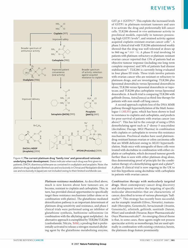
New, improved platinum drugs. Many tens of platinum analogues have entered the clinic in the past 30 years and interest in this strategy has waxed and, in particular, waned, as early optimism has faded to clinical demise (for example, with tetraplatin). However, since the turn of the new millennium, this strategy seems to finally be proving successful.
Oxaliplatin (1R,2R-diaminocyclohexane oxalato-platinum(II)) is based on the 1,2-diaminocyclohexane (DACH) carrier ligand and was originally described in the late 1970s37 (FIG. 4). It is a more water-soluble deriva-tive of the failed drug tetraplatin. Interestingly, oxali-platin showed a differing pattern of sensitivity to that of cisplatin in the NCI 60-cell human tumour panel38. In addition: in contrast to cisplatin and carboplatin, the accumulation of oxaliplatin seems to be less dependent on the copper transporter CTR1 (REF. 14); MMR recog-nition proteins do not recognize oxaliplatin-induced DNA adducts29; some differences exist in the structure of oxaliplatin-induced 1,2-intrastrand DNA crosslinks39; and oxaliplatin retains activity against some cancer cells with acquired resistance to cisplatin40.
Early clinical trials with oxaliplatin, which were con-ducted in France, revealed modest single-agent activity in patients with colorectal cancer (10% objective response rate from >100 patients)41, but a more promising level of activity when used in combination with 5-fluorouracil (5FU) and leucovorin (LV) (58% objective response rate from 93 patients)42. Before these studies were reported, colon cancer had been widely acknowledged in the medical community as being insensitive to platins.
Between the years 2000 and 2004, four independent large phase III trials all demonstrated that oxaliplatin is active against metastatic colon cancer when used in the 5FU–LV combination (TABLE 1). In the first of these studies, patients with advanced colon cancer received 5FU with LV with or without oxaliplatin, as first-line treatment43. The addition of oxaliplatin significantly improved antitumour efficacy (median progression-free survival of 6.1 months without, versus 8.7 months with oxaliplatin added, p=0.048). Another study in patients with previously untreated
Box 2 | Why can’t all cancers respond to cisplatin like testicular cancer?
The introduction of cisplatin-containing regimens in the mid-1970s (with vinblastine and bleomycin) for men with metastatic testicular cancer changed the cure rate from 5% to 60%; the subsequent substitution of vinblastine with etoposide has pushed cure rates to around 80% (REF. 99). In the context of cancer chemotherapy for adult solid tumours, particularly metastatic ones, these are unusually high response and cure rates. Cell lines derived from testicular cancer were also shown to be intrinsically hypersensitive to cisplatin, in comparison to lines derived from bladder cancer100.
We now have some answers to why this occurs: the hypersensitivity often relates to a reduced DNA-repair capacity in response to platinum–DNA adducts. Tumour cells that were originally derived from a patient with testicular cancer were repeatedly exposed to cisplatin in vitro, thereby generating acquired resistance to the drug. Although the amount of platinum binding to DNA was similar in both cell lines, the resistant cells removed platinum from their DNA at the normal rate, but the parent cells had an inherent defect, and repaired DNA at only half this rate101. Subsequent studies showed that testicular cancer cell lines were also defective, in comparison to bladder cancer cell lines, at removing platinum from specific genes (so-called gene-specific repair)102. Using cell extracts to examine the specific DNA-repair pathway, nucleotide-excision repair (NER), it was shown that extracts from testicular cancer cells had low constitutive NER capacity and, in particular, low levels of the protein XPA (xeroderma pigmentosum (XP), complementation group A)103. Further studies have shown low levels of XPA and two other NER proteins, XPF (XP, complementation group F) and ERCC1 (excision repair cross-complementing-1), in testicular versus several additional tumour types104. Together, these studies reveal that testicular cancer cells often possess a low DNA-repair capability, and so, upon exposure to cisplatin, will undergo relatively more apoptosis.
R E V I E W S
NATURE REVIEWS | CANCER VOLUME 7 | AUGUST 2007 | 577
colon cancer used bolus 5FU with LV followed by a 22-hour infusion of 5FU, alone or with oxaliplatin44. Again, the group receiving oxaliplatin had significantly longer progression-free survival (median 9.0 versus 6.2 months, p=0.0003) and a better response rate (50.7% versus 22.3%, p=0.0001). In a third trial, patients who had progressed on bolus 5FU with LV and irinotecan (Camptosar, Pfizer; IFL), received bolus and infusional 5FU and LV (LV5FU2), single-agent oxaliplatin, or the combination of LV5FU2 and oxaliplatin (FOLFOX4). The FOLFOX4 regimen had significantly better clinical activity as measured by objective response rate (9.9% versus 0% for LV5FU2, p<0.0001) and median time to progression (4.6 months versus 2.7 months for LV5FU2, p<0.0001)45. Finally, patients with previously untreated metastatic colorectal cancer received either IFL or oxaliplatin and infused 5FU with LV (FOLFOX) or irinotecan and oxaliplatin (IROX). Significantly higher response rates and increased median time to progression and median survival times were observed for the FOLFOX arm (for example, median survival of 19.5 months versus 15.0 months for IFL and 17.4 months for IROX)46. Oxaliplatin became the third pla-tin to be approved by the US FDA in 2002 (TABLE 1).
Satraplatin (Spectrum Pharmaceuticals/GPC Biotech) and picoplatin (Poniard Pharmaceuticals, see
below) emerged from a continuation of the ICR/JM collaboration after the development of carboplatin. Satraplatin (bis-acetato-ammine-dichloro-cyclohexy-lamine platinum(IV), JM216) (FIG. 4) was originally developed to be an orally active version of carboplatin (that is, to possess a carboplatin rather than cispla-tin-like toxicity profile). Preclinical studies showed that the drug possessed good antitumour activity by the oral route, at least comparable to intravenously administered cisplatin or carboplatin, in mice with human ovarian cancer xenografts47, especially when administered over a 5-consecutive-day schedule48. It also retained activity in human cancer cells with acquired cisplatin-resistance, in which resistance was due to reduced platinum transport49. In vivo satraplatin is biotransformed to around six products, the major one being JM118 (cis-amminedichloro-cyclohexylamine-platinum(II)50. JM118 retained activity in cells that had lost the copper-influx transporter CTR1 (REF. 51), and has been shown to bind DNA in a very similar manner to that described for cisplatin52 and to be repaired by the NER pathway with similar kinetics53.
Early clinical trials with satraplatin demonstrated the feasibility of administering a platin by the oral route, especially when dosed on a daily basis for 5 consecutive days54. Satraplatin showed promising clinical activity in
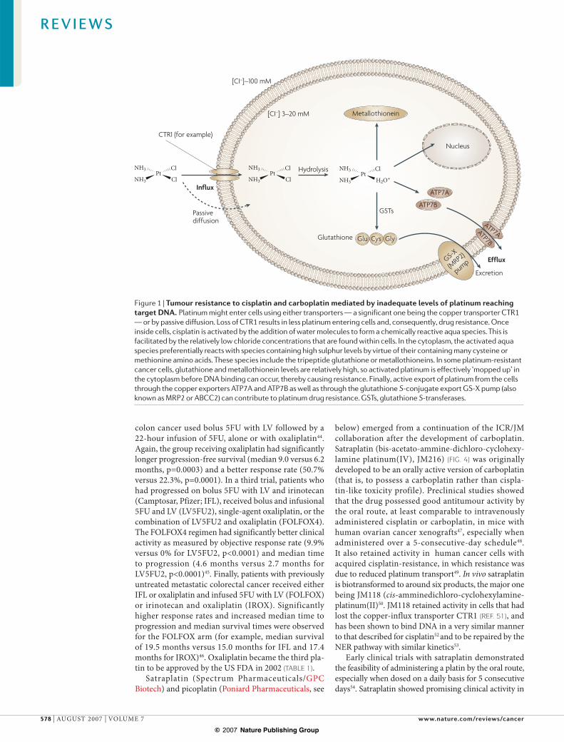
Figure 1 | Tumour resistance to cisplatin and carboplatin mediated by inadequate levels of platinum reaching target DNA. Platinum might enter cells using either transporters — a significant one being the copper transporter CTR1 — or by passive diffusion. Loss of CTR1 results in less platinum entering cells and, consequently, drug resistance. Once inside cells, cisplatin is activated by the addition of water molecules to form a chemically reactive aqua species. This is facilitated by the relatively low chloride concentrations that are found within cells. In the cytoplasm, the activated aqua species preferentially reacts with species containing high sulphur levels by virtue of their containing many cysteine or methionine amino acids. These species include the tripeptide glutathione or metallothioneins. In some platinum-resistant cancer cells, glutathione and metallothionein levels are relatively high, so activated platinum is effectively ‘mopped up’ in the cytoplasm before DNA binding can occur, thereby causing resistance. Finally, active export of platinum from the cells through the copper exporters ATP7A and ATP7B as well as through the glutathione S-conjugate export GS-X pump (also known as MRP2 or ABCC2) can contribute to platinum drug resistance. GSTs, glutathione S-transferases.
R E V I E W S
578 | AUGUST 2007 | VOLUME 7 www.nature.com/reviews/cancer
Increased nucleotide-excision repair (for example, increased ERCC1)
Loss of DNA mismatch repair(for example, loss of MLH1 expression)
Bypass of DNA adducts Decreased apoptosis
Increased tolerance Increased removal from DNA
NN
N
O
HH
C NN
N
NO
N
H
HH
G
Guanine N7 positionPt
NH3
NH3 PtNH3
NH3
XPFERCC1
RPA
XPA XPC
XPG
XPD XPBTFB5
TFIIH
HR23B
PMS2 MLH1
MSH3/6MSH2
Hazard ratioThe relative risk of experiencing a particular event; an HR of 0.6 means that one group has a 40% lower risk than the other group.
a small randomized trial in 50 patients with hormone-refractory prostate cancer; median overall survival was 14.9 months in patients receiving satraplatin plus prednisone, versus 11.9 months for prednisone alone (hazard ratio (HR) of 0.84)55–57. Subsequently, a phase III trial involving a similar comparison in 900 patients who had failed previous chemotherapy was completed. Final progression-free-survival data, released by the sponsor-ing company (GPC Biotech), showed that satraplatin significantly reduced the risk of disease progression, irrespective of the type of previous chemotherapy (HR of 0.6, p=0.0000003)56. These data have recently prompted the submission of a new drug application to the FDA for this indication. Combination trials are also ongoing with paclitaxel, erlotinib (Tarceva, Genentech), capecitabine (Xeloda, Roche) or radiotherapy.
Picoplatin (cis-amminedichloro, 2-methylpyridine, platinum (II); JM473) (FIG. 4), the third and final drug to emerge from the ICR/JM collaboration, was rationally
designed to provide steric bulk around the platinum centre58. This was suggested, and subsequently shown, to lead to a relative reduction in inactivation by thiol-containing species such as glutathione59 and metal-lothionein23, in comparison to cisplatin. Picoplatin retains activity against a wide range of cisplatin-resistant58 and oxaliplatin-resistant60 cells in vitro, which was independ-ent of whether resistance was due to reduced transport, increased cytoplasmic detoxification or increased DNA repair. It also possesses antitumour activity in vivo by both the intravenous and oral routes61; in addition, synergy has been demonstrated for picoplatin when used in combi-nation with paclitaxel62. Picoplatin has shown evidence of antitumour activity in phase II trials of ‘platinum-sensitive’ ovarian cancer63 and cisplatin-resistant small cell lung cancer (response rate of 15.4% in one trial64 and a median overall survival of 26.7 weeks in a recently completed second trial). Based on these data, the spon-soring company, Poniard, is planning a phase III trial of
Figure 2 | Tumour resistance to cisplatin and carboplatin mediated after DNA binding. Once the activated aqua platinum species (see FIG. 1 and note that this is the same for cisplatin and carboplatin) has entered the nucleus, preferential covalent binding to the nitrogen on position 7 of guanine occurs. The major covalent bis-adduct that is formed involves adjacent guanines on the same strand of DNA (the intrastrand crosslink); a minor adduct involves binding to guanines on opposite DNA strands (the interstrand crosslink). The main removal pathway for these DNA adducts is that of nucleotide-excision repair (NER); increased NER, especially through increased activity of the endonuclease protein ERCC1 (excision repair cross-complementing-1) can occur in tumours, and can lead to platinum drug resistance (as adducts are removed before apoptotic signalling pathways are triggered). In addition, resistance can occur through increased tolerance to platinum–DNA adducts — even though the DNA adducts are formed — either through loss of DNA mismatch repair, bypassing of adducts by polymerase ! and ", or through downregulation of apoptotic pathways. BAX, BCL2-associated X protein; BID, BH3 interacting domain death agonist; HR23B, human Rad23B; MLH1, MutL homologue 1; MSH2/3/6, MutS homologue 2/3/6; PCNA, proliferating cell nuclear antigen; PMS2, postmeiotic segregation increased-2; RPA, replication protein A; TFB5, tenth subunit of TFIIH; TFIIH, general transcription factor IIH; XPA/B/C/D/F/G, xeroderma pigmentosum (XP), complementation group A/B/C/D/F/G.
R E V I E W S
NATURE REVIEWS | CANCER VOLUME 7 | AUGUST 2007 | 579
picoplatin in patients with small-cell lung cancer; trials are also ongoing for picoplatin in combination with docetaxel (Taxotere, Sanofi-Aventis) against prostate cancer and with 5FU with LV against colorectal cancer (Poniard Pharmaceuticals).
The dose-limiting toxicities of these new platins are generally different from those observed with cisplatin. For example, in contrast to cisplatin and carboplatin, the dose-limiting toxicity of oxaliplatin is a cumulative sensory peripheral neuropathy that is exacerbated by exposure to cold41. Clinical studies with satraplatin and picoplatin have revealed dose-limiting toxicities similar to those of carboplatin, with reversible thrombocytopenia and neutropenia observed but no marked nephrotoxicity or neurotoxicity55,65.
In addition to oxaliplatin, satraplatin and picoplatin, another interesting platinum drug that entered phase I clinical trials in the late 1990s was the ‘rule-breaking’ cationic trinuclear agent BBR3464 (REF. 66). This drug forms interstrand crosslinks over a much longer range than cisplatin or carboplatin — that is, across up to six DNA base pairs — and encouraged the concept of designing platinum drugs with different modes of DNA binding. However, the clinical development of BBR3464 was stopped after initial phase II trials showed a relatively poor response rate. Furthermore, nedaplatin (254-S, cis-diammine-glycolatoplatinum(II)) is a platinum agent that is closely related in chemical structure to cisplatin, and has been used exclusively in Japan for the past 15 years to treat various cancers, such as head and neck, ovarian and lung cancer67. Improved delivery of platinum to tumours. The strat-egy of using delivery vehicles to selectively transport more of a tumour-killing agent to tumours is attrac-tive, and has now been clinically validated with the cytotoxics doxorubicin (liposomal doxorubicin; Doxil, Ortho Biotech Products68 and paclitaxel (nanoparti-cle albumin-bound paclitaxel; Abraxane, Abraxis BioScience)69. Unsurprisingly, given their clinical
importance, liposomal preparations of cisplatin-like molecules have been prepared, and one, SPI-77, has been tested clinically70. Although there was minimal toxicity with SPI-77, it had only modest antitumour activity in comparison to cisplatin; this might reflect the challenge of not only having to deliver platinum to the tumour in a relatively inactive form, but also the subsequent need to achieve good release and activation. The current lead platinum liposomal drug is based on the DACH stable ligand found in oxaliplatin (DACH-L-NDDP; Aroplatin, Antigenics)71 (FIG. 4). A phase II trial in 20 patients with advanced colorectal cancer that was refractory to 5FU with LV, capecitabine or irinotecan, reported that DACH-L-NDDP was well tolerated and produced a modest tumour response rate of 5.6%, which is broadly comparable to that of single-agent oxaliplatin in this patient population71. Trials are continuing with a reformulated product in an attempt to improve the antitumour activity of the agent.
A second strategy, which is also being tested in early-stage clinical development, is to link a platinum-based drug to a water-soluble, biocompatible co-polymer, such as hydroxypropylmethacrylamide (HPMA), in order to exploit the enhanced permeability and retention (EPR) effect of macromolecules in tumours. As with the lipo-some approach, initial clinical studies have progressed from using a cisplatin-like platin (AP5280)72 to ongoing trials with a derivative that is based on the DACH carrier ligand, AP5346 (ProLindac, Access Pharmaceuticals)73 (FIG. 4). Preclinical studies showed that AP5346 possessed superior antitumour activity to oxaliplatin in a mouse model of melanoma and a xenograft model of human ovarian carcinoma. Furthermore, at equitoxic doses, AP5346 delivered 16.3-fold more platinum to the tumour, and 14.2-fold more platinum to tumour DNA, than oxaliplatin73. Another feature was that the rate of release of platinum for AP5346 was sevenfold higher at pH 5.4 versus pH 7.4. As the extracellular pH of solid tumours has been shown to often be more acidic than normal tis-sues, this might also explain, in part, the increased tumour delivery of this agent. A phase I clinical trial with AP5346 recommended a dose of 640 mg m–2 for progressing to phase II, and reported two partial responses (in patients with metastatic melanoma and ovarian cancer)74. Phase II trials are ongoing in patients with recurrent ovarian or head and neck cancers.
Finally, in particular situations, such as in patients with ovarian cancer, localized platinum-drug admin-istration through intraperitoneal injection might be applied. In support of this strategy, a recently completed phase III trial of over 400 patients (who had undergone surgery for ovarian cancer), compared the activity of intravenous paclitaxel on day 1 plus intravenous cispla-tin on day 2 with that of intravenous paclitaxel followed by intraperitoneally administered cisplatin on day 2 and intraperitoneally administered paclitaxel on day 8 (REF.
75). Overall survival was significantly improved in the intraperitoneal arm (median of 65.6 months versus 49.7 months, p=0.03). This was the third such randomized trial showing a clinical advantage with intraperitoneal cisplatin in the treatment of ovarian cancer, which might encourage its further application.
Figure 3 | Major ongoing strategies to circumvent cisplatin and carboplatin resistance. Resistance can be tackled by: increasing the levels of platinum reaching tumours (for example, liposomal platinum products) thereby resulting in greater killing; combining existing platinum drugs with molecularly targeted drugs (for example, bevacizumab); using novel platinum drugs such as oxaliplatin that are capable of circumventing cisplatin-mediated resistance mechanisms; and using other drugs either alone (for example, TLK286) or in combination (for example, decitabine), which exploit particular cisplatin-mediated resistance mechanisms. GSH, reduced glutathione; GST, glutathione S-transferase.
R E V I E W S
580 | AUGUST 2007 | VOLUME 7 www.nature.com/reviews/cancer
Broader spectrum of activity; retention against acquired resistance to cisplatin
Broader spectrum ofantitumour activity
Improved delivery
R=C9H19
Improved safety
PtCl
NH3
NH3
Cl
PtO
NH3
NH3
O
O
OCarboplatin (1982)
Oxaliplatin (1986)
Pt
H2N
NH2
(R)(R1)
O
OO
O
Pt
H2N
NH2
O
O
O
RR
O
DACH-L-NDDP(Aroplatin) (2004)
PtCl
N
H3N
Cl
Picoplatin (JM473) (1997)
PtCl
NH2
NH3
Cl
O
O
O
O
Satraplatin (JM216) (1993)
AP5346 (ProLindac) (2004)
NH2Pt
H2N
ON
NH
HN
O
O
NH
O
HN
OH
O n
O
–ONa+
O
O
Platinum-resistance modulation. As described above, much is now known about how tumours are, or become, resistant to cisplatin and carboplatin. This, in turn, has provided clinical opportunities to specifically target these resistance mechanisms (either alone or in combination with platins). The glutathione-mediated detoxification pathway is an important determinant of platinum-drug sensitivity and resistance, and phase I clinical trials were performed using an inhibitor of glutathione synthesis, buthionine sulfoximine (in combination with the alkylating agent melphalan). An alternative approach is exemplified by TER286/TLK286 (canfosfamide; Telcyta, Telik), a prodrug that is prefer-entially activated to release a nitrogen mustard alkylat-ing agent by the glutathione-metabolizing enzyme,
GST pi-1 (GSTP1)76. This exploits the increased levels of GSTP1 in platinum-resistant tumours and uses it to activate the drug and preferentially kill cancer cells. TLK286 showed in vivo antitumour activity in preclinical models, especially in tumours possess-ing high GSTP1 levels76, and retained activity against acquired cisplatin-resistant ovarian cancer cells77. A phase I clinical trial with TLK286 administered weekly showed that the drug was well tolerated at doses up to 960 mg m–2 (REF. 78). A phase II trial involving 34 patients with platinum-refractory or platinum-resistant ovarian cancer reported that 15% of patients had an objective tumour response (including one long-term complete response) and 50% of patients had disease stabilization79. TLK286 is currently being evaluated in four phase III trials. Three trials involve patients with ovarian cancer who are resistant or refractory to platinum drugs, and are investigating: TLK286 plus liposomal doxorubicin versus liposomal doxorubicin alone; TLK286 versus liposomal doxorubicin or topo-tecan; and TLK286 plus carboplatin versus liposomal doxorubicin. A fourth trial is comparing TLK286 with gefitinib (Iressa, AstraZeneca) as third-line therapy for patients with non-small-cell lung cancer.
A second approach exploits loss of the DNA MMR pathway through hypermethylation of the MutL homo-logue-1 (MLH1) gene, which has been shown to lead to resistance to cisplatin and carboplatin, and predicts for poor survival of patients with ovarian cancer (see above)31. This has led to the concept of using a DNA demethylating agent such as 2$-deoxy-5-azacytidine (decitabine; Dacoge, MGI Pharma) in combination with cisplatin or carboplatin to reverse this resistance mechanism. Preclinical studies have used platinum-drug-resistant human ovarian or colon carcinoma cells that are MMR deficient owing to MLH1 hypermeth-ylation. Nude mice with xenografts of these cells were treated with decitabine in combination with either cis-platin or carboplatin, which decreased tumour growth further than is seen with either platinum drug alone, thus demonstrating proof of principle for the combi-nation therapy of a demethylating agent and a platin80. A phase II clinical trial is now ongoing in the UK to test this hypothesis using decitabine with carboplatin in patients with ovarian cancer.
Combination therapy with molecularly targeted drugs. Most contemporary cancer drug discovery and development involves the targeting of specific molecular abnormalities that are characteristic of cancer, described in terms of various phenotypic ‘hall-marks’81. This strategy has recently been successful; see for example: imatinib (Glivec, Novartis), trastuzu-mab (Herceptin, Genentech), bevacizumab (Avastin, Genentech), erlotinib, gefitinib, sunitinib (Sutent, Pfizer) and sorafenib (Nexavar, Bayer Pharmaceuticals/Onyx Pharmaceuticals)82. An emerging clinical theme is that, in some cases, these agents might not possess spectacular activity as monotherapy, but are used opti-mally in combination with existing cytotoxics; herein, the platinum drugs feature prominently.
Figure 4 | The current platinum drug ‘family tree’ and generalized rationale underlying their development. Dates indicate when each drug was first given to patients. DACH, diaminocyclohexane as contained in oxaliplatin and aroplatin. Note: the platinum drugs lobaplatin (limited use and exclusively in China) and Nedaplatin (limited use and exclusively in Japan) are not included owing to their limited worldwide use.
R E V I E W S
NATURE REVIEWS | CANCER VOLUME 7 | AUGUST 2007 | 581
HER2An oncogene belonging to the EGFR family that has an important role in around a quarter of all breast cancers.
Bevacizumab, a humanized monoclonal antibody that targets vascular endothelial growth factor (VEGF), did not possess marked single-agent antitumour efficacy in phase II trials. But it has subsequently been shown to significantly improve responses and survival of patients with non-small-cell lung cancer when added to car-boplatin–paclitaxel combination chemotherapy83. The median survival was 12.3 months in the bevacizumab combination arm compared to 10.3 months in the chemo-therapy-alone group (HR for death of 0.79, p=0.003). In 2006, the FDA granted approval of bevacizumab for use in combination with carboplatin and paclitaxel in patients with unresectable, locally advanced, recurrent or metastatic, non-squamous, non-small-cell lung cancer. Also in patients with non-small-cell lung cancer, recently released phase II data show that the vascular-disrupting drug (that is, an agent that targets established tumour vasculature rather than the anti-angiogenics that target neo-vasculature) dimethylxanthenone-4-acetic acid (DMXAA, AS1404) might have clinical activity when used in combination with carboplatin and paclitaxel84.
The approved platinum drugs have traditionally not found widespread utility in the treatment of advanced breast cancer. However, recent data indicate that, under some circumstances, platins might be useful. First, preclinical data showed that a mouse antibody against ERBB2 (also known as HER2) synergized with cisplatin through a mechanism involving inhibition of the repair of platinum induced DNA damage85. Subsequently, 3 phase II clinical trials in patients with ERBB2-positive breast cancer using the anti-ERBB2 humanized antibody trastuzumab in combination with cisplatin or carbopla-tin and docetaxel, have shown promising clinical activ-ity86,87. Second, platins might be particularly useful in breast cancers harbouring BRCA1 or BRCA2 mutations
(approximately 5–10% of cases) as mouse-derived Brca1-negative cell lines have been shown to be fivefold hyper-sensitive to cisplatin, compared with wild-type cells88. This seems to be related to a lower DNA-repair capacity, and a clinical trial is ongoing to test this hypothesis89.
Preclinical data also indicate that platins might use-fully be combined with mammalian target of rapamycin (mTOR, also known as FRAP1) inhibitors, such as the rapamycin derivative RAD001 (everolimus; Certican, Novartis). Inhibition of mTOR by RAD001 was shown to sensitize cancer cells to cisplatin by increasing cisplatin-induced apoptosis; this related to inhibiting p53-induced p21 expression90. Other approaches such as short-hairpin-RNA-based screening91, cDNA-microarray screening92 and genome-wide expression profiling or in silico analyses93 might reveal additional drug targets for sensitizing tumours to cisplatin or carboplatin.
Future directions and prospectsThe three approved platinum drugs, cisplatin, carbo-platin and oxaliplatin, continue to have a major role in contemporary medical oncology. Additional platins such as satraplatin and picoplatin might further broaden their applicability to tumour types such as prostate cancer and small-cell lung cancer, respectively. There will probably be continued and extended use of platin-containing regimens with the new generation of molecularly tar-geted therapies, as exemplified by the recent approval of bevacizumab in combination with carboplatin and paclitaxel in patients with lung cancer; breast cancer provides additional possibilities. Improved tumour-delivery strategies and co-administration with specific modulators of prevalent platin-resistance mechanisms might also provide future clinical benefits.
1. Rosenberg, B., VanCamp, L., Krigas, T. Inhibition of cell division in Escherichia coli by electrolysis products from a platinum electrode. Nature 205, 698–699 (1965).
2. Rosenberg, B., VanCamp, L., Trosko, J. E., Mansour, V. H. Platinum compounds: a new class of potent antitumour agents. Nature 222, 385–386 (1969).
3. Siddik, Z. H. Cisplatin: mode of cytotoxic action and molecular basis of resistance. Oncogene 22, 7265–7279 (2003).
4. Chaney, S. G., Campbell, S. L., Temple, B., Bassett, E., Wu, Y., Faldu, M. Protein interactions with platinum–DNA adducts: from structure to function. J. Inorg. Biochem. 98, 1551–1559 (2004).
5. Eastman, A. Activation of programmed cell death by anticancer agents: cisplatin as a model system. Cancer Cells 2, 275–280 (1990).
6. Cvitkovic, E., Spaulding, J., Bethune, V., Martin, J., Whitmore, W. F. Improvement of cis-dichlorodiammineplatinum (NSC 119875): therapeutic index in an animal model. Cancer 39, 1357–1361 (1977).
7. Harrap, K. R. Preclinical studies identifying carboplatin as a viable cisplatin alternative. Cancer Treat. Rev. 12, 21–33 (1985).
8. Knox, R. J., Friedlos, F., Lydall, D. A., Roberts, J. J. Mechanism of cytotoxicity of anticancer platinum drugs: evidence that cis-diamminedichloroplatinum(II) and cis-diammine-(1,1-cyclobutanedicarboxylato)platinum(II) differ only in the kinetics of their interaction with DNA. Cancer Res. 46, 1972–1979 (1986).
9. Aabo, K., et al. Chemotherapy in advanced ovarian cancer: four systematic meta-analyses of individual patient data from 37 randomized trials. Advanced Ovarian Cancer Trialists’ Group. Br. J. Cancer 78, 1479–1487 (1998).
10. Kelland, L. R., et al. Mechanism-related circumvention of cis-diamminedichloro platinum(II) acquired resistance using two pairs of human ovarian carcinoma cell lines by ammine/amine platinum(IV) dicarboxylates. Cancer Res. 52, 3857–3864 (1992).
11. Gately, D. P., Howell, S. B. Cellular accumulation of the anticancer agent cisplatin: a review. Br. J. Cancer 67, 1171–1176 (1993).
12. Ishida, S., Lee, J., Thiele, D. J., Herskowitz, I. Uptake of the anticancer drug cisplatin mediated by the copper transporter Ctr1 in yeast and mammals. Proc. Natl Acad. Sci. USA 99, 14298–14302 (2002).This work identifies the role of the protein CTR1, normally involved in copper homeostasis, in transporting cisplatin into cells.
13. Katano, K., et al. Acquisition of resistance to cisplatin is accompanied by changes in the cellular pharmacology of copper. Cancer Res. 62, 6559–6565 (2002).
14. Holzer, A. K., Manorek, G. H., Howell, S. B. Contribution of the major copper influx transporter CTR1 to the cellular accumulation of cisplatin, carboplatin and oxaliplatin. Molec. Pharmacol. 70, 1390–1394 (2006).
15. Holzer, A. K., Howell, S. B. The internalization and degradation of human copper transporter 1 following cisplatin exposure. Cancer Res. 66, 10944–10952 (2006).
16. Safaei, R., Holzer, A. K., Katano, K., Samimi, G., Howell, S. B. The role of copper transporters in the development of resistance to Pt drugs. J. Inorg. Chem. 98, 1607–1613 (2004).
17. Samimi, G., et al. Increased expression of the copper efflux transporter ATP7A mediates resistance to cisplatin, carboplatin and oxaliplatin in ovarian cancer cells. Clin. Cancer Res. 10, 4661–4669 (2004).
18. Mistry, P., Kelland, L. R., Abel, G., Sidhar, S., Harrap, K. R. The relationships between glutathione, glutathione-S-transferase and cytotoxicity of platinum drugs and melphalan in eight human ovarian carcinoma cell lines. Br. J. Cancer 64, 215–220 (1991).One of the first preclinical studies identifying a correlation between increased levels of glutathione and resistance to cisplatin and carboplatin in ovarian carcinoma cells.
19. Ishikawa, T. The ATP-dependent glutathione S-conjugate export pump. Trends Biochem. Sci. 17, 463–468 (1992).
20. Lewis, A. D., Hayes, J. D., Wolf, C. R. Glutathione and glutathione-dependent enzymes in ovarian adenocarcinoma cell lines derived from a patient before and after the onset of drug resistance: intrinsic differences and cell cycle effects. Carcinogenesis, 9, 1283–1287 (1988).
21. Yang, P., Ebbert, J. O., Sun, Z., Weinshilboum, R. M. Role of the glutathione metabolic pathway in lung cancer treatment and prognosis: a review. J. Clin. Oncol. 24, 1761–1769 (2006).
22. Kelley, S. L. et al. Overexpression of metallothionein confers resistance to anticancer drugs. Science 241, 1813–1815 (1988).
23. Holford, J., Beale, P. J., Boxall, F. E., Sharp, S. Y., Kelland, L. R. Mechanisms of drug resistance to the platinum complex ZD0473 in ovarian cancer cell lines. Eur. J. Cancer 36, 1984–1990 (2000).
R E V I E W S
582 | AUGUST 2007 | VOLUME 7 www.nature.com/reviews/cancer
24. Johnson, S., et al. Relationship between platinum–DNA adduct formation and removal and cisplatin cytotoxicity in cisplatin-sensitive and -resistant human ovarian cancer cells. Cancer Res. 54, 5911–5916 (1994).
25. Ferry, K. V., Hamilton, T. C., Johnson, S. W. Increased nucleotide excision repair in cisplatin-resistant ovarian cancer cells: role of ERCC1-XPF. Biochem. Pharmacol. 60, 1305–1313 (2000).
26. Chang, I. Y., et al. Small interfering RNA-induced suppression of ERCC1, enhances sensitivity of human cancer cells to cisplatin. Biochem. Biophys. Res. Commun. 327, 225–233 (2005).
27. Dabholkar, M., Bostick-Bruton, F., Weber, C., Bohr, V. A., Egwuagu, C., Reed, E. ERCC1 and ERCC2 expression in malignant tissues from ovarian cancer patients. J. Natl. Cancer Inst. 84, 1512–1517 (1992).An original clinical translational study linking overexpression of the NER DNA-repair pathway gene ERCC1 to poor response to platinum-based chemotherapy in patients with ovarian cancer.
28. Reed, E. ERCC1 and clinical resistance to platinum-based therapy. Clin. Cancer Res. 11, 6100–6102 (2005).
29. Fink, D. et al. The role of DNA mismatch repair in platinum drug resistance. Cancer Res. 56, 4881–4886 (1996).
30. Zdraveski, Z. Z., Mello, J. A., Farinelli, C. K., Essigmann, J. M., Marinus, M. G. MutS preferentially recognises cisplatin- over oxaliplatin-modified DNA. J. Biol. Chem. 277, 1255–1260 (2002).
31. Gifford, G., Paul, J., Vasey, P. A., Kaye, S. B., Brown, R. The acquisition of hMLH1 methylation in plasma DNA after chemotherapy predicts poor survival for ovarian cancer patients. Clin. Cancer Res. 10, 4420–4426 (2004).A clinical translational study in ovarian cancer patients showing that loss of function of the DNA-mismatch-repair pathway through hypermethylation of the hMLH1 gene after chemotherapy, predicts poor survival.
32. Helleman, J., et al. Mismatch repair and treatment resistance in ovarian cancer. BMC Cancer 6, 201 (2006).
33. Bassett, E., et al. Frameshifts and deletions during in vitro translesion synthesis past Pt-DNA adducts by DNA polymerases ! and ". DNA repair 1, 1003–1016 (2002).
34. Albertella, M. R., Green, C. M., Lehmann, A. R., O’Connor, M. J. A role for polymerase " in the cellular tolerance to cisplatin-induced damage. Cancer Res. 65, 9799–9806 (2005).Experiments indicating that inhibition of DNA polymerase " could increase the anticancer effectiveness of cisplatin.
35. Gadducci, A., Cosio, S., Muraca, S., Genazzani, A. R. Molecular mechanisms of apoptosis and chemosensitivity to platinum and paclitaxel in ovarian cancer: biological data and clinical implications. Eur. J. Gynaecol. Oncol. 23, 390–396 (2002).
36. Kelland, L. R. Overcoming resistance to platinum therapy in patients with advanced cancer. Am. J. Cancer 1, 247–255 (2002).
37. Kidani, Y., Inagaki, K., Iigo, M., Hoshi, A., Kuretani, K. Antitumor activity of 1,2-diaminocyclohexane-platinum complexes against sarcoma-180 ascites form. J. Med. Chem. 21, 1315–1318 (1978).
38. Rixe, O., et al. Oxaliplatin, tetraplatin, cisplatin and carboplatin: spectrum of activity in drug-resistant cell lines and in the cell lines of the National Cancer Institute’s Anticancer Drug Screen panel. Biochem. Pharmacol. 52, 1855–1865 (1996).
39. Spingler, B., Whittington, D. A., Lippard, S. J. 2.4Å crystal structure of an oxaliplatin 1,2-d(GpG) intrastrand cross-link in a DNA dodecamer duplex. Inorg. Chem. 40, 5596–5602 (2001).
40. Raymond, E., Faivre, S., Chaney, S., Woynarowski, J., Cvitkovic, E. Cellular and molecular pharmacology of oxaliplatin. Mol. Cancer Ther. 1, 227–235 (2002).
41. Machover, D., et al. Two consecutive phase II studies of oxaliplatin (L-OHP) for treatment of patients with advanced colorectal carcinoma who were resistant to previous treatment with fluoropyrimidines. Ann. Oncol. 7, 95–98 (1996).
42. Levi, F., et al. A chronopharmacologic phase II clinical trial with 5-fluorouracil, folinic acid, and oxaliplatin using an ambulatory multichannel programmable pump. High antitumour effectiveness against metastatic colorectal cancer. Cancer 69, 893–900 (1992).
43. Giacchetti, S., et al. Phase III multicenter randomized trial of oxaliplatin added to chronomodulated fluorouracil–leucovorin as first-line treatment of metastatic colorectal cancer. J. Clin. Oncol. 18, 136–147 (2000).
44. De Gramont, A., et al. Leucovorin and fluorouracil with or without oxaliplatin as first-line treatment in advanced colorectal cancer. J. Clin. Oncol. 18, 2938–2947 (2000).
45. Rothenberg, M. L., et al. Superiority of oxaliplatin and fluorouracil–leucovorin compared with either therapy alone in patients with progressive colorectal cancer after irinotecan and fluorouracil–leucovorin: interim results of a Phase III trial. J. Clin. Oncol. 21, 2059–2069 (2003).
46. Goldberg, R. M., et al. A randomized controlled trial of fluorouracil plus leucovorin, irinotecan, and oxaliplatin combinations in patients with previously untreated metastatic colorectal cancer. J. Clin. Oncol. 22, 23–29 (2004).A key clinical phase III study showing significantly improved survival with oxaliplatin, fluorouracil/leucovorin (FOLFOX regimen) in patients with previously untreated metastatic colorectal cancer.
47. Kelland, L. R., et al. Preclinical antitumor evaluation of Bis-acetato-ammine-dichloro-cyclohexylamine platinum(IV): an orally active platinum drug. Cancer Res. 53, 2581–2586 (1993).The first paper demonstrating, in preclinical tumour models, the feasibility of achieving antitumour activity with an orally administered platin, JM216/satraplatin.
48. McKeage, M. J. et al. Schedule dependency of orally administered Bis-acetato-ammine-dichloro-cyclohexylamine-platinum(IV) (JM216) in vivo. Cancer Res. 54, 4118–4122 (1994).
49. Sharp, S. Y., Rogers, P. M., Kelland, L. R. Transport of cisplatin and Bis-acetato-ammine-dichlorocyclohexylamine platinum(IV) (JM216) in human ovarian carcinoma cell lines: identification of a plasma membrane protein associated with cisplatin resistance. Clin. Cancer Res. 1, 981–989 (1995).
50. Kelland, L. R. An update on satraplatin: the first orally available platinum anticancer drug. Exp. Opin. Invest. Drugs 9, 1373–1382 (2000).
51. Samimi, G., Howell, S. B. Modulation of the cellular pharmacology of JM118, the major metabolite of satraplatin, by copper influx and efflux transporters. Cancer Chemother. Pharmacol. 57, 781–788 (2006).
52. Silverman, A. P., Bu, W., Cohen, S. M., Lippard, S. J. 2.4-Å crystal structure of the asymmetric platinum complex [Pt(ammine)(cyclohexylamine)]2+ bound to a dodecamer DNA duplex. J. Biol. Chem. 277, 49743–49749 (2002).
53. Reardon, J. T., Vaisman, A., Chaney, S. G., Sancar, A. Efficient nucleotide excision repair of cisplatin, oxaliplatin, and bis-aceto-ammine-dichloro-cyclohexylamine-platinum(IV) (JM216) platinum intrastrand DNA diadducts. Cancer Res. 59, 3968–3971 (1999).
54. McKeage, M. J., et al. Phase I and pharmacokinetic study of an oral platinum complex given daily for 5 days in patients with cancer. J. Clin. Oncol. 15, 2691–2700 (1997).
55. Choy, H. Satraplatin: an orally available platinum analog for the treatment of cancer. Expert Rev. Anticancer Ther. 6, 973–982 (2006).
56. McKeage, M. J. Satraplatin in hormone-refractory prostate cancer and other tumour types: pharmacological properties and clinical evaluation. Drugs 67, 859–869 (2007).
57. Sternberg, C. N., et al. Phase III trial of Satraplatin, an oral platinum plus prednisone vs. prednisone alone in patients with hormone-refractory prostate cancer. Oncology, 68, 2–9 (2005).A pivotal clinical study demonstrating the potential clinical benefit of satraplatin in patients with hormone-refractory prostate cancer.
58. Holford, J., Sharp, S. Y., Murrer, B. A., Abrams, M., Kelland, L. R. In vitro circumvention of cisplatin resistance by the novel sterically hindered platinum complex AMD473. Br. J. Cancer 77, 366–373 (1998).The first studies with JM473/picoplatin, showing retention of activity in vitro against several cisplatin-resistant tumour cell lines of defined mechanisms of resistance.
59. Holford, J., et al. Chemical, biochemical and pharmacological activity of the novel sterically hindered platinum co-ordination complex, cis-[amminedichloro(2-methylpyridine)] platinum(II) (AMD473). Anticancer Drug Design 13, 1–18 (1998).
60. Sharp, S. Y., O’Neill, C. F., Rogers, P. M., Boxall, F. E, Kelland, L. R. Retention of activity by the new generation platinum agent AMD0473 in four human tumour cell lines possessing acquired resistance to oxaliplatin. Eur. J. Cancer 38, 2309–2315 (2002).
61. Raynaud, F. I. et al. Cis-amminedichloro(2-methylpyridine) platinum (II) (AMD473), a novel sterically hindered platinum complex: in vivo activity, toxicology and pharmacokinetics in mice. Clin. Cancer Res. 3, 2063–2074 (1997).
62. Rogers, P., Boxall, F. E., Allot, C. P., Stephens, T. C., Kelland, L. R. Sequence-dependent synergism between the new generation platinum agent ZD0473 and paclitaxel in cisplatin-sensitive and -resistant human ovarian carcinoma cell lines. Eur. J. Cancer 38, 1653–1660 (2002).
63. Gore, M. E., et al. A phase II trial of ZD0473 in platinum-pretreated ovarian cancer. Eur. J. Cancer, 38, 2416–2420 (2002).
64. Treat, J., et al. ZD0473 treatment in lung cancer: an overview of the clinical trial results. Eur. J. Cancer 38, S13–S18 (2002).A summary of early-phase clinical studies of picoplatin in patients with lung cancer, demonstrating potential utility in cisplatin-resistant small-cell lung cancer.
65. Beale, P. et al. A phase I clinical and pharmacological study of cis-diamminedichloro(2-methylpyridine) platinum II (AMD473). Br. J. Cancer 88, 1128–1134 (2003).
66. Brabec, V. et al. DNA modifications by a novel bifunctional trinuclear platinum phase I anticancer agent. Biochemistry 38, 6781–6790 (1999).
67. Akaza, H. et al. Phase II study of cis-diammine(glycolato)platinum, 254–S, in patients with advanced germ-cell testicular cancer, prostatic cancer, and transitional-cell carcinoma of the urinary tract. Cancer Chemother. Pharmacol. 31, 187–192 (1992).
68. Gordon, A. N., et al. Long-term survival advantage for women treated with pegylated liposomal doxorubicin compared with topotecan in a phase 3 randomized study of recurrent and refractory epithelial ovarian cancer. Gynecol. Oncol. 95, 1–8 (2004).
69. Gradishar, W. J., et al. Phase III trial of nanoparticle albumin-bound paclitaxel compared with polyethylated castor oil-based paclitaxel in women with breast cancer. J. Clin. Oncol. 23, 7794–7803 (2005).
70. White, S. C. et al. Phase II study of SPI-77 (sterically stabilised liposomal cisplatin) in advanced non-small cell lung cancer. Br. J. Cancer, 95, 822–828 (2006).
71. Dragovich, T., Mendelson, D., Kurtin, S., Richardson, K., Von Hoff, D., Hoos, A. A Phase 2 trial of the liposomal DACH platinum L-NDDP in patients with therapy-refractory advanced colorectal cancer. Cancer Chemother. Pharmacol. 58, 759–764 (2006).
72. Rademaker-Lakhai, J. M., et al. A Phase I and pharmacological study of the platinum polymer AP5280 given as an intravenous infusion once every 3 weeks in patients with solid tumors. Clin. Cancer Res. 10, 3386–3395 (2004).
73. Rice, J. R., Gerberich, J. L., Nowotnik, D. P., Howell, S. B. Preclinical efficacy and pharmacokinetics of AP5346, a novel diaminocyclohexane-platinum tumor-targeting drug delivery system. Clin. Cancer Res. 12, 2248–2254 (2006).
74. Campone, M., et al. Phase I and pharmacokinetic trial of AP5346, a DACH-platinum-polymer conjugate, administered weekly for three out of every 4 weeks to advanced solid tumor patients. Cancer Chemother. Pharmacol. 17 Feb 2007 (doi:10.1007/s00280-006-0397-0).
75. Armstrong, D. K., et al. Intraperitoneal cisplatin and paclitaxel in ovarian cancer. N. Engl. J. Med. 354, 34–43 (2006).The most recent of 3 randomized trials in patients with ovarian cancer indicating an improvement in tumour response rates through intraperitoneal administration of platinum-based chemotherapy.
76. Morgan, A. S., et al. Tumor efficacy and bone-marrow sparing properties of TER286, a cytotoxin activated by glutathione S-transferase. Cancer Res. 58, 2568–2575 (1998).
77. Townsend, D. M., Shen, H., Staros, A. L., Gate, L., Tew, K. D. Efficacy of a glutathione S-transferase %-activated prodrug in platinum-resistant ovarian cancer cells. Mol. Cancer Ther. 1, 1089–1095 (2002).
R E V I E W S
NATURE REVIEWS | CANCER VOLUME 7 | AUGUST 2007 | 583
78. Rosen, L. S., Phase I study of TLK286 (Telcyta) administered weekly in advanced malignancies. Clin. Cancer Res. 10, 3689–3698 (2004).
79. Kavanagh, J. J., et al. Multi-institutional phase 2 study of TLK286 (TELCYTA, a glutathione S-transferase P1–1 activated glutathione analog prodrug) in patients with platinum and paclitaxel refractory or resistant ovarian cancer. Int. J. Gynecol. Cancer 15, 593–600 (2005).
80. Plumb, J. A., Strathdee, G., Sludden, J., Kaye, S. B., Brown, R. Reversal of drug resistance in human tumor xenografts by 2$-deoxy-5-azacytidine-induced demethylation of the hMLH1 gene promoter. Cancer Res. 60, 6039–6044 (2000).
81. Hanahan, D., Weinberg, R. A. The hallmarks of cancer. Cell 100, 57–70 (2000).
82. Collins, I., Workman, P. New approaches to molecular cancer therapeutics. Nature Chem. Biol. 2, 689–700 (2006).
83. Sandler, A., et al. Paclitaxel–carboplatin alone or with bevacizumab for non-small cell lung cancer. N. Engl. J. Med. 355, 2542–2550 (2006).The first demonstration of clinical proof of principle for the use of platinum-based chemotherapy (carboplatin) in combination with a molecularly targeted drug (bevacizumab).
84. McKeage, M. J., Kelland, L. R. 5,6-dimethylxanthenone-4-acetic acid (DMXAA). Clinical potential in combination with taxane-based chemotherapy. Am. J. Cancer, 5, 155–162 (2006).
85. Pietras, R. J., Fendly, B. M., Chazin, V. R., Pegram, M. D., Howell, S. B., Slamon, D. J. Antibody to HER-2/neu receptor blocks DNA repair after cisplatin in human breast and ovarian cancer cells. Oncogene, 9, 1829–1838 (1994).
86. Pegram, M. D., et al. Results of two open-label, multicenter phase II studies of docetaxel, platinum salts, and trastuzumab in HER2-positive advanced breast cancer. J. Natl. Cancer Inst. 96, 759–769 (2004).
87. Hurley, J., et al. Docetaxel, cisplatin and trastuzumab as primary systemic therapy for human epidermal growth factor receptor 2-positive locally advanced breast cancer. J. Clin. Oncol. 24, 1831–1838 (2006).
88. Bhattacharyya, A., Ear, U. S., Koller, B. H., Weichselbaum, R. R., Bishop, D. K. The breast cancer susceptibility gene BRCA1 is required for subnuclear assembly of Rad51 and survival following treatment
with the DNA cross-linking agent cisplatin. J. Biol. Chem. 275, 23899–23903 (2000).A preclinical study showing that breast cancer cells harbouring BRCA1 mutations are hypersensitive to cisplatin.
89. Turner, N., Tutt, A., Ashworth, A. Targeting the DNA repair defect of BRCA tumours. Curr. Opin. Pharmacol. 5, 388–393 (2005).
90. Beuvink, I, et al. The mTOR inhibitor RAD001 sensitizes tumor cells to DNA-damaged induced apoptosis through inhibition of p21 translation. Cell 120, 747–759 (2005).
91. Morgan-Lappe, S., et al. RNAi-based screening of the human kinome identifies Akt-cooperating kinases: a new approach to designing efficacious multitargeted kinase inhibitors. Oncogene, 25, 1340–1348 (2006).
92. Roberts, D., et al. Identification of genes associated with platinum drug sensitivity and resistance in human ovarian cancer cells. Br. J. Cancer 92, 1149–1158 (2005).
93. Olivero, M., et al. Genes regulated by hepatocyte growth factor as targets to sensitize ovarian cells to cisplatin. Mol. Cancer Ther. 5, 1126–1135 (2006).
94. Davies, M. S., Berners-Price, S. J., Hambley, T. W. Rates of platination of –AG- and –GA- containing double-stranded oligonucleotides: effect of chloride concentration. J. Inorg. Biochem. 79, 167–172 (2000).
95. Fichtinger-Schepman, A. M., van der Veer, J. L., den Hartog, J. H., Lohman, P. H., Reedijk, J. Adducts of the antitumor drug cis-diamminedichloroplatinum(II) with DNA: formation, identification, and quantitation. Biochemistry, 24, 707–713 (1985).
96. Takahara, P. M., Rosenzweig, A. C., Frederick, C. A., Lippard, S. J. Crystal structure of double-stranded DNA containing the major adduct of the anticancer drug cisplatin. Nature 377, 649–652 (1995).
97. Huang, H., Zhu, L., Reid, B. R., Drobny, G. P., Hopkins, P. B. Solution structure of a cisplatin-induced interstrand cross-link. Science 270, 1842–1845 (1995).
98. Teuben, J. M., Bauer, C., Wang, A. H., Reedijk, J. Solution structure of a DNA duplex containing a cis-diammineplatinum(II) 1,3-d(GTG) intrastrand cross-link, a major adduct in cells treated with the anticancer drug carboplatin. Biochemistry 38, 12305–12312 (1999).
99. Einhorn, L. H. Curing metastatic testicular cancer. Proc. Natl Acad. Sci. USA 99, 4592–4595 (2002).
100. Walker, M. C., Parris, C. N., Masters, J. R. Differential sensitivities of human testicular and bladder tumor cell lines to chemotherapeutic drugs. J. Natl Cancer Inst. 79, 213–216 (1987).
101. Kelland, L. R., et al. Establishment and characterization of an in vitro model of acquired resistance to cisplatin in a human testicular nonseminomatous germ cell line. Cancer Res. 52, 1710–1716 (1992).
102. Koberle, B. et al. DNA repair capacity and cisplatin sensitivity of human testis tumour cells. Int. J. Cancer 70, 551–555 (1997).
103. Koberle, B., Masters, J. R., Hartley, J. A., Wood, R. D. Defective repair of cisplatin-induced DNA damage caused by reduced XPA protein in testicular germ cell tumours. Curr. Biol. 9, 273–276 (1999).The identification of a molecular defect in the NER DNA-repair pathway causing hypersensitivity of at least some testicular cancers to cisplatin.
104. Welsh, C., Day, R., McGurk, C., Masters, J. R. W., Wood, R. D. Koberle, B. Reduced levels of XPA, ERCC1 and XPF DNA repair proteins in testis tumor cell lines. Int. J. Cancer 110, 352–361 (2004).
AcknowledgementsThe author wishes to thank K. Harrap and members of his laboratory, from 1987 to 1997. Studies on what became satraplatin and picoplatin during this period were supported by what is now Cancer Research UK.
Competing interests statementThe author declares competing financial interests: see web version for details.
FURTHER INFORMATIONCancer Research Technology: http://www.cancertechnology.comGPC Biotech: http://www.gpc-biotech.comPoniard Pharmaceuticals: http://www.poniard.comTelik Inc.: http://www.telik.comAccess to this links box is available online.
R E V I E W S
584 | AUGUST 2007 | VOLUME 7 www.nature.com/reviews/cancer