MEDICAL EDUCATION Classic Versus Millennial Medical Lab Anatomy BRION BENNINGER, 1,2,3,4,5,6,7,8,9 * NIK MATSLER, 1 AND TAYLOR DELAMARTER 1 1 Department of Medical Anatomical Sciences, Western University of Health Sciences, COMP–Northwest, Lebanon, Oregon 2 Department of Neuromuscular Medicine, Western University of Health Sciences, COMP–Northwest, Lebanon, Oregon 3 Department of Family Practice, Western University of Health Sciences, COMP–Northwest, Lebanon, Oregon 4 College of Dental Medicine, Pomona, California 5 Department of Orthopaedics, Samaritan Health Services, Corvallis, Oregon 6 Department of General Surgery, Samaritan Health Services, Corvallis, Oregon 7 Department of Oral Maxillofacial Surgery, Oregon Health and Science University, Portland, Oregon 8 Department of Surgery, Oregon Health and Science University, Portland, Oregon 9 Department of Orthopaedics and Rehabilitation, Oregon Health and Science University, Portland, Oregon This study investigated the integration, implementation, and use of cadaver dissection, hospital radiology modalities, surgical tools, and AV technology dur- ing a 12-week contemporary anatomy course suggesting a millennial labora- tory. The teaching of anatomy has undergone the greatest fluctuation of any of the basic sciences during the past 100 years in order to make room for the me- teoric rise in molecular sciences. Classically, anatomy consisted of a 2-year methodical, horizontal, anatomy course; anatomy has now morphed into a 12- week accelerated course in a vertical curriculum, at most institutions. Surface and radiological anatomy is the language for all clinicians regardless of spe- cialty. The objective of this study was to investigate whether integration of full- body dissection anatomy and modern hospital technology, during the anatomy laboratory, could be accomplished in a 12-week anatomy course. Literature search was conducted on anatomy text, journals, and websites regarding con- temporary hospital technology integrating multiple image mediums of 37 embalmed cadavers, surgical suite tools and technology, and audio/visual tech- nology. Surgical and radiology professionals were contracted to teach during the anatomy laboratory. Literature search revealed no contemporary studies integrating full-body dissection with hospital technology and behavior. About 37 cadavers were successfully imaged with roentograms, CT, and MRI scans. Students were in favor of the dynamic laboratory consisting of multiple activity sessions occurring simultaneously. Objectively, examination scores proved to be a positive outcome and, subjectively, feedback from students was over- whelmingly positive. Despite the surging molecular based sciences consuming much of the curricula, full-body dissection anatomy is irreplaceable regarding both surface and architectural, radiological anatomy. Radiology should not be a small adjunct to understand full-body dissection, but rather, full-body dissec- tion aids the understanding of radiology mediums. The millennial anatomy dis- section laboratory should consist of, at least, 50% radiology integration during *Correspondence to: Brion Benninger, Oregon Health and Sci- ence University, Oregon, USA. E-mail: [email protected]Received 15 February 2013; Accepted 26 March 2013 Published online in Wiley Online Library (wileyonlinelibrary.com). DOI: 10.1002/ca.22260 V V C 2013 Wiley Periodicals, Inc. Clinical Anatomy 00:000–000 (2013)
Transcript
MEDICAL EDUCATION
Classic Versus Millennial Medical Lab AnatomyBRION BENNINGER,1,2,3,4,5,6,7,8,9* NIK MATSLER,1 AND TAYLOR DELAMARTER1
1Department of Medical Anatomical Sciences, Western University of Health Sciences, COMP–Northwest,Lebanon, Oregon
2Department of Neuromuscular Medicine, Western University of Health Sciences, COMP–Northwest,Lebanon, Oregon
3Department of Family Practice, Western University of Health Sciences, COMP–Northwest, Lebanon,Oregon
4College of Dental Medicine, Pomona, California5Department of Orthopaedics, Samaritan Health Services, Corvallis, Oregon
6Department of General Surgery, Samaritan Health Services, Corvallis, Oregon7Department of Oral Maxillofacial Surgery, Oregon Health and Science University, Portland, Oregon
8Department of Surgery, Oregon Health and Science University, Portland, Oregon9Department of Orthopaedics and Rehabilitation, Oregon Health and Science University, Portland, Oregon
This study investigated the integration, implementation, and use of cadaverdissection, hospital radiology modalities, surgical tools, and AV technology dur-ing a 12-week contemporary anatomy course suggesting a millennial labora-tory. The teaching of anatomy has undergone the greatest fluctuation of any ofthe basic sciences during the past 100 years in order to make room for the me-teoric rise in molecular sciences. Classically, anatomy consisted of a 2-yearmethodical, horizontal, anatomy course; anatomy has now morphed into a 12-week accelerated course in a vertical curriculum, at most institutions. Surfaceand radiological anatomy is the language for all clinicians regardless of spe-cialty. The objective of this study was to investigate whether integration of full-body dissection anatomy and modern hospital technology, during the anatomylaboratory, could be accomplished in a 12-week anatomy course. Literaturesearch was conducted on anatomy text, journals, and websites regarding con-temporary hospital technology integrating multiple image mediums of 37embalmed cadavers, surgical suite tools and technology, and audio/visual tech-nology. Surgical and radiology professionals were contracted to teach duringthe anatomy laboratory. Literature search revealed no contemporary studiesintegrating full-body dissection with hospital technology and behavior. About37 cadavers were successfully imaged with roentograms, CT, and MRI scans.Students were in favor of the dynamic laboratory consisting of multiple activitysessions occurring simultaneously. Objectively, examination scores proved tobe a positive outcome and, subjectively, feedback from students was over-whelmingly positive. Despite the surging molecular based sciences consumingmuch of the curricula, full-body dissection anatomy is irreplaceable regardingboth surface and architectural, radiological anatomy. Radiology should not be asmall adjunct to understand full-body dissection, but rather, full-body dissec-tion aids the understanding of radiology mediums. The millennial anatomy dis-section laboratory should consist of, at least, 50% radiology integration during
*Correspondence to: Brion Benninger, Oregon Health and Sci-ence University, Oregon, USA. E-mail: [email protected]
Received 15 February 2013; Accepted 26 March 2013
Published online in Wiley Online Library(wileyonlinelibrary.com). DOI: 10.1002/ca.22260
VVC 2013 Wiley Periodicals, Inc.
Clinical Anatomy 00:000–000 (2013)
full-body dissection. This pilot study is an example of the most comprehensiveintegration of full-body dissection, radiology, and hospital technology. Clin.Anat. 00:000–000, 2013. VC 2013 Wiley Periodicals, Inc.
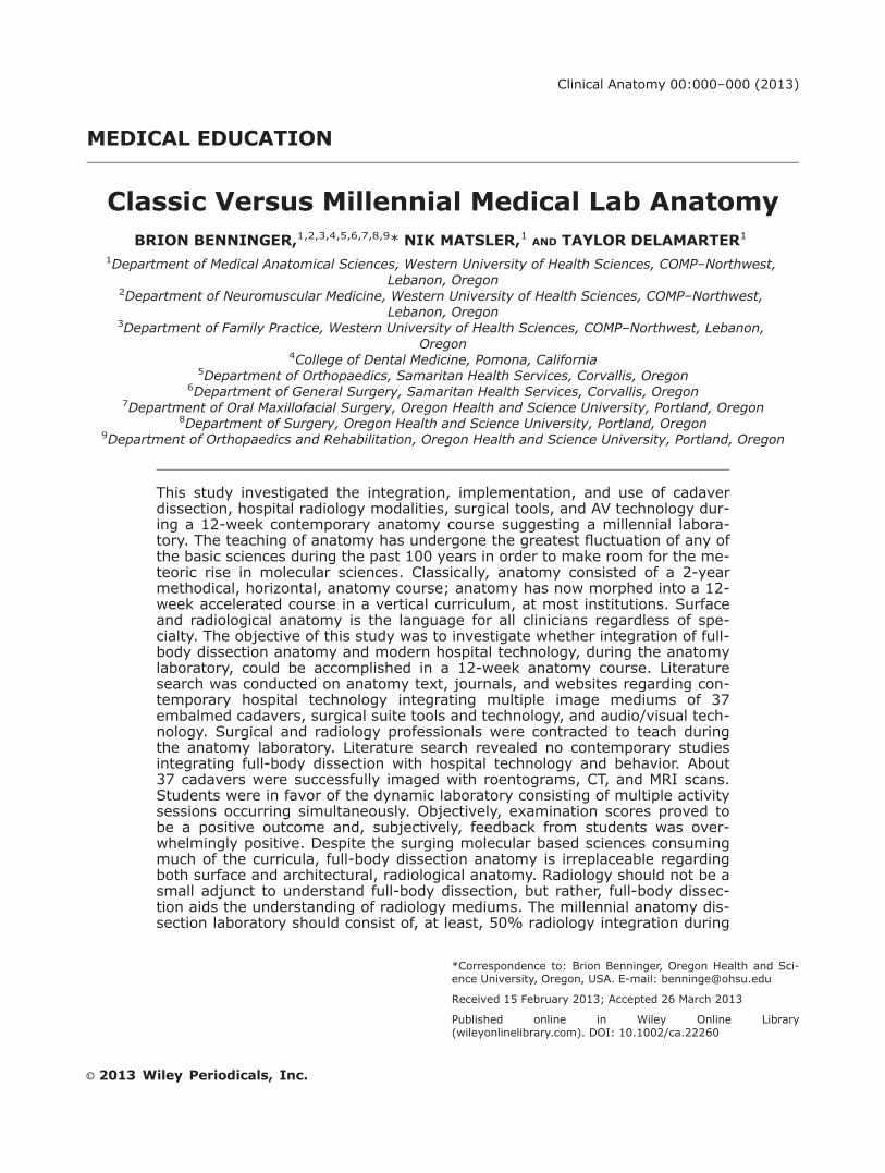
Historically, anatomy for health care students con-sisted of daily didactic lectures with complimentarylaboratory dissection hours covering the first and sec-ond years of basic sciences during a horizontal curric-ulum. As technology and science have progressed, thebasic science curriculum has had to integrate andadjust tremendously. This has placed increasing pres-sure on minimizing the time for anatomy training atmost institutions, distilling it down to an approxi-mately 12-week course. Recently, trends have devel-oped, especially amongst the European schools, tomove away from full-body dissection, utilizing instead:2D and 3D computer models; body painting; claymodeling; and viewed prosections (McLachlan, 2004;McMenamin, 2008; Tam et al., 2009). These trendshave surfaced in response to the lack of allotted timeduring an anatomy course and the increasing cost ofoperating a full-body dissection laboratory. Whilestudies are still being conducted on the efficacy ofcourses that do not involve full-body dissection, it iscertain that resident directors have noticed studentsare matriculating into various specialties with an in-credible lack of palpation and functional anatomicalconfidence regarding operating and invasive technolo-gies (Biasutto et al., 2006). Full-body dissection isparamount concerning anatomy education; however,with the meteoric rise in technology, it appears neces-sary for contemporary hospital equipment and tech-nology to be integrated early into students’ curriculumto be successful in today’s clinical environment (Jas-trow and Vollrath, 2003; Older 2004; McLachlan andPatten, 2006; Brenton et al., 2007; Korf et al., 2008)(Fig. 1). This study investigated if both dissection andtechnology could be integrated for an optimal anat-omy course within a vertically integrated curriculum.
METHODS
Literature search was conducted on anatomy texts,journals, websites, and specialty texts/journalsregarding the integration of hospital technology. Radi-ology: roentograms, CT scans, MRI, and ultrasonogra-phy was performed on 37 (F: 19 M: 18, ages 55–89,avg 78.35) embalmed cadavers. Anatomy dissectionlaboratory technology: audio/visual 8 3 8 foot projec-tor screens; flat-screen, 50’ HDTV’s (computerready); wireless audio system built into the dissectionlaboratory; portable, stacked laparoscopic machinewith insufflator; five different, mobile light boxes forprinted film viewing, along with one built in, six panellight box; two portable ultra sound machines; and
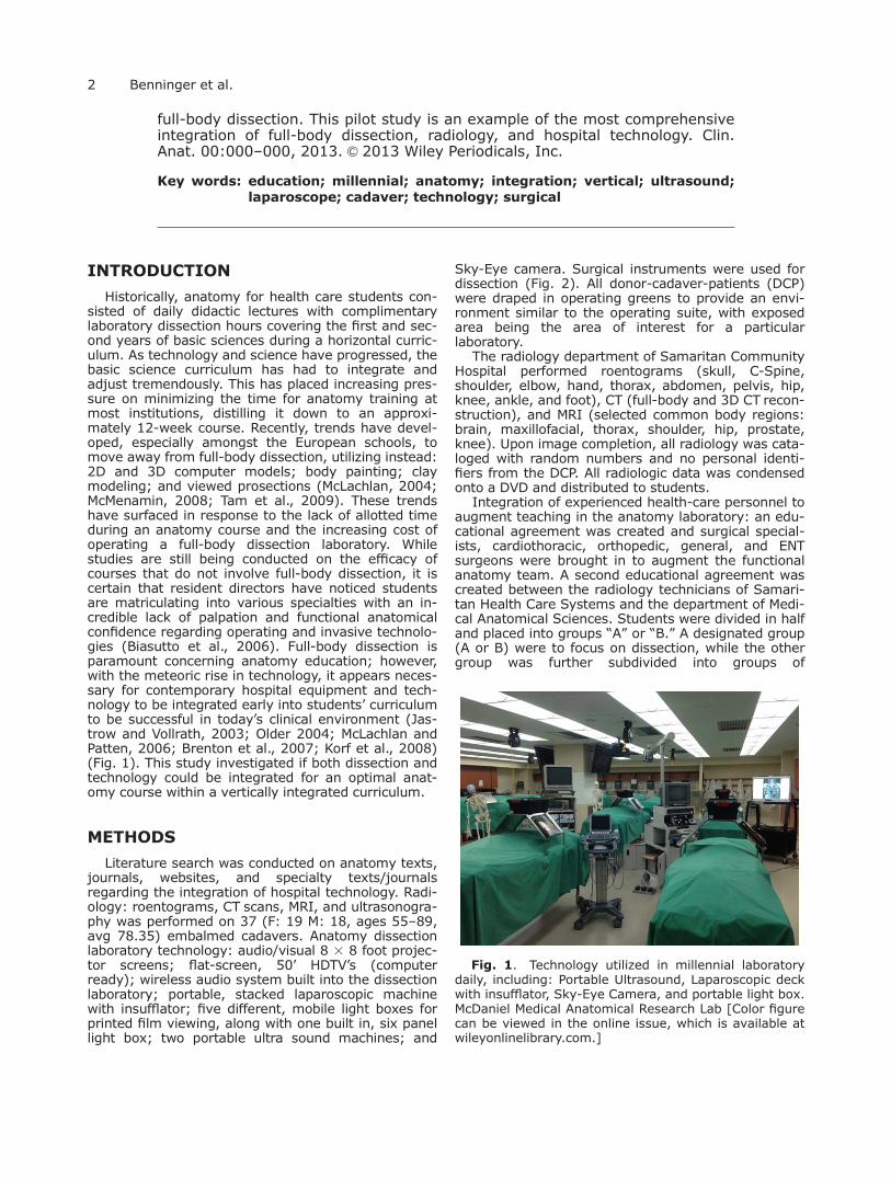
Sky-Eye camera. Surgical instruments were used fordissection (Fig. 2). All donor-cadaver-patients (DCP)were draped in operating greens to provide an envi-ronment similar to the operating suite, with exposedarea being the area of interest for a particularlaboratory.
The radiology department of Samaritan CommunityHospital performed roentograms (skull, C-Spine,shoulder, elbow, hand, thorax, abdomen, pelvis, hip,knee, ankle, and foot), CT (full-body and 3D CT recon-struction), and MRI (selected common body regions:brain, maxillofacial, thorax, shoulder, hip, prostate,knee). Upon image completion, all radiology was cata-loged with random numbers and no personal identi-fiers from the DCP. All radiologic data was condensedonto a DVD and distributed to students.
Integration of experienced health-care personnel toaugment teaching in the anatomy laboratory: an edu-cational agreement was created and surgical special-ists, cardiothoracic, orthopedic, general, and ENTsurgeons were brought in to augment the functionalanatomy team. A second educational agreement wascreated between the radiology technicians of Samari-tan Health Care Systems and the department of Medi-cal Anatomical Sciences. Students were divided in halfand placed into groups “A” or “B.” A designated group(A or B) were to focus on dissection, while the othergroup was further subdivided into groups of
Fig. 1. Technology utilized in millennial laboratorydaily, including: Portable Ultrasound, Laparoscopic deckwith insufflator, Sky-Eye Camera, and portable light box.McDaniel Medical Anatomical Research Lab [Color figurecan be viewed in the online issue, which is available atwileyonlinelibrary.com.]
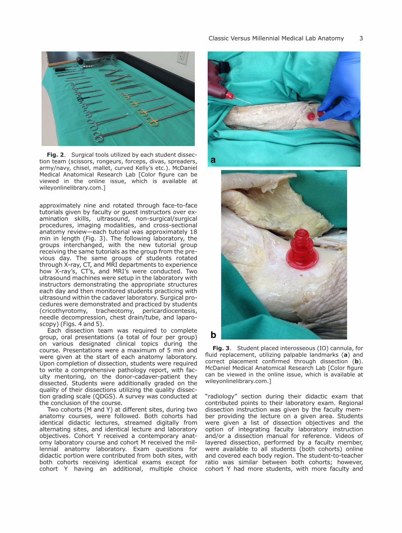
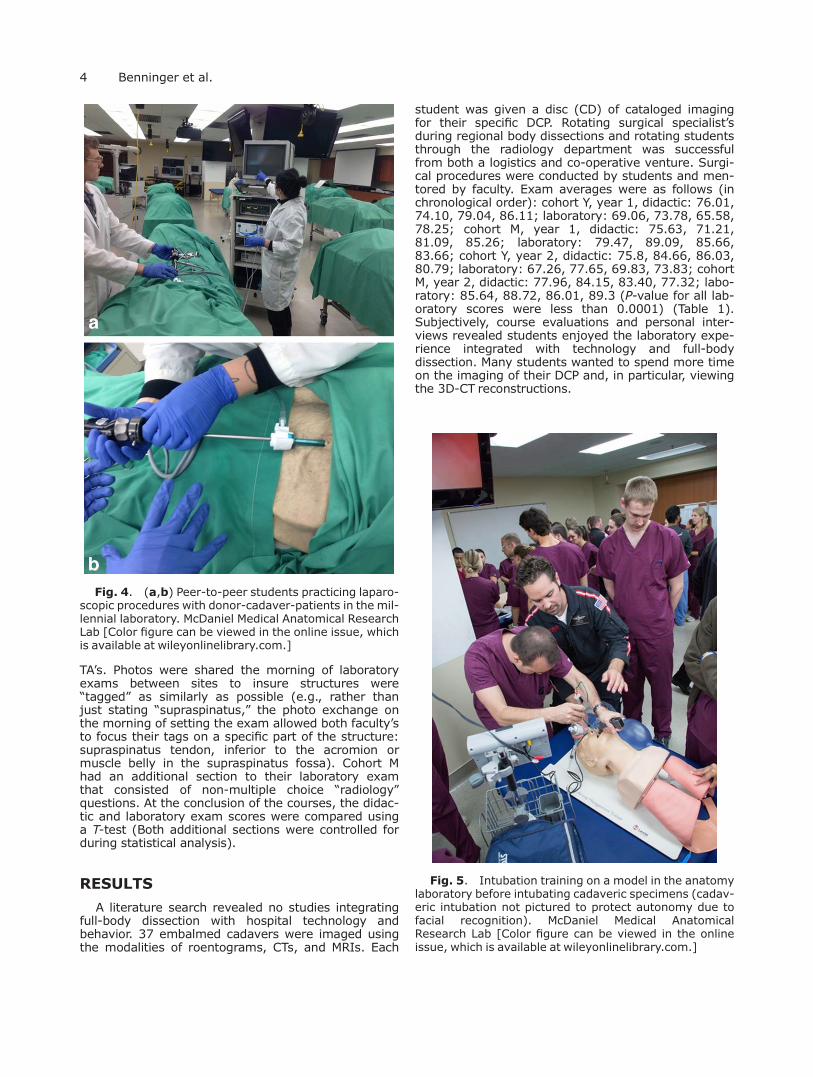
approximately nine and rotated through face-to-facetutorials given by faculty or guest instructors over ex-amination skills, ultrasound, non-surgical/surgicalprocedures, imaging modalities, and cross-sectionalanatomy review—each tutorial was approximately 18min in length (Fig. 3). The following laboratory, thegroups interchanged, with the new tutorial groupreceiving the same tutorials as the group from the pre-vious day. The same groups of students rotatedthrough X-ray, CT, and MRI departments to experiencehow X-ray’s, CT’s, and MRI’s were conducted. Twoultrasound machines were setup in the laboratory withinstructors demonstrating the appropriate structureseach day and then monitored students practicing withultrasound within the cadaver laboratory. Surgical pro-cedures were demonstrated and practiced by students(cricothyrotomy, tracheotomy, pericardiocentesis,needle decompression, chest drain/tube, and laparo-scopy) (Figs. 4 and 5).
Each dissection team was required to completegroup, oral presentations (a total of four per group)on various designated clinical topics during thecourse. Presentations were a maximum of 5 min andwere given at the start of each anatomy laboratory.Upon completion of dissection, students were requiredto write a comprehensive pathology report, with fac-ulty mentoring, on the donor-cadaver-patient theydissected. Students were additionally graded on thequality of their dissections utilizing the quality dissec-tion grading scale (QDGS). A survey was conducted atthe conclusion of the course.
Two cohorts (M and Y) at different sites, during twoanatomy courses, were followed. Both cohorts hadidentical didactic lectures, streamed digitally fromalternating sites, and identical lecture and laboratoryobjectives. Cohort Y received a contemporary anat-omy laboratory course and cohort M received the mil-lennial anatomy laboratory. Exam questions fordidactic portion were contributed from both sites, withboth cohorts receiving identical exams except forcohort Y having an additional, multiple choice
“radiology” section during their didactic exam thatcontributed points to their laboratory exam. Regionaldissection instruction was given by the faculty mem-ber providing the lecture on a given area. Studentswere given a list of dissection objectives and theoption of integrating faculty laboratory instructionand/or a dissection manual for reference. Videos oflayered dissection, performed by a faculty member,were available to all students (both cohorts) onlineand covered each body region. The student-to-teacherratio was similar between both cohorts; however,cohort Y had more students, with more faculty and
Fig. 2. Surgical tools utilized by each student dissec-tion team (scissors, rongeurs, forceps, divas, spreaders,army/navy, chisel, mallet, curved Kelly’s etc.). McDanielMedical Anatomical Research Lab [Color figure can beviewed in the online issue, which is available atwileyonlinelibrary.com.]
Fig. 3. Student placed interosseous (IO) cannula, forfluid replacement, utilizing palpable landmarks (a) andcorrect placement confirmed through dissection (b).McDaniel Medical Anatomical Research Lab [Color figurecan be viewed in the online issue, which is available atwileyonlinelibrary.com.]
TA’s. Photos were shared the morning of laboratoryexams between sites to insure structures were“tagged” as similarly as possible (e.g., rather thanjust stating “supraspinatus,” the photo exchange onthe morning of setting the exam allowed both faculty’sto focus their tags on a specific part of the structure:supraspinatus tendon, inferior to the acromion ormuscle belly in the supraspinatus fossa). Cohort Mhad an additional section to their laboratory examthat consisted of non-multiple choice “radiology”questions. At the conclusion of the courses, the didac-tic and laboratory exam scores were compared usinga T-test (Both additional sections were controlled forduring statistical analysis).
RESULTS
A literature search revealed no studies integratingfull-body dissection with hospital technology andbehavior. 37 embalmed cadavers were imaged usingthe modalities of roentograms, CTs, and MRIs. Each
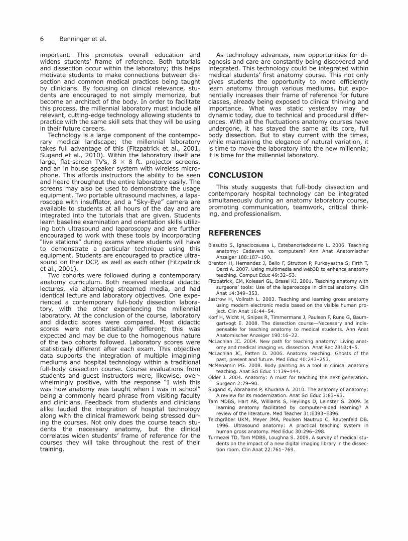
student was given a disc (CD) of cataloged imagingfor their specific DCP. Rotating surgical specialist’sduring regional body dissections and rotating studentsthrough the radiology department was successfulfrom both a logistics and co-operative venture. Surgi-cal procedures were conducted by students and men-tored by faculty. Exam averages were as follows (inchronological order): cohort Y, year 1, didactic: 76.01,74.10, 79.04, 86.11; laboratory: 69.06, 73.78, 65.58,78.25; cohort M, year 1, didactic: 75.63, 71.21,81.09, 85.26; laboratory: 79.47, 89.09, 85.66,83.66; cohort Y, year 2, didactic: 75.8, 84.66, 86.03,80.79; laboratory: 67.26, 77.65, 69.83, 73.83; cohortM, year 2, didactic: 77.96, 84.15, 83.40, 77.32; labo-ratory: 85.64, 88.72, 86.01, 89.3 (P-value for all lab-oratory scores were less than 0.0001) (Table 1).Subjectively, course evaluations and personal inter-views revealed students enjoyed the laboratory expe-rience integrated with technology and full-bodydissection. Many students wanted to spend more timeon the imaging of their DCP and, in particular, viewingthe 3D-CT reconstructions.
Fig. 4. (a,b) Peer-to-peer students practicing laparo-scopic procedures with donor-cadaver-patients in the mil-lennial laboratory. McDaniel Medical Anatomical ResearchLab [Color figure can be viewed in the online issue, whichis available at wileyonlinelibrary.com.]
Fig. 5. Intubation training on a model in the anatomylaboratory before intubating cadaveric specimens (cadav-eric intubation not pictured to protect autonomy due tofacial recognition). McDaniel Medical AnatomicalResearch Lab [Color figure can be viewed in the onlineissue, which is available at wileyonlinelibrary.com.]
Anatomy remains as one of the core building blocksthat make up the foundation upon which medicine isbuilt. Thorough understanding of the architecture ofthe body is paramount and can provide a platform fordifferential diagnosis, irrespective of specialty (Older,2004). However, anatomy does not come in discretepackets; it is a study of patterns and variation, of dif-ference in sameness throughout sub-populations. De-spite trends to move away from it, full-bodydissection still stands as the most efficient way to gainan understanding of the fluidity of anatomy and theinherent variation. Regardless of specialty, compre-hensive understanding and interpretation of radiologi-cal mediums is vital in today’s medical practice.Therefore, anatomy could be taught concurrently withradiology and given the same emphasis, offering theopportunity to students to understand both subjectsfrom the others’ perspective. Many health care curric-ula include full-body dissection in their laboratory andeither teaches radiology as a separate course or aftera significant break of time from anatomy dissection.
This break can create a disconnection between stu-dents and their anatomy knowledge, forcing them tospend as much time learning radiology as they dorelearning anatomy. Integrating radiology with dissec-tion, especially with imaging of the students’ specificdonor-cadaver-patient, allows the students to seecommon patterns of anatomy and pathological var-iance reflected in the physical and radiological. This“body-to-image” mentality allows students a more ef-ficient understanding of anatomy through imaginingand imagining through anatomy. Previous studiesintegrating imaging with first year anatomy dissectionlaboratories have shown that students respond posi-tively to the combination of anatomy and imaging andconsistently request more access to imaging outsideof structured laboratory time (Turmezei et al., 2009).These studies were limited, however, to “stock” imag-ing and the imaging was confined to the laboratoryonly; within the millennial laboratory, students check-out the imaging associated with their DCP and haveaccess to it at their convenience.
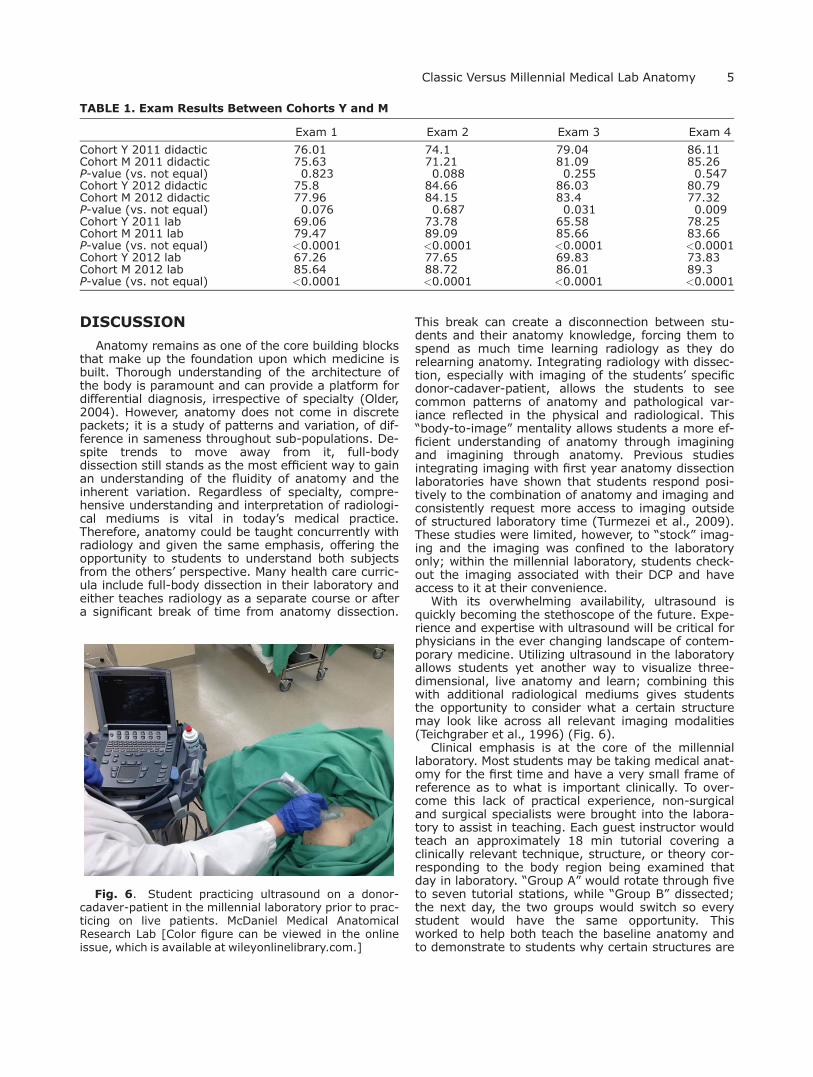
With its overwhelming availability, ultrasound isquickly becoming the stethoscope of the future. Expe-rience and expertise with ultrasound will be critical forphysicians in the ever changing landscape of contem-porary medicine. Utilizing ultrasound in the laboratoryallows students yet another way to visualize three-dimensional, live anatomy and learn; combining thiswith additional radiological mediums gives studentsthe opportunity to consider what a certain structuremay look like across all relevant imaging modalities(Teichgraber et al., 1996) (Fig. 6).
Clinical emphasis is at the core of the millenniallaboratory. Most students may be taking medical anat-omy for the first time and have a very small frame ofreference as to what is important clinically. To over-come this lack of practical experience, non-surgicaland surgical specialists were brought into the labora-tory to assist in teaching. Each guest instructor wouldteach an approximately 18 min tutorial covering aclinically relevant technique, structure, or theory cor-responding to the body region being examined thatday in laboratory. “Group A” would rotate through fiveto seven tutorial stations, while “Group B” dissected;the next day, the two groups would switch so everystudent would have the same opportunity. Thisworked to help both teach the baseline anatomy andto demonstrate to students why certain structures are
Fig. 6. Student practicing ultrasound on a donor-cadaver-patient in the millennial laboratory prior to prac-ticing on live patients. McDaniel Medical AnatomicalResearch Lab [Color figure can be viewed in the onlineissue, which is available at wileyonlinelibrary.com.]
TABLE 1. Exam Results Between Cohorts Y and M
Exam 1 Exam 2 Exam 3 Exam 4
Cohort Y 2011 didactic 76.01 74.1 79.04 86.11Cohort M 2011 didactic 75.63 71.21 81.09 85.26P-value (vs. not equal) 0.823 0.088 0.255 0.547Cohort Y 2012 didactic 75.8 84.66 86.03 80.79Cohort M 2012 didactic 77.96 84.15 83.4 77.32P-value (vs. not equal) 0.076 0.687 0.031 0.009Cohort Y 2011 lab 69.06 73.78 65.58 78.25Cohort M 2011 lab 79.47 89.09 85.66 83.66P-value (vs. not equal) <0.0001 <0.0001 <0.0001 <0.0001Cohort Y 2012 lab 67.26 77.65 69.83 73.83Cohort M 2012 lab 85.64 88.72 86.01 89.3P-value (vs. not equal) <0.0001 <0.0001 <0.0001 <0.0001
important. This promotes overall education andwidens students’ frame of reference. Both tutorialsand dissection occur within the laboratory; this helpsmotivate students to make connections between dis-section and common medical practices being taughtby clinicians. By focusing on clinical relevance, stu-dents are encouraged to not simply memorize, butbecome an architect of the body. In order to facilitatethis process, the millennial laboratory must include allrelevant, cutting-edge technology allowing students topractice with the same skill sets that they will be usingin their future careers.
Technology is a large component of the contempo-rary medical landscape; the millennial laboratorytakes full advantage of this (Fitzpatrick et al., 2001,Sugand et al., 2010). Within the laboratory itself arelarge, flat-screen TV’s, 8 3 8 ft. projector screens,and an in house speaker system with wireless micro-phone. This affords instructors the ability to be seenand heard throughout the entire laboratory easily. Thescreens may also be used to demonstrate the usageequipment. Two portable ultrasound machines, a lapa-roscope with insufflator, and a “Sky-Eye” camera areavailable to students at all hours of the day and areintegrated into the tutorials that are given. Studentslearn baseline examination and orientation skills utiliz-ing both ultrasound and laparoscopy and are furtherencouraged to work with these tools by incorporating“live stations” during exams where students will haveto demonstrate a particular technique using thisequipment. Students are encouraged to practice ultra-sound on their DCP, as well as each other (Fitzpatricket al., 2001).
Two cohorts were followed during a contemporaryanatomy curriculum. Both received identical didacticlectures, via alternating streamed media, and hadidentical lecture and laboratory objectives. One expe-rienced a contemporary full-body dissection labora-tory, with the other experiencing the millenniallaboratory. At the conclusion of the course, laboratoryand didactic scores were compared. Most didacticscores were not statistically different; this wasexpected and may be due to the homogenous natureof the two cohorts followed. Laboratory scores werestatistically different after each exam. This objectivedata supports the integration of multiple imaginingmediums and hospital technology within a traditionalfull-body dissection course. Course evaluations fromstudents and guest instructors were, likewise, over-whelmingly positive, with the response “I wish thiswas how anatomy was taught when I was in school”being a commonly heard phrase from visiting facultyand clinicians. Feedback from students and cliniciansalike lauded the integration of hospital technologyalong with the clinical framework being stressed dur-ing the courses. Not only does the course teach stu-dents the necessary anatomy, but the clinicalcorrelates widen students’ frame of reference for thecourses they will take throughout the rest of theirtraining.
As technology advances, new opportunities for di-agnosis and care are constantly being discovered andintegrated. This technology could be integrated withinmedical students’ first anatomy course. This not onlygives students the opportunity to more efficientlylearn anatomy through various mediums, but expo-nentially increases their frame of reference for futureclasses, already being exposed to clinical thinking andimportance. What was static yesterday may bedynamic today, due to technical and procedural differ-ences. With all the fluctuations anatomy courses haveundergone, it has stayed the same at its core, fullbody dissection. But to stay current with the times,while maintaining the elegance of natural variation, itis time to move the laboratory into the new millennia;it is time for the millennial laboratory.
CONCLUSION
This study suggests that full-body dissection andcontemporary hospital technology can be integratedsimultaneously during an anatomy laboratory course,promoting communication, teamwork, critical think-ing, and professionalism.
REFERENCES
Biasutto S, Ignaciocaussa L, Estebancriadodelrio L. 2006. Teachinganatomy: Cadavers vs. computers? Ann Anat AnatomischerAnzeiger 188:187–190.
Brenton H, Hernandez J, Bello F, Strutton P, Purkayastha S, Firth T,Darzi A. 2007. Using multimedia and web3D to enhance anatomyteaching. Comput Educ 49:32–53.
Fitzpatrick, CM, Kolesari GL, Brasel KJ. 2001. Teaching anatomy withsurgeons’ tools: Use of the laparoscope in clinical anatomy. ClinAnat 14:349–353.
Jastrow H, Vollrath L. 2003. Teaching and learning gross anatomyusing modern electronic media based on the visible human pro-ject. Clin Anat 16:44–54.
Korf H, Wicht H, Snipes R, Timmermans J, Paulsen F, Rune G, Baum-gartvogt E. 2008. The dissection course—Necessary and indis-pensable for teaching anatomy to medical students. Ann AnatAnatomischer Anzeiger 190:16–22.
McLachlan JC. 2004. New path for teaching anatomy: Living anat-omy and medical imaging vs. dissection. Anat Rec 281B:4–5.
McLachlan JC, Patten D. 2006. Anatomy teaching: Ghosts of thepast, present and future. Med Educ 40:243–253.
McMenamin PG. 2008. Body painting as a tool in clinical anatomyteaching. Anat Sci Educ 1:139–144.
Older J. 2004. Anatomy: A must for teaching the next generation.Surgeon 2:79–90.
Sugand K, Abrahams P, Khurana A. 2010. The anatomy of anatomy:A review for its modernization. Anat Sci Educ 3:83–93.
Tam MDBS, Hart AR, Williams S, Heylings D, Leinster S. 2009. Islearning anatomy facilitated by computer-aided learning? Areview of the literature. Med Teacher 31:E393–E396.
Teichgr€aber UKM, Meyer JMA, Poulsen Nautrup C, Rautenfeld DB.1996. Ultrasound anatomy: A practical teaching system inhuman gross anatomy. Med Educ 30:296–298.
Turmezei TD, Tam MDBS, Loughna S. 2009. A survey of medical stu-dents on the impact of a new digital imaging library in the dissec-tion room. Clin Anat 22:761–769.