29
1 JOÃO NUNO BICHO BEATO CLINICAL ASSESSMENT OF ROD-CONE DYSTROPHY PATIENTS CARRYING RHODOPSIN MUTATIONS Faculdade de Medicina da Universidade de Coimbra Março 2011
| Date post: | 10-Nov-2018 |
| Category: |
Documents |
| Upload: | vuongtuong |
| View: | 213 times |
| Download: | 0 times |

1
JOÃO NUNO BICHO BEATO
CLINICAL ASSESSMENT OF ROD-CONE
DYSTROPHY PATIENTS CARRYING RHODOPSIN
MUTATIONS
Faculdade de Medicina da Universidade de Coimbra
Março 2011

2
INDEX
LIST OF ILLUSTRATIONS ........................................................................................ 3
LIST OF TABLES ........................................................................................................ 3
LIST OF ABBREVIATIONS ....................................................................................... 4
ABSTRACT ................................................................................................................. 5
RESUMO ..................................................................................................................... 6
KEY WORDS............................................................................................................... 7
INTRODUCTION ........................................................................................................ 8
POPULATION AND METHODS .............................................................................. 10
RESULTS ................................................................................................................... 11
DISCUSSION ............................................................................................................. 21
ACKNOWLEDGEMENTS ........................................................................................ 26
REFERENCES ........................................................................................................... 27

3
LIST OF ILLUSTRATIONS
Figure 1.1 - Direct sequencing of the coding region of exon 1, patient AAV.
Figure 1.2- Direct sequencing of the coding region of exon 1, patient MBC.
Figure 2- Pedigree of patient AAV who carries a c.180T>C transition leading to a
p.Y60H substitution (simplex RP case).
Figure 3- OptomapR images showing 200º fundus pictures.
Figure 4- Spectral-domain OCT images line passing through the central macular area.
Figure 5- Auto-fluorescence images.
Figure 6- Pedigree of patient who carries a c.207 C>T transition leading to a p.R69C
substitution in patient with presumed autosomal dominant RP.
Figure 7- Fundus photography.
Figure 8- OptomapR images showing 200º fundus pictures.
Figure 9- Spectral domain OCT.
Figure 10- Auto-fluorescence images.
Figure 11- Humphrey visual field (10/2).
Figure 12- Schematic Model of human rhodopsin showing the locations of mutations
described in rhodopsin, reported to date.
LIST OF TABLES
Table I- Sequence variation detected in RHO of patients.
Table II- Clinical information from individuals with RHO variations.

4
LIST OF ABBREVIATIONS
adRP – Autosomal dominant Retinitis Pigmentosa
arRP – Autosomal recessive Retinitis Pigmentosa
CSNB – Congenital Stationary Night Blindness
ERG – Electroretinography
GPCR – G protein-coupled receptor
IOL – Intraocular lens
OCT – Optical Coherence Tomography
OD – Right eye
OS – Left eye
RCD – Rod-Cone Dystrophy
RHO – Rhodopsin gene
ROS – Rod outer segment
RPE – Retinal pigment epithelium
XLRP – X-linked Retinitis Pigmentosa

5
ABSTRACT
Rod-cone dystrophies (RCD) are a heterogeneous group of genetic retinal
disorders characterized by the progressive loss of rod and cone photoreceptors, leading
in most cases to severe visual impairment. It is one of the most common inherited
diseases of the retina with a unique set of clinical characteristics that make it a complex
disease associated to distinct inheritance patterns.
Mutations in the rhodopsin gene (RHO) are suggested to be the most common
cause of autosomal dominant retinitis pigmentosa (adRP); nevertheless, the prevalence
of RHO mutations in the Portuguese population has not been established. In this study,
direct cycle sequencing was used to analyze all five coding exons and adjacent intronic
regions of the RHO gene in 48 Portuguese probands with different forms of non X-
linked RP (XLRP).
Two novel RHO missense mutations were identified in 2 of the 48 unrelated
tested probands; the c.180 T>C transition (exon 1) leading to a p.Y60H substitution,
identified in patient AAV, is located at the cytoplasmic end of the first transmembrane
domain, whereas the c.207 C>T transition (exon1) leading to a p.R69C substitution,
identified in patient MBC, is located in the first intra-cytoplasmic loop. Both mutation
replace important amino acid residues that interfere with protein folding (class II
mutations).
The mutation frequency of this Portuguese sample is 4,16% (2/48) which is not
concordant with earlier studies in other Caucasian populations. This is probably due to
the geographic isolation for many centuries and high consanguineous rates in our
population.
Complete clinical assessment disclosed typical autosomal dominant cases but
with early-onset of symptoms, which might be related to the position and function of the
6
amino acid replaced in the protein. Differences related to rhythms of progression of the
disease could be explained by differences in the genetic background or environmental
factors.
RESUMO
A distrofia de bastonetes e cones é um grupo heterogéneo de doenças genéticas
da retina que são caracterizadas pela perda progressiva dos fotoreceptores, geralmente
provocando graves perturbações da visão. É uma das doenças hereditárias da retina mais
frequentes com um conjunto único de características clínicas que a tornam uma doença
complexa associada a diferentes padrões de hereditariedade. As mutações no gene da
rhodopsina (RHO) são, provavelmente, a causa mais frequente de Retinopatia
Pigmentada autossómica dominante, contudo, a prevalência na população Portuguesa
não está estabelecida.
Neste estudo, utilizamos a sequenciação directa para analizar os cinco exões e
regiões intrónicas adjacentes do gene da rodopsina (RHO) em 48 probandos portugueses
com diferentes formas de RP não ligadas ao cromossoma X.
Identificámos duas novas mutações missense no gene da rodopsina em 2 dos 48
probandos testados; a transição c.180 T>C (exão 1) que provoca uma substituição
p.Y60H, identificada no paciente AAV, está localizada na extremidade citoplasmática
do primeiro domínio transmembranar; enquanto, a transição c.207 C>T (exão1) que
provoca uma substituição p.R69C, identificada no paciente MBC, está localizada na
primeira loop intra-citoplasmática. Ambas as mutações provocam a substituição de
aminoácidos importantes que interferem com a estrutura terciária da proteína (mutações
de classe II).

7
A prevalência de mutações na amostra de Portugueses é 4,16% (2/48), sendo
inferior a estudos anteriores em populações caucasianas. Isto, provavelmente, é devido
ao isolamento geográfico durante vários séculos e a uma alta taxa de consanguinidade
na nossa população.
A avaliação clínica completa revelou casos típicos de retinopatia pigmentada
autossómica dominante, contudo com um início precoce dos sintomas, o que pode estar
ligado à posição e função dos aminoácidos substituídos na proteína. As diferenças
relacionadas com o ritmo de progressão da doença podem ser explicadas por diferenças
no background e factores ambientais.
KEY WORDS
Autosomal dominant RP (adRP), Genotype/phenotype correlation, Missense
mutations, Photoreceptors degeneration, Rhodopsin gene (RHO), Rod-cone dystrophies
(RCD), Portuguese population

8
INTRODUCTION
Rhodopsin is a G protein-coupled receptor (GPCR) - family A [1], with a seven
α-helical transmembrane architecture, that is covalently bound via a protonated Schiff
base to the light sensitive chromophore 11-cis-retinal, which is the only light-sensitive
protein in the visual transduction cascade. [2-3]
The importance of rhodopsin arises from its primary role in vision (initiation of
phototransduction cascade). It constitutes up to 85% of the total amount of protein in the
rod outer segment (ROS) [4] and is present both in the plasma membrane and in the
lamellar sides of the disks.
Using somatic cell hybrid studies, Nathans and Hogness assigned the human
Rhodopsin gene (RHO, MIM #180380), which consists of five exons, to 3q21-ter. [5]
Mutations in the RHO were first described in 1990. [6-7]
Although there are reports of autosomal recessive Retinitis Pigmentosa (arRP)
[8-11], congenital stationary night blindness (CSNB) [12-15] and retinitis punctata
albescens [16], almost all mutations in the RHO cause autosomal dominant RP (adRP).
[17] In the adRP, the second most frequent mode of inheritance of RP (15% to 20%),
20-25% of families have mutations in rhodopsin. [17]
So far, over 120 mutations have been found in the RHO gene in association with
RP. [17] They are located in all three domains of rhodopsin, namely the intradiscal, the
transmembrane and the cytoplasmic domains.
Soon after the identification of mutations in RHO, additional studies with
transgenic mice indicated that defective folding (class II) [18-19] and transport (class I)
[20-21] of rhodopsin to the membrane are the primary defects in adRP. [22-26]
RP belongs to the group of pigmentary retinopathies, a generic name that covers
all retinal dystrophies presented with a loss of photoreceptors and retinal pigment

9
deposits. [27] The word ‘‘retinitis’’ is a misnomer because retinal inflammation does
not play a prominent role in the disease’s pathophysiology [28] and the word
"pigmentosa" refers to an associated discoloration of the retina, which is detectable on
eye examination.
RP is the leading cause of inherited retinal degeneration - associated blindness
worldwide [28-33] with a prevalence approximately 1 in 3,000 to 1 in 5,000 individuals
[28, 30-33], affecting approximately 1.5 million people. [32, 34-35]
The most common form of RP is a rod-cone dystrophy (RCD), characterized by
the primary degeneration of rods followed by a secondary loss of cone sensitivity in the
later stages. Patients typically present a history of night blindness followed by a mid-
peripheral visual field loss. In the later stages of the disease, cone degeneration becomes
more evident with the loss of central vision acuity and color vision defects. RP is
usually non-syndromic but there are also many syndromic forms, the most frequent
being Usher syndrome. [36-37]
Degeneration of photoreceptors associated with RP, although stimulated by
various processes, is primarily genetically programmed. [28-34] Despite reports of
families where the RP phenotype follows a non-mendelian inheritance pattern [38-42]
the vast majority are inherited as mendelian traits. Most cases are monogenic, but the
disease is nevertheless very heterogeneous genetically; and most genes involved in the
disease are linked to only one form of inheritance (exceptions, mutation NRL, RP1 and,
exceptionally, RHO). [43]
AdRP are usually the mildest forms (slowest progression), with some cases
starting after the age of 50 [44], however severe disease can also appear. [45] Most
pedigrees show complete penetrance, and yet, adRP can vary greatly from individual to
individual even within the same pedigree. [44]

10
Objective measures of photoreceptor sensitivity, such as electroretinogram, are
much more reliable than symptoms for diagnosis of RP and grading its severity.
In this study, we propose to identify prevalence of RHO mutations in Portuguese
patients with non-X linked forms of RP. Then, perform a complete clinical assessment
including novel techniques for better structural and functional assessment of retinal
degeneration with special care given to the study of rod and cone photoreceptors.
Finally, identify potential genotype/phenotype correlation of patients with different
mutations on the RHO gene.
POPULATION AND METHODS
Patients with RP/RCD were collected from our Center of Excellence for
Hereditary Eye Diseases from de Department of Ophthalmology, University Hospital of
Coimbra, between 1995 and 2010. A total of 48 probands with adRP, arRP, unknown
patterns of inheritance, and cases without a family history (isolated) were collected
during this period.
Detailed phenotypic characterization was performed, including family history,
geographic provenance, best-corrected visual acuity (BCVA), slit-lamp examination,
fundus examination using a non-contact 78 D lens. Fundus images were acquired in
accordance to the International accepted guidelines using a Zeiss fundus camera with
VISUPACTM
Digital Imaging System (Carl Zeiss, Meditec, Jena, Germany) and a Pan-
Retinal camera (OptomapR) (Optos plc, Dunfermline, Scotland, UK). Visual fields
were assessed using a Humphrey Visual Field Analyzer i-Series (Carl Zeiss Ophthalmic
Systems Inc, Dunblin, CA, USA), in accordance with the manufacturer’s guidelines.
Ganzfeld electroretinography (ERG) was performed in accordance with the ISCEV
(International Society for Clinical Electrophysiology of Vision) guidelines. Clinical

11
assessment was completed with fundus autofluorescence imaging and optical coherence
tomography (OCT) (HRAII and Spectralis OCT, respectively; Heidelberg Engineering,
Dossenheim, Germany).
Peripheral blood samples with EDTA anticoagulant were collected from each
patient. Genomic DNA was extracted using an Automated Extractor (BioRobots EZI,
Qiagen, Hilden, Germany). The exons of the RHO gene, including the intron-exon
boundaries, were PCR-amplified with previously described primers. [46]Sequencing
reactions were performed using the 4-dye terminator cycle sequencing ready reaction kit
(Big Dye DNA sequencing kit, Applied Biosystems, Foster City, CA, USA). Sequence
products were resolved in a ABI Prism 3130 (Applied Biosystems).
This study was approved by the local ethics committee and followed the tenets
of the Declaration of Helsinki. Informed consent was obtained from the participating
individuals or their guardians prior to the collection of clinical data and genomic
samples.
RESULTS
We screened the major gene for adRP, the RHO gene, for underlying rod-cone
dystrophy by direct sequencing of the coding exons and flanking intronic regions in
each proband. Two novel RHO missense mutations were identified, representing a c.180
T>C transition (exon 1) leading to a p.Y60H substitution (Fig. 1.1) and a c.207 C>T
transition (exon1) leading to a p.R69C substitution (Fig. 1.2) in 2 of the 48 unrelated
tested probands. Thus, the overall allele mutation frequency of this Portuguese sample
is 4,16% (2/48) (Table I). Both mutations affect highly conserved amino acid residues
and are not present in the healthy control population.

12
Figure 1.1 - Direct sequencing of the coding region of exon 1, patient AAV. A- Heterozygous missense
mutation (TAC60CAC); B- Normal sequence around codon 60 of exon 1.
Figure 1.2- Direct sequencing of the coding region of exon 1, patient MBC. 1. A- Heterozygous missense
mutation (CGC69TGC); B- Normal sequence around codon 69 of exon 1.
Codon 60 – T>C Heterozygous
(Y60H) Exon 1 Codon 60 – Normal sequence
A B
Codon 69 – T>C Heterozygous
(R69C) Exon 1 Codon 69 – Normal sequence
A B

13
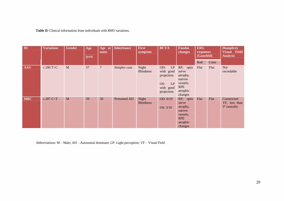
Table I- Sequence variation detected in RHO of patients.
Both probands with heterozygous RHO mutations have clinical symptoms and
signs of RP. They were available for clinical investigation, and the examination results
are summarized in Table II.
AAV is a 57 year-old single male, born to non-consanguineous parents and no
past family history of retinopathies (Fig. 2). His first disease symptoms started around
age 7 with complaints of night blindness. Changes in the visual field were first noted
during adolescence, with slow constriction of visual fields in parallel with progressive
loss of vision. Photophobia became a problem after the fourth decade of life. At present
his best corrected visual acuity is light perception with good projection for both eyes.
Ophthalmic examination disclosed abolished pupillary reflexes, absence of
nystagmus, normal ocular motility with orthotropia for near and distance. Slit-lamp
examination revealed bilateral pseudophakia with posterior chamber intra-ocular lens
(IOL) and transparent posterior capsules. Dilated fundus examination (Fig. 3) depicted a
pale optic disc, extremely narrow retinal vessels, scattered atrophy of the retinal
pigment epithelium (RPE) with macular involvement and bone spicules distributed in
the mid- and far periphery. This clinical picture is symmetrical in both eyes.
Complete phenotypical characterization included spectral-domain OCT (Fig. 4)
that revealed significant disorganization of the RPE/photoreceptor interface, granular
deposits in the outer retina and thinning of the neurosensory retina. Autofluorescence
imaging (Fig. 5) demonstrated a macular hyperfluorescent ring surrounded by globular
Variation Effect Prediction Frequency
in patients
Note
c.180 T>C p.Y60H Damaging 1/48 Novel
c.207 C>T p.R69C Damaging 1/48 Novel

14
areas of complete RPE atrophy, very thin retinal vessels and relative hypofluorescence
in the perifoveal area. Ganzfleld ERG was completely flat. We did not perform
multifocal ERG in this patient. Very limited visual acuity did not allow the use of
Humphrey Visual field analysis.
Figure 2- Pedigree of patient AAV who
carries a c.180T>C transition leading to a
p.Y60H substitution (simplex RP case).
Patients II-1(AAV) is heterozygous for the
mutation; unaffected family members are
I-1 and I-2.
Figure 3- OptomapR images showing 200º fundus pictures. 3A (Right eye) Significant optic pallor, thin
vessels, densely pigmented mid-periphery and scattered pigment bone spicules in the far periphery.
Relative preservation of the inferior-nasal mid-periphery (less pigmented) is observed. 3B (Left eye) a
symmetrical picture is observed for the contralateral fundus.
3A) 3B)
57
I
II
I - 1 I - 2
II - 1
Male Proband
Female Affected
Unaffected Deceased

15
Figure 4- Spectral-domain OCT images line passing through the central macular area. Relative
preservation of retinal thickness in the perifovea contrast with significant peripheral atrophy (macula).
Disorganization of outer retinal layers. Inset 4A right eye; inset 4B left eye.
Figure 5- Auto-fluorescence images. 5A: right eye; 5B: Left eye. Macular hyperfluorescent ring
surrounded by globular areas of complete RPE atrophy, very thin retinal vessels and relative
hypofluorescence in the perifoveal area. Left eye displays a thicker and more hyperfluorescent ring.
4A)
4A)
4B)
5A)
5B)

16
MBC is a 59 year-old married male, born to non-consanguineous parents.
Family history showed that his two elder brothers also had RP, but the youngest was
unaffected; by questionnaire, his parents do not seem to be affected and have distinct
geographical origin (Fig. 6). Symptoms included early-onset night vision disturbances
(night blindness before age 10), constricted visual fields by confrontation and
asymmetric decreased vision starting at the third decade of life. Photophobia became a
problem after the fifth decade of life representing a mild to moderate impairment. At
present his best corrected visual acuity is OD (right eye): 6:10 and OS (left eye): 3:10.
Complete ophthalmic assessment disclosed reduced pupillary reflexes, absence
of nystagmus, normal ocular motility with orthotropia for near and distance. Slit-lamp
examination revealed bilateral pseudophakia with posterior chamber IOL and
transparent posterior capsules. Dilated fundus examination (Fig. 7 and 8) reveals patchy
and asymmetrical areas of chorioretinal atrophy, optic disc pallor, narrow retinal blood
vessels and bone spicule deposits distributed in the mid- and far periphery. This clinical
picture is symmetrical in both eyes.
Further phenotypical characterization included spectral-domain OCT (Fig. 9)
that revealed significant disorganization of the outer and inner layers of the
neurosensory retina, a thick posterior hyaloid membrane/fibroglial proliferation that
eliminates the typical central foveal pit and granular deposits in the outer retina. The
loss of the outer retinal structures in atrophic areas is associated with increased deep
backscatter and subretinal pseudocystic lesions that seem to correspond to vascular
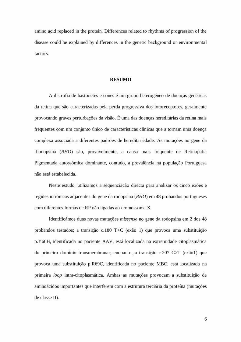
structures. Autofluorescence imaging (Fig. 10) revealed areas of granular RPE clumps
with hyperautofluorescence interspersed with globular atrophic areas of RPE and outer
retina. No typical hyperfluorescent ring was detected in this patient. Ganzfleld ERG was

17
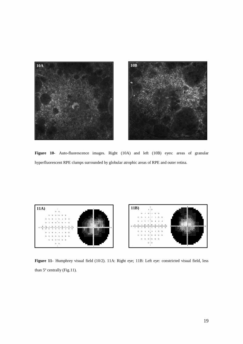
completely flat. We did not perform multifocal ERG in this patient. Humphrey Visual
field analysis demonstrated constricted visual field, less than 5º centrally (Fig.11).
Figure 6- Pedigree of patient who carries a
c.207 C>T transition leading to a p.R69C
substitution in patient with presumed
autosomal dominant RP. Patients II-1
(MBC) is heterozygous for the mutation;
II-1 and II-2 are affected and unaffected
family members are I-1, I-2 and II-4.
Figure 7- Fundus photography: 7A: Right eye: optic atrophy, significantly thin retinal vessels, patch
atrophy of RPE; beaten bronze macula; peripheral pigmented bone spicules. 7B Left eye: same aspect;
noteworthy the fact that there is paravascular pigment clumping along the inferior temporal arcade and
also the superior temporal arcade.
7B)
7A)
59
I
II
II - 1
I - 1 I - 2
II - 2 II - 3 II - 4
Male Proband
Female Affected
Unaffected Deceased

18
Figure 8- OptomapR images showing 200º fundus pictures. 8A (Right eye) Optic atrophy, thin vessels,
pigmented mid-periphery and scattered pigment bone spicules in the far periphery. 8B (Left eye) a
symmetrical picture is observed for the contralateral fundus.
Figure 9- Spectral domain OCT. 9A (Right eye) and 9B (Left eye) revealed significant disorganization of
the outer and inner layers of the retina, a thick posterior hyaloid membrane/fibroglial membrane, no
central foveal pit and granular deposits in the outer retina. Sub-RPE increased deep backscatter and
subretinal pseudocystic lesions correspond to choroidal vascular structures.
8A)
8B)
9A)
9B)
19
Figure 10- Auto-fluorescence images. Right (10A) and left (10B) eyes: areas of granular
hyperfluorescent RPE clumps surrounded by globular atrophic areas of RPE and outer retina.
Figure 11- Humphrey visual field (10/2). 11A: Right eye; 11B: Left eye: constricted visual field, less
than 5º centrally (Fig.11).
10A
)
10B
)
11A)
11B)
20
Table II- Clinical information from individuals with RHO variations.
Abbreviations: M – Male; AD – Autosomal dominant; LP- Light perception; VF – Visual Field
ID Variations Gender Age
(yrs)
Age at
onset
Inheritance First
symptom
BCVA Fundus
changes
ERG
responses
(Ganzfeld)
Humphrey
Visual Field
Analysis
Rod Cone
AAV c.180 T>C M 57 7 Simplex case Night
Blindness
OD: LP
with good
projection
OS: LP
with good
projection
RP; optic
nerve
atrophy,
narrow
vessels;
RPE atrophic
changes
Flat Flat Not
recordable
MBC c.207 C>T M 59 50 Presumed AD Night
Blindness OD: 6/10
OS: 3/10
RP; optic
nerve
atrophy,
narrow
vessels;
RPE
atrophic
changes
Flat Flat Constricted
VF, less than
5º centrally

21
DISCUSSION
The present study provides a useful clue regarding the frequency of RHO
mutations in the Portuguese population. The genetic screening reported here has
identified two novel RHO missense mutations in 2 of the 48 unrelated tested probands.
The c.180 T>C transition (exon 1) leading to a p.Y60H substitution, identified in patient
AAV, is located at the cytoplasmic end of the first transmembrane domain, whereas the
c.207 C>T transition (exon1) leading to a p.R69C substitution, identified in patient
MBC, is located in the first intra-cytoplasmic loop (Fig. 12).
Figure 12- Schematic Model of human rhodopsin showing the locations of mutations described in
rhodopsin, reported to date. Two novel mutations reported here are identified with arrows (adapted from
Preising 07.2000).
Most of the rhodopsin mutations identified to date in subjects with adRP have
been found in only one or just a few families. [6-7, 22, 47-50]

22
Although RHO is the first gene implicated [6-7] and probably the most studied
gene in RP, the great number of rare mutations suggests that many additional mutations
in the rhodopsin gene remain to be discovered.
The finding of 2 novel clinically significant RHO mutations among 48 (2/48,
4,16%) probands is not concordant with earlier estimates of 16% to 28,5% of frequency
among caucasians with adRP. [47, 49, 51-52] However, there is strong evidence for
ethnic variations in the mutation frequency of RHO [53-56], as it has been observed
with other genetic eye diseases, namely Leber Congenital Amaurosis; this could be the
result of a relative geographic isolation for many centuries or due to the high
consanguineous rate in our population and the fact that RHO gene mutations are
primarily related to AD forms of RP. Although we are dealing with a relatively small
patient population, it would be expected to find a higher percentage of mutations.
Both mutations affect highly conserved amino acid residues and are not present
in the healthy control population. Some insight into possible mechanisms responsible
for the ensuing retinal degeneration may be derived from considerations regarding the
amino acids affected by these mutations.
The c.180 T>C transition (exon 1), leads to a substitution of a tyrosine, which is
an aromatic amino acid with nonpolar and hydrophobic characteristics, for a histidine,
which is a basic amino acid with polar (positively charged) and hydrophilic
characteristics, might alter drastically the structure of the protein and the stability in the
bilayer lipid membrane. Histidine has quite unique structure and functional properties
sharing no resemblance with other amino acids. It is rather ambiguous whether it prefers
to be buried in the protein core or exposed to solvents. Also, histidine is the most
common amino acid in protein functional centers and binding sites, which could explain
why the change may potentially render inadequate rhodopsin activity.

23
The c.207 C>T transition (exon1), leads to a substitution of an arginine, which is
a basic amino acid with polar (positively charged) and hydrophilic characteristics, for a
cysteine, which is a neutral and small amino acid, probably disturbing the structure of
the first intradiscal loop and consequently the tertiary structure of the mutant rhodopsin.
Cysteine is known to be frequently involved in disulphide bonds that stabilize the
protein structure, especially important in extracellular domains; however, in this case it
may still be involved in the formation of disulfide bonds and/or protein interactions. In
the intracellular environment, cysteines may still play a key structural role. Their
sulfydryl side-chain is excellent for metal-binding, such as zinc, thus compromising
protein function.
According to disease mechanisms described in adRP [57], the novel mutations
described here, belong to class II (defective protein folding) the most common in adRP
mutations. Class I mutants, affecting the c-terminal region, fold normally in cell cultures
but are not correctly transported into the outer segments in vivo. [57] Bioinformatic
analysis and crystallography studies give further insight into the functional
consequences of amino acid substitutions.
To our knowledge, only three mutations were identified nearby. The p.Thr58Arg
[58-59] (Fig.12), a cytosine-to-guanine (C-to-G) transversion mutation in the second
nucleotide of codon 58 of the RHO gene, causing a substitution of the amino acid
arginine for a threonine, showed regional predilection for pigmentary changes in the
inferior and inferonasal quadrants of the retina, as well as visual scotomas
predominantly in the superior hemifields (sector RP). This clearly differs from the
phenotype observed in our affected probands.
The p.Gln64ter mutation (Fig.12) [60] represents a nonsense mutation that is
able to cause adRP, suggesting that synthesis of a rhodopsin fragment consisting of the

24
first 63 amino acids damages the rod photoreceptors. Cellular damage could result from
disruption of the lipid bilayer structure or from interference with the folding or transport
of other proteins.
The in-frame 12-bp deletion of codons 68 to 71 [48] occurs in the cytoplasmic
loop connecting the first and second transmembrane helices (Fig. 12). This is the most
conserved region on the cytoplasmic surface and has been suggested to be a point of
interaction with cytoplasmic proteins. [61] However, it seems possible that the removal
of these amino acids has an effect on protein folding in addition to any functional
significance this region may have in the signal transduction pathway.
Rhodopsin mutations have been reported in association with other retinal
phenotypes. Autosomal recessive Retinitis Pigmentosa (arRP) [8-11] is caused by
mutations in the cytoplasmic and extracellular domains, what might suggest that they
have a more damaging effect compared with mutations in the transmembrane domains.
Congenital stationary night blindness (CSNB) [12-15] has also been described, as the
result of mutations in the extracellular end of the second and seven transmembrane
domains, strengthening the hypothesis described above. The two novel mutations
described here, are located in the cytoplasmic end of the first transmembrane domain
and in the first intracytoplasmic loop; this may underlie the early observed disease onset
in our probands.
Suspicion of autosomal dominant pattern of inheritance usually occurs in the
presence of mild sporadic cases [27]. In 10 to 40 percent of all cases of retinitis
pigmentosa, only one person in a family is affected - simplex case. It can be difficult to
determine the inheritance pattern in those cases because affected individuals may have
no affected relatives or may be unaware of other family members with the disease.
Simplex cases can also result from a new gene mutation that is not present in other

25
family members. Multiplex cases correspond to 2 or more affected family members
(typically siblings) who have no pre-existing family history, which seems to be the case
of patient MBC. Segregation analysis is still pending to confirm the etiology of our
finding.
Although the typical manifestations present between adolescence and early
adulthood, the age of onset has been documented to range from infancy to adulthood
[62]. Due to the remarkable variation in how aware individuals are of their visual loss,
the age of onset of symptoms is an imprecise measure of disease severity and gives little
or no indication of when photoreceptor degeneration actually begins. [43]
Both probands report early-onset night vision disturbances accompanied by
progressive loss of peripheral visual field. Photophobia became a problem after several
years. There is no history of consanguinity or retinopathies. Clinically, they presented
typical features of RP, including retinal vessels attenuation, bone spicule deposition,
and a waxy appearance of the optic disc. Usually early-onset and severe forms of RP
with myopia in male are associated with X-linked RP (XLRP). [27] Perhaps, the
position of the substituted amino acids in the protein, and the side chain polarity of the
substituted amino acids may explain the similarity of phenotypes.
AdRP, in most cases, is a long lasting disease that typically evolves over several
decades with good overall long-term prognosis. Even at terminal stages, the disease
progression remains slow. [27] The phenotypic variability seen between probands at age
50 could be explained by differences in genetic background or by environmental
factors, even though both of them are of European decent and have comparable
lifestyles.
This study contributed to emphasize the importance of RHO mutation screening
in patients with RCD, since we identified two novel missense mutations. Also it gives

26
an overview of its prevalence in a Portuguese population. It was possible to attest the
phenotypic variability associated with rhodopsin mutations and the need to improve our
understanding of disease mechanisms to offer genetic counseling.
ACKNOWLEDGEMENTS
I would like to thank my professor and friend Eduardo Silva for all the work and
support during the elaboration of this paper.
27
REFERENCES
1. Mirzadegan, T., et al., Sequence analyses of G-protein-coupled receptors: similarities to rhodopsin. Biochemistry, 2003. 42(10): p. 2759-67.
2. Wald, G., Vitamin A in the retina. Nature, 1933. 132: p. 351-71. 3. Wald, G., Carotenoids and the Visual Cycle. J Gen Physiol, 1935. 19(2): p. 351-71. 4. van Soest, S., et al., Retinitis pigmentosa: defined from a molecular point of view. Surv
Ophthalmol, 1999. 43(4): p. 321-34. 5. Nathans, J. and D.S. Hogness, Isolation and nucleotide sequence of the gene encoding
human rhodopsin. Proc Natl Acad Sci U S A, 1984. 81(15): p. 4851-5. 6. Dryja, T.P., et al., Mutations within the rhodopsin gene in patients with autosomal
dominant retinitis pigmentosa. N Engl J Med, 1990. 323(19): p. 1302-7. 7. Dryja, T.P., et al., A point mutation of the rhodopsin gene in one form of retinitis
pigmentosa. Nature, 1990. 343(6256): p. 364-6. 8. Rosenfeld, P.J., et al., A null mutation in the rhodopsin gene causes rod photoreceptor
dysfunction and autosomal recessive retinitis pigmentosa. Nat Genet, 1992. 1(3): p. 209-13.
9. Greenberg, J., L. Roberts, and R. Ramesar, A rare homozygous rhodopsin splice-site mutation: the issue of when and whether to offer presymptomatic testing. Ophthalmic Genet, 2003. 24(4): p. 225-32.
10. Kumaramanickavel, G., et al., Missense rhodopsin mutation in a family with recessive RP. Nat Genet, 1994. 8(1): p. 10-1.
11. Bayes, M., et al., Autosomal recessive retinitis pigmentosa in Spain: evaluation of four genes and two loci involved in the disease. Clin Genet, 1996. 50(5): p. 380-7.
12. Dryja, T.P., et al., Heterozygous missense mutation in the rhodopsin gene as a cause of congenital stationary night blindness. Nat Genet, 1993. 4(3): p. 280-3.
13. Zeitz, C., et al., Identification and functional characterization of a novel rhodopsin mutation associated with autosomal dominant CSNB. Invest Ophthalmol Vis Sci, 2008. 49(9): p. 4105-14.
14. al-Jandal, N., et al., A novel mutation within the rhodopsin gene (Thr-94-Ile) causing autosomal dominant congenital stationary night blindness. Hum Mutat, 1999. 13(1): p. 75-81.
15. Rao, V.R., G.B. Cohen, and D.D. Oprian, Rhodopsin mutation G90D and a molecular mechanism for congenital night blindness. Nature, 1994. 367(6464): p. 639-42.
16. Souied, E., et al., Retinitis punctata albescens associated with the Arg135Trp mutation in the rhodopsin gene. Am J Ophthalmol, 1996. 121(1): p. 19-25.
17. RETNET. March 20, 2011]; Available from: http://www.sph.uth.tmc.edu/Retnet/. 18. Sung, C.H., C.M. Davenport, and J. Nathans, Rhodopsin mutations responsible for
autosomal dominant retinitis pigmentosa. Clustering of functional classes along the polypeptide chain. J Biol Chem, 1993. 268(35): p. 26645-9.
19. Kaushal, S. and H.G. Khorana, Structure and function in rhodopsin. 7. Point mutations associated with autosomal dominant retinitis pigmentosa. Biochemistry, 1994. 33(20): p. 6121-8.
20. Sung, C.H., et al., A rhodopsin gene mutation responsible for autosomal dominant retinitis pigmentosa results in a protein that is defective in localization to the photoreceptor outer segment. J Neurosci, 1994. 14(10): p. 5818-33.
21. Sung, C.H. and A.W. Tai, Rhodopsin trafficking and its role in retinal dystrophies. Int Rev Cytol, 2000. 195: p. 215-67.
22. Sung, C.H., et al., Functional heterogeneity of mutant rhodopsins responsible for autosomal dominant retinitis pigmentosa. Proc Natl Acad Sci U S A, 1991. 88(19): p. 8840-4.

28
23. Goto, Y., et al., Functional abnormalities in transgenic mice expressing a mutant rhodopsin gene. Invest Ophthalmol Vis Sci, 1995. 36(1): p. 62-71.
24. Naash, M.I., et al., Simulation of human autosomal dominant retinitis pigmentosa in transgenic mice expressing a mutated murine opsin gene. Proc Natl Acad Sci U S A, 1993. 90(12): p. 5499-503.
25. Roof, D.J., M. Adamian, and A. Hayes, Rhodopsin accumulation at abnormal sites in retinas of mice with a human P23H rhodopsin transgene. Invest Ophthalmol Vis Sci, 1994. 35(12): p. 4049-62.
26. Andres, A., P. Garriga, and J. Manyosa, Altered functionality in rhodopsin point mutants associated with retinitis pigmentosa. Biochem Biophys Res Commun, 2003. 303(1): p. 294-301.
27. Hamel, C., Retinitis pigmentosa. Orphanet J Rare Dis, 2006. 1: p. 40. 28. Weleber R. Retinitis pigmentosa and allied disorders. In: Ryan S, O.T., Schachat A, eds.
Retina. 2nd ed. St. Louis: Mosby-Year Book, Inc; 1994:334-40., ed. 29. Marshall J, H.J.R.p.P.J.B.L.C.-. 30. Berson E. Retinitis pigmentosa and allied diseases. In: Albert DM, J.F., eds. Principles
and practice of ophthalmology: clinical practice, Vol 3, 2nd ed. Philadelphia: WB Saunders; 1994:1214.
31. Ammann, F., D. Klein, and A. Franceschetti, Genetic and epidemiological investigations on pigmentary degeneration of the retina and allied disorders in Switzerland. J Neurol Sci, 1965. 2(2): p. 183-96.
32. Haim, M., Epidemiology of retinitis pigmentosa in Denmark. Acta Ophthalmol Scand Suppl, 2002(233): p. 1-34.
33. Boughman, J.A., P.M. Conneally, and W.E. Nance, Population genetic studies of retinitis pigmentosa. Am J Hum Genet, 1980. 32(2): p. 223-35.
34. Berson, E.L., Retinitis pigmentosa. The Friedenwald Lecture. Invest Ophthalmol Vis Sci, 1993. 34(5): p. 1659-76.
35. Berson, E.L., Retinitis pigmentosa: unfolding its mystery. Proc Natl Acad Sci U S A, 1996. 93(10): p. 4526-8.
36. Keats, B.J. and S. Savas, Genetic heterogeneity in Usher syndrome. Am J Med Genet A, 2004. 130A(1): p. 13-6.
37. Kimberling, W.J., D. Orten, and S. Pieke-Dahl, Genetic heterogeneity of Usher syndrome. Adv Otorhinolaryngol, 2000. 56: p. 11-8.
38. Mansergh, F.C., et al., Retinitis pigmentosa and progressive sensorineural hearing loss caused by a C12258A mutation in the mitochondrial MTTS2 gene. Am J Hum Genet, 1999. 64(4): p. 971-85.
39. Kajiwara, K., E.L. Berson, and T.P. Dryja, Digenic retinitis pigmentosa due to mutations at the unlinked peripherin/RDS and ROM1 loci. Science, 1994. 264(5165): p. 1604-8.
40. Rivolta, C., E.L. Berson, and T.P. Dryja, Paternal uniparental heterodisomy with partial isodisomy of chromosome 1 in a patient with retinitis pigmentosa without hearing loss and a missense mutation in the Usher syndrome type II gene USH2A. Arch Ophthalmol, 2002. 120(11): p. 1566-71.
41. Rivolta, C., et al., Retinitis pigmentosa and allied diseases: numerous diseases, genes, and inheritance patterns. Hum Mol Genet, 2002. 11(10): p. 1219-27.
42. Thompson, D.A., et al., Retinal dystrophy due to paternal isodisomy for chromosome 1 or chromosome 2, with homoallelism for mutations in RPE65 or MERTK, respectively. Am J Hum Genet, 2002. 70(1): p. 224-9.
43. Hartong, D.T., E.L. Berson, and T.P. Dryja, Retinitis pigmentosa. Lancet, 2006. 368(9549): p. 1795-809.
44. Holopigian, K., et al., Rates of change differ among measures of visual function in patients with retinitis pigmentosa. Ophthalmology, 1996. 103(3): p. 398-405.

29
45. Berson, E.L., et al., Natural course of retinitis pigmentosa over a three-year interval. Am J Ophthalmol, 1985. 99(3): p. 240-51.
46. Keen, J., et al., Rapid detection of single base mismatches as heteroduplexes on Hydrolink gels. Trends Genet, 1991. 7(1): p. 5.
47. Dryja, T.P., et al., Mutation spectrum of the rhodopsin gene among patients with autosomal dominant retinitis pigmentosa. Proc Natl Acad Sci U S A, 1991. 88(20): p. 9370-4.
48. Keen, T.J., et al., Autosomal dominant retinitis pigmentosa: four new mutations in rhodopsin, one of them in the retinal attachment site. Genomics, 1991. 11(1): p. 199-205.
49. Sung, C.H., et al., Rhodopsin mutations in autosomal dominant retinitis pigmentosa. Proc Natl Acad Sci U S A, 1991. 88(15): p. 6481-5.
50. Inglehearn, C.F., et al., A 3-bp deletion in the rhodopsin gene in a family with autosomal dominant retinitis pigmentosa. Am J Hum Genet, 1991. 48(1): p. 26-30.
51. Ziviello, C., et al., Molecular genetics of autosomal dominant retinitis pigmentosa (ADRP): a comprehensive study of 43 Italian families. J Med Genet, 2005. 42(7): p. e47.
52. Sullivan, L.S., et al., Prevalence of disease-causing mutations in families with autosomal dominant retinitis pigmentosa: a screen of known genes in 200 families. Invest Ophthalmol Vis Sci, 2006. 47(7): p. 3052-64.
53. Zhang, X.L., et al., Mutational analysis of the rhodopsin gene in Chinese ADRP families by conformation sensitive gel electrophoresis. Life Sci, 2006. 78(13): p. 1494-8.
54. Chan, W.M., et al., Rhodopsin mutations in Chinese patients with retinitis pigmentosa. Br J Ophthalmol, 2001. 85(9): p. 1046-8.
55. Gandra, M., et al., Retinitis pigmentosa: mutation analysis of RHO, PRPF31, RP1, and IMPDH1 genes in patients from India. Mol Vis, 2008. 14: p. 1105-13.
56. Fujiki, K., et al., Point mutations of rhodopsin gene found in Japanese families with autosomal dominant retinitis pigmentosa (ADRP). Jpn J Hum Genet, 1992. 37(2): p. 125-32.
57. Mendes, H.F. and M.E. Cheetham, Pharmacological manipulation of gain-of-function and dominant-negative mechanisms in rhodopsin retinitis pigmentosa. Hum Mol Genet, 2008. 17(19): p. 3043-54.
58. Moore, A.T., et al., Abnormal dark adaptation kinetics in autosomal dominant sector retinitis pigmentosa due to rod opsin mutation. Br J Ophthalmol, 1992. 76(8): p. 465-9.
59. Fishman, G.A., et al., Ocular findings associated with a rhodopsin gene codon 58 transversion mutation in autosomal dominant retinitis pigmentosa. Arch Ophthalmol, 1991. 109(10): p. 1387-93.
60. Macke, J.P., et al., Identification of novel rhodopsin mutations responsible for retinitis pigmentosa: implications for the structure and function of rhodopsin. Am J Hum Genet, 1993. 53(1): p. 80-9.
61. Applebury, M.L. and P.A. Hargrave, Molecular biology of the visual pigments. Vision Res, 1986. 26(12): p. 1881-95.
62. Shintani, K., D.L. Shechtman, and A.S. Gurwood, Review and update: current treatment trends for patients with retinitis pigmentosa. Optometry, 2009. 80(7): p. 384-401.


