Archives of Disease in Childhood, 1978, 53, 366-374 Clinical applications of serum carcinoembryonic antigen and alpha-fetoprotein levels in children with solid tumours J. R. MANN, G. E. LAKIN, J. C. LEONARD, H. A. RAWLINSON, S. G. N. RICHARDSON, J. J. CORKERY, A. H. CAMERON, AND K. J. SHAH From Birmingham Children's Hospital; Department of Experimental Pathology, University of Birmingham; and Department of Medical Genetics, Royal Hospital for Sick Children, Glasgow SUMMARY A study was carried out on serum carcinoembryonic antigen (CEA) and alpha-feto- protein (AFP) levels, both measured by radioimmunoassay, in 88 children with malignant solid tumours and in 26 children with nonmalignant disorders, who presented during the years 1973-77. Slightly or moderately raised CEA levels were found at presentation in 11 of 66 children with malignant tumours, in 2 others with recurrent tumours, and in 4 children with nonmalignant disorders. Raised CEA levels generally indicated advanced malignant disease, often affecting the liver, or other hepatic disorders, but were not associated with a specific tumour type. Except in the first months of life, significantly raised AFP levels were detected only in 11 patients with yolk sac-derived tumours, or hepatomas, and in one child with tyrosinosis who later developed a malignant hepatoma. Serial measurements of AFP accurately reflected the clinical response to treatment and in 2 patients indicated recurrence before this could be detected clinically. Carcinoembryonic antigen (CEA), a glycoprotein, was first found as an apparently specific surface antigen associated with adenocarcinoma of the human colon by Gold and Freedman (1965). However, although about 70% of patients with gastrointestinal malignancy have raised levels, raised CEA has since been found in patients with other adult cancers (Booth et al., 1973; Neville and Laurence, 1974) and certain nonmalignant dis- orders. Nevertheless, serial studies have shown that in adult patients with CEA-releasing colorectal carcinomas serum levels fall if treatment is successful and rise again when metastases occur (Booth et al., 1974). Studies of serum CEA in children with neuroblastoma (Reynoso et al., 1972; Wang et al., 1974; Frens et al., 1976) showed that serum CEA levels were usually raised when active tumour was present and fell when treatment was successful. That CEA may act as a tumour marker in paediatric malignancy was also suggested by Felberg et al. (1976), who found raised levels in retinoblastoma patients. Alpha-fetoprotein (AFP), a fetal protein chemi- cally similar to albumin, was first identified by Received 7 October 1977 Pedersen in 1944. Raised serum levels were described in association with hepatomas in rodents (Abelev et al., 1963) and humans (Tatarinov, 1964) and in patients with malignant gonadal teratoblastomas (Abelev et al., 1967). Raised levels have also been found in patients with certain nonmalignant dis- orders (Neville and Cooper, 1976). Early studies (Abelev et al., 1967; Masopust et al., 1968; Mawas et al., 1969) of over 300 children with malignant tumours, using comparatively insensitive bidimen- sional immunodiffusion methods, showed detectably raised levels in a proportion of children with terato- mata, gonadal embryonal carcinomata, hepato- blastoma, and hepatocellular carcinoma, but in none with the commoner tumours such as neuro- blastoma and nephroblastoma. Radioimmunoassay has increased the sensitivity of the test by more than 1000-fold, and has permitted the detection of AFP in sera from most children with yolk sac-derived and hepatic malignancies. Studies in some 30 paediatric cancer patients suggest that AFP monitoring by this sensitive method may be of value during treatment (Hagesawa et al., 1972, 1973; Takahashi, 1973; Itoh et al., 1974; Kohn and Weaver, 1974; Teilum et al., 1974; Pick et al., 1975; N0rgaard-Pedersen et al., 1975, 366 on 4 June 2018 by guest. Protected by copyright. http://adc.bmj.com/ Arch Dis Child: first published as 10.1136/adc.53.5.366 on 1 May 1978. Downloaded from
Transcript
Archives of Disease in Childhood, 1978, 53, 366-374
Clinical applications of serum carcinoembryonicantigen and alpha-fetoprotein levels in children withsolid tumoursJ. R. MANN, G. E. LAKIN, J. C. LEONARD, H. A. RAWLINSON, S. G. N. RICHARDSON,J. J. CORKERY, A. H. CAMERON, AND K. J. SHAH
From Birmingham Children's Hospital; Department ofExperimental Pathology, University ofBirmingham; and Department of Medical Genetics, Royal Hospital for Sick Children, Glasgow
SUMMARY A study was carried out on serum carcinoembryonic antigen (CEA) and alpha-feto-protein (AFP) levels, both measured by radioimmunoassay, in 88 children with malignant solidtumours and in 26 children with nonmalignant disorders, who presented during the years 1973-77.Slightly or moderately raised CEA levels were found at presentation in 11 of 66 children withmalignant tumours, in 2 others with recurrent tumours, and in 4 children with nonmalignantdisorders. Raised CEA levels generally indicated advanced malignant disease, often affecting theliver, or other hepatic disorders, but were not associated with a specific tumour type.
Except in the first months of life, significantly raised AFP levels were detected only in 11 patientswith yolk sac-derived tumours, or hepatomas, and in one child with tyrosinosis who later developed amalignant hepatoma. Serial measurements of AFP accurately reflected the clinical response totreatment and in 2 patients indicated recurrence before this could be detected clinically.
Carcinoembryonic antigen (CEA), a glycoprotein,was first found as an apparently specific surfaceantigen associated with adenocarcinoma of thehuman colon by Gold and Freedman (1965).However, although about 70% of patients withgastrointestinal malignancy have raised levels,raised CEA has since been found in patients withother adult cancers (Booth et al., 1973; Neville andLaurence, 1974) and certain nonmalignant dis-orders. Nevertheless, serial studies have shown thatin adult patients with CEA-releasing colorectalcarcinomas serum levels fall if treatment is successfuland rise again when metastases occur (Booth et al.,1974). Studies of serum CEA in children withneuroblastoma (Reynoso et al., 1972; Wang et al.,1974; Frens et al., 1976) showed that serum CEAlevels were usually raised when active tumour waspresent and fell when treatment was successful. ThatCEA may act as a tumour marker in paediatricmalignancy was also suggested by Felberg et al.(1976), who found raised levels in retinoblastomapatients.
Alpha-fetoprotein (AFP), a fetal protein chemi-cally similar to albumin, was first identified by
Received 7 October 1977
Pedersen in 1944. Raised serum levels were describedin association with hepatomas in rodents (Abelevet al., 1963) and humans (Tatarinov, 1964) and inpatients with malignant gonadal teratoblastomas(Abelev et al., 1967). Raised levels have also beenfound in patients with certain nonmalignant dis-orders (Neville and Cooper, 1976). Early studies(Abelev et al., 1967; Masopust et al., 1968; Mawaset al., 1969) of over 300 children with malignanttumours, using comparatively insensitive bidimen-sional immunodiffusion methods, showed detectablyraised levels in a proportion of children with terato-mata, gonadal embryonal carcinomata, hepato-blastoma, and hepatocellular carcinoma, but innone with the commoner tumours such as neuro-blastoma and nephroblastoma. Radioimmunoassayhas increased the sensitivity of the test by more than1000-fold, and has permitted the detection of AFPin sera from most children with yolk sac-derived andhepatic malignancies.
Studies in some 30 paediatric cancer patientssuggest that AFP monitoring by this sensitive methodmay be of value during treatment (Hagesawa et al.,1972, 1973; Takahashi, 1973; Itoh et al., 1974;Kohn and Weaver, 1974; Teilum et al., 1974;Pick et al., 1975; N0rgaard-Pedersen et al., 1975,
366
on 4 June 2018 by guest. Protected by copyright.
http://adc.bmj.com
/A
rch Dis C
hild: first published as 10.1136/adc.53.5.366 on 1 May 1978. D
Clinical applications of serum carcinoembryonic antigen 367
1976; Tsuchida et al., 1975; Palmer et al., 1976;Urano et al., 1976; Shirai et al., 1976; Sakashita et al.,1976; Schoenfeld et al., 1976; Grigor et al., 1977).The purpose of our study was to define the fre-
quency of raised serum CEA and AFP levels, bothmeasured by radioimmunoassay, in children withmalignant solid tumours, and to assess the clinicalvalue of serial measurements in monitoring response
to therapy and detecting tumour recurrence. We alsoreport the findings in 25 children with nonmalignantdisorders.
Patients and methods
Eighty-eight children referred during 1973-77 fortreatment of malignant solid tumours and 26children being investigated or treated for what provedto be nonmalignant conditions were studied. Inpatients found to have raised CEA or AFP levelsserial venepunctures were performed to assess therelationship between the serum levels and responseto therapy. In other patients blood forCEA and AFPlevels was taken during venepuncture for otherinvestigations, or for the administration of cytotoxicdrugs. The patients' ages ranged from 5 days to 15years, 11 being aged less than one year, and theirdiagnoses are shown in Tables 1 and 2.CEA levels were measured by the double-antibody
technique, the 95% confidence limits of normalhealthy adults being 0-15 ng/ml, standard error 1 9(Booth et al., 1974). In some patients a comparisonwas made, using the same sera, with CEA levelsmeasured by the Z-gel method (Lo Gerfo et al.,1971), the normal upper limit in healthy adults being2 5 ng/ml, the reproducibility of the CEA Rocheassay being 0 5 ng/ml at this level.AFP levels, normally less than 25 ng/ml in adults
(Grigor et al., 1977), were measured by radio-immunoassay. In the first 18 patients AFP
estimations were done in the Department of CancerStudies, University ofBirmingham; in this laboratorythe levels in healthy adult males and nonpregnantfemales were in the range 1-19 ng/ml (mean ± SD9 3 + 8-6). Sera from subsequent patients weretested in the Department of Medical Genetics,Glasgow, using the method of Vince et al. (1975), thelower limit of sensitivity varying from <16-7 to<5 *0 ng/ml (conversion of these results to units/l x10-3 using the international reference preparation(first British Standard Human Cord Serum 72/227)can be done by multiplying by 1 * 5).
Results
CEA. In the majority of patients the CEA levelsmeasured by the double-antibody technique werenormal (Fig. 1). Details of the 17 patients withlevels >15 ng/ml are given in Table 3. 8 of the 17
Fanconi's anaemia 2Twin of child with tumour 2Tyrosinosts 1Other nonmalignant disorders 6
Total 26
Table 3 Patients with raised serum CEA levels(>15 nglml)Age Sex CEA level Diagnosis(years) (ng/ml)
13.0 M 34.5 Fanconi's anaemia; jaundice;oxymetholone therapy; died 1 yearlater of leukaemia
7 w M 29.5 Multiple osteomyelitis4.4 M 18.0 Tyrosinosis; hepatoma (but included with
nonmalignant disorders in Fig. 1)1.8 M 21-3 Hamartoma of liver3.9 M 19-0 Histiocytic lymphoma; jaundice10.2 F 18.0 Histiocytic lymphoma15.8 F 19-0 Relapsed Hodgkin's disease; hepatic
involvement7.1 M 16-5 StageIWilns'stumour4-7 F 19.8 Stagem Wilms's tumour; hepatic failure2.5 M 17-3 Stage H Wilms's tumour7.5 F 15.5 Thoracic mesenchymal tumour0.9 M 25.3 Stage IV neuroblastoma; hepatic
involvement11.7 F 15.3 Ovarian embryonal carcinoma1.8 M 21.3 Orchioblastoma (see text)2.7 F 23.3 Hepatoma (bile duct tumour); jaundice;
incomplete resection; CEA 25 beforedeath
1.3 F 31 Hepatoblastoma, recurrent2.0 M 17.5 Pulmonary blastoma
on 4 June 2018 by guest. Protected by copyright.
http://adc.bmj.com
/A
rch Dis C
hild: first published as 10.1136/adc.53.5.366 on 1 May 1978. D
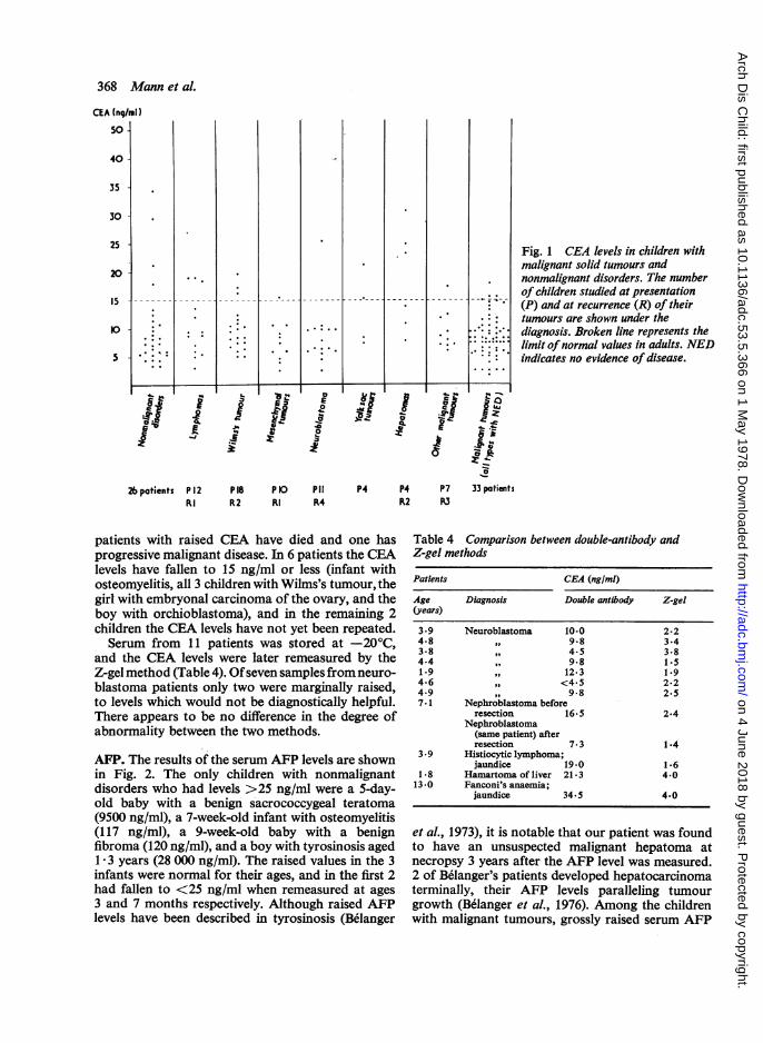
Fig. 1 CEA levels in children withmalignant solid tumours andnonmalignant disorders. The numberof children studied at presentation(P) and at recurrence (R) of theirtumours are shown under thediagnosis. Broken line represents thelimit ofnormal values in adults. NEDindicates no evidence of disease.
patients with raised CEA have died and one hasprogressive malignant disease. In 6 patients the CEAlevels have fallen to 15 ng/ml or less (infant withosteomyelitis, all 3 children with Wilms's tumour, thegirl with embryonal carcinoma of the ovary, and theboy with orchioblastoma), and in the remaining 2children the CEA levels have not yet been repeated.Serum from 11 patients was stored at -20°C,
and the CEA levels were later remeasured by theZ-gel method (Table 4). Ofseven samples from neuro-blastoma patients only two were marginally raised,to levels which would not be diagnostically helpful.There appears to be no difference in the degree ofabnormality between the two methods.
AFP. The results of the serum AFP levels are shownin Fig. 2. The only children with nonmalignantdisorders who had levels >25 ng/ml were a 5-day-old baby with a benign sacrococcygeal teratoma(9500 ng/ml), a 7-week-old infant with osteomyelitis(117 ng/ml), a 9-week-old baby with a benignfibroma (120 ng/ml), and a boy with tyrosinosis agedI * 3 years (28 000 ng/ml). The raised values in the 3infants were normal for their ages, and in the first 2had fallen to <25 ng/ml when remeasured at ages3 and 7 months respectively. Although raised AFPlevels have been described in tyrosinosis (Belanger
Table 4 Comparison between double-antibody andZ-gel methods
1-8 Hamartoma of liver 21.3 4.013.0 Fanconi's anaemia;
jaundice 34.5 4.0
et al., 1973), it is notable that our patient was foundto have an unsuspected malignant hepatoma atnecropsy 3 years after the AFP level was measured.2 of Belanger's patients developed hepatocarcinomaterminally, their AFP levels paralleling tumourgrowth (Belanger et al., 1976). Among the childrenwith malignant tumours, grossly raised serum AFP
33 patients
on 4 June 2018 by guest. Protected by copyright.
http://adc.bmj.com
/A
rch Dis C
hild: first published as 10.1136/adc.53.5.366 on 1 May 1978. D
Fig. 2 AFP levels inchildren with malignantsolid tumnours andnonmalignant disorders.The number of childrenstudied at presentation(P) and atrecurrence (R) oftheir tumours areshown under thediagnosis. NEDindicates no evidenceof disease. * = levelsmeasured in Glasgow;o = levels measuredin Birmingham.
26 patients P12 P17
EOc -o
*~-6 .
Rl R2 Rl R3 Rl R2 R3
was found only in children with yolk sac-derived(malignant teratoma, orchioblastoma, ovarian em-bryonal carcinoma) or hepatic tumours (Table 5).A slight rise in serum AFP was present in 2 boys
aged 5 and 9 5 years with alveolar rhabdomyosar-coma (42 and 45 ng/ml), and in a boy aged 4 yearswith neuroblastoma (36 ng/ml). The AFP subse-quently fell below 25 ng/ml in all 3 boys althoughonly the first child's tumour responded to treatment.
A level of 43 ng/ml was detected in a 9-week-oldbaby with pelvic fibrosarcoma, and his AFP re-mained for some time above the adult normalrange (37 ng/ml at age 3 and 5 5 months) but wasundetectable by 7 months of age, when there wasstill no evidence of recurrent tumour. None of theother children with malignant tumours had raisedAFP levels at presentation, at recurrence, or aftersuccessful treatment.
IiAJJJU .1. I X.
100000.
1000
1000-
100'
0
00
21.<10 ::
I.:: : {ii i*Ioi
on 4 June 2018 by guest. Protected by copyright.
http://adc.bmj.com
/A
rch Dis C
hild: first published as 10.1136/adc.53.5.366 on 1 May 1978. D
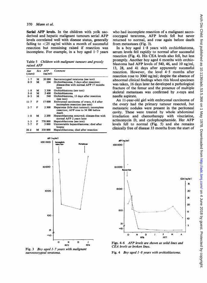
Serial AFP levels. In the children with yolk sac-derived and hepatic malignant tumours serial AFPlevels correlated well with disease status, generallyfalling to <25 ng/ml within a month of successfulresection but remaining raised if resection wasincomplete. For example, in a boy aged 1-7 years
Table 5 Children with malignant tumours and grosslyraised AFPAge Sex AFP Comment(years) (ng/ml)
1.7 M 20 000 Sacrococcygeal teratoma (see text)0.9 M 204 Orchioblastoma, 5 days after resection;
disease-free with normal AFP 17 monthslater
1.8 M 2 300 Orchioblastoma (see text)5.0 M 5 400 Orchioblastoma0.5 M 540 Orchioblastoma, 13 days after resection
(see text)11.7 F 17 000 Embryonal carcinoma of ovary, 6 d after
incomplete resection (see text)2.7 F 2 300 Hepatoma (bile duct tumour); incomplete
resection; AFP rose to 54 300 beforedeath
1.8 M 2 200 Hepatoblastoma removed; disease-free withnormal AFP 2 years later
1.3 F 778 000 Hepatoblastoma (see text)7.7 F 3 000 Unresectable hepatoblastoma; died after
biopsy26 d M 330 000 Hepatoblastoma; died after resection
who had incomplete resection of a malignant sacro-coccygeal teratoma, AFP levels fell but neverreturned to normal, and rose again before deathfrom metastases (Fig. 3).
In a boy aged 18 years with orchioblastoma,serum levels fell rapidly to normal after successfulresection (Fig. 4). His CEA levels also fell, but lesspromptly. Another boy aged 6 months with orchio-blastoma had AFP levels of 540, 46, and 10 ng/ml,13, 28, and 41 days after apparently successfulresection. However, the level 4*5 months afterresection rose to 3060 ng/ml; despite the absence ofabnormal clinical findings when this blood specimenwas taken, 16 days later he developed a pathologicalfracture of the femur and the presence of multipleskeletal metastases was confirmed by x-rays andneedle aspirate.An 11-year-old girl with embryonal carcinoma of
the ovary had the primary tumour resected, butmetastatic nodules were present in the peritonealcavity. These were treated by whole abdominalirradiation and chemotherapy with vincristine,actinomycin D, and cyclophosphamide. Her AFPlevels fell to normal (Fig. 5) and she remainsclinically free of disease 33 months from the start of
AFP (ng/ml)100 000] Incomplete
rmect.on
10000
1000
100
MetetassesAFP (nq/ml)
100000 -
1 0000 -
Re
l00 I
100
25 -
A§o25j
'10 -
0 N D J1973 1974
Fig. 3 Boy aged 1 7 years with malignantsacrococcygeal teratoma.
secton
aIA (nq/ml )
- 35
- 30
- 25
- 20
- IS
I l
5
0 N D1976
i F1977
M A
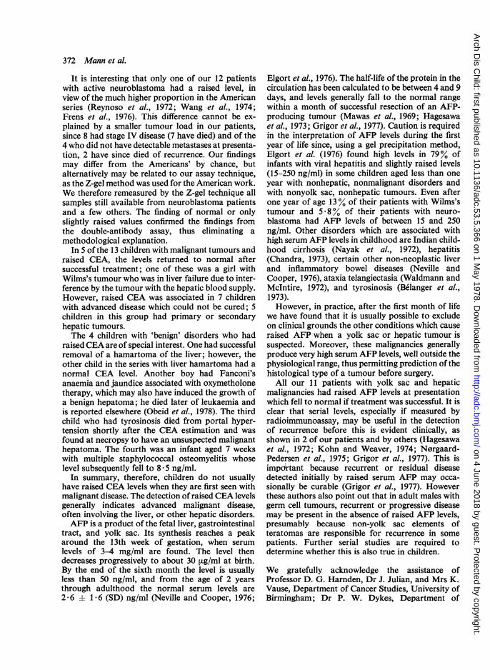
Figs. 4-6 AFP levels are shown as solid lines andCEA levels as broken lines.
Fig. 4 Boy aged I *8 years with orchioblastoma.
on 4 June 2018 by guest. Protected by copyright.
http://adc.bmj.com
/A
rch Dis C
hild: first published as 10.1136/adc.53.5.366 on 1 May 1978. D
Fig. 5 Girl aged 11 years with embryonal carcinomaof ovary.
treatment, chemotherapy having been stopped after24 months.Apparently complete resection of a hepatoma in a
girl aged 13 years was followed by a sharp fall inserum AFP levels (Fig. 6). Postoperatively rightphrenic palsy occurred but thoracotomy revealed nocause. Her AFP levels did not return to normal andthen rose again. Extensive investigations, includingchest and skeletal radiography, bone marrowaspirate, liver scan, hepatic angiography, and anotherlaparotomy failed to demonstrate recurrence.However, 2 months later metastases were visible inboth lungs on chest x-rays. Chemotherapy withactinomycin D and adriamycin led to temporarydisappearance of the metastases and a fall in AFPlevel, but she also became jaundiced. As the AFPlevels rose the pulmonary metastases again becamevisible. The jaundice faded after actinomycin andadriamycin therapy were stopped but AFP continuedto rise and the pulmonary metastases slowly en-larged, despite treatment with cyclophosphamide,and she died. Her CEA levels also reflected theclinical course, though less closely than did the AFP.
Discussion
The best established biochemical marker of paedia-tric malignancy is the raised urinary catecholamineexcretion in patients with neuroblastoma (Marsdenand Steward, 1976). The usefulness of measuring
M A J J A S O N D J F M A
1976 1977
Fig. 6 Girl aged 1 3 years with hepatoblastoma.
CEA levels in paediatric malignancy has not beenwell assessed, although 16 of 19 children with activeneuroblastoma were reported to have raised levelswhich fell to normal if treatment was successful(Reynoso et al., 1972; Wang et al., 1974; Frens et al.,1976). 4 of 5 retinoblastoma patients had raised CEAlevels which fell after enucleation, and raised levelswere found in 29-7% of their nonaffected familymembers (Felberg et al., 1976).
In our study we have found raised CEA levels in13 of 68 children with malignant solid tumours. Ingeneral the rise has been small and no patient hadlevels in the range (>50 ng/ml) commonly found inadults with gastrointestinal cancer (Booth et al.,1973). There was no specificity for any tumour type,a small proportion of children with lymphomas,Wilms's tumour, mesenchymal tumours, neuro-blastoma, yolk sac tumours, hepatomas, and othertumours having raised levels.
interrupted.tae reacion
Chemotherpy
Radotherapy
on 4 June 2018 by guest. Protected by copyright.
http://adc.bmj.com
/A
rch Dis C
hild: first published as 10.1136/adc.53.5.366 on 1 May 1978. D
It is interesting that only one of our 12 patientswith active neuroblastoma had a raised level, inview of the much higher proportion in the Americanseries (Reynoso et al., 1972; Wang et al., 1974;Frens et al., 1976). This difference cannot be ex-plained by a smaller tumour load in our patients,since 8 had stage IV disease (7 have died) and of the4 who did not have detectable metastases at presenta-tion, 2 have since died of recurrence. Our findingsmay differ from the Americans' by chance, butalternatively may be related to our assay technique,as the Z-gel method was used for the American work.We therefore remeasured by the Z-gel technique allsamples still available from neuroblastoma patientsand a few others. The finding of normal or onlyslightly raised values confirmed the findings fromthe double-antibody assay, thus eliminating amethodological explanation.
In 5 of the 13 children with malignant tumours andraised CEA, the levels returned to normal aftersuccessful treatment; one of these was a girl withWilms's tumour who was in liver failure due to inter-ference by the tumour with the hepatic blood supply.However, raised CEA was associated in 7 childrenwith advanced disease which could not be cured; 5children in this group had primary or secondaryhepatic tumours.The 4 children with 'benign' disorders who had
raisedCEA are of special interest. One had successfulremoval of a hamartoma of the liver; however, theother child in the series with liver hamartoma had anormal CEA level. Another boy had Fanconi'sanaemia and jaundice associated with oxymetholonetherapy, which may also have induced the growth ofa benign hepatoma; he died later of leukaemia andis reported elsewhere (Obeid et al., 1978). The thirdchild who had tyrosinosis died from portal hyper-tension shortly after the CEA estimation and wasfound at necropsy to have an unsuspected malignanthepatoma. The fourth was an infant aged 7 weekswith multiple staphylococcal osteomyelitis whoselevel subsequently fell to 8 *5 ng/ml.
In summary, therefore, children do not usuallyhave raised CEA levels when they are first seen withmalignant disease. The detection ofraised CEA levelsgenerally indicates advanced malignant disease,often involving the liver, or other hepatic disorders.AFP is a product of the fetal liver, gastrointestinal
tract, and yolk sac. Its synthesis reaches a peakaround the 13th week of gestation, when serumlevels of 3-4 mg/ml are found. The level thendecreases progressively to about 30 Vg/ml at birth.By the end of the sixth month the level is usuallyless than 50 ng/ml, and from the age of 2 yearsthrough adulthood the normal serum levels are2-6 ±t 1 6 (SD) ng/ml (Neville and Cooper, 1976;
Elgort et al., 1976). The half-life of the protein in thecirculation has been calculated to be between 4 and 9days, and levels generally fall to the normal rangewithin a month of successful resection of an AFP-producing tumour (Mawas et al., 1969; Hagesawaet al., 1973; Grigor et al., 1977). Caution is requiredin the interpretation of AFP levels during the firstyear of life since, using a gel precipitation method,Elgort et al. (1976) found high levels in 79% ofinfants with viral hepatitis and slightly raised levels(15-250 ng/ml) in some children aged less than oneyear with nonhepatic, nonmalignant disorders andwith nonyolk sac, nonhepatic tumours. Even afterone year of age 13% of their patients with Wilms'stumour and 5 8% of their patients with neuro-blastoma had AFP levels of between 15 and 250ng/ml. Other disorders which are associated withhigh serum AFP levels in childhood are Indian child-hood cirrhosis (Nayak et al., 1972), hepatitis(Chandra, 1973), certain other non-neoplastic liverand inflammatory bowel diseases (Neville andCooper, 1976), ataxia telangiectasia (Waldmann andMcIntire, 1972), and tyrosinosis (Belanger et al.,1973).However, in practice, after the first month of life
we have found that it is usually possible to excludeon clinical grounds the other conditions which causeraised AFP when a yolk sac or hepatic tumour issuspected. Moreover, these malignancies generallyproduce very high serum AFP levels, well outside thephysiological range, thus permitting prediction of thehistological type of a tumour before surgery.
All our 11 patients with yolk sac and hepaticmalignancies had raised AFP levels at presentationwhich fell to normal if treatment was successful. It isclear that serial levels, especially if measured byradioimmunoassay, may be useful in the detectionof recurrence before this is evident clinically, asshown in 2 of our patients and by others (Hagesawaet al., 1972; Kohn and Weaver, 1974; N0rgaard-Pedersen et al., 1975; Grigor et al., 1977). This isimpdcrtant because recurrent or residual diseasedetected initially by raised serum AFP may occa-sionally be curable (Grigor et al., 1977). Howeverthese authors also point out that in adult males withgerm cell tumours, recurrent or progressive diseasemay be present in the absence of raised AFP levels,presumably because non-yolk sac elements ofteratomas are responsible for recurrence in somepatients. Further serial studies are required todetermine whether this is also true in children.
We gratefully acknowledge the assistance ofProfessor D. G. Hamden, Dr J. Julian, and Mrs K.Vause, Department of Cancer Studies, University ofBirmingham; Dr P. W. Dykes, Department of
on 4 June 2018 by guest. Protected by copyright.
http://adc.bmj.com
/A
rch Dis C
hild: first published as 10.1136/adc.53.5.366 on 1 May 1978. D
Experimental Pathology, University of Birmingham;Professor M. A. Ferguson-Smith, Department ofMedical Genetics, Royal Hospital for Sick Children,Glasgow; Dr J. G. Ratcliffe, Department of Bio-chemistry, Glasgow Royal Infirmary; Mr P. Gornall,Mr A. Gourevitch, and Dr B. S. B. Wood, Bir-mingham Children's Hospital; Dr A. J. Banks,Queen Elizabeth Hospital, Birmingham; Dr P. M.Jones, Royal Manchester Children's Hospital; andall the paediatricians and surgeons in the WestMidlands who referred patients to us.
References
Abelev, G. I., Perova, S. D., Khramkova, N. I., Postinkova,S. I., and Irlin, J. S. (1963). Production of embryonalalpha-globulin by transplantable mouse hepatomas.Transplantation, 1, 174-180.
Abelev, G. I., Assecritova, I. V., Kraevsky, N. A., Perova,S. D., and Perevodchikova, N. I. (1967). Embryonalserum alpha-globulin in cancer patients; diagnostic value.International Journal of Cancer, 2, 551-558.
B6langer, L., Belanger, M., Prive, L., Larochelle, J., Tremblay,M., and Aubin, G. (1973). Tyrosin6mie hereditaire etalpha-1-foetoproteine. I. Interet clinique de l'alpha-foeto-proteine dans la tyrosinemie h6r6ditaire. Pathologieet Biologie, 21, 449-455.
Belanger, L., Larochelle, J., Belanger, M., and Prive, L.(1976). Tyrosinosis: hereditary persistence of alpha-1-fetoprotein. Onco-developmental Gene Expression, pp. 155-158. Ed. by W. H. Fishman and S. Sell. Academic Press,New York.
Booth, S. N., King, J. P. G., Leonard, J. C., and Dykes, P. W.(1973). Serum carcinoembryonic antigen in clinicaldisorders. Gut, 14, 794-799.
Booth, S. N., Jamieson, G. C., King, J. P. G., Leonard, J.,Oates, G. D., and Dykes, P. W. (1974). Carcinoembryonicantigen in management of colorectal carcinoma. BritishMedical Journal, 3, 183-187.
Chandra, R. K. (1973). Hepatitis antigen and alpha-feto-protein in neonatal hepatitis. Archives of Disease inChildhood, 48, 157-158.
Elgort, D. A., Abelev, G. I., Durnov, L. A., Pashkov, I. V.,Levina, D. M., Laskina, A. V., Izrailskaya, M. A., Kharit,I. O., Ter-Grigorova, E. N., and Reizis, A. R. (1976).Alpha-fetoprotein in the diagnosis of tumours in children.(In Russian). Vestnik Akademii Meditsinskikn Nauk SSSR,No. 2, 31-44.
Felberg, N. T., Michelson, J. B., and Shields, J. A. (1976).CEA family syndrome. Abnormal carcinoembryonicantigen (CEA) levels in asymptomatic retinoblastoma familymembers. Cancer, 37, 1397-1402.
Frens, D. B., Bray, P. F., Wu., J. T., and Lahey, M. E. (1976).The carcinoembryonic antigen assay: prognostic value inneural crest tumours. Journal ofPediatrics, 88, 591-594.
Gold, P., and Freedman, S. 0. (1965). Demonstration oftumour-specific antigens in human colonic carcinomata byimmunological tolerance and absorption techniques.Journal of Experimental Medicine, 121, 439-462.
Grigor, K. M., Detre, S. I., Kohn, J., and Neville, A. M.(1977). Serum alpha-foetoprotein levels in 153 malepatients with germ cell tumours. British Journal of Cancer,35, 52-58.
Hasegawa, H., Ise, T., Otsuki, H., Okura, H., and Mukojima,T. (1972). Adriamycin therapy in a child with malignantsacrococcygeal teratoma with special reference to serum
alpha-fetoprotein. Japanese Journal of Clinical Oncology,12, 133-138.
Hasegawa, H., Mukojima, T., Hattoni, N., Sano, R., andHirota, T. (1973). Embryonal carcinoma and alpha-fetoprotein with special reference to hepatoblastoma.Alpha-fetoprotein and Hepatoma, pp. 129-139. Ed. byH. Hirai and T. Miyaji. Gann Monograph on CancerResearch 14. University of Tokyo Press, Tokyo.
Itoh, T., Shirai, T., Naka, A., and Matsumoto, S. (1974).Yolk sac tumour and alpha-fetoprotein: clinicopatho-logical study of four cases. Gann, 65, 215-226.
Kohn, J., and Weaver, P. C. (1974). Serum alphai-feto-protein in hepatocellular carcinoma. Lancet, 2, 334-337.
Lo Gerfo, P., Krupey, J., and Hansen, H. J. (1971). Demon-strations of an antigen common to several varieties ofneoplasia. New England Journal of Medicine, 285, 138-141.
Marsden, H. B., and Steward, J. K. (1976). (Editors.)Tumours in Children, pp. 206-217. Recent Results inCancer Research No. 13. Springer, Berlin.
Masopust, J., Kithier, K., Radl, J., Koutecky, J., andKotal, L. (1968). Occurrence of fetoprotein in patientswith neoplasms and non-neoplastic diseases. InternationalJournal of Cancer, 3, 364-373.
Mawas, C., Kohen, M., Lemerle, J., Buffe, D., Schweisguth,D., and Burtin, P. (1969). Serum alpha-fetoprotein(fetuin) in children with malignant ovarian or testicularteratomas. International Journal of Cancer, 4, 76-79.
Nayak, N. C., Malaviya, A. N., Chawla, V., and Chandra,R. K. (1972). Alpha-fetoprotein in Indian childhoodcirrhosis. Lancet, 1, 68-69.
Neville, A. M., and Cooper, E. H. (1976). Biochemicalmonitoring of cancer. A review. Annals of Clinical Bio-chemistry, 13, 283-305.
Neville, A. M., and Laurence, D. J. R. (1974). The Carcino-embryonic Antigen (CEA). Present Position and Proposalsfor Future Investigation. UICC Technical Report Series,Volume 12. Geneva.
Norgaard-Pedersen, B. Albrechtsen, F., and Teilum, G.(1975). Serum alpha-foetoprotein as a marker for endo-dermal sinus tumour (yolk sac tumour) or a vitellinecomponent of teratocarcinoma. Acta Pathologica etMicrobiologica Scandinavica, 83A, 573-589.
N0rgaard-Pedersen, B., Hertz, H., Sell, A., and Tygstrup, I.(1976). Infantile endodermal sinus tumours (yolk sactumours) and alpha-fetoprotein. Onco-developmental GeneExpression, pp. 379-385. Ed. by W. H. Fishman and S.Sell. Academic Press, New York.
Obeid, D. A., Hill, F. G. H., Wood, B. S. B., Harnden, D. G.,and Mann, J. R. (1978). Fanconi anaemia, oxymetholone,hepatoma and leukaemia (in preparation).
Palmer, P. E., Safaii, H., and Wolfe, H. J. (1976). Alphl-antitrypsin and alpha-fetoprotein. Protein markers inendodermal sinus (yolk sac tumours). American Journal ofClinical Pathology, 65, 575-582.
Pedersen, K. 0. (1944). Fetuin, a new globulin isolated fromserum. Nature, 154, 575.
Pick, A. I., Schoenfeld, Y., Schreibman, S., Weiss, H., andBen-Bassat, M. (1975). Alpha-fetoprotein assay. Signi-ficance and clinical applications. New York State Journalof Medicine, 75, 1403-1409.
Reynoso, G., Chu, T. M., Holyoke, D., Cohen, E., Nemoto,T., Wang, J. J., Chuang, J., Guinan, P., and Murphy, G. P.(1972). Carcinoembryonic antigen in patients with differentcancers. Journal of the American Medical Association. 220,361-365.
Sakashita, S., Hirai, H., Nishi, S., Nakamura, K., andTsuji, I. (1976). Alpha-fetoprotein synthesis in tissueculture of human testicular tumours and an examinationof experimental yolk sac tumours in the rat. CancerResearch, 36, 4232-4235.
on 4 June 2018 by guest. Protected by copyright.
http://adc.bmj.com
/A
rch Dis C
hild: first published as 10.1136/adc.53.5.366 on 1 May 1978. D
Schoenfeld, Y., Pick, A. I., Schreibman, S., Kessler, H., andDintzman, M. (1976). Serum alpha fetal protein in a threeyear old child with hepatoma. Tumori, 62, 407-414.
Shirai, T., Itoh, T., Yoshiki, T., Novo, T., Tomino, Y., andHayasaka, T. (1976). Immunofluorescent demonstrationof alpha-fetoprotein and other plasma proteins in yolk sactumour. Cancer, 38, 1661-1667.
Takahashi, M. (1973). Cytodiagnosis of primary liver cellcarcinoma with reference to alpha-fetoprotein. (in Japan-ese). Japanese Journal of Clinical Pathology, 21, 769-772.
Tatarinov, I. S. (1964). Detection of embryospecific alpha-globulin in the blood sera of patients with primary livercancer. (In Russian). Voprosy Meditsinskoi Khimii, 10,90-91.
Teilum, G., Albrechtsen, R., and N0rgaard-Pedersen, B.(1974). Demonstration of alpha-fetoprotein synthesis in atesticular yolk sac carcinoma by immunofluorescentlocalisation. Acta Pathologica et Microbiologica Scan-dinavica, 82A, 586-588.
Tsuchida, Y., Urano, Y., Endo, Y., Ohmi, K., Hashizume,K., Saito, S., Ishida, M., and Hasegawa, H. (1975). Astudy on alpha-fetoprotein and endodermal sinus tumour.Journal ofPediatric Surgery, 10, 501-506.
Urano, Y., Endo, Y., and Tsuchida, Y. (1976). Alpha-fetoprotein synthesis in teratocarcinoma: immunopatho-logical studies of yolk sac tumor. Onco-developmentalGene Expression, pp. 131-137. Ed. by W. H. Fishman andS. Sell. Academic Press, New York.
Vince, J. D., McManus, T. J., Ferguson-Smith, M. A., andRatcliffe, J. G. (1975). A semi-automated serum alpha-fetoprotein radioimmunoassay for prenatal spina bifidascreening. British Journal of Obstetrics and Gynaecology,82, 718-727.
Waldmann, T. A., and Maclntire, K. R. (1972). Serum alpha-fetoprotein levels in patients with ataxia-telangiectasia.Lancet, 2, 1112-1115.
Wang, J-J., Sinks, L. F., and Chu, T. M. (1974). Carcino-embryonic antigen in patients with neuroblastoma. Journalof Surgical Oncology, 6, 211-217.
Correspondence to Dr J. R. Mann, Department ofHaematology, Birmingham Children's Hospital,Ladywood Middleway, Birmingham B16 8ET.
on 4 June 2018 by guest. Protected by copyright.
http://adc.bmj.com
/A
rch Dis C
hild: first published as 10.1136/adc.53.5.366 on 1 May 1978. D