Objective. Keratosis pilaris (KP) is a common condition which can frequently be cosmetically disturbing. Topical treatments can beused with limited efficacy. The objective of this study is to evaluate the effectiveness and safety of fractional carbon dioxide (CO
2)
laser for the treatment of KP. Patients and Methods. A prospective, randomized, single-blinded, intraindividual comparative studywas conducted on adult patients with KP. A single session of fractional CO
2laser was performed to one side of arm whereas the
contralateral side served as control. Patients were scheduled for follow-up at 4 and 12 weeks after treatment. Clinical improvementwas graded subjectively by blinded dermatologists. Patients rated treatment satisfaction at the end of the study. Results. Twentypatients completed the study. All patients stated that the laser treatment improved KP lesions. At 12-week follow-up, 30% of lesionson the laser-treated side had moderate to good improvement according to physicians’ global assessment (𝑝 = 0.02). Keratoticpapules and hyperpigmentation appeared to respond better than the erythematous component. Four patients with Fitzpatrick skintype V developed transient pigmentary alteration. Conclusions. Fractional CO
2laser treatment may be offered to patients with KP.
Dark-skinned patients should be treated with special caution.
1. Introduction
Keratosis pilaris (KP) is a common disorder of keratinization.It is characterized bymultiple tiny follicular keratotic papulesthat may have surrounding erythema. Hyperpigmentationcan sometimes occur especially in individuals with dark-complexioned skin. KP mainly involves the extensor arms,back, anterior thighs, face, and buttock.The exact prevalenceis difficult to estimate but could be found up to 50% of thegeneral population [1, 2]. Although KP has no impact ongeneral health, its influence on the quality of life arises espe-cially for those with lesions on the exposed areas. Treatmentoptions include emollients, keratolytics, and topical steroidswhen necessary [3]. However, the result is highly variableand recurrence following treatment discontinuation is oftenproblematic.
Over the past decade, attempts to eradicate KP throughvarious laser and light-based therapy have been investigated.This includes 532 nm potassium titanyl phosphate laser;
595 nm pulsed dye laser; 1064 nm Q-switched Nd:YAG laser;long-pulse 1064 nm Nd:YAG laser; combination of 595 nmpulse dye laser, long-pulse 755 nm alexandrite laser, andmicrodermabrasion [4–9]. To the best of our knowledge,there has never been a report on the effectiveness of fractionalcarbon dioxide laser (CO
2) for the treatment of KP. We
hypothesize that fractional CO2laser could remove the
keratotic component and brown pigmentation of KP. Theobjective of this study is to evaluate the efficacy and safetyof fractional CO
2laser for the treatment of KP.
2. Materials and Methods
This study is a prospective, randomized, single-blinded,intraindividual comparative study. The study protocol hasbeen approved by Mahidol University Institutional ReviewBoard for Human Subject Research (Protocol number015821).The study confirmed the guidelines of the declaration
Hindawi Publishing CorporationBioMed Research InternationalVolume 2016, Article ID 1928540, 6 pageshttp://dx.doi.org/10.1155/2016/1928540
2 BioMed Research International
of Helsinki. All study subjects had obtained the informconsent at the enrollment.
2.1. Patient. Subjects were recruited from an outpatient der-matology clinic at a university-based hospital (RamathibodiHospital, Mahidol University, Bangkok, Thailand). Healthypatients aged 18 years or older with the presence of KP onboth sides of arms were eligible. Patients with history ofkeloid or hypertrophic scar and those who were pregnantor lactating were excluded from the study. Patients whoreceived topical medications or emollients within 1 monthor had performed any laser treatment or dermabrasion forKP within past 6 months were also excluded. In addition, weeliminated patients with prior history of medication affectingkeratinization (e.g., isotretinoin and acitretin) within the past3 years from this trial.
After enrollment, informed consent had been obtainedand demographic data was recorded. Using the table ofrandomization (a table of random numbers), the left side andright side of the armswere randomly allocated to receive lasertreatment. One side received fractional CO
2laser therapy
(side A) and the other (side B) did not receive laser treatment.
2.2. Treatment Regimens. Fractional CO2laser therapy using
a 10,600 nm eCO2laser (Lutronic Corporation, Goyang,
Republic of Korea) was performed to the lesions on side A.Thiswas a single session laser treatment.The settingswere thepulse energy of 24–30mJ and spot density of 300 spots/cm2in static mode; 2 passes were delivered using a 300-densitytip. To minimized pain, local anesthetic cream (a eutecticmixture of local anesthetics, Astra Zeneca LP, Wilmington,DE) was applied under occlusion for 30 minutes beforethe laser treatment and air cooling with a cold air coolingdevice (CRIOjet AIR Mini CRIO Medizintechnik GmbH,Birkenfeld, Germany) at a cooling level of 4 was used duringthe laser therapy.
After the laser treatment, petrolatum ointment wasapplied to the lesions on side A twice a day for 5 days. Toavoid confounding effect of petrolatum ointment, the lesionson side B also received petrolatum ointment twice daily for5 days. The patients were appointed for follow-up on the 4thand 12th week after the last treatment (Figure 1).
2.3. Outcome Evaluation. Standard digital photograph wastaken at baseline, 4 weeks, and 12 weeks after the lasttreatment. Two dermatologists who did not perform the laserprocedure evaluated the response through digital images.Global improvement score, keratotic papules, hyperpigmen-tation, and erythemawere evaluated using the grading systemfor improvement. The grading scale is as follows: grade −4,>75% worsening; grade −3, 51–75% worsening; grade −2,26–50% worsening; grade −1, 1–25% worsening; grade 0, nochange; grade 1, 1–25% improvement (minimal); grade 2, 26–50% improvement (moderate); grade 3, 51–75% improvement(good); grade 4, >75% improvement (excellent).
Patient satisfaction was assessed at the end of study(12th week of follow-up). They were asked to rate theoverall improvement and satisfaction by grading score, that
4 weeks of follow-up 12 weeks of follow-up0
0.20.40.60.8
11.21.41.6
Mea
n im
prov
emen
t sco
re
LaserControl
0.90 (±0.97)
0.45 (±0.60)0.70 (±1.03)
0.20 (±0.41)
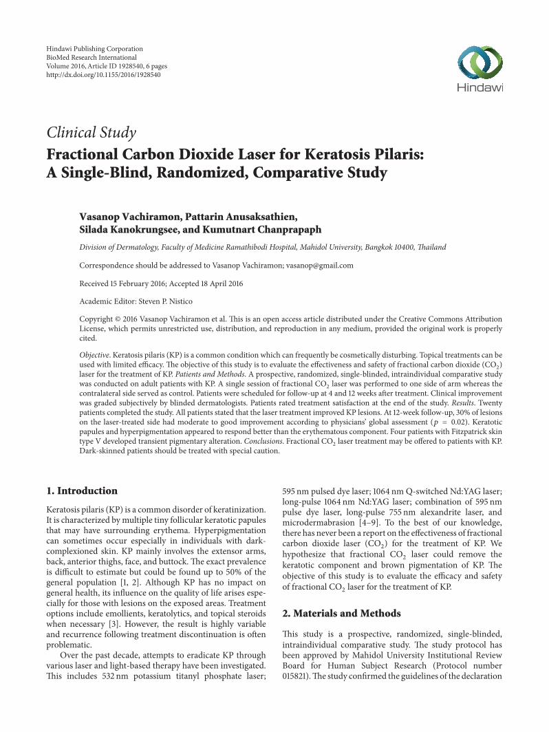
Figure 1: Mean improvement score at 4-week and 12-week follow-up.
is, 0, unsatisfied; 1, poor; 2, fair; 3, satisfied; 4, extremelysatisfied. Pain scorewas also evaluated immediately after lasertreatment using 10-point visual analogue scale (VAS, 0–10),in which 0 indicated no pain at all and 10 indicated extremepain. Possible adverse events (e.g., erythema, burning sen-sation, hyperpigmentation, hypopigmentation, scarring, andinfection) were recorded at every visit.
2.4. Statistical Analyses. Categorical variables were expressedas percentages. Continuous variables (e.g., physician gradingscore and VAS) were expressed as mean ± standard deviationor median (range). 𝑡-test was used to compare continuousdata between two dependent samples. All analyses wereperformed using STATA version 13 (Stata Corp., CollegeStation, Texas, USA). A 𝑝 value of 0.05 or less was consideredstatistically significant.
3. Results
Twenty-four eligible patients with KP on both arms wereenrolled in this study. Four patients lost to follow-up at 12weeks after treatment for reasons unrelated to laser treatment(i.e., inconvenient follow-up schedule). Twenty patients com-pleted the 12-week study and were included in the analysis(20 arms on side A and 20 arms on side B). Twelve were men(60%) and 8 were women (40%). The median age of subjectwas 27 years old (19–32). The median age of KP onset was 15years old (10–22). Eight patientswere Fitzpatrick skin type III,5 patients were Fitzpatrick skin type IV, and 7 patients wereFitzpatrick skin type V.
3.1. Global Assessments. At 4 weeks after treatment, grade 2or more improvement was achieved in 4 arms on side A and1 arm on side B (𝑝 = 0.097). At 12 weeks of follow-up, 6 armson side A achieved grade 2 or more improvement but no KPlesion on side B achieved such response (𝑝 = 0.02). None ofthe patients experienced worsening of the lesions (Table 1).
In terms of mean improvement score, the score was 0.90(±0.97) on side A and 0.45 (±0.60) on side B (𝑝 = 0.08) at 4weeks after treatment.Themean improvement scores on sideA and side B at 12 weeks of follow-up were 0.7 (±1.03) and 0.2(±0.41), respectively (𝑝 = 0.05) (Figures 1 and 2). There was
BioMed Research International 3
Table 1: Global assessment by nontreating dermatologists.
Grading 4 weeks of follow-up, 𝑛 = 20 12 weeks of follow-up, 𝑛 = 20Side A Side B Side A Side B
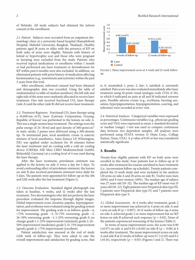
Figure 2: Photograph of patient before (a, d); at 4-week follow-up (b, e); at 12-week follow-up (c, f). Left arm was treated with fractional CO2
laser (a, b, c) and right arm was served as control (d, e, f). Left arm showed grade 3 improvement at 12-week follow-up (c).
no statistically significant difference according to sex, age, ordisease duration.
3.2. Keratotic Papules. The evaluators assessed overallimprovement of keratotic papules but the actual number wasnot counted. Similar to global assessments, higher grade ofimprovement (grade 2 or more) was found in 4 arms on sideA and 1 arm on side B at 4 weeks of follow-up. At 12 weeks
of follow-up, at least grade 2 improvement was achieved in5 arms on side A and 1 arm on side B. No study subjectsexperienced worsening of keratotic papules (Table 2).
3.3. Hyperpigmentation. Grade 2 or more improvement inhyperpigmentation was achieved in 5 patients on side A at4 weeks of follow-up. At 12 weeks of follow-up, there were 6arms on side A that achieved grade 2 or more improvement.
4 BioMed Research International
Table 2: Assessment of keratotic papules by nontreating dermatologists.
Grading 4 weeks of follow-up, 𝑛 = 20 12 weeks of follow-up, 𝑛 = 20Side A Side B Side A Side B
Table 4: Patiens’ satisfaction at 12 weeks of follow-up.
Patients’ grading Side A, 𝑛 = 20 Side B, 𝑛 = 200 (unsatisfied) — 51 (poor) 8 112 (fair) 5 43 (satisfied) 5 —4 (extremely satisfied) 2 —Side A: fractional CO2 laser treated side.Side B: control side.
Majority of lesions on side B showed no improvement interms of pigmentation at both the 4th and 12th week offollow-up (Table 3).
3.4. Erythema. Grade 3 improvement in erythema wasachieved on 2 arms of side A at 4 weeks of follow-up butno lesions on side B achieved similar response. At 12 weeks,grade 2 and grade 3 improvement were found in 2 patientseach on side A. None of KP lesions on side B achieved grade2, 3, or 4 improvement at 12weeks. No patients hadworseningof erythema after laser.
3.5. Patient Satisfaction. All patients rated the lesions asimproved on side A. Improvement of seven lesions on side A(35%) was marked as satisfied or extremely satisfied whereasno lesion on side Bwas rated as satisfied or extremely satisfied(𝑝 = 0.08) (Table 4).
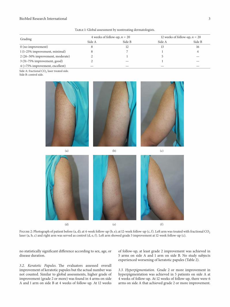
3.6. Adverse Events. Pain was observed on the laser-treatedside in all patients. The mean pain score was 4.2 (±2.6).
Figure 3: Hypopigmentation occurred on the laser-treated side at4-week follow-up.
Erythema and burning sensation were also observed in alllaser-treated lesions.Themedian duration of erythema was 2days (1–5) and median duration of burning sensation was 24hours (1–72). Two patients experienced hyperpigmentationon laser-treated side at 4 weeks of follow-up, both of whichhad spontaneous improvement at 12 weeks after treatment.Hypopigmentation developed on 2 lesions of side A: thefirst lesion spontaneously improved at 4-week follow-upand another lesion improved without any treatment at 12-week follow-up (Figure 3). No infection or scarring wasobserved. All patients who developed hyperpigmentation orhypopigmentation were Fitzpatrick skin type V.
BioMed Research International 5
4. Discussion
KP is a very common condition among the general popula-tion but few treatment options with promising and sustainedresults exist. Although it is often asymptomatic, it can causepruritus in some patients. In addition, the affected skinresembling gooseflesh resulting in unsightly appearance maylead to psychological distress. Treatment options are variabledue to various aspects of the lesions. In most cases, reassur-ance and general skin care focusing on avoiding skin drynessare required. Emollients, keratolytic agents, topical steroids,chemical or mechanical peels, vitamin D3 analogues, andtopical or systemic retinoids may be used [8]. Various laseror light-based therapies targeting different components of thelesion have been tried with variable success rate [4, 5, 10, 11].Despite the advantage of low cost, topical treatments havelimited success rate, as they may not target all components ofKP (e.g., hyperkeratosis, hyperpigmentation, and erythema).In addition, irritation from high-concentrated keratolyticagents can be intolerable to some patients. According tothe histopathologic nature of KP, epidermal hyperkeratosis,hypergranulosis, and plugging of individual hair folliclesare seen [3, 12]. Mild superficial perivascular lymphocyticinflammatory infiltrates can be found which reflex the ery-thematous component of someKP lesions. In Fitzpatrick skintypes III–V (i.e., Asian skin), brown pigmentation is oftenseen [7]. In this study, the rationale for the use of fractionalCO2is to target the excess keratinous plug and the brown
pigments.In this study, we demonstrated that a single session of
fractional CO2laser treatment results in moderate to good
improvement of KP lesions in some patients by global assess-ment. Although merely 30% of the patient had moderate togood improvement at the end of the study, to the patients’perception, all stated that the laser treatment improved theappearance of their lesions.
Taking particular elements of KP into account, keratoticpapules and hyperpigmentation appear to respond betterthan erythematous components. Evidently, higher numbersof patients rated their keratotic lesions and hyperpigmenta-tion as moderate or good response on both 4 weeks and12weeks of follow-up. This result supports our hypothesisthat fractional CO
2laser treatment could eradicate excess
keratosis and pigmentation.Regarding the laser parameter in this study, the pulse
energy of 24–30mJ with a spot density of 300 spots/cm2 wasdelivered into 2 passes. This setting was capable of causingmaximum ablative depth of approximately 530–580𝜇m andthe width of coagulative zone is 243𝜇m per dot, whereas theepidermal thickness of the shoulder and upper extremitiesis approximately 81–135 𝜇m [13, 14]. With this setting, webelieve that certain parts of each keratotic papule would bedestroyed. However, due to disparity in size and depth ofkeratotic papules, absolute ablative depth may be variable.Hence, some may be completely ablated and others may onlybe partially ablated.
There wasmarginal degree of improvement on KP lesionsof control side (side B). This is probably due to the emollient
effect from petrolatum ointment. As also demonstrated themean improvement score of side B at the 12th week offollow-up was less than that of the 4th week of follow-up. Therefore, emollients are proven beneficial in KP whengiven continuously. Cessation of emollientmay cause gradualrecurrence. This same effect also occurred on side A, wherethe mean improvement score at the 4th week was better thanthe 12thweek. For this reason, application of emollient shouldbe advised to all patients, regardless of laser therapy.
Previous studies have evaluated the effectiveness of vari-ous lasers intended to attack specified targets such as 532 nmpotassium titanyl phosphate laser and 595 nm pulsed dyelaser aimed to reduce KP-associated erythema [4, 5, 10].According to a study by Alcantara Gonzalez et al., all10 patients with keratosis rubra pilaris or keratosis pilarisatrophicans faciei achieved more than 75% improvementin erythema after 2–7 sessions of 595 nm pulsed dye laser[10]. In patients with pronounced pigmentary componentof KP, 1064 nm Q-switched Nd:YAG laser may be used[6, 7]. Park et al. evaluated the efficacy of 1064 nm Q-switched Nd:YAG laser on pigmented KP, 41.7% of patientsshowed more than 50% improvement in dyspigmentation[6]. Recently, Saelim et al. successfully treated KP with long-pulsed 1064 nmNd:YAG laser [9]. A significant improvementin global assessment, erythema, and keratotic papules wasnoted after 3 sessions of long-pulsed 1064 nm Nd:YAGlaser at 4-week interval. The proposed mechanism of long-pulsed 1064 nm Nd:YAG laser was to reduce the size of theaffected hair follicles. According to a study by Lee et al.,the combination of 595 nm pulsed dye laser, long-pulsed755 nm alexandrite laser, and microdermabrasion to target 3components of KP resulted in marked improvement of KPin 51.7% of patients [8]. Therefore, the most suitable choicefor the treatment of KP mainly depends on the pronouncedand problematic component. Combination of different lasersor modalities may be most beneficial to patients with mul-tiple elements of KP. However, lasers hold several limita-tions such as pain, stinging sensation, erythema, purpura,hyper- and hypopigmentation, and high cost [4–10]. Inaddition, recurrence of the lesions may occur after treatmentcessation.
The limitations of this study are the small sample size andthe short follow-up time of 3 months.Therefore, a conclusioncannot be drawn as to how long the laser effect wouldlast and whether recurrence would occur. Moreover, we didnot count the actual keratotic lesions and skin roughnesswas inaccessible through the evaluation by 2D photography.Finally, our study was performed in Asian subjects withFitzpatrick skin types III–V; hence, this laser setting cannotbe applied to all skin types.
In conclusion, we demonstrated that fractional CO2laser
can be used as an alternative treatment for KP in somepatients, particularly in the presence of marked keratoticcomponents. However, special caution should be given topatients with greater Fitzpatrick skin type, as pigmentaryalteration can occur as a sequel. Further studies are neededto find the optimum parameter, appropriate frequency, andsuitable treatment sessions of fractional CO
2laser for KP.
6 BioMed Research International
Competing Interests
The authors declare that there are no competing interestsregarding the publication of this paper.
References
[1] A. W. Arnold and S. A. Buechner, “Keratosis pilaris andkeratosis pilaris atrophicans faciei,” Journal der DeutschenDermatologischen Gesellschaft, vol. 4, no. 4, pp. 319–323, 2006.
[2] B. Mevorah, A. Marazzi, and E. Frenk, “The prevalence ofaccentuated palmoplantar markings and keratosis pilaris inatopic dermatitis, autosomal dominant ichthyosis and controldermatological patients,” British Journal of Dermatology, vol.112, no. 6, pp. 679–685, 1985.
[3] S. Hwang and R. A. Schwartz, “Keratosis pilaris: a commonfollicular hyperkeratosis,”Cutis, vol. 82, no. 3, pp. 177–180, 2008.
[4] G. Dawn, M. Urcelay, M. Patel, and A. M. M. Strong, “Keratosisrubra pilaris responding to potassium titanyl phosphate laser,”British Journal of Dermatology, vol. 147, no. 4, pp. 822–824, 2002.
[5] K. M. Kaune, E. Haas, S. Emmert, M. P. Schon, and M. Zutt,“Successful treatment of severe keratosis pilaris rubra with a595-nm pulsed dye laser,” Dermatologic Surgery, vol. 35, no. 10,pp. 1592–1595, 2009.
[6] J. Park, B. J. Kim, M. N. Kim, and C. K. Lee, “A pilot study ofQ-switched 1064-nm Nd:YAG laser treatment in the keratosispilaris,” Annal of Dermatology, vol. 23, no. 3, pp. 293–298, 2011.
[7] S. Kim, “Treatment of pigmented keratosis pilaris in Asianpatients with a novel Q-switched Nd:YAG laser,” Journal ofCosmetic and Laser Therapy, vol. 13, no. 3, pp. 120–122, 2011.
[8] S. J. Lee, M. J. Choi, Z. Zheng, W. S. Chung, Y. K. Kim, and S.B. Cho, “Combination of 595-nm pulsed dye laser, long-pulsed755-nmalexandrite laser, andmicrodermabrasion treatment forkeratosis pilaris: retrospective analysis of 26 Korean patients,”Journal of Cosmetic and Laser Therapy, vol. 15, no. 3, pp. 150–154, 2013.
[9] P. Saelim, M. Pongprutthipan, S. Pootongkam, V. Jariyaset-havong, and P. Asawanonda, “Long-pulsed 1064-nm Nd:YAGlaser significantly improves keratosis pilaris: a randomized,evaluator-blind study,” Journal of Dermatological Treatment, vol.24, no. 4, pp. 318–322, 2013.
[10] J. Alcantara Gonzalez, P. Boixeda, M. T. Truchuelo Dıez, andB. Fleta Asın, “Keratosis pilaris rubra and keratosis pilarisatrophicans faciei treated with pulsed dye laser: report of 10cases,” Journal of the European Academy of Dermatology andVenereology, vol. 25, no. 6, pp. 710–714, 2011.
[11] R. Rodrıguez-Lojo, J. D. Pozo, J. M. Barja, F. Pineyro, andL. Perez-Varela, “Keratosis pilaris atrophicans: treatment withintense pulsed light in four patients,” Journal of Cosmetic andLaser Therapy, vol. 12, no. 4, pp. 188–190, 2010.
[12] P. Sallakachart and Y. Nakjang, “Keratosis pilaris: a clinico-histopathologic study,” Journal of the Medical Association ofThailand, vol. 70, no. 7, pp. 386–389, 1987.
[13] M. Huzaira, F. Rius, M. Rajadhyaksha, R. R. Anderson, and S.Gonzalez, “Topographic variations in normal skin, as viewed byin vivo reflectance confocal microscopy,” Journal of InvestigativeDermatology, vol. 116, no. 6, pp. 846–852, 2001.
[14] J. Sandby-Møller, T. Poulsen, and H. C. Wulf, “Epidermalthickness at different body sites: relationship to age, gender,pigmentation, blood content, skin type and smoking habits,”Acta Dermato-Venereologica, vol. 83, no. 6, pp. 410–413, 2003.