Celiac Disease and Nonceliac Gluten SensitivityA ReviewMaureen M. Leonard, MD, MMSc; Anna Sapone, MD, PhD; Carlo Catassi, MD, MPH; Alessio Fasano, MD
C eliac disease is a chronic, small-intestinal immune-mediated enteropathy initiated by exposure to dietarygluten in genetically predisposed individuals and charac-
terized by specific autoantibodies against tissue transglutaminase2 (anti-tTG2), endomysium, and/or deamidated gliadin peptide.1
Although up to 40% of the population carries the genotype HLA-DQ2or HLA-DQ8, which is required for the development of celiac dis-ease, only 2% to 3% of HLA-DQ2 or HLA-DQ8 carriers subsequentlydevelop celiac disease.2 Celiac disease, once considered a rela-tively rare gastrointestinal condition affecting almost exclusivelyyoung white children, can develop at any age and can affect almostany race. Celiac disease was first described by Samuel Gee in 1887.Wheat was hypothesized as the possible offending agent byWilliam Dicke in 1941.3
The epidemiology, clinical presentation, pathophysiology, andmanagement of the disease have changed since its initial descrip-
tion. There is strong evidence that celiac disease is an autoimmunedisease triggered by the ingestion of gluten present in wheat, bar-ley, and rye in genetically predisposed individuals. The prevalenceof celiac disease in the general population is 1%, with regional dif-ferences (Table 1).4 Celiac disease can affect any human organ or tis-sue (Table 1 and Table 2).6
Nonceliac gluten sensitivity is a term used to describe individu-als who have intestinal signs or symptoms, extraintestinal signsor symptoms, or both, related to ingestion of gluten-containinggrains (Table 2), with improvement when these are removed froma patient’s diet. The frequency of nonceliac gluten sensitivityis unknown owing to the lack of validated biomarkers, but it isthought to be more common than celiac disease. Wheat allergy, thethird gluten-related disorder, which will not be addressed in thisreview, is defined as an adverse type-2 helper T-cell immunologicreaction to wheat proteins and typically presents soon after wheat
IMPORTANCE The prevalence of gluten-related disorders is rising, and increasing numbers ofindividuals are empirically trying a gluten-free diet for a variety of signs and symptoms. Thisreview aims to present current evidence regarding screening, diagnosis, and treatment forceliac disease and nonceliac gluten sensitivity.
OBSERVATIONS Celiac disease is a gluten-induced immune-mediated enteropathycharacterized by a specific genetic genotype (HLA-DQ2 and HLA-DQ8 genes) andautoantibodies (antitissue transglutaminase and antiendomysial). Although theinflammatory process specifically targets the intestinal mucosa, patients may presentwith gastrointestinal signs or symptoms, extraintestinal signs or symptoms, or both,suggesting that celiac disease is a systemic disease. Nonceliac gluten sensitivityis diagnosed in individuals who do not have celiac disease or wheat allergy but whohave intestinal symptoms, extraintestinal symptoms, or both, related to ingestionof gluten-containing grains, with symptomatic improvement on their withdrawal. Theclinical variability and the lack of validated biomarkers for nonceliac gluten sensitivity makeestablishing the prevalence, reaching a diagnosis, and further study of this conditiondifficult. Nevertheless, it is possible to differentiate specific gluten-related disorders fromother conditions, based on currently available investigations and algorithms. Clinicianscannot distinguish between celiac disease and nonceliac gluten sensitivity by symptoms,as they are similar in both. Therefore, screening for celiac disease must occur beforea gluten-free diet is implemented, since once a patient initiates a gluten-free diet,testing for celiac disease is no longer accurate.
CONCLUSIONS AND RELEVANCE Celiac disease and nonceliac gluten sensitivity are common.Although both conditions are treated with a gluten-free diet, distinguishing between celiacdisease and nonceliac gluten sensitivity is important for long-term therapy. Patients withceliac disease should be followed up closely for dietary adherence, nutritional deficiencies,and the development of possible comorbidities.
Author Affiliations: Center for CeliacResearch and Treatment, Division ofPediatric Gastroenterology andNutrition, MassGeneral Hospital forChildren, Boston, Massachusetts(Leonard, Sapone, Catassi, Fasano);Celiac Research Program, HarvardMedical School, Boston,Massachusetts (Leonard, Sapone,Catassi, Fasano); Shire, Lexington,Massachusetts (Sapone); EuropeanBiomedical Research InstituteSalerno, Salerno, Italy (Catassi,Fasano); Department of Pediatrics,Università Politecnica delle Marche,Ancona, Italy (Catassi).
Corresponding Author: AlessioFasano, MD, Center for CeliacResearch and Mucosal Immunologyand Biology Research Center,Massachusetts General Hospital East,Bldg 114, 16th St (Mail Stop 114-3503),Charlestown, MA 02129-4404([email protected]).
Section Editors: Edward Livingston,MD, Deputy Editor, and Mary McGraeMcDermott, MD, Senior Editor.
Clinical Review & Education
JAMA | Review
jama.com (Reprinted) JAMA August 15, 2017 Volume 318, Number 7 647
ingestion, with signs of anaphylaxis such as swelling or itching ofthe mouth, throat, and skin; nasal congestion; watery eyes; and dif-ficulty breathing. Wheat allergy is more common in children, withreported prevalence between 2% and 9% in children and 0.5%and 3% in adults.8
This review provides an evidence-based update of the patho-physiology, diagnosis, treatment, and implications of celiac dis-ease and nonceliac gluten sensitivity.
MethodsThe Cochrane Library (January 15, 2010, to April 10, 2017),MEDLINE (January 15, 2010, to April 10, 2017), and Google
Scholar (January 15, 2010, to April 18, 2017) were searched usingthe search terms coeliac, celiac, non-celiac, non-coeliac, gluten,and wheat sensitivity, alone and in combination. Publications inthe past 5 years were selected in addition to commonly refer-enced and highly regarded older publications. Reference lists ofarticles identified by this search strategy were selected. Reviewarticles and book chapters were cited to provide readers withadditional details and sources of additional references.
ResultsPathophysiologyGluten as Environmental Trigger of Gluten-Related DisordersGluten is a mixture of gliadins and glutenins, complex pro-teins unusually rich in prolines and glutamines that are notcompletely digestible by intestinal enzymes.9 The final productof this partial digestion is a mix of peptides that can triggerhost responses (increased intestinal permeability and innate+/− adaptive immune response) that closely resemble thoseinstigated by the exposure to gastrointestinal pathogens10-13
(Figure 1).
Normal Phsyiologic Events That Contribute to the Pathogenesis ofCeliac Disease and Nonceliac Gluten Sensitivity
Gluten Translocation From Lumen to Lamina Propria (Paracellular vsTranscellular) | Previous studies have shown that gliadin cancause an immediate and transient increase in gut permeability.9,13
This permeating effect is secondary to the binding of spe-cific undigestible gliadin fragments to the CXCR3 chemokinereceptor with subsequent release of zonulin, a modulator of inter-cellular tight junctions (Figure 1).14 This process takes place in allindividuals who ingest gluten. For the majority, these events donot lead to abnormal consequences. However, these same eventscan lead to an inflammatory process in genetically predisposedindividuals when the immunologic surveillance system mistakenlyrecognizes gluten as a pathogen. Thus, this normal physiologicprocess is also essential to the development of celiac disease andnonceliac gluten sensitivity in at-risk individuals. Additionally,there is evidence that during the acute phase of celiac disease,gluten can also cross the intestinal barrier through the transcellu-lar pathway via transferrin receptor CD71, once tolerance to glu-ten has been lost15 (Figure 1).
The Innate Immune Response | Innate immunity plays a criticalrole in initiating celiac disease and possibly nonceliac glu-ten sensitivity. Cytokines such as interleukin (IL) 15 and interferonalfa can prime the innate immune response by polarizingdendritic cells and intraepithelial lymphocyte function.15,16
These mucosal events, along with the breach of the epithelialbarrier function secondary to the gliadin-mediated zonulin re-lease,14 lead to the passage of undigested peptides fromthe gut lumen to the lamina propria. Once gliadin crosses theepithelial barrier, neutrophil recruitment through IL8 pro-duction11,12,17 or a direct neutrophil chemoattractant effect18
causes a loss of tolerance to gluten in genetically susceptible in-dividuals (Figure 1).
Table 1. Prevalence of Celiac Disease in the General Populationand in At-Risk Groupsa
Prevalence, %
General Population
Algeria 5.6
Argentina 0.6
Australia 0.4
Brazil 0.5
Burkina Fasu 0
Egypt 0.5
Finland 1.0-2.4
Germany 0.2
India 0.3-1.0
Iran 0.5-1.0
Ireland 0.8
Italy 0.9-1.0
Libya 0.8
The Netherlands 0.5
New Zealand 1.2
Portugal 0.7
Russia 0.2
Spain 0.3-1.4
Sweden 0.5-2.9
Tunisia 0.6
Turkey 0.6-1.0
United Kingdom 0.9-1.5
United States 0.3-0.9
Mean (weighted) 1.0
At-Risk Groups
Type 1 diabetes 3-12
Autoimmune thyroid disease 3
Autoimmune liver disease 13.5
Down syndrome 5.5
Turner syndrome 6.5
Williams syndrome 9.5
IgA deficiency 3
IgA nephropathy 4
Juvenile idiopathic arthritis 1.5-2.5
a Modified from Husby et al.4 Data on at-risk groups were collected fromdifferent, Western populations.5 A prevalence range indicates that more than 1study is available.
Specific Events in Celiac Disease PathogenesisThe Celiac Disease Adaptive Immune ResponseThe adaptive immune response is the consequence of a highly spe-cific interplay between selected gluten peptides and major histo-compatibility complex class II HLA-DQ2/8–restricted T-cell anti-gens and plays a role in celiac disease pathogenesis.19,20 The contactof CD4+ T cells in the lamina propria with gluten induces their acti-vation and proliferation, leading to production of proinflammatorycytokines, metalloproteases, and keratinocyte growth factor, whichinduces cryptal hyperplasia and villous blunting secondary to intes-tinal epithelial cell death induced by intraepithelial lymphocytes.20,21
Celiac disease crypt hyperplasia has been hypothesized to be the con-sequence of an imbalance between continuous tissue damage dueto the mucosal autoimmune insult described above and inability ofthe stem cells to compensate (Figure 1).
Specific Events in Nonceliac Gluten Sensitivity PathogenesisThe pathophysiology of nonceliac gluten sensitivity remains largelyundetermined. In addition to gluten, α-amylase/trypsin inhibitors aresuggested to play a key role in the innate immune response of gluten-related disorders.22 A study by Sapone et al23 found that gluten-sensitive individuals without celiac disease have a significant reduc-tion in T-regulatory cell markers compared with control patients andpatients with celiac disease and an increase in the α and β classes ofintraepithelial lymphocytes, with no increase in adaptive immunity-related gut mucosal gene expression. These findings suggest an im-portant role of the intestinal innate immune system in the patho-genesis of nonceliac gluten sensitivity without an adaptive immuneresponse.24 This hypothesis is also supported by the lack of enter-opathy with villous blunting in nonceliac gluten sensitivity, a fea-ture detected in celiac disease as a sign of HLA-driven adaptiveimmune response.
Clinical PresentationCeliac DiseaseHistorically, the classic presentation of celiac disease had been mal-absorption manifesting as diarrhea and poor growth in child-hood.25 It was presumed that the disease appeared when glutenwas introduced, and the timing of the presentation of symptomsvaried according to the intensity of the immune response. How-ever, the development of highly sensitive and specific noninvasivetests6,26 facilitated a more accurate measurement of celiac diseaseprevalence, identified at-risk individuals and groups, and helped toestablish that celiac disease is a systemic autoimmune diseaseand that onset can occur at any age,27 presenting with gastrointes-tinal manifestations, extraintestinal manifestations, or both(Table 2).28,29
Intestinal ManifestationsGastrointestinal symptoms are more common in the pediatric agegroup. Children younger than 3 years are likely to present with di-arrhea, loss of appetite, abdominal distention, and poor growth.30
Older children and adults may present with diarrhea, bloating, con-stipation, abdominal pain, or weight loss.31,32
Extraintestinal ManifestationsThe etiology of extraintestinal manifestations is attributable to a com-bination of chronic inflammation, nutrient deficiencies, and possi-
bly an adaptive immune response spreading from the intestinal mu-cosa to other tissues and organs. Poor growth, short stature, ordelayed puberty may be the only presenting symptoms of pediat-ric celiac disease.6 Dental enamel defects are common in childrenwho develop celiac disease before age 7 years.33 Iron-deficiency ane-mia is a common presentation of celiac disease and is seen in 32%of adults and 9% of children.7,34,35 In women, studies suggest an in-creased risk of miscarriage.36,37 In addition to dermatitis herpeti-formis, dermatologic conditions such as urticaria, psoriasis, and dryskin are more frequent in patients with celiac disease.38 Up to 22%
Table 2. Gastrointestinal and Extraintestinal Manifestations of CeliacDisease and Nonceliac Gluten Sensitivitya
Symptoms
Presence of Symptoms
Celiac DiseasebNonceliac GlutenSensitivity
Intestinal
Abdominal pain, % + (27.8) +
Anorexia + −
Bloating + +
Constipation, % + (20.2) +
Diarrhea, % + (35.3) +
Flatulence + +
Lactose intolerance + −
Nausea + −
Gastroesophageal reflux + −
Weight loss + −
Vomiting + −
Extraintestinal
Anemia, % + (32) +
Anxiety + +
Arthralgia, % + (29.3) +
Arthritis, % + (1.5) +
Ataxia + +
Dental enamel hypoplasia + −
Delayed puberty + −
Dermatitis herpetiformis + −
Depression + +
Elevated liver enzymes + −
Rash (eg, eczema) + +
Fatigue, % + (26.3) +
Cloudiness of consciousness + +
Headache + +
Infertility + (1.5) −
Irritability + +
Iron-deficiency anemia + −
Mouth sores + −
Myalgias + +
Osteoporosis, % + (5.5) −
Pancreatitis + −
Peripheral neuropathy, % + (0.7) +
Short stature, % + (1.0) −
a Sources: Lionetti and Catassi5 and Fasano et al.6
b Prevalence of celiac disease at presentation indicated in parentheses whereavailable.5,7
Figure 1. Mucosal Innate and Adaptive Immune Responses Involved in Celiac Disease Pathogenesis
Normal duodenal mucosa
I N T E S T I N A LE P I T H E L I A LC E L L S ( I E C )
I E C
L U M E N
Environmental factors
L A M I N A P R O P R I A
L A M I N A P R O P R I A
L U M E N
Crypt
M U SM U SM U SM U SM U SM U SM U S C U LC U LC U LC U LC U LC U LC A R IA R IA R IA R IA RR S MS MS MS M U C OU C OU C OU C OU C OU C OU C O S AS AS AAS AS AS A
L A ML A ML A M I N AI N AI N AN AI N AN AA P RP RRRP RP RP RP RP R O P RO P RO P RO P RO P RO P RP RO P RO I AI AI AII AI AA
L U ML U ML U E NNN L U M E N
Villus
Epithelium
Celiac disease enteropathy
CD71 in basal position
Neutrophils
Gobletcell
Intraepitheliallymphocyte (IEL)
oof f tigtigghtht t jununununjununjunjunnnccctictic oononnon
Gluten fragment ZonulinEGFRCXCR3
ReReleasea eofof zononuliulinn
PParParParParaaaccceeea eeellullullullulluluuulululularrrlarrlalalalalaaa ppaspaspaspasp sssasaagagagsageeeooof of of o ggglgluglululuululul ttteenennnt nnfrafrafrafrafraf ggmgmgmegmmgmegmgm nnntnttssnn
A, Mucosa with normal 3:1 villous-crypt ratio (left) and with structural features ofceliac disease (right). B, Competent tight junctions and CD71 receptor expressed onIEC basal membrane. C, Specific undigested gluten fragments bind to CXCR3receptor with subsequent release of zonulin and increased paracellular passage ofgluten fragments. D, In genetically susceptible individuals, the presence of glutenfragments in the lamina propria triggers an innate immune response (left),culminating in tissue transglutaminase (tTG) release from damaged cells. Deamidated
gluten fragments are presented to CD4+ T cells (right), with subsequent activation ofboth TH2 response leading to B-cell proliferation and TH1 response leading to therelease of proinflammatory cytokines, migration of natural killer (NK) cells to the gutepithelium, and increased IELs, and ultimately to the insult of IEC and CD71 expressionontheapicalsideofIECwithsubsequentadditionalpassageofglutenfragmentsthroughthe transcellular pathway. EGFR, epidermal growth factor receptor; IL, interleukin;INF, interferon; KGF, keratinocyte growth factor; TCR, T-cell receptor.
of patients with celiac disease have neurologic manifestations, psy-chiatric manifestations, or both.39,40 Peripheral neuropathy is fre-quent, compared with healthy controls. The etiology may be attrib-utable to nutritional deficiencies such as deficient vitamin B12, chronicinflammation, or an immune-based mechanism.40 In one study, neu-ropathy was diagnosed in 0.7% of patients with celiac disease, com-pared with 0.3% of controls.41
Refractory celiac disease is defined as persistent or recurrentmalabsorptive symptoms and villous atrophy despite strict adher-ence to a gluten-free diet for at least 6 to 12 months. Patients withrefractory celiac disease may go on to develop uncommon but se-vere complications such as ulcerative jejunitis and enteropathy-associated T-cell lymphoma.42
Nonceliac Gluten SensitivityThe clinical symptoms of nonceliac gluten sensitivity begin after theingestion of gluten-containing grains. Symptoms improve or disap-pear with withdrawal of these grains from the diet, and symptomsreappear after gluten challenge, usually within hours or days. Theclinical gastrointestinal presentation of nonceliac gluten sensitivityis characterized by abdominal pain, bloating, bowel irregularity (di-arrhea, constipation, or both), while extraintestinal manifestationsinclude patient report of a “foggy brain,” which is described as slowedthinking, memory disturbance, or reduced level of alertness, alongwith headache, joint and muscle pain, fatigue, depression, leg or armnumbness, dermatitis (eczema or skin rash), and anemia (Table 2).8,43
Assessment and DiagnosisCeliac DiseaseThere has been an increase in the availability and use of accurate non-invasive tools for the diagnosis of celiac disease in the last 20 years.Their performance has been recently and comprehensivelyreviewed.44,45 Recommendations for the use of each test and thesensitivity and specificity are summarized in Table 3. In practice, mea-surement of serum IgA antibodies to tissue transglutaminase(anti-tTG) (or IgG class in patients with IgA deficiency) is an excel-lent screening procedure with high sensitivity and specificity and isconsidered the first screening test that should be ordered in pa-tients in whom celiac disease is suspected. The IgA antiendomysialantibody determination is 98% specific for active celiac disease, butit should be used only as a confirmatory test because of cost and sub-jective interpretation, which may contribute to the more variablesensitivity.44 Deamidated gliadin peptides—antibodies of the IgGclass—have a sensitivity and specificity close to IgA anti-tTG anti-bodies and should be used as the initial screening test for patientswith IgA deficiency. Given the highly accurate tests available, the first-generation antinative gliadin antibody test should no longer be used
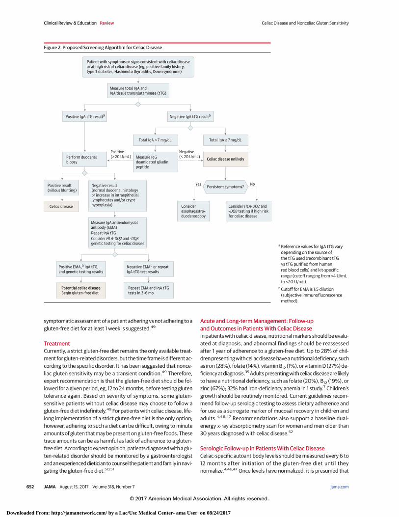
to evaluate for celiac disease.44 HLA-DQ2 and HLA-DQ8 determina-tion may be useful when there is discrepancy between serologic stud-ies and histologic findings to better assess whether celiac disease ispossible, because the disease, except in rare cases, cannot developin individuals who are negative for HLA-DQ2 and HLA-DQ8.44 Figure 2provides a diagnostic approach to celiac disease.
Villous blunting on the small-intestinal biopsy can definitivelyestablish the diagnosis of celiac disease and is recommended by theNorth American Society for Pediatric Gastroenterology, Hepatol-ogy, and Nutrition (NASPGHAN) and the American College ofGastroenterology (ACG).46,47 The characteristic histologic changesassociated with celiac disease include an increased number of in-traepithelial lymphocytes (>25 per 100 enterocytes), elongation ofthe crypts, and partial to total villous atrophy.48 The European Societyfor Pediatric Gastroenterology and Hepatology (ESPGHAN) has pro-posed an algorithm for children who meet specific criteria, for whombiopsy is not recommended.4 For the biopsy to be omitted, chil-dren must have signs and symptoms suggesting celiac disease, a posi-tive anti-tTG antibodies finding with a level greater than 10 timesthe upper limit of normal, a positive antiendomysial antibody find-ing obtained at a different time than the anti-tTG antibodies find-ing, and a HLA genotype compatible with celiac disease.4 Specificdiagnostic procedures such as double-balloon enteroscopy, video-capsule endoscopy, or magnetic resonance imaging are rarely usedand may be indicated in the workup of complicated celiac diseasewhen there are discrepancies between serology and histology re-sults or when a patient with celiac disease has persistent or wors-ening symptoms despite following a gluten-free diet.
Nonceliac Gluten SensitivityNo specific biomarkers have yet been identified and validated fornonceliac gluten sensitivity. Clinicians should suspect nonceliac glu-ten sensitivity in a patient who presents with gastrointestinal or ex-traintestinal symptoms (Table 2) that appear to improve with a glu-ten-free diet. Since these symptoms also can be seen with celiacdisease and, to a lesser extent, with wheat allergy, these conditionsneed to be preliminarily excluded with serologic and histologic evi-dence to focus on the suspicion of nonceliac gluten sensitivity. Untilbiomarkers are identified and validated, the diagnosis of nonceliacgluten sensitivity is confirmed in a research setting with a double-blind crossover gluten challenge.49 In a clinical setting, clinicians maysuggest a blinded gluten challenge during which a patient is givenapproximately 8 g of gluten (corresponding to approximately 2slices of bread) or placebo for 1 week each, separated by a 1-weekgluten-free washout period. Symptoms are monitored throughoutthe challenge.49 A blinded challenge is not generally feasible in a clini-cal setting; therefore, for patients with fluctuating symptoms,
Table 3. Range of Sensitivity and Specificity and Use of Current Serologic Tests for Celiac Diseasea
SerologicStudy
%
Application in Clinical PracticeSensitivity SpecificityIgA tTG 73.9-100 77.8-100 First-line testing to screen for celiac diseaseb
IgG DGP 80.1-96.9 86.0-96.9 First-line testing for celiac disease in patients with IgA deficiency
IgA EMA 82.6-100 94.7-100 Second-line confirmatory test to screen for celiac disease
IgG tTG 12.6-99.3 86.3-100 Not recommended for routine use because of poor sensitivity comparedwith IgG DGP
IgA DGP 80.7-95.1 86.3-93.1 Not recommended for routine use because of poor sensitivity and specificitycompared with IgA tTG and IgA EMA
Abbreviations: EMA, antiendomysialantibody; DGP, deamidated gliadinpeptide; tTG, tissuetransglutaminase.a Adapted from Thawani et al.41
b Should be sent with a baseline IgAlevel initially to ensure there is noIgA deficiency.
symptomatic assessment of a patient adhering vs not adhering to agluten-free diet for at least 1 week is suggested.49
TreatmentCurrently, a strict gluten-free diet remains the only available treat-ment for gluten-related disorders, but the time frame is different ac-cording to the specific disorder. It has been suggested that nonce-liac gluten sensitivity may be a transient condition.49 Therefore,expert recommendation is that the gluten-free diet should be fol-lowed for a given period, eg, 12 to 24 months, before testing glutentolerance again. Based on severity of symptoms, some gluten-sensitive patients without celiac disease may choose to follow agluten-free diet indefinitely.49 For patients with celiac disease, life-long implementation of a strict gluten-free diet is the only option;however, adhering to such a diet can be difficult, owing to minuteamounts of gluten that may be present on gluten-free foods. Thesetrace amounts can be as harmful as lack of adherence to a gluten-free diet. According to expert opinion, patients diagnosed with a glu-ten-related disorder should be monitored by a gastroenterologistand an experienced dietician to counsel the patient and family in navi-gating the gluten-free diet.50,51
Acute and Long-term Management: Follow-upand Outcomes in Patients With Celiac DiseaseIn patients with celiac disease, nutritional markers should be evalu-ated at diagnosis, and abnormal findings should be reassessedafter 1 year of adherence to a gluten-free diet. Up to 28% of chil-dren presenting with celiac disease have a nutritional deficiency, suchas iron (28%), folate (14%), vitamin B12 (1%), or vitamin D (27%) de-ficiency at diagnosis.35 Adults presenting with celiac disease are likelyto have a nutritional deficiency, such as folate (20%), B12 (19%), orzinc (67%); 32% had iron-deficiency anemia in 1 study.7 Children’sgrowth should be routinely monitored. Current guidelines recom-mend follow-up serologic testing to assess dietary adherence andfor use as a surrogate marker of mucosal recovery in children andadults.4,46,47 Recommendations also support a baseline dual-energy x-ray absorptiometry scan for women and men older than30 years diagnosed with celiac disease.52
Serologic Follow-up in Patients With Celiac DiseaseCeliac-specific autoantibody levels should be measured every 6 to12 months after initiation of the gluten-free diet until theynormalize.4,46,47 Once levels have normalized, it is presumed that
Figure 2. Proposed Screening Algorithm for Celiac Disease
Measure total IgA and IgA tissue transglutaminase (tTG)
Positive IgA tTG resulta Negative IgA tTG resulta
Celiac disease unlikelyPerform duodenal biopsy
Positive result(villous blunting)
Positive EMA,b IgA tTG, and genetic testing results
Negative EMAb or repeat IgA tTG test results
Negative result(normal duodenal histology or increase in intraepithelial lymphocytes and/or crypt hyperplasia)
Measure IgA antiendomysial antibody (EMA)Repeat IgA tTGConsider HLA-DQ2 and -DQ8 genetic testing for celiac disease
Consider esophagastro-duodenoscopy
Consider HLA-DQ2 and -DQ8 testing if high risk for celiac disease
Celiac disease
Total IgA < 7 mg/dL
Measure IgG deamidated gliadin peptide
Total IgA ≥ 7 mg/dL
Patient with symptoms or signs consistent with celiac disease or at high risk of celiac disease (eg, positive family history, type 1 diabetes, Hashimoto thyroiditis, Down syndrome)
Potential celiac diseaseBegin gluten-free diet
Repeat EMA and IgA tTG tests in 3-6 mo
Negative(< 20 U/mL)
NoYes
Positive(≥ 20 U/mL)
Persistent symptoms?
a Reference values for IgA tTG varydepending on the source ofthe tTG used (recombinant tTGvs tTG purified from humanred blood cells) and kit-specificrange (cutoff ranging from <4 U/mLto <20 U/mL).
b Cutoff for EMA is 1:5 dilution(subjective immunofluorescencemethod).
mucosal recovery, secondary to dietary adherence, has occurred. Al-though current guidelines according to the ACG, NASPGHAN, andESPGHAN endorse and recommend the use of serology as a markerof dietary adherence and mucosal recovery, these tests have notbeen validated for this purpose.4,46,47 Furthermore, recent work hasreported that the sensitivity of IgA tTG to identify persistent enter-opathy after a patient with celiac disease has started a gluten-freediet is 43% to 83%.53,54
Intestinal Biopsy in Patients With Celiac DiseaseCurrently available serologic tests may be inadequate to predictmucosal recovery in patients with celiac disease who follow agluten-free diet.54,55 In the United States, the Food and DrugAdministration has prioritized the need to develop accurate surro-gate end points to assess mucosal recovery in patients with celiacdisease. Although data evaluating pediatric mucosal recovery arelimited, previous data suggested a more complete and faster heal-ing time in children compared with adults.56 More recent data sug-gested that 5% to 19% of children with celiac disease who follow agluten-free diet may have persistent enteropathy despite treat-ment with a gluten free diet for at least 1 year.53,54 Irrespective ofIgA tTG levels, 25% to 40% of adults did not achieve mucosalrecovery after 2 years of following a gluten-free diet.57,58 Persistentvillous blunting was more common in older and male patients andless common in patients with higher educational attainment.59
Given these findings, a follow-up endoscopy to ensure mucosalrecovery in adult patients is recommended in some celiac centers.More studies are needed to evaluate the long-term complicationsassociated with persistent villous blunting in adults and childrenwith celiac disease before universal follow-up and treatment regi-mens are described. At this time, available treatment options forpatients diagnosed with celiac disease with persistent villous blunt-ing despite a gluten-free diet include implementation of the glutencontamination elimination diet, which offers only fresh fruits, veg-etables, meat, and limited condiments or medical therapy withimmunosuppressants, such as budesonide.60,61 These interven-tions should be offered and monitored by a gastroenterologist withexpertise in celiac disease, because both symptom improvementand resolution of the enteropathy are considered in the responseto treatment.
Prognosis of Celiac DiseaseImplementation of the gluten-free diet is the only treatment for ce-liac disease and therefore acts to prevent possible morbidity and pos-sible mortality associated with untreated celiac disease.
MorbidityResearch by Cosnes et al62 suggested that the implementation of agluten-free diet in patients with celiac disease may be associated witha protective effect against autoimmune disease such as thyroid dis-ease. Researchers reported a lower cumulative risk of developingsubsequent autoimmune disease in patients following a gluten-free diet compared with patients diagnosed with celiac disease notfollowing a gluten-free diet for at least 10 years (6% [±2%] vs 15.6%[±5.9%], respectively).62 Additionally, 2 Italian studies63,64 sug-gest that a gluten-free diet may decrease the prevalence of thyroidautoantibodies; however, whether it protects against hypothyroid-ism or hyperthyroidism remains to be established. Men with undi-
agnosed celiac disease have an increased rate of osteoporosis andhypothyroidism and had a lower body mass index and lower levelsof ferritin and cholesterol compared with women with undiag-nosed celiac disease.65 Last, infertility and recurrent abortion are re-ported as possible adverse pregnancy outcomes.66
MortalityA number of studies have examined mortality in undiagnosed ce-liac disease. Some showed increased mortality, while others didnot.67-69 A recent meta-analysis suggested that the overall malig-nancy risk in patients with diagnosed celiac disease was not el-evated, compared with the risk in general population–basedcontrols.70 However, individual cancers, such as lymphoprolifera-tive cancer and gastrointestinal cancers,71,72 may still be positivelyassociated with celiac disease.
Practical QuestionsShould Family Members of People With Celiac Disease Be Tested?First-degree family members of patients with celiac disease have upto a 15- to 25-fold higher frequency of developing celiac disease basedon their genetics, compared with individuals without a first-degreefamily member with celiac disease.6,73 For that reason, the ACG,NASPGHAN, and ESPGHAN suggest screening first-degree familymembers with or without signs or symptoms concerning for celiacdisease.4,46,47 Recommendations include initiation of testing by age3 years and, if serologic testing results are negative, repeating testingthroughout a patient’s lifetime. The US Preventive Services Task Forcerecently released recommendations that screening asymptomaticpatients with or without a known increased risk of celiac disease, in-clusive of family members, is not recommended, based on inadequateevidence regarding benefits and risks of this screening.74,75
When Should Clinicians Check for HLA-DQ2 and HLA-DQ8?Clinically, genetic testing for HLA-DQ2 and HLA-DQ8 may be help-ful to determine whether patients in high-risk groups, such as fam-ily members or patients with comorbid conditions such as autoim-mune thyroid disease, need screening for celiac disease. It also maybe used for patients already following a gluten-free diet who havenot been accurately assessed for celiac disease and are hesitant toreintroduce gluten into their diet for an accurate diagnostic reevalu-ation. In these cases, if patients do not carry the HLA-DQ2 orHLA-DQ8 genes, they would not require further diagnostic workupto evaluate for celiac disease. Celiac genetic testing may be usefulin complicated cases such as in patients with signs and symptomssuggestive of celiac disease and with compatible histology but se-ronegative serology to solidify a diagnosis.2 If patients are found tohave compatible celiac genetics but other test results are normal,this places them among the nearly 40% of the general populationthat carries one of these genes. These patients should continue tofollow a gluten-containing diet unless they are diagnosed witha gluten-related disorder.
How Does One Make a Diagnosis of Celiac Disease? What SerologicTests Should Be Used?Clinicians should start with serologic testing in patients presentingwith signs and symptoms compatible with celiac disease or thosewho belong to a high-risk group (Table 2). Signs, symptoms, familyhistory, and serologic results should be considered according to the
algorithm shown in Figure 2 to make a diagnosis. At the time thatceliac disease serology is tested, patients should have been follow-ing a gluten-containing diet for at least some months.
What Are the Most Common Manifestationsat Celiac Disease Presentation?The clinical presentation of celiac disease is heterogeneous; thus,there is not a “typical” presentation. Testing for celiac disease shouldbe considered in patients who have gastrointestinal manifestationssuch as abdominal pain, bloating, diarrhea, and constipation. Extrain-testinal manifestations such as anemia, joint pain, osteoporosis, pe-ripheral neuropathy, fatigue, and headache are frequent (Table 2).In pediatric patients, testing should be considered for those with slowor poor growth, delayed puberty, or tooth enamel defects.
How Common Is Iron-Deficiency Anemia or Osteoporosis thePresenting Sign or Symptom? Which Patients With Iron-DeficiencyAnemia or Osteoporosis Should Be Screened for Celiac Disease?At the time of diagnosis, approximately 28% and 9% of childrenmay present with iron deficiency and iron-deficiency anemia,respectively.35 Up to 32% of adults present with iron-deficiency ane-mia, making it the second most common clinical presentation afterdiarrhea.7,76 Patients with otherwise unexplained iron-deficiencyanemia, in those who do not respond to oral iron therapy, should betested for celiac disease. In adults, osteoporosis may be present in10% of patients at celiac disease diagnosis.76 However, the fre-quency with which patients present with osteoporosis is likely under-reported, since most cases of osteoporosis will not be apparent un-til a clinical complication such as a spontaneous fracture occurs.Therefore, physicians should have a low threshold for testing for
celiac disease in children and adults with unexplained, spontane-ous, or repetitive fracture.
Can a Gluten-Free Diet Be a Treatment for SituationsThat Do Not Belong to Gluten-Related Disorders?There is no scientific evidence to suggest that a gluten-free diet ispart of a healthier lifestyle or can be helpful to treat overweight orobesity. The incomplete digestibility of gluten (see “Pathophysiol-ogy” section) may explain why some people report nonspecific im-provement in well-being after starting the gluten-free diet. Further-more, gluten-containing cereals, particularly wheat, are also a primarysource of FODMAPs (fermentable oligosaccharides, disaccharides,and monosaccharides and polyols), a group of highly fermentablebut poorly absorbed short-chain carbohydrates and polyols.The reduction of FODMAPs associated with the gluten-free diet mayexplain, at least in part, why some patients affected with irritablebowel symptoms may report amelioration of their symptoms afterstarting a gluten-free diet.77 Self-diagnosis of nonceliac gluten sen-sitivity should be discouraged to avoid misdiagnosis and inappro-priate treatment.
ConclusionsCeliac disease and nonceliac gluten sensitivity are common. Al-though both conditions are treated with a gluten-free diet, distin-guishing between celiac disease and nonceliac gluten sensitivity isimportant for long-term therapy. Patients with celiac disease shouldbe followed up closely for dietary adherence, nutritional deficien-cies, and the development of possible comorbidities.
ARTICLE INFORMATION
Accepted for Publication: July 3, 2017.
Author Contributions: Drs Leonard and Fasanohad full access to all of the data in the study andtake responsibility for the integrity of the data andthe accuracy of the data analysis.Concept and design: All authors.Acquisition, analysis, or interpretation of data:Leonard.Drafting of the manuscript: All authors.Critical revision of the manuscript for importantintellectual content: All authors.Administrative, technical, or material support:Leonard.Supervision: Sapone, Catassi, Fasano.
Conflict of Interest Disclosures: All authors havecompleted and submitted the ICMJE Form forDisclosure of Potential Conflicts of Interest.Dr Catassi reported receiving fees for scientificconsultancy from Dr Schär Institute. Dr Fasanoreported receiving a grant for sponsored researchfrom INOVA Diagnostics; receiving speaking feesfrom Mead Johnson Nutrition; and that he iscofounder of, and a stockholder in, AlbaTherapeutics, which makes products for thetreatment of autoimmune and inflammatorydiseases. No other authors reported disclosures.
Submissions: We encourage authors to submitpapers for consideration as a Review. Pleasecontact Edward Livingston, MD, at [email protected] or Mary McGraeMcDermott, MD, at [email protected].
REFERENCES
1. Ludvigsson JF, Leffler DA, Bai JC, et al. The Oslodefinitions for coeliac disease and related terms. Gut.2013;62(1):43-52.
2. Leonard MM, Serena G, Sturgeon C, Fasano A.Genetics and celiac disease: the importance ofscreening. Expert Rev Gastroenterol Hepatol. 2015;9(2):209-215.
3. Dicke W. Simple dietary treatment for thesyndrome of Gee-Herter. Ned Tijdschr Geneeskd.1941;85:1715-1716.
4. Husby S, Koletzko S, Korponay-Szabó IR, et al;ESPGHAN Working Group on Coeliac DiseaseDiagnosis; ESPGHAN Gastroenterology Committee;European Society for Pediatric Gastroenterology,Hepatology, and Nutrition. European Society forPediatric Gastroenterology, Hepatology, andNutrition guidelines for the diagnosis of coeliacdisease. J Pediatr Gastroenterol Nutr. 2012;54(1):136-160.
5. Lionetti E, Catassi C. Co-localization of glutenconsumption and HLA-DQ2 and -DQ8 genotypes,a clue to the history of celiac disease. Dig Liver Dis.2014;46(12):1057-1063.
6. Fasano A, Berti I, Gerarduzzi T, et al. Prevalenceof celiac disease in at-risk and not-at-risk groups inthe United States: a large multicenter study. ArchIntern Med. 2003;163(3):286-292.
7. Wierdsma NJ, van Bokhorst-de van der SchuerenMA, Berkenpas M, Mulder CJ, van Bodegraven AA.Vitamin and mineral deficiencies are highly
prevalent in newly diagnosed celiac diseasepatients. Nutrients. 2013;5(10):3975-3992.
8. Sapone A, Bai JC, Ciacci C, et al. Spectrum ofgluten-related disorders: consensus on newnomenclature and classification. BMC Med.2012;10:13.
9. Silano M, Vincentini O, De Vincenzi M. Toxic,immunostimulatory and antagonist gluten peptidesin celiac disease. Curr Med Chem. 2009;16(12):1489-1498.
10. Shan L, Molberg Ø, Parrot I, et al. Structuralbasis for gluten intolerance in celiac sprue. Science.2002;297(5590):2275-2279.
11. Jelínková L, Tucková L, Cinová J, Flegelová Z,Tlaskalová-Hogenová H. Gliadin stimulates humanmonocytes to production of IL-8 and TNF-alphathrough a mechanism involving NF-kappaB. FEBSLett. 2004;571(1-3):81-85.
12. Lammers KM, Khandelwal S, Chaudhry F, et al.Identification of a novel immunomodulatory gliadinpeptide that causes interleukin-8 release in achemokine receptor CXCR3-dependent manneronly in patients with coeliac disease. Immunology.2011;132(3):432-440.
13. Picarelli A, Di Tola M, Sabbatella L, et al. 31-43amino acid sequence of the alpha-gliadin inducesanti-endomysial antibody production during in vitrochallenge. Scand J Gastroenterol. 1999;34(11):1099-1102.
14. Sturgeon C, Fasano A. Zonulin, a regulator ofepithelial and endothelial barrier functions, and its
involvement in chronic inflammatory diseases.Tissue Barriers. 2016;4(4):e1251384.
15. Matysiak-Budnik T, Moura IC, Arcos-Fajardo M,et al. Secretory IgA mediates retrotranscytosis ofintact gliadin peptides via the transferrin receptorin celiac disease. J Exp Med. 2008;205(1):143-154.
16. Kim SM, Mayassi T, Jabri B. Innate immunity:actuating the gears of celiac disease pathogenesis.Best Pract Res Clin Gastroenterol. 2015;29(3):425-435.
17. Barone MV, Troncone R, Auricchio S. Gliadinpeptides as triggers of the proliferative andstress/innate immune response of the celiac smallintestinal mucosa. Int J Mol Sci. 2014;15(11):20518-20537.
18. Lammers KM, Chieppa M, Liu L, et al. Gliadininduces neutrophil migration via engagement of theformyl peptide receptor, FPR1. PLoS One. 2015;10(9):e0138338.
19. Stamnaes J, Sollid LM. Celiac disease:autoimmunity in response to food antigen. SeminImmunol. 2015;27(5):343-352.
20. Pagliari D, Urgesi R, Frosali S, et al.The interaction among microbiota, immunity, andgenetic and dietary factors is the condicio sine quanon celiac disease can develop. J Immunol Res.2015;2015:123653.
21. Hüe S, Mention JJ, Monteiro RC, et al. A directrole for NKG2D/MICA interaction in villous atrophyduring celiac disease. Immunity. 2004;21(3):367-377.
22. Junker Y, Zeissig S, Kim SJ, et al. Wheat amylasetrypsin inhibitors drive intestinal inflammation viaactivation of toll-like receptor 4. J Exp Med. 2012;209(13):2395-2408.
23. Sapone A, Lammers KM, Mazzarella G, et al.Differential mucosal IL-17 expression in twogliadin-induced disorders: gluten sensitivity and theautoimmune enteropathy celiac disease. Int ArchAllergy Immunol. 2010;152(1):75-80.
24. Mansueto P, Seidita A, D’Alcamo A, Carroccio A.Non-celiac gluten sensitivity: literature review. J AmColl Nutr. 2014;33(1):39-54.
26. Hill ID. What are the sensitivity and specificityof serologic tests for celiac disease? do sensitivityand specificity vary in different populations?Gastroenterology. 2005;128(4)(suppl 1):S25-S32.
27. Catassi C, Kryszak D, Bhatti B, et al. Naturalhistory of celiac disease autoimmunity in a USAcohort followed since 1974. Ann Med. 2010;42(7):530-538.
28. Fasano A, Catassi C. Clinical practice: celiacdisease. N Engl J Med. 2012;367(25):2419-2426.
29. Leffler DA, Green PH, Fasano A. Extraintestinalmanifestations of coeliac disease. Nat RevGastroenterol Hepatol. 2015;12(10):561-571.
30. Vivas S, Ruiz de Morales JM, Fernandez M,et al. Age-related clinical, serological, andhistopathological features of celiac disease. Am JGastroenterol. 2008;103(9):2360-2365.
31. Ludvigsson JF, Ansved P, Fälth-Magnusson K,et al. Symptoms and signs have changed in Swedishchildren with coeliac disease. J PediatrGastroenterol Nutr. 2004;38(2):181-186.
32. Reilly NR, Aguilar K, Hassid BG, et al. Celiacdisease in normal-weight and overweight children:clinical features and growth outcomes following agluten-free diet. J Pediatr Gastroenterol Nutr. 2011;53(5):528-531.
33. Rashid M, Zarkadas M, Anca A, Limeback H.Oral manifestations of celiac disease: a clinical guidefor dentists. J Mich Dent Assoc. 2011;93(10):42-46.
34. Harper JW, Holleran SF, Ramakrishnan R,Bhagat G, Green PH. Anemia in celiac disease ismultifactorial in etiology. Am J Hematol. 2007;82(11):996-1000.
35. Wessels MM, van Veen II, Vriezinga SL, PutterH, Rings EH, Mearin ML. Complementary serologicinvestigations in children with celiac disease isunnecessary during follow-up. J Pediatr. 2016;169:55-60.
37. Tersigni C, Castellani R, de Waure C, et al. Celiacdisease and reproductive disorders: meta-analysisof epidemiologic associations and potentialpathogenic mechanisms. Hum Reprod Update.2014;20(4):582-593.
38. Ludvigsson JF, Lindelöf B, Zingone F, Ciacci C.Psoriasis in a nationwide cohort study of patientswith celiac disease. J Invest Dermatol. 2011;131(10):2010-2016.
39. Hadjivassiliou M, Aeschlimann P, Strigun A,Sanders DS, Woodroofe N, Aeschlimann D.Autoantibodies in gluten ataxia recognize a novelneuronal transglutaminase. Ann Neurol. 2008;64(3):332-343.
40. Briani C, Zara G, Alaedini A, et al. Neurologicalcomplications of celiac disease and autoimmunemechanisms: a prospective study. J Neuroimmunol.2008;195(1-2):171-175.
41. Thawani SP, Brannagan TH III, Lebwohl B, GreenPH, Ludvigsson JF. Risk of neuropathy among28,232 patients with biopsy-verified celiac disease.JAMA Neurol. 2015;72(7):806-811.
42. Rubio-Tapia A, Murray JA. Classification andmanagement of refractory coeliac disease. Gut.2010;59(4):547-557.
43. Volta U, Tovoli F, Cicola R, et al. Serologicaltests in gluten sensitivity (nonceliac glutenintolerance). J Clin Gastroenterol. 2012;46(8):680-685.
44. Giersiepen K, Lelgemann M, Stuhldreher N,et al; ESPGHAN Working Group on Coeliac DiseaseDiagnosis. Accuracy of diagnostic antibody tests forcoeliac disease in children: summary of an evidencereport. J Pediatr Gastroenterol Nutr. 2012;54(2):229-241.
45. Husby S, Murray JA. Diagnosing coeliac diseaseand the potential for serological markers. Nat RevGastroenterol Hepatol. 2014;11(11):655-663.
46. Hill ID, Dirks MH, Liptak GS, et al; NorthAmerican Society for Pediatric Gastroenterology,Hepatology and Nutrition. Guideline for thediagnosis and treatment of celiac disease inchildren: recommendations of the North AmericanSociety for Pediatric Gastroenterology, Hepatologyand Nutrition. J Pediatr Gastroenterol Nutr. 2005;40(1):1-19.
47. Rubio-Tapia A, Hill ID, Kelly CP, Calderwood AH,Murray JA; American College of Gastroenterology.ACG clinical guidelines: diagnosis and managementof celiac disease. Am J Gastroenterol. 2013;108(5):656-676.
48. Hill PG, Holmes GK. Coeliac disease: a biopsyis not always necessary for diagnosis. AlimentPharmacol Ther. 2008;27(7):572-577.
49. Fasano A, Sapone A, Zevallos V, Schuppan D.Nonceliac gluten sensitivity. Gastroenterology.2015;148(6):1195-1204.
50. Snyder J, Butzner JD, DeFelice AR, et alEvidence-informed expert recommendations forthe management of celiac disease in children.Pediatrics. 2016;138(3):e20153147.
51. Pietzak MM. Follow-up of patients with celiacdisease: achieving compliance with treatment.Gastroenterology. 2005;128(4)(suppl 1):S135-S141.
52. Pantaleoni S, Luchino M, Adriani A, et al. Bonemineral density at diagnosis of celiac disease andafter 1 year of gluten-free diet. ScientificWorldJournal.2014;2014:173082.
53. Vécsei E, Steinwendner S, Kogler H, et al.Follow-up of pediatric celiac disease: value ofantibodies in predicting mucosal healing, aprospective cohort study. BMC Gastroenterol. 2014;14:28.
54. Leonard MM, Weir DC, DeGroote M, et al. Valueof IgA tTG in predicting mucosal recovery inchildren with celiac disease on a gluten-free diet.J Pediatr Gastroenterol Nutr. 2017;64(2):286-291.
55. Mahadev S, Murray JA, Wu TT, et al. Factorsassociated with villus atrophy in symptomaticcoeliac disease patients on a gluten-free diet.Aliment Pharmacol Ther. 2017;45(8):1084-1093.
56. Dickey W, Hughes DF, McMillan SA.Disappearance of endomysial antibodies in treatedceliac disease does not indicate histologicalrecovery. Am J Gastroenterol. 2000;95(3):712-714.
57. Ciacci C, Cirillo M, Cavallaro R, Mazzacca G.Long-term follow-up of celiac adults on gluten-freediet: prevalence and correlates of intestinaldamage. Digestion. 2002;66(3):178-185.
58. Rubio-Tapia A, Rahim MW, See JA, Lahr BD, WuTT, Murray JA. Mucosal recovery and mortality inadults with celiac disease after treatment with agluten-free diet. Am J Gastroenterol. 2010;105(6):1412-1420.
59. Lebwohl B, Murray JA, Rubio-Tapia A, GreenPH, Ludvigsson JF. Predictors of persistent villousatrophy in coeliac disease: a population-basedstudy. Aliment Pharmacol Ther. 2014;39(5):488-495.
60. Hollon JR, Cureton PA, Martin ML, Puppa EL,Fasano A. Trace gluten contamination may play arole in mucosal and clinical recovery in a subgroupof diet-adherent non-responsive celiac diseasepatients. BMC Gastroenterol. 2013;13:40.
61. Jamma S, Leffler DA, Dennis M, et al. Smallintestinal release mesalamine for the treatment ofrefractory celiac disease type I. J Clin Gastroenterol.2011;45(1):30-33.
62. Cosnes J, Cellier C, Viola S, et al; GroupeD’Etude et de Recherche Sur la Maladie Coeliaque.Incidence of autoimmune diseases in celiac disease:protective effect of the gluten-free diet. ClinGastroenterol Hepatol. 2008;6(7):753-758.
63. Ventura A, Neri E, Ughi C, Leopaldi A, Città A,Not T. Gluten-dependent diabetes-related and
thyroid-related autoantibodies in patients withceliac disease. J Pediatr. 2000;137(2):263-265.
64. Toscano V, Conti FG, Anastasi E, et al.Importance of gluten in the induction of endocrineautoantibodies and organ dysfunction inadolescent celiac patients. Am J Gastroenterol.2000;95(7):1742-1748.
65. Bai D, Brar P, Holleran S, Ramakrishnan R,Green PH. Effect of gender on the manifestations ofceliac disease: evidence for greater malabsorptionin men. Scand J Gastroenterol. 2005;40(2):183-187.
66. Martinelli P, Troncone R, Paparo F, et al. Coeliacdisease and unfavourable outcome of pregnancy. Gut.2000;46(3):332-335.
67. Rubio-Tapia A, Kyle RA, Kaplan EL, et al.Increased prevalence and mortality in undiagnosedceliac disease. Gastroenterology. 2009;137(1):88-93.
68. Canavan C, Logan RF, Khaw KT, West J. Nodifference in mortality in undetected coeliacdisease compared with the general population:
a UK cohort study. Aliment Pharmacol Ther. 2011;34(8):1012-1019.
69. Godfrey JD, Brantner TL, Brinjikji W, et al.Morbidity and mortality among older individualswith undiagnosed celiac disease. Gastroenterology.2010;139(3):763-769.
70. Tio M, Cox MR, Eslick GD. Meta-analysis:coeliac disease and the risk of all-cause mortality,any malignancy and lymphoid malignancy. AlimentPharmacol Ther. 2012;35(5):540-551.
71. Elfström P, Granath F, Ye W, Ludvigsson JF. Lowrisk of gastrointestinal cancer among patients withceliac disease, inflammation, or latent celiacdisease. Clin Gastroenterol Hepatol. 2012;10(1):30-36.
72. Green PH, Fleischauer AT, Bhagat G, Goyal R,Jabri B, Neugut AI. Risk of malignancy in patientswith celiac disease. Am J Med. 2003;115(3):191-195.
73. Lionetti E, Castellaneta S, Francavilla R, et al;SIGENP (Italian Society of Pediatric
Gastroenterology, Hepatology, and Nutrition)Working Group on Weaning and CD Risk.Introduction of gluten, HLA status, and the risk ofceliac disease in children. N Engl J Med. 2014;371(14):1295-1303.
75. Choung RS, Murray JA. The US PreventiveServices Task Force recommendation on screeningfor asymptomatic celiac disease: a dearth ofevidence. JAMA. 2017;317(12):1221-1223.
76. Green PH, Krishnareddy S, Lebwohl B. Clinicalmanifestations of celiac disease. Dig Dis. 2015;33(2):137-140.
77. Catassi G, Lionetti E, Gatti S, Catassi C. The lowFODMAP diet: many question marks for a catchyacronym. Nutrients. 2017;9(3):E292.