Roche Molecular Systems, Inc. cobas® 4800 BRAF V600 Mutation Test Pleasanton, CA 94588-2722 Module 3: Labeling Draft Package Insert I cobas® 4800 BRAF V600 Mutation Test c(obase 2 FOR IN VITRO DIAGNOSTIC USE. 3 cobas® DNA Sample Preparation Kit 24 Tests P/N: 05985536190 4, 5 cobas® 4800 BRAF V600 Mutation Test IRgAF 1 24 Tests P/N: 05985579190 6 7 NOTICE: The purchase of this product allows the purchaser to use it for amplification and detection of nucleic acid 8 sequences by polymerase chain reaction (PCR) and related processes for human in vitro diagnostics for the specific intended 9 use stated in this Package Insert. No general patent or other license of any kind other than this specific right of use from 10 purchase is granted hereby outside of the indications for use stated here. II TABLE OF CONTENTS 12 TABLE OF CONTENTS...... ................. .... .. ...... ... ..... ....... .... 13 INTENDED USE ....................... ................................. ..................... 14 SUMMARY AND EXPLANATION OF THE TEST........................... ....................... 3 I5 PRINCIPLES OF THE PROCEDURE...........................................3 1 6 Specimen Preparation . e o .. ... .... .... ..... ....... .... .......... ... 3 17 PCR Amplification and Detectio ............. ............ 18 T arget Selection .................... .. . .. .... ....... . .... ...... ... . .. 3 19 Target A m plification .................................................. 20 Automated Real-time Detection.... .................... ............. ..... .. ... .. 4 21 Selective A m plification . ...... ..... ....... ... ..... ...... ........ .... .. .4 22 REAG ENTTS ............................ HL . U MS...... . ............. ........ .. 4 23 WARNINGS AND PRECAUTIONS................ 7 24 STORAGE AND HANDLING REQUIREMENTS ............ ............ 8 25 MATERIALS ROVID PROVIDED. ..................... ............................................... 8 26 MATERIALS REQUIRED BUT NOT PRO VIDED.............................................. .... 9 27 Instrumentation and Sofiware..............I......... ............ 1 28 SPECIMEN COLLECTION, TRANSPORT AND STORAGE ........................ ...................... 1 29 A. Specim en Collection ....................... . . . ... .... . . . . .... . . . . . . . . 10 30 B. Specim en Transport ........................ . ..... .. I .... . . . . ... ... .. 1 31 C . Specim en Storage ................................ .... . ...... ... .. 1 32 INSTRUCTIONS FOR USE I........ .............. .... ..................... .10 3 3 R un Size .. ..... .... ...... ... .... ....... .... .... .... .... ...... .0 34 W orkflow ............. ........... 10 35 R eagent Preparation ............................ .... ... ... .... ... ....... .... ... ..... ...... ..... ... .... .. . 0 36 Deparaffinization of FFPET Sections Mounted on Slides ................................... ............... 11 37 Deparaffinization of FFPET Sections not Mounted on Slides....................... ... ................... 11 38 D N A 1solation ........................ . .... . ....... ... ... ... .. 12 39 D N A Q uantitation........................... ....................... 13 40 AM PLIFICATION AND DETECTION ................................................. 13 41 Instrum ent Set-U p ............................................... 13 42 T est O rder Set-U p .................................................................... .............................. .............. . .................................. I3 43 Dilution Calculation of Specimen DNA Stock at Concentrations from 5 ng/pL to 35 ng/pL ................................................ 14 44 Dilution Calculation of Specimen DNA Stock at Concentrations >35 ng/IL ......................... .4........................ 14 45 Specimen Dilution......................................... ..................................... ............. 14 46 Preparation of W orking M aster M ix (M M X) .......................... ..................... 15 47 Addition of Controls and Specimens ......................... ................... I 5 PMA M100022 CONFIDENTIAL AND PROPRIETARY Draft Package Insert Print Date: August 29, 2011 Page 1 of 30
Transcript
Roche Molecular Systems, Inc. cobas® 4800 BRAF V600 Mutation TestPleasanton, CA 94588-2722 Module 3: Labeling
Draft Package Insert
I cobas® 4800 BRAF V600 Mutation Test c(obase2 FOR IN VITRO DIAGNOSTIC USE.
3 cobas® DNA Sample Preparation Kit 24 Tests P/N: 05985536190
7 NOTICE: The purchase of this product allows the purchaser to use it for amplification and detection of nucleic acid8 sequences by polymerase chain reaction (PCR) and related processes for human in vitro diagnostics for the specific intended9 use stated in this Package Insert. No general patent or other license of any kind other than this specific right of use from
10 purchase is granted hereby outside of the indications for use stated here.
26 MATERIALS REQUIRED BUT NOT PRO VIDED.............................................. ....9
27 Instrumentation and Sofiware..............I......... ............ 128 SPECIMEN COLLECTION, TRANSPORT AND STORAGE ........................ ...................... 1
29 A . Specim en Collection ....................... . . . ... .... . . . . .... . . . . . . . . 1030 B. Specim en Transport ........................ . ..... .. I .... . . . . ... ... .. 131 C . Specim en Storage ................................ .... . ...... ... .. 132 INSTRUCTIONS FOR USE I........ .............. .... ..................... .10
3 3 R un Size .. ..... .... ...... ... .... ....... .... .... .... .... ...... .034 W orkflow ............. ........... 1035 R eagent Preparation ............................ .... ... ... .... ... ....... .... ... ..... .... .. ..... ... .... .. . 036 Deparaffinization of FFPET Sections Mounted on Slides ................................... ............... 1137 Deparaffinization of FFPET Sections not Mounted on Slides....................... ... ................... 1138 D N A 1solation ........................ . .... . ....... ... ... ... .. 1239 D N A Q uantitation........................... ....................... 1340 AM PLIFICATION AND DETECTION ................................................. 13
41 Instrum ent Set-U p ............................................... 1342 T est O rder Set-U p .................................................................... .............................. .............. . .................................. I343 Dilution Calculation of Specimen DNA Stock at Concentrations from 5 ng/pL to 35 ng/pL ................................................ 1444 Dilution Calculation of Specimen DNA Stock at Concentrations >35 ng/IL ......................... .4........................ 1445 Specimen Dilution......................................... ..................................... ............. 1446 Preparation of W orking M aster M ix (M M X) .......................... ..................... 1547 Addition of Controls and Specimens ......................... .................... I 5
PMA M100022 CONFIDENTIAL AND PROPRIETARY Draft Package InsertPrint Date: August 29, 2011 Page 1 of 30
Roche Molecular Systems, Inc. cobase 4800 BRAF V600 Mutation TestPleasanton, CA 94588-2722 Module 3: Labeling
Draft Package Insert
48 Starting PC R ............................... . . . . . . I .649 INTERPRETATION OF RESULTS ............. ........... 1650 Retesting of Specimens w ith Invalid Results.......................... ........................ 175 1 Q UALITY CONTROL ... 1........ ............. I752 BRAF Mutant Control ....................... ............................................ ........................ 1753 BRA F W ild-Type Control .... ........... ......... .... .....................1754 PROCEDURAL PRECAUTIONS............ . .......... 17
55 PROCEDURAL LI M ITA TION ................................... ....................... . 1756 NON-CLINICAL PERFORMANCE EVALUATION........... ...................................... ..I.8
57 Analytical Sensitivity - Limit of Blank (LoB)......................... .I........................ I758 Analytical Sensitivity- Limit of Detection (LoD) ............................................... 18
59 Analytical Sensitivity Using Specimen Blends................................................1960 Analytical Sensitivity Using FFPET Specimens........................ . 9..................... 19
61 Analytical Sensitivity Using Cell Line Blend ....................... ........................ 1962 M inim al Tum or Content..... ......... .......... .................... ....... 2063 C ross-R eactivity .... .... .... ............. ...... ......... .... ......... ... ....... ........... . ... ...2 164 BRAF Non-V600E Melanoma FFPET Specimens ......................... 2....................... 22
65 BRAF Non-V600E Plasmids .................................................. 2266 Plasm ids of BRA F Hom ologs ......................... . .......................... 2367 Skin-related M icroorganism s.. ... ..-. ... ......... .. ..... ........ ... ..... . .. . .23
PMA M100022 CONFIDENTIAL AND PROPRIETARY Draft Package InsertPrint Date: August 29, 2011 Page 2 of 30
Roche Molecular Systems, Inc. cobaso 4800 BRAF V600 Mutation TestPleasanton, CA 94588-2722 Module 3: Labeling
Draft Package Insert
73 INTENDED USE
74 The cobaso 4800 BRAF V600 Mutation Test is an in vitro diagnostic device intended for the qualitative detection of the BRAF V600E75 mutation in DNA extracted from formalin-fixed, paraffin-embedded human melanoma tissue. The cobas® 4800 BRAF V600 Mutation76 Test is a real-time PCR test on the cobas 4800 system, and is intended to be used as an aid in selecting melanoma patients whose tumors77 carry the BRAF V600E mutation for treatment with vemurafenib.
78 SUMMARY AND EXPLANATION OF THE TEST
79 Activating mutations of the proto-oncogene BRAF occur in many human cancers, including malignant melanoma, colorectal cancer,80 ovarian cancer, and thyroid cancer. 1,2 BRAF mutations have been identified in 40%-60% of malignant melanomas.3 Mutations are also81 common in benign nevi,4 suggesting that such mutations are a very early event. The discovery of such somatic mutations in the BRAF82 gene in melanoma and other human tumors has helped to elucidate the central role of the BRAT kinase in signaling pathways that83 control cellular proliferation, differentiation and cell death. In normal cells, BRAF is part of a highly regulated signaling pathway that84 mediates the effects of growth factor receptors (such as EGFR) through RAS. RAF, MEK and ERK. Oncogenic mutations in BRAF85 result in a gain of kinase function, rendering the RAF-MEK-ERK pathway constitutively active in the absence of the typical growth86 factors.
87 The majority of BRAF mutations in melanoma and other human tumors occur in codon 600.' The predominant mutation at codon 600 is88 the V600E mutation (GTG>GAG). A number of dinucleotide mutations affecting codon 600 [V600K (GTG>AAG), V600R89 (GTG>AGG), V600E2 (GTG>GAA), and V600D (GTG>GAT)] have also been observed less commonly, primarily in melanoma and90 rarely in other tumors, such as colorectal cancer. The cobas' 4800 BRAF V600 Mutation Test is a real-time PCR assay designed to91 detect the presence of the V600E (T1799A) mutation. The cobas" 4800 BRAF V600 Test is used as a companion diagnostic test for92 vemurafenib, a compound which inhibits the mutant V600E version of BRAF. Clinical trials of vemurafenib in patients with advanced93 melanoma have shown that patients with a V600E-mutant tumor are likely-to experience clinical benefit from the compound67.
94 PRINCIPLES OF THE PROCEDURE
95 The cobas® 4800 BRAF V600 Mutation Test is based on two processes: (1) manual specimen preparation to obtain genomic DNA from96 formalin-fixed, paraffin-embedded tissue (FFPET); (2) PCR amplification and detection of target DNA using a complementary primer97 pair and two oligonucleotide probes labeled with different fluorescent dyes. One probe is designed to detect the wild-type BRAF V60098 sequence and one is designed to detect the V600E mutation sequence. Two external run controls are provided and the wild-type allele99 serves as an internal, full process control.
100 Specimen Preparation
101 FFPET specimens are processed and genomic DNA isolated using the cobas® DNA Sample Preparation Kit, a manual specimen102 preparation based on nucleic acid binding to glass fibers. A deparaffinized 5-pm section of an FFPET specimen is lysed by incubation at103 an elevated temperature with a protease and chaotropic lysis/binding buffer that releases nucleic acids and protects the released genomic104 DNA from DNases. Subsequently, isopropanol is added to the lysis mixture that is then centrifuged through a column with a glass fiber105 filter insert. During centrifugation, the genomic DNA is bound to the surface of the glass fiber filter. Unbound substances, such as salts,106 proteins and other cellular impurities, are removed by centrifugation. The adsorbed nucleic acids are washed and then cluted with an107 aqueous solution. The amount of genomic DNA is spectrophotometrically determined and adjusted to a fixed concentration to be added108 to the amplification and detection mixture. The target DNA is then amplified and detected on the cobas z 480 analyzer using the109 amplification and detection reagents provided in the cobas® 4800 BRAF V600 Mutation Test kit.
110 PCR Amplification and Detection
Ill Target Selection
112 The cobas® 4800 BRAF V600 Mutation Test uses primers that define a l16-base pair sequence of human genomic DNA containing the113 BRAF codon 600 site in exon 15. The entire BRAF gene is not amplified. The cobas® 4800 BRAF V600 Mutation Test is designed to14 detect the nucleotide (T1799A) change in the BRAF gene which results in a valine-to-glutamic acid substitution at codon 600 (V600E).
115 BRAF wild-type and mutant DNA target-specific, fluorescent dye-labeled TaqMan probes bind to the wild-type and mutant sequences,116 respectively. The wild-type and mutant sequences are detected using a dedicated optical channel for each sequence.
117 Target Amolification
118 Thermus species Z05 DNA polymerase is utilized for target amplification. First, the PCR reaction mixture is heated to denature the119 genomic DNA and expose the primer target sequences. As the mixture cools, the upstream and downstream primers anneal to the target120 DNA sequences. The Z05 DNA Polymerase, in the presence of divalent metal ion and excess dNTPs, extends each annealed primer,
PMA M100022 CONFIDENTIAL AND PROPRIETARY Draft Package InsertPrint Date: August 29, 2011 Page 3 of 30
Roche Molecular Systems, Inc. cobass 4800 BRAF V600 Mutation TestPleasanton, CA 94588-2722 Module 3: Labeling
Draft Package Insert
121 thus synthesizing a second DNA strand. This completes the first cycle of PCR, yielding a double-stranded DNA copy of the targeted122 116-basepair region of the BRAF gene. This process is repeated for a number of cycles, with each cycle effectively doubling the amount123 of amplicon DNA. Amplification occurs only in the region of the BRAF gene between the primers.
124 Automated Real-time Detection
125 The cobas" 4800 BRAF V600 Mutation Test utilizes real-time PCR technology. Each target-specific, oligonucleotide probe in the126 reaction is labeled with a fluorescent dye that serves as a reporter, and with a quencher molecule that absorbs (quenches) fluorescent127 emissions from the reporter dye within an intact probe. During each cycle of amplification, probe complementary to the single-stranded128 DNA sequence in the amplicon binds and is subsequently cleaved by the 5' to 3' nuclease activity of the Z05 DNA Polymerase. Once129 the reporter dye is separated from the quencher by this nuclease activity, fluorescence of a characteristic wavelength can be measured130 when the reporter dye is excited by the appropriate spectrum of light. Two different reporter dyes are used to label the target-specific131 BRAF wild-type (WT) probe and the BRAF V600E mutation probe. Amplification of the two BRAF sequences can be detected132 independently in a single reaction well by measuring fluorescence at the two characteristic wavelengths in dedicated optical channels.
133 Selective Amplification
134 Selective amplification of target nucleic acid from the specimen is achieved in the cobas* 4800 BRAF V600 Mutation Test by the use135 of AmpErase (uracil-N-glycosylase) enzyme and deoxyuridine triphosphate (dUTP). The AmpErase enzyme recognizes and catalyzes136 the destruction of DNA strands containing deoxyuridine, but not DNA containing thymidine. Deoxyuridine is not present in naturally137 occurring DNA, but is always present in amplicon due to the use of dUTP as one of the nucleotide triphosphates in the Reaction Mix138 reagent; therefore, only amplicon contains deoxyuridine. Deoxyuridine renders contaminating amplicon susceptible to destruction by139 AmpErase enzyme prior to amplification of the target DNA. The AmpErase enzyme, which is included in the Reaction Mix reagent,140 catalyzes the cleavage of deoxyuridine-containing DNA at the deoxyuridine residues by opening the deoxyribose chain at the Cl-141 position. When heated in the first thermal cycling step at alkaline pH, the amplicon DNA chain breaks at the position of the142 deoxyuridine, thereby rendering the DNA non-amplifiable. The AmpErase enzyme is inactive at temperatures above 550C, i.e.,143 throughout the thermal cycling steps, and therefore does not destroy target amplicon.
144 REAGENTS
145 cobas* DNA Sample Preparation Kit 24 Tests146 (P/N: 05985536190)
PMA M100022 CONFIDENTIAL AND PROPRIETARY Draft Package InsertPrint Date: August 29, 2011 Page 6 of 30
Roche Molecular Systems, Inc. cobase 4800 BRAF V600 Mutation TestPleasanton, CA 94588-2722 Module 3: Labeling
Draft Package Insert
233 DNA SD 2 xl mL234 (DNA Specimen Diluent)
235 Tris-HCI buffer236 0.09% Sodium azide237 WARNINGS AND PRECAUTIONS
238 A. FOR IN VITRO DIAGNOSTIC USE.
239 B. This test is for use with formalin-fixed, paraffin-embedded tissue specimens.
240 C. Do not pipette by mouth.
241 D. Do not eat, drink or smoke in laboratory work areas.
242 E. Avoid microbial and DNA contamination of reagents.
243 F. Dispose of unused reagents and waste in accordance with country, federal, state and local regulations.
244 G. Do not use kits after their expiration dates.
245 H. Do not pool reagents from different kits or lots.
246 1. Material Safety Data Sheets (MSDS) are available on request from your local Roche office.
247 J. Gloves must be worn and must be changed between handling specimens and cobas" 4800 reagents to prevent contamination.
248 K. To avoid contamination of the working Master Mix with DNA specimens, Amplification and Detection should be performed in an249 area separated from DNA Isolation. The amplification and detection work area should be thoroughly cleaned before working250 Master Mix preparation. For proper cleaning, all surfaces including racks and pipettors should be thoroughly wiped with 0.5%251 Sodium hypochlorite* solution followed by wiping with a 70% ethanol solution.
252 L. DNA PBB, WB I and WB II contain guanidine hydrochloride. If liquid containing this buffer is spilled, clean with suitable253 laboratory detergent and water. If a spill occurs with potentially infectious agents, clean the affected area first with laboratory254 detergent and water, and then with 0.5% sodium hypochlorite*. If spills occur on the cobas z 480 analyzer, follow the instructions255 in the cobaso 4800 system Operator's Manual.
256 *NOTE: Commercial liquid household bleach typically contains sodium hypochlorite at a concentration of 5.25%. A 1:10 dilution of257 household bleach will produce a 0.5% sodium hypochlorite solution.
258 M. Specimens should be handled as infectious using safe laboratory procedures such as those outlined in Biosafety in Microbiological259 and Biomedical Laboratories' and in the CLSI Document M29-A3' 0.
260 N. DNA TLB and DNA PBB contain Triton X-100, an irritant to mucous membranes. Avoid contact with eyes, skin and mucous261 membranes.
262 0. DNA TLB, DNA PB, DNA EB, RXNMIX, MGAC, BRAF MUT, BRAF WT, and DNA SD contain sodium azide.263 Sodium azide may react with lead and copper plumbing to form highly explosive metal azides. While disposing of sodium azide264 containing solutions down laboratory sinks, flush the drains with a large volume of cold water to prevent azide buildup.
265 P. Xylene is a hazardous chemical and should be used in a chemical hood. Discard into chemical waste in accordance with local, state,266 and federal regulations.
267 Q. Wear eye protection, laboratory coats and disposable gloves when handling any reagents. Avoid contact of these materials with the268 skin, eyes or mucous membranes. If contact does occur, immediately wash with large amounts of water. Burns can occur if left269 untreated. If spills occur, dilute with water before wiping dry.
270 R. All disposable items are single use. Do not reuse.
271 S. Do not use disposable items beyond their expiration date.
272 T. Do not use sodium hypochlorite solution (bleach) for cleaning the cobas z 480 analyzer. Clean the cobas z 480 analyzer273 according to procedures described in the cobaso 4800 system Operator's Manual.
274 U. For additional warnings, precautions and procedures to reduce the risk of contamination for the cobas z 480 analyzer, consult the275 cobas® 4800 system Operator's Manual.
PMA M100022 CONFIDENTIAL AND PROPRIETARY Draft Package InsertPrint Date: August 29, 2011 Page 7 of 30 7
Roche Molecular Systems, Inc. cobas" 4800 BRAF V600 Mutation TestPleasanton, CA 94588-2722 Module 3: Labeling
Draft Package Insert
276 V. The use of sterile disposable pipets and DNase-free pipet tips is recommended.
277 STORAGE AND HANDLING REQUIREMENTS
278 A. Do not freeze reagents.
279 B. Store DNA TLB, DNA PBB, WB I, WB II, DNA EB, FT and CT at 15-30'C. Once opened, these reagents are stable for up280 to 8 uses over 90 days or until the expiration date, whichever comes first.
281 C. Store PIK at 15-300C. After addition of sterile, nuclease-free water to PK, store unused reconstituted PIK in 450 pL aliquots at -282 20'C. Once reconstituted, PIK must be used within 90 days or until the expiration date, whichever comes first.
283 D. After addition of absolute ethanol, store WB I and WB II at 15-300C. These Working Solutions are stable for up to 8 uses over284 90 days or until the expiration date, whichever comes first.
285 E. Store RXNMIX, MGAC, BRAF OM, BRAF MUT, BRAF WT, and DNA SD at 2-80 C. Once opened, these reagents are286 stable for up to 4 uses over 60 days or until the expiration date, whichever comes first.
287 F. BRAF OM and working Master Mix (prepared by the addition of BRAF OM and MGAC to RXNMIX) should be protected288 from prolonged exposure to light.
289 G. Working Master Mix (prepared by the addition of BRAF OM and MGAC to RXNMIX) must be stored at 2-80 C in the dark.290 The prepared specimens and controls must be added within I hour of preparation of the working Master Mix (MMX).
291 H. Processed specimens are stable for up to 24 hours at 15-30 0C, up to 14 days at 2-80C or frozen at -200C for up to 60 days. The292 processed specimens (extracted DNA) are also stable after undergoing up to 4 freeze thaws.
293 1. Amplification must be started within I hour from the time that the processed specimens and controls are added to the working294 Master Mix (prepared by the addition of BRAF OM and MGAC to RXNMIX).
295 MATERIALS PROVIDED
296 A. cobast DNA Sample Preparation Kit 24 Tests
297 (P/N: 05985536190)298 DNA TLB299 (DNA Tissue Lysis Buffer)
300 PIK301 (Proteinase K)
302 DNA PBB303 (DNA Paraffin Binding Buffer)
304 WBI305 (DNA Wash Buffer I)
306 WBl I307 (DNA Wash Buffer II)308 DNA EB309 (DNA Elution Buffer)310 FT311 (Filter tubes with caps)312 CT313 (Collection Tubes)
314 B. cobas 4800 BRAF V600 Mutation Test 24 Tests IRAFu 1315 (P/N:05985579190)
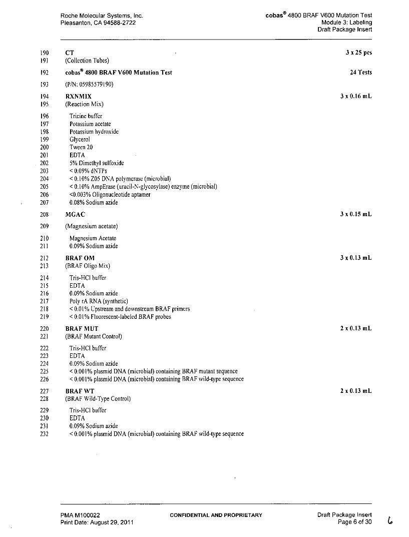
316 RXNMIX317 (Reaction Mix) (Cap with Natural Button)
318 MGAC319 (Magnesium acetate) (Cap with Yellow Button)
PMA M100022 CONFIDENTIAL AND PROPRIETARY Draft Package InsertPrint Date: August 29, 2011 Page 8 of 30
Roche Molecular Systems, Inc. cobas" 4800 BRAF V600 Mutation TestPleasanton, CA 94588-2722 Module 3: Labeling
Draft Package Insert
320 BRAF OM321 (BRAF Oligo Mix) (Black Cap with White Button)
322 BRAF MUT323 (BRAF Mutant Control) (Cap with Red Button)
324 BRAF WT325 (BRAF Wild-Type Control) (Cap with Blue Button)
326 DNA SD327 (DNA Specimen Diluent) (Cap with Purple Button)
328 MATERIALS REQUIRED BUT NOT PROVIDED
329 * Xylene (ACS, 98.5% xylenes)
330 * Absolute Ethanol (for Molecular Biology)
331 * Isopropanol (ACS, 995%)
332 * Sterile Nuclease-free Water (for Molecular Biology)
333 * Sterile disposable, serological pipettes: 5 and 25 mL
334 * cobas* 4800 System Microwell Plate (AD-Plate) and Sealing Film (Roche P/N 05232724001)
335 * Adjustable Pipettors*: (capacity 10 pL. 20 pL, 200 AL and 1000 pL) with aerosol barrier or positive displacement336 DNase-free tips
337 * Pipette Aid (Drummond P/N: 4-000-100 or equivalent)
338 * Bench top microcentrifuge capable of 20,000 x g
339 * Two (2) Dry Heat Blocks capable of heating microcentrifuge tubes to 560C and 900C**
341 * NanoDrop UV-Vis Spectrophotometer (Thermo Scientific ND-1000 or ND-2000)**
342 * Vortex mixer**
343 * Microcentrifuge tube racks
344 * Disposable gloves, powderless
345 * Calibrated Thermometers for Dry Heat Block**
346 * Waterbath** capable of maintaining 370C
347 * Single edge blade or similar
348 * Pipettors should be maintained according to the manufacturer's instructions and accurate within 3% of stated volume. Aerosol349 barrier or positive displacement DNase-free tips must be used where specfied to prevent specimen degradation and cross-350 contamination.
351 **All equipment should be properly maintained according to the manufacturer's instructions.
PMA M100022 CONFIDENTIAL AND PROPRIETARY Draft Package InsertPrint Date: August 29, 2011 Page 9 of 30
Roche Molecular Systems, Inc. cobas® 4800 BRAF V600 Mutation TestPleasanton, CA 94588-2722 Module 3: Labeling
Draft Package Insert
352 Instrumentation and Software
353 * cobas z 480 analyzer
354 * cobas@ 4800 SR2 System Control Unit with OSXP image
355 * cobas@ 4800 SR2 System Software version 2.0
356 * BRAF Analysis Package Software version 1.0
357 * Barcode Reader (Roche P/N 05339910001)
358 * Printer HP P2055d (Roche P/N 05704375001)
359 SPECIMEN COLLECTION, TRANSPORT AND STORAGE
360 NOTE: Handle all specimens as if they are capable of transmitting infectious agents.
361 A. Specimen Collection
362 FFPET specimens have been validated for use with the cobas® 4800 BRAF V600 Mutation Test.
363 B. Specimen Transport
364 FFPET specimens can be transported at 15-30'C. Transportation of FFPET specimens must comply with country, federal, state and365 local regulations for the transport of etiologic agents'.
366 C. Specimen Storage
367 Stability of FFPET specimens stored at 15-30'C for up to 9 months after the date of collection has been confirmed. 5 pm sections368 mounted on slides may be stored at 15-30 0C for up to 60 days.
369 INSTRUCTIONS FOR USE
370 NOTE: All reagents except RXNMIX, MGAC, and BRAF OM must be at ambient temperature prior to use. The RXN371 MIX, MGA C, and BRAF OM may be taken directly from 2-80C storage to prepare working Master Mix.
372 NOTE: Only melanoma FFPET sections of 5 pm thickness containing at least 50% tumor content are to be used in the373 cobas® 4800 BRAF V600 Mutation Test. Any specimen containing less tian 50% tumor content should be374 macro-dissected prior to deparaffinization.
375 NOTE: Refer to the cobas® 4800 system Operator's Manual for detailed operating instructions for the cobas® z 480376 analyzer.
377 NOTE: Dry heat blocks, capable of heating microcentrifuge tubes, should be turned on and set at 560C and 900C.
378 Run Size
379 The cobas® 4800 BRAF V600 Mutation Test kit is designed to run from a minimum of 3 specimens plus controls up to a maximum380 of 24 specimens plus controls. Fewer than 3 specimens plus controls can be run at one time, but may result in an insufficient volume of381 reagents to run a total of 24 specimens plus controls with the kit. The cobas® 4800 BRAF V600 Mutation Test contains reagents382 sufficient for 8 runs of 3 specimens plus controls. One replicate of the cobas® 4800 BRAF V600 Mutation Test Mutant Control383 [BRAF MU] and one replicate of the cobas® 4800 BRAF V600 Mutation Test Wild-type Control [BRAF WT] are required to384 perform each run (see "Quality Control" section).
385 Workflow
386 NOTE: cobas® 4800 BRAF V600 Mutation Test can be used for up to 24 specimens in a run.
387 NOTE: To maximize reagent use, a test run should include a minimum of three (3) patient specimens plus controls.
388 The cobas® 4800 BRAF V600 Mutation Test consists of manual sample preparation using the cobas® DNA Sample389 Preparation Kit followed by amplification/detection on the cobas® z 480 analyzer using the cobas' 4800 BRAF V600 Mutation390 Test kit. Run size can be from one specimen plus controls to 24 specimens plus controls.
391 Reagent Preparation
PMA M100022 CONFIDENTIAL AND PROPRIETARY Draft Package InsertPrint Date: August 29, 2011 Page 10 of 30 1e.
Roche Molecular Systems, Inc. cobas® 4800 BRAF V600 Mutation TestPleasanton, CA 94588-2722 Module 3: Labeling
Draft Package Insert
392 1. Reconstitute Proteinase K (PK) by adding 4.5 mL of sterile (PCR grade) water to the vial using a sterile, disposable 5-mL393 serological pipette. Mix by inverting the vial 5 to 10 times. Dispense 450-pL aliquots of reconstituted PK into 1.5 mL Safe-Lock394 microcentrifuge tubes and store at -200 C. If the Proteinase K has already been reconstituted and frozen, thaw a sufficient number395 of aliquots to process the number of specimens to be run prior to,deparaffinization (70pL of reconstituted PK is required for each396 specimen).
397 2. All solutions stored at 15-30 0C should be clear. If precipitate is present in any reagent, warm the solution in a 370 C water bath until398 the precipitate dissolves. Do not use until all precipitate has been dissolved.
399 3. Prepare working DNA Wash Buffer I (WB 1) by adding 15 mL of absolute ethanol to the bottle of WB 1. Mix by inverting the400 bottle 5 to 10 times. Note on the bottle that ethanol has been added and the date. Store working WB I at 150C to 300C.
401 4. Prepare working DNA Wash Buffer II (WB II) by adding 50 mL of absolute ethanol to the bottle of WB II. Mix by inverting the402 bottle 5 to 10 times. Note on the bottle that ethanol has been added and the date. Store working WB II at 150C to 300C.
403 Deparaffinization of FFPET Sections Mounted on Slides
404 Note: Xylene is a hazardous chemical. All steps for deparaffinization should be performed under a chemical hood. See405 Warnings and Precautions
406 A. Add a slide with mounted 5 pm FFPET section to a container with sufficient xylene to cover tissue, and soak for 5 minutes.
407 B. Transfer slide to container with sufficient absolute ethanol to cover tissue and soak for 5 minutes.
408 C. Remove slide and allow section to air dry completely (5 to 10 minutes).
409 D. Perform macrodissection if specimen contains <50% tumor content
410 E. Label one 1.5-mL Safe-Lock microcentrifuge tube for each specimen with the specimen identification information.
411 F. Add 180 ML DNA TLB into the labeled 1.5-mL Safe-Lock microcentrifuge tube.
412 G. Add 70 pL of reconstituted PK to the 1.5-mL Safe-Lock microcentrifuge tube containing DNA TLB
413 H. Scrape the tissue off the slide and immerse into the DNA TLB and PK mixture
414 1. Continue to Step A of the DNA Isolation procedure.
415 Deparaffinization of FFPET Sections not Mounted on Slides
416 Note: Xylene is a hazardous chemical All steps for deparaffinization should be performed under a chemical hood. See417 Warnings and Precautions
418 A. Macrodissection for specimens that contains <50% tumor content is required. Place one 5-Mm FFPET section into a 1.5-mL Safe-419 Lock microcentrifuge tube labeled with the specimen identification information for each specimen.
420 B. Add 500 pL Xylene to a Safe-Lock microcentrifuge tube containing the FFPET section.
421 C, Mix well by vortexing for 10 seconds.
422 D. Let the tube stand for 5 minutes at 150C-30 0C.
423 E. Add 500 pL absolute ethanol and mix by vortexing for 10 seconds.
424 F. Let the tube stand for 5 minutes at 150C-30 0C.
425 G. Centrifuge at 16,000 x g to 20,000 x g for 2 minutes and remove supernatant without disturbing the pellet. Discard the supernatant426 into chemical waste.
427 H. Add I mL absolute ethanol and vortex for 10 seconds.
428 1. Centrifuge at 16,000 x g to 20,000 x g for 2 minutes and remove the supernatant without disturbing the pellet. Discard the429 supernatant into chemical waste.
430 NOTE: If the pellet is floating in the remaining supernatant, spin again for 1 minute at 16,000x g to 20,000 x g. Remove431 any remaining supernatant
432 J. Dry the tissue pellet for 10 minutes at 560C in a heating block with tubes open.
PMA M100022 CONFIDENTIAL AND PROPRIETARY Draft Package InsertPrint Date: August 29, 2011 Page 11 of 30
Roche Molecular Systems, Inc. cobas® 4800 BRAF V600 Mutation TestPleasanton, CA 94588-2722 Module 3: Labeling
Draft Package Insert
433 NOTE: Make sure the ethanol is completely evaporated and pellet is dry before proceeding to the next step.
434 NOTE: If needed, dry pellets can be stored up to 24 hours at 2 - 80 .
435 K. Resuspend tissue pellet in 180 pL of DNA Tissue Lysis Buffer (DNA TLB).
436 L. Add 70 pL of reconstituted PK.
437 M. Continue to Step A of the DNA Isolation procedure.
438 DNA Isolation
439 A. Vortex tube with specimen/DNA TLB/PK mixture for 30 seconds.
440 NOTE: The tissue must be fully immersed in the DNA TLB/PK mixture.
441 B. Place tube in 560C dry heat block and incubate for 60 minutes.
442 C. Vortex the tube for 10 seconds.
443 NOTE: The tissue must be fully immersed in the DNA TLB/PK mixture.
444 D. Place tube in 900C dry heat block and incubate for 60 minutes.
445 NOTE: During the incubation, prepare the required number of filter tubes (F) with hinged caps by placing onto446 collection tubes (C) and label each FT/CT unit with proper identification oil the cap of each FT.
447 NOTE: Each specimen will need I FT, 3 CT and one elation tube (1.5 mL microcentrifuge tube).
448 NOTE: During the incubation, label the required number of elution tubes (1.5 mL microcentrifuge tubes) with proper449 specimen identification information.
450 E. Allow the tube to cool to 150C-30 0C. After cooling, pulse centrifuge to collect any excess liquid from the cap.
451 F. Add 200 ML DNA PBB and mix by pipetting up and down 3 times.
452 G. Incubate tube at 150C-30 0C for 10 minutes.
453 H. Add 100 gL isopropanol and mix lysate by pipetting up and down 3 times.
454 1. Transfer all of the lysate into the appropriately labeled FT/CT unit.
455 J. Centrifuge FT/CT units at 8,000 x g for 1 minute.
456 K. Place FT onto a new CT. Discard the flow-through from the old CT into chemical waste and properly dispose of the old CT.
457 L. Add 500 ML working WB I to the FT.
458 NOTE: Preparation of working WB I is described in the Reagent Preparation section.
459 M. Centrifuge FT/CT units at 8,000 x g for I minute.
460 N. Discard the flow-through in each CT into chemical waste. Place FT back into the same CT.
461 0. Add 500 pL working WB II to the FT.
462 NOTE: Preparation of working WB II is described in the Reagent Preparation section.
463 P. Centrifuge FT/CT units at 8,000 x g for I minute.
464 Q. Place FT onto a new CT. Discard the flow-through from the old CT into chemical waste and properly dispose of the old CT.
465 R. Centrifuge FT/CT unit at 16,000 to 20,000 x g for I minute to dry the filter membrane.
466 S. Place the FT tube into an elution tube (1.5 mL microcentrifuge tube) pre-labeled with specimen identification information. Discard467 the flow-through from the old CT into chemical waste and properly dispose of the old CT.
468 T. Add 100 pL DNA EB to the center of the FT membrane without touching the FT membrane.
469 U. Incubate FT with elution tube at 150C-30 0C for 5 minutes.
PMA M100022 CONFIDENTIAL AND PROPRIETARY Draft Package InsertPrint Date: August 29, 2011 Page 12 of 3O
Roche Molecular Systems, Inc. cobaso 4800 BRAF V600 Mutation TestPleasanton, CA 94588-2722 Module 3: Labeling
Draft Package Insert
470 V. Centrifuge FT with elution tube at 8,000 x g for I minute to collect eluate into the elution tube (pre-labeled 1.5-mL471 microcentrifuge tube). Properly dispose of the FT. The eluate is the DNA stock.
472 W. Close caps on elution tubes. Continue with Step A in the DNA Quantitation section.
473 NOTE: DNA quantitation should be performed immediately after the DNA Isolation procedure and prior to storage.
474 DNA Quantitation
475 A. Mix each DNA stock by vortexing for 5 seconds before quantitation.
476 B. Quantify DNA by a NanoDrop UV-Vis Spectrophotometer (ND-1000 or ND-2000) according to the manufacturer's protocol. Use477 DNA EB as the blank for the instrument. An average of 2 readings is necessary. The two measurements should be within + 10%478 of each other when the DNA concentration readings are 20.0 ng/pL. For DNA concentration readings <20.0 ng/ul, the two479 measurements should be within + 2.0 ng/pL.
480 C. DNA stock concentration must be 5 ng/pL to perform the cobaso 4800 BRAF V600 Mutation Test.
481 NOTE: Each DNA stock must have a minimum concentration of 5 ng/pL to perform the cobas@ 4800 BRAF V600482 Mutation Test. If the concentration of a DNA stock is < 5 ng/pL, the DNA Isolation Procedure should be483 repeated for that specimen using two 5 pm FFPET sections. Continue with Step A of "Deparaffinization of484 FFPET Sections Mounted on Slides" or Step A of "Deparaffinization of FFPET Sections Not Mounted on485 Slides" combining the tissue from both slides/sections into one tube. Continue with the DNA Isolation procedure.486 If the DNA stock is still < 5 ng/ptL, another FFPET specimen may need to be requested from the referring487 clinical site.
488 NOTE: Store undiluted DNA stock at 20C - 80Cfor up to 2 weeks or at -20 0C for up to 60 days
489 AMPLIFICATION AND DETECTION
490 NOTE: To avoid contamination of the working Master Mix with DNA specimens, Amplification and Detection should be491 performed in an area separated from DNA Isolation. The amplification and detection work area should be492 thoroughly cleaned before working Master Mix preparation. For proper cleaning, all surfaces including racks493 and pipettors should be thoroughly wiped with 0.5% sodium hypochlorite solution followed by wiping with a 70%494 ethanol solution. Commercial liquid household bleach typically contains sodium iypochlorite at a concentration495 of 5.25%. A 1:10 dilution of household bleach will produce a 0.5% sodium hypochlorite solution.
496 Instrument Set-Up
497 Refer to the cobas 4800 Instrument Operator's Manual for detailed instruction for the cobas z 480 set-up.
498 Test Order Set-Up
499 Refer to the cobas 4800 system Operator's Manual Software Version 2.0 for cobas® BRAF V600 Mutation Test (cobas@ BRAF500 Operator's Manual) for detailed instructions on the BRAF workflow steps.
PMA M100022 CONFIDENTIAL AND PROPRIETARY Draft Package InsertPrint Date: August 29, 2011 Page 13 of 30
Roche Molecular Systems, Inc. cobaso 4800 BRAF V600 Mutation TestPleasanton, CA 94588-2722 Module 3: Labeling
Draft Package Insert
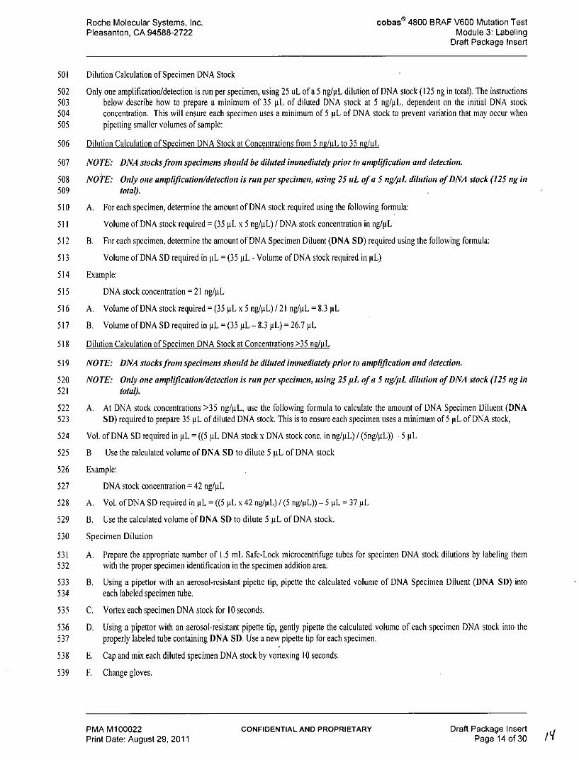
501 Dilution Calculation of Specimen DNA Stock
502 Only one amplification/detection is run per specimen, using 25 uL ofa 5 ng/pL dilution of DNA stock (125 ng in total). The instructions503 below describe how to prepare a minimum of 35 pL of diluted DNA stock at 5 ng/gL, dependent on the initial DNA stock504 concentration. This will ensure each specimen uses a minimum of 5 pL of DNA stock to prevent variation that may occur when505 pipetting smaller volumes of sample:
506 Dilution Calculation of Specimen DNA Stock at Concentrations from 5 ng/iL to 35 nF/iL
507 NOTE: DNA stocks front specimens should be diluted immediately prior to amplification and detection.
508 NOTE: Only one amplification/detection is run per specimen, using 25 uL of a 5 ng/uL dilution of DNA stock (125 ng in509 total).
510 A. For each specimen, determine the amount of DNA stock required using the following formula:
511 Volume of DNA stock required= (35 pL x 5 ng/gL) /DNA stock concentration in ng/pL
512 B. For each specimen, determine the amount of DNA Specimen Diluent (DNA SD) required using the following formula:
513 Volume of DNA SD required in pL = (35 gL - Volume of DNA stock required in pL)
514 Example:
515 DNA stock concentration = 21 ng/ML
516 A. Volume of DNA stock required = (35 ML x 5 ng/uL) / 21 ng/pL = 8.3 pL
517 B. Volume of DNA SD required in pL = (35 gL - 8.3 ML) = 26.7 pL
518 Dilution Calculation of Specimen DNA Stock at Concentrations >35 nduL
519 NOTE: DNA stocks from specimens should be diluted immediately prior to amplification and detection.
520 NOTE: Only one amplification/detection is run per specimen, using 25 pL of a 5 ng/pL dilution of DNA stock (125 ng in521 total).
522 A. At DNA stock concentrations >35 ng/pL, use the following formula to calculate the amount of DNA Specimen Diluent (DNA523 SD) required to prepare 35 pL of diluted DNA stock. This is to ensure each specimen uses a minimum of 5 pL of DNA stock,
524 Vol. of DNA SD required in pL = ((5 ML DNA stock x DNA stock conc. in ng/L) / (5ng/AL))- 5 pL
525 B Use the calculated volume of DNA SD to dilute 5 pL of DNA stock
526 Example:
527 DNA stock concentration = 42 ng/L
528 A. Vol. of DNA SD required in pL = ((5 ML x 42 ng/pL) / (5 ng/pL)) -5 pL = 37 pL
529 B. Use the calculated volume of DNA SD to dilute 5 ML of DNA stock.
530 Specimen Dilution
531 A. Prepare the appropriate number of 1.5 mL Safe-Lock microcentrifuge tubes for specimen DNA stock dilutions by labeling them532 with the proper specimen identification in the specimen addition area.
533 B. Using a pipettor with an aerosol-resistant pipette tip, pipette the calculated volume of DNA Specimen Diluent (DNA SD) into534 each labeled specimen tube.
535 C. Vortex each specimen DNA stock for 10 seconds.
536 D. Using a pipettor with an aerosol-resistant pipette tip, gently pipette the calculated volume of each specimen DNA stock into the537 properly labeled tube containing DNA SD. Use a new pipette tip for each specimen.
538 E. Cap and mix each diluted specimen DNA stock by vortexing 10 seconds.
539 F. Change gloves.
PMA M100022 CONFIDENTIAL AND PROPRIETARY Draft Package InsertPrint Date: August 29, 2011 Page 14 of 30 19
Roche Molecular Systems, Inc. cobas I4800 BRAF V600 Mutation TestPleasanton, CA 94588-2722 Module 3: Labeling
Draft Package Insert
540 Preparation of Working Master Mix (MMX)
541 NOTE: The BRA F Oligo Mix and working MMX are light-sensitive. All open mixtures of BRAF OM and working MMX542 should beprotectedfrom prolonged exposure to light
543 A. Calculate the volume of RXNMIX required using the following formula:
544 Volume of RXNMIX required = (Number of Specimens + 2 Controls + 1) x 10 ML
545 B. Calculate the volume of BRAF OM required using the following formula:
546 Volume of BRAF OM required = (Number of Specimens + 2 Controls + 1) x 8 pL
547 C. Calculate the volume of MGAC required using the following formula:
548 Volume of MCAC required = (Number of Specimens + 2 Controls + 1) x 7 pL
549 Table I may be used to determine volumes of each reagent needed for the preparation of working MMX based on the number of550 specimens included in the run.
551 Table )
Volumes of Reagents Needed for Working MMx# of Specimens*
553 D. Remove appropriate number of RXNMIX, BRAF OM and MGAC vials from 20 -8'C storage. Vortex each reagent for 5554 seconds to collect liquid at the bottom of the tube before use. Label a sterile microcentrifuge tube as MMX for the working Master555 Mix (MMX).
556 E. Add the calculated volume of RXNMIX to the labeled MMX tube.
557 F. Add the calculated volume of BRAF OM to the labeled MMX tube.
558 G. Add the calculated volume of MGAC to the labeled MMX tube.
559 H. Vortex tube for 5 seconds to assure adequate mixing.
560 NOTE: Use only cobas@ 4800 System Microwell Plate (AD-Plate) and Sealing Film (Roche P/N 05232724001)
561 1. Carefully add 25 uL of working MMX to each reaction well of the microwell plate (AD-plate) that is needed for the run. Do not562 allow the pipette tip to touch the plate outside that well.
563 Addition of Controls and Specimens
564 A. Add 25 uL of BRAF MUT Control to well A01 of the microwell plate (AD-plate) and mix well using pipettor to aspirate and565 dispense within the well a minimum of two times.
566 B. Using a new pipette tip, add 25 uL of BRAF WT Control to well 80 of the microwell plate (AD-plate) and mix well using567 pipettor to aspirate and dispense within the well a minimum of two times.
568 NOTE: Each run must contain both a BRAF MUT Control in position A01 and a BRA F WT Control in position B01 or569 the run will be invalidated by the cobas z 480 analyzer.
PMA M100022 CONFIDENTIAL AND PROPRIETARY Draft Package InsertPrint Date: August 29, 2011 Page 15 of 30
Roche Molecular Systems, Inc. cobas® 4800 BRAF V600 Mutation TestPleasanton, CA 94588-2722 Module 3: Labeling
Draft Package Insert
570 NOTE: Change gloves as needed to protect against specimen-to-specimen contamination and external PCR reaction tube571 contamination.
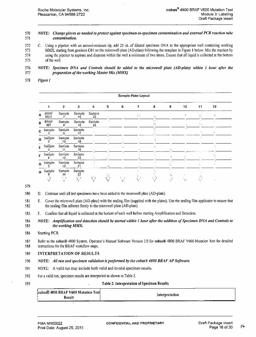
572 C. Using a pipettor with an aerosol-resistant tip, add 25 uL of diluted specimen DNA to the appropriate well containing working573 MMX, starting from position COI on the microwell plate (AD-plate) following the template in Figure I below. Mix the reaction by574 using the pipettor to aspirate and dispense within the well a minimum of two times. Ensure that all liquid is collected at the bottom575 of the well.
576 NOTE: Specimen DNA and Controls should be added to the microwell plate (AD-plate) within I hour after the577 preparation of the working Master Mi& (MMX)
578 Figure 1
Sample Plate Layout
1 2 3 4 5 6 7 8 9 10 11 12
A BRAF Sample Sample SampleMUT 7 15 23
B BRAF Sam ple Sample SampleWT 8 16 24
C Sample Sample Sample19 17
D Saiple Sample Sample2 10 18
E SaMple Sample Sample3 11 19
F Saniple Sample Sample4 12 20
G Sample Saimple Sample5 13 21
H Sample Sainple Sample6 14 22
V ' v v
579
580 D. Continue until all test specimens have been added to the microwell plate (AD-plate).
581 E. Cover the microwell plate (AD-plate) with the sealing film (supplied with the plates). Use the sealing film applicator to ensure that582 the sealing film adheres firmly to the microwell plate (AD-plate).
583 F. Confirm that all liquid is collected at the bottom of each well before starting Amplification and Detection.
584 NOTE: Amplification and detection should be started within I hour after tie addition of Specimen DNA and Controls to585 the working MMX
586 Starting PCR
587 Refer to the cobas@ 4800 System, Operator's Manual Software Version 2.0 for cobas@ 4800 BRAF V600 Mutation Test for detailed588 instructions for the BRAF workflow steps.
589 INTERPRETATION OF RESULTS
590 NOTE: All run and specimen validation is performed by the cobas@ 4800 BRAF AP Software.
591 NOTE: A valid run may include both valid and invalid specimen results.
592 For a valid run, specimen results are interpreted as shown in Table 2.
593 . Table 2: Interpretation of Specimen Results
cobas@ 4800 BRAF V600 Mutation Test InterpretationResult
PMA M100022 CONFIDENTIAL AND PROPRIETARY Draft Package InsertPrint Date: August 29, 2011 Page 16 of 30
Roche Molecular Systems, Inc. cobas® 4800 BRAF V600 Mutation TestPleasanton, CA 94588-2722 Module 3: Labeling
Draft Package Insert
Mutation Detected V600E Mutation Detected in the BRAF codon 600 site in exon 15
Mutation Not Detected* V600E Mutation Not Detected in the BRAF codon 600 site in exon 15
Result is invalid.Invalid Repeat the testing of specimens with invalid results following the instructions outlined in
the "Retesting of Specimens with Invalid Results" section below.
Failed Failed run due to hardware or software failure
594 * A Mutation Not Detected result does not preclude the presence of a mutation in the BRAF codon 600 site since results depend595 on percent mutant sequences, adequate specimen integrity, absence of inhibitors, and sufficient DNA to be detected.
596 All of the patients in the phase III study N025026 (BRIM3) sponsored by Hoffmann-La Roche were selected using the597 cobas@ 4800 BRAF V600 Mutation Test. Enrollment in the study was limited to patients whose melanoma tissue tested598 positive by the test. Therefore it is not known whether patients who test negative by the cobas@ 4800 BRAF V600599 Mutation Test will benefit from vemurafenib (Zelboraf TM) treatment.
600
601 Retesting of Specimens with Invalid Results
602 A. Repeat dilution of the invalid specimen DNA stock starting from "Dilution Calculation of Specimen DNA Stock" and603 "Specimen Dilution" procedures in the "AMPLIFICATION and DETECTION" section.
604 Note: If there is not enough specimen DNA stock remaining to perform a new dilution of the DNA stock, obtain a new605 5-ium section of tissue and start with the "Deparaffinization of FFPET Sections Mounted on Slides" or606 "Deparaffinization of FFPET Sections not Mounted on Slides" procedure, then: proceed with Step B below.
607 B. After performing the DNA stock dilution to 5 ng/pL described in "Specimen Dilution", perform an additional 1:2 dilution by608 taking 20 ML of the diluted DNA stock and adding 20 ML of DNA Specimen Diluent (DNA SD).
609 C. Continue with "Preparation of Working Master Mix (MMx)" and the remainder of the amplification and detection procedure.
610 Note: If the specimen remains invalid after retesting at a 1:2 dilution, repeat the entire test procedure for that specimen,611 starting with Deparaffinization and DNA Isolation using a new 5-jim FFPET section. The standard 25 pL of612 DNA at 5 ng/uL (without further dilution) should be used for amplification and detection.
613 QUALITY CONTROL
614 The cobas® 4800 BRAF V600 Mutation Test Mutant (BRAF MUT) Control and Wild-type (BRAF WT) Control are included in615 each run. A run is valid if both the BRAF MUT Control well (A01) and the BRAF WT Control well (BOI) have a valid control616 status. In order for a mutant control to be called valid, the Ct value needs to be between 23.5 to 30.5 in the mutant channel, and between617 23.5 to 29.0 in the wild-type channel. In order for a wild-type control to be called valid, the Ct value needs to be >43 in the mutant618 channel and between 31.5 to 38.5 in the wild-type channel. If either the BRAF MUT Control or BRAF WT Control is invalid, the619 run must be repeated. Prepare a fresh dilution of the previously isolated specimen DNA stock to set up a new microwell plate (AD-plate)620 with controls for amplification and detection.
621 BRAF Mutant Control
622 The BRAF MUT Control result must be 'Valid'. If the BRAF MUT Control results are consistently invalid, contact your local Roche623 office for technical assistance.
624 BRAF Wild-Type Control
625 The BRAF WT Control result must be 'Valid'. If the BRAF WT Control results are consistently invalid, contact your local Roche626 office for technical assistance.
627 PROCEDURAL PRECAUTIONS
628 As with any test procedure, good laboratory technique is essential to the proper performance of this assay. Due to the high analytical629 sensitivity of PCR-based tests, care should be taken to keep reagents and amplification mixtures free of contamination.
630 PROCEDURAL LIMITATIONS
PMA M100022 CONFIDENTIAL AND PROPRIETARY Draft Package InsertPrint Date: August 29, 2011 Page 17 of 30
Roche Molecular Systems, Inc. cobas" 4800 BRAF V600 Mutation TestPleasanton, CA 94588-2722 Module 3: Labeling
Draft Package Insert
631 1. Test only the indicated specimen types. The cobas® 4800 BRAF V600 Mutation Test has been validated for use only with632 melanoma FFPET specimens.
633 2. The cobas® 4800 BRAF V600 Mutation Test has been validated using only the cobas® DNA Sample Preparation Kit (Roche P/N:634 05985536190) to extract genomic DNA.
635 3. Detection of a mutation is dependent on the number of mutant sequence copies present in the specimen and may be affected by636 specimen integrity, amount of isolated DNA, and the presence of interfering substances.
637 4. Reliable results are dependent on adequate specimen fixation, transport, storage and processing. Follow the procedures in this638 Package Insert and in the cobaso 4800 System Operator's Manual.
639 5. The addition of AmpErase enzyme into the cobas® 4800 BRAF V600 Master Mix enables selective amplification of target DNA;640 however, good laboratory practices and careful adherence to the procedures specified in this Package Insert are necessary to avoid641 contamination of reagents.
642 6. Use of this product must be limited to personnel trained in the techniques of PCR and the use of the cobas® 4800 System v2.0.
643 7. Only the cobas® 4800 System v2.0 has been validated for use with this product. No other PCR system has been validated with this644 product.
645 8. Though rare, mutations and variants within the regions of the BRAF gene covered by the primers or probes used in the cobas®646 4800 BRAF V600 Mutation Test may result in failure to amplify the BRAF V600 allele or detect the presence of mutation in647 codon 600.
648 9. The presence of PCR inhibitors may cause false negative or invalid results.
649 10. Melanin is a known inhibitor of PCR reactions. The DNA sample preparation kit removes melanin from the specimen during650 extraction; however, melanin in a specimen may still cause invalid results. If melanin inhibition is suspected, repeat testing using a651 1:2 dilution is suggested as described in "Retesting of Specimens with Invalid Results".
652 11. The cobas® 4800 BRAF V600 Mutation Test shows limited cross-reactivity with non-V600E mutant specimens (V600K, V600D,653 and V600E2). Refer to the Non-clinical Performance Evaluation section for more details.
654 12. FFPET specimens containing degraded DNA may affect the ability of the test to detect the V600E mutation.
655 13. The cobas 4800 BRAF mutation test is a qualitative test. The test is not for quantitative measurements of mutation.
656 NON-CLINICAL PERFORMANCE EVALUATION
657 For the nonclinical studies described below, % tumor was assessed by pathology review and melanin content was assessed658 by pathology review. Bi-directional Sanger sequencing was used to select the specimens for testing. The % mutation was659 determined using a parallel sequencing method.
660 Analytical Sensitivity - Limit of Blank (LoB)
661 To assess performance of the cobas 4800 BRAF V600 Mutation Test in the absence of template and to ensure that a blank662 sample, or a sample with an excess of 100% wild-type DNA, does not generate an analytical signal that might indicate a663 low concentration of mutation, samples with no template and 100% BRAF wild-type DNA were evaluated. No detectable664 Ct results were identified in the mutant channel in the presence of 100% BRAF wild-type DNA or in either channel665 following a no template sample. Additionally, no detectable Ct was identified in the wild-type channel when testing a666 100% BRAF V600E sample.
667 Analytical Sensitivity- Limit of Detection (LoD)
668 The minimum amount of input DNA that produces correct results 95% of the time was assessed using dilution panels prepared from669 three types of specimens:
670 * Specimen blends prepared by mixing DNA stocks obtained from BRAF V600E mutant FFPET specimens and BRAF wild-type671 FFPET specimens to achieve specific mutation levels.
672 * Individual FFPET DNA stocks prepared from three BRAF V600E mutant FFPET specimens.
673 * Cell line blend prepared by mixing DNA stocks obtained from a BRAF V600E mutant cell line and a BRAF wild-type cell line.
PMA M100022 CONFIDENTIAL AND PROPRIETARY Draft Package InsertPrint Date: August 29, 2011 Page 18 of 30
Roche Molecular Systems, Inc. cobass 4800 BRAF V600 Mutation TestPleasanton, CA 94588-2722 Module 3: Labeling
Draft Package Insert
674 Analytical Sensitivity Usina Specimen Blends
675 BRAF V600E mutant FFPET specimen DNA stocks were blended with BRAF wild-type FFPET specimen DNA stocks to achieve one676 specimen at -10%, three specimens at -5%, and one specimen at -3% mutation level. One BRAF wild-type specimen was also tested.677 Each of the five specimen blends with V600E mutation (but not the wild-type specimen) was then diluted to produce panel members at678 a range of DNA concentrations (125 ng to 0.3 ng in a 25 uL volume).
679 Eight (8) replicates of each panel member were run using each of 3 cobas® 4800 BRAF V600 Mutation Test kit lots (n=24/panel680 member). This study demonstrated that in FFPET specimen blends, the cobas@ 4800 BRAF V600 Mutation Test can detect the BRAF681 V600E mutation at >5% mutation level using the standard input of 125 ng/25 ML. The -10% blend correctly called all replicates at a 31682 ng/25 ML dilution. The 3% FFPET blend at 125 ng/25 pL was not detected.
683 Analytical Sensitivity Using FFPET Specimens
684 To confirm the 5% claim in patient specimens, forty-eight individual 5 pm sections from each of 3 BRAF V600E mutant FFPET685 specimens containing 5%, 6%, and 12%, mutation levels were individually processed using 3 lots of cobaso DNA Sample Preparation686 Kit to isolate the DNA. To assess the impact of melanin on the assay, one specimen (6% mutation) had a high melanin concentration.687 Serial dilutions of the DNA from each section were prepared to produce a set of 6 panel members at a range of DNA concentrations688 (125 ng to 0.5 ng in a 25 pL volume).
689 Sixteen (16) replicates of each panel member were run using each of 3 cobas® 4800 BRAF V600 Mutation Test kit lots (n=48/panel690 member).
691 The study demonstrated that the cobas® 4800 BRAF V600 Mutation Test can detect the BRAF V600E mutation in actual clinical692 FFPET specimens at 5% mutation level using the standard input of 125 ng/25 pL.
693 Analytical Sensitivity Using Cell Line Blend
694 DNA stocks from two melanoma cell lines [SK-MEL 28 (BRAF V600E mutant) and SK-MEL 2 (BRAF wild-type)] were blended to695 achieve a sample at 5% mutation. Three separate dilution panels containing from 125 ng/25 pL to 0.5 ng/25 pL DNA were prepared.696 Twenty (20) replicates of each panel member were tested, using each of 3 cobas® 4800 BRAF V600 Mutation Test kit lots (60697 replicates total). Sensitivity was determined by the lowest amount of DNA that gave a BRAF V600E "Mutation Detected" rate of at698 least 95%. The results of the study are shown in Table 3 below.
PMA M100022 CONFIDENTIAL AND PROPRIETARY Draft Package InsertPrint Date: August 29, 011 Page 19 of 30
Roche Molecular Systems, Inc. cobas® 4800 BRAF V600 Mutation TestPleasanton, CA 94588-2722 Module 3: Labeling
Draft Package Insert
699 Table 3:700 Sensitivity of the cobas@ 4800 BRA F V600 Mutation Test using Cell Line Blend
Mean Percent Amount of DNA in the Panel "Mutation Detected" RateMutation Member (n=60)
125.0 ng/25 gL 97%
31.3 ng/25 pL 100%
15.6ng/25 pL 95%
Cell Line Blend 5% 7.8 ng/25 pL 98%
3.9 ng/25 gL 95%
2.0 ng/25 pL 82%
1.0 ng/25 gL 78%
0.5 ng/25 pL 77%
701
702 The Test gave a 95% "Mutation Detected" rate at 3.9 ng/25pL, which represents a 1:32 dilution of the recommended DNA input of 125703 ng/25pL.
704 Genomic Input Range:
705 The recommended DNA input for the cobas@ 4800 BRAF V600 Mutation Test is 125 ng. Various genomic DNA input amounts may706 result from DNA quantitation errors and/or variation in the amount of degraded DNA. To evaluate the effects of various genomic DNA707 input amounts, genomic DNA was extracted from II melanoma FFPET specimens, selected for their mutation status and level of708 pigmentation, and serially diluted with sample input representing 250 ng, 125 ng, 62.5 ng, and 31.3 ng/ 25 ML. All 4 DNA levels were709 evaluated using 2 lots. The expected results were obtained for all genomic DNA input levels.
710 Minimal Tumor Content
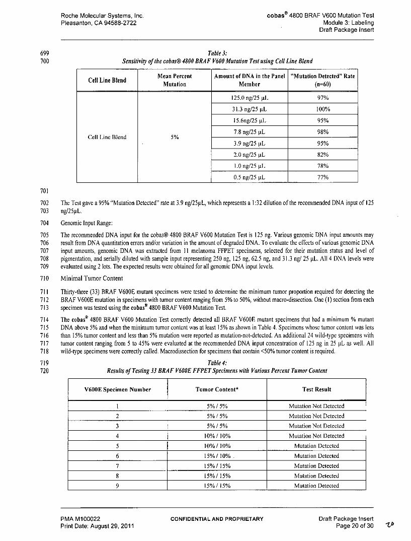
711 Thirty-three (33) BRAF V600E mutant specimens were tested to determine the minimum tumor proportion required for detecting the712 BRAF V600E mutation in specimens with tumor content ranging from 5% to 50%, without macro-dissection. One (1) section from each713 specimen was tested using the cobas® 4800 BRAF V600 Mutation Test.
714 The cobas® 4800 BRAF V600 Mutation Test correctly detected all BRAF V600E mutant specimens that had a minimum % mutant715 DNA above 5% and when the minimum tumor content was at least 15% as shown in Table 4. Specimens whose tumor content was less716 than 15% tumor content and less than 5% mutation were reported as mutation-not-detected. An additional 24 wild-type specimens with717 tumor content ranging from 5 to 45% were evaluated at the recommended DNA input concentration of 125 ng in 25 pL as well. All718 wild-type specimens were correctly called. Macrodissection for specimens that contain <50% tumor content is required.
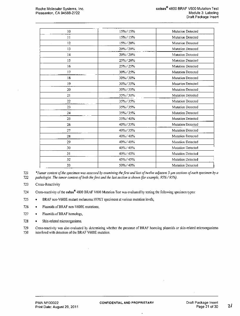
719 Table 4:720 Results of Testing 33 BRAF V600E FFPET Specimens with Various Percent Tumor Content
V600E Specimen Number Tumor Content* Test Result
1 5%1 5% Mutation Not Detected
2 5% / 5% Mutation Not Detected
3 5% / 5% Mutation Not Detected
4 10% / 10% Mutation Not Detected
5 10% / 10% Mutation Detected
6 15%/ 10% . Mutation Detected
7 15%/ 15% Mutation Detected
8 15%! 15% Mutation Detected
9 15%1 15% Mutation Detected
PMA M100022 CONFIDENTIAL AND PROPRIETARY Draft Package InsertPrint Date: August 29, 2011 Page 20 of 30
Roche Molecular Systems, Inc. cobasI 4800 BRAF V600 Mutation TestPleasanton, CA 94588-2722 Module 3: Labeling
Draft Package Insert
10 15% / 15% Mutation Detected
11 15% / 15% Mutation Detected
12 15% / 20% Mutation Detected
13 20% / 20% Mutation Detected
14 20% / 20% Mutation Detected
I5 25% /20% Mutation Detected
16 25%/25% Mutation Detected
17 30% / 25% Mutation Detected
18 30%/30% Mutation Detected
19 30%/35% Mutation Detected
20 30% /35% Mutation Detected
21 35%130% Mutation Detected
22 35% / 35% Mutation Detected
23 35% / 35% Mutation Detected
24 35% /35% Mutation Detected
25 35% / 40% Mutation Detected
26 40% / 35% Mutation Detected
27 40% / 35% Mutation Detected
28 40% / 40% Mutation Detected
29 40% / 40% Mutation Detected
30 40% / 40% Mutation Detected
31 40% / 45% Mutation Detected
32 45% / 45% Mutation Detected
33 50%/40% Mutation Detected
721 *Tumor content of the specimen was assessed by examining the first and last oftwelve adjacent 5-pm sections ofeach specimen by a722 pathologist. The tumor content of both the first and the last section is shown (for example, 95% / 95%).
723 Cross-Reactivity
724 Cross-reactivity of the cobaso 4800 BRAF V600 Mutation Test was evaluated by testing the following specimen types:
725 * BRAF non-V600E mutant melanoma FFPET specimens at various mutation levels,
726 * Plasmids of BRAF non-V600E mutations,
727 * Plasmids of BRAF homologs,
728 * Skin-related microorganisms.
729 Cross-reactivity was also evaluated by determining whether the presence of BRAF homolog plasmids or skin-related microorganisms730 interfered with detection of the BRAF V600E mutation.
PMA M100022 CONFIDENTIAL AND PROPRIETARY Draft Package InsertPrint Date: August 29, 2011 Page 21 of 30
Roche Molecular Systems, Inc. cobass 4800 BRAF V600 Mutation TestPleasanton, CA 94588-2722 Module 3: Labeling
Draft Package Insert
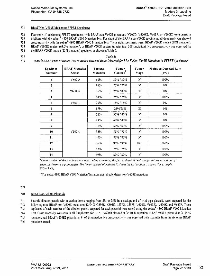
731 BRAF Non-V600E Melanoma FFPET Specimens
732 Fourteen (14) melanoma FFPET specimens with BRAF non-V600E mutations (V600D, V600E2, V600R, or V600K) were tested in733 triplicate with the cobas® 4800 BRAF V600 Mutation Test. For eight of the BRAF non-V600E specimens, all three replicates showed734 cross-reactivity with the cobas® 4800 BRAF V600 Mutation Test. These eight specimens were: BRAF V600D mutant (18% mutation),735 BRAF V600E2 mutant (68.0% mutation), or BRAF V600K mutant (greater than 30% mutation). No cross-reactivity was observed for736 the BRAF V600R mutant (23% mutation) specimen as shown in Table 5.
737 Table 5:738 cobas@ BRAF V600 Mutation Test Mutation Detected Rates Observedfor BRAF Non- V600E Mutations in FFPET Specimens*
aTumor content of the specimen was assessed by examining the first and last oftwelve adjacent 5-pm sections ofeach specimen by a pathologist The tumor content of both the first and the last section is shown (for example,95%/95%).*The cobas 4800 BRAF V600 Mutation Test does not reliably detect non-V600E mutations
739
740 BRAF Non-V600E Plasmids
741 Plasmid dilution panels with mutation levels ranging from 5% to 75% in a background of wild-type plasmid, were prepared for the742 following nine BRAF non-V600E mutations: D594G, G596R, K601E, L597Q, L597S, V600D, V600E2, V600K, and V600R. Three743 replicates of each member of the dilution panels prepared for each plasmid were tested using the cobas® 4800 BRAF V600 Mutation744 Test. Cross-reactivity was seen in all 3 replicates for BRAF V600D plasmid at > 10 % mutation, BRAF V600K plasmid at ) 35 %745 mutation, and BRAF V600E2 plasmid at 65 % mutation. No cross-reactivity was observed with plasmids from the six other BRAF746 mutations tested.
PMA M100022 CONFIDENTIAL AND PROPRIETARY Draft Package InsertPrint Date: August 29, 2011 Page 22 of 30 2-
Roche Molecular Systems, Inc. cobass 4800 BRAF V600 Mutation TestPleasanton, CA 94588-2722 Module 3: Labeling
Draft Package Insert
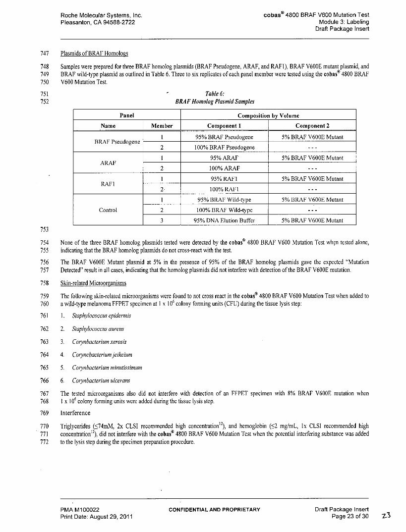
747 Plasmids of BRAF Homologs
748 Samples were prepared for three BRAF homolog plasmids (BRAF Pseudogene, ARAF, and RAFI), BRAF V600E mutant plasmid, and749 BRAF wild-type plasmid as outlined in Table 6. Three to six replicates of each panel member were tested using the cobas® 4800 BRAF750 V600 Mutation Test.
754 None of the three BRAF homolog plasmids tested were detected by the cobas® 4800 BRAF V600 Mutation Test when tested alone,755 indicating that the BRAF homolog plasmids do not cross-react with the test.
756 The BRAF V600E Mutant plasmid at 5% in the presence of 95% of the BRAF homolog plasmids gave the expected "Mutation757 Detected" result in all cases, indicating that the homolog plasmids did not interfere with detection of the BRAF V600E mutation.
758 Skin-related Microorganisms
759 The following skin-related microorganisms were found to not cross react in the cobas® 4800 BRAF V600 Mutation Test when added to760 a wild-type melanoma FFPET specimen at I x 10' colony forming units (CFU) during the tissue lysis step:
761 1. Staphylococcus epidermis
762 2. Staphylococcus aureus
763 3. Corynbacterium xerosis
764 4. Corynebacterium jeikeium
765 5. Corynbacterium minutissimum
766 6. Corynbacterium ulcerans
767 The tested microorganisms also did not interfere with detection of an FFPET specimen with 8% BRAF V600E mutation when768 1 x 10' colony forming units were added during the tissue lysis step.
769 Interference
770 Triglycerides (574mM, 2x CLSI recommended high concentration'), and hemoglobin (52 mg/mL, lx CLSI recommended high771 concentration 2), did not interfere with the cobaso 4800 BRAF V600 Mutation Test when the potential interfering substance was added772 to the lysis step during the specimen preparation procedure.
PMA M100022 CONFIDENTIAL AND PROPRIETARY Draft Package InsertPrint Date: August 29, 2011 Page 23 of 30 7-
Roche Molecular Systems, Inc. cobas" 4800 BRAF V600 Mutation TestPleasanton, CA 94588-2722 Module 3: Labeling
Draft Package Insert
773 Necrotic tissue
774 The ability of the cobas@ 4800 BRAF V600 Mutation Test to perform correctly when samples have high necrotic tissue content was775 tested using 27 V600E or WT melanoma FFPET specimens that contained from 10% to 95% necrotic tissue. Samples were selected776 based on their BRAF V600E mutation status, necrotic tissue content, and percent mutation. Of the 27 specimens selected, II were777 BRAF V600E mutant, and 16 were BRAF wild-type. The percent mutation ranged from 4% to 65%. The tumor content and percentage778 of necrotic tissue in each specimen was reviewed by a pathologist. Samples were run in duplicate using I lot of reagents. For the 11779 BRAF V600E specimens, mutation detected results were obtained for all samples except one whose % mutation was below the780 threshold of detection (4%). The specimen with 95% necrotic tissue whose % mutation was 10% was correctly called mutation detected.781 Mutation not detected results were obtained for all wild-type specimens.
782 Table 7:783 Summary ofcobas 4800 BRA F V600 Mutation Test Performance with High Necrotic Melanoma FFPET Specimens
Specimen % Necrosis Test Results (n = 2)
15% Mutation Detected/Mutation Detected
15% Mutation Detected/Mutation Detected
20% Mutation Detected/Mutation Detected
25% Mutation Detected/Mutation Detected
25% Mutation Detected/Mutation Detected
V600E 30% Mutation Detected/Mutation Detected
30% Mutation not Detected/Mutation not Detected
55% Mutation Detected/Mutation Detected
55% Mutation Detected/Mutation Detected
60% Mutation Detected/Mutation Detected
95% Mutation Detected/Mutation Detected
784
785 Melanin
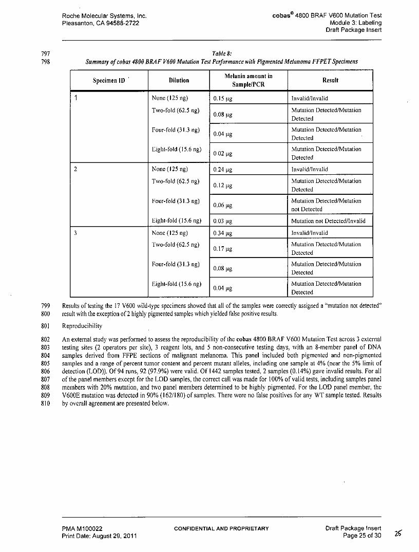
786 The impact of high concentrations of endogenous melanin was evaluated using highly pigmented melanoma FFPET samples. A total of787 41 unique FFPET melanoma tumor tissue specimens were selected based upon their level of pigmentation: 33 were highly pigmented, 3788 were from African Americans and 5 were lightly pigmented for comparison. DNA was extracted from the tissue and melanin789 concentration was determined for each sample. A single replicate of the DNA stock from each of the two sections obtained from each of790 the 41 specimens was tested. Three specimens produced invalid results. One specimen produced a mutation not detected result but this791 specimen was determined to be below the limit of detection. The 3 specimens with "Invalid," results were used to prepare the792 recommended concentration of DNA for the test as well as two-fold, four-fold, and eight-fold dilutions of the recommended DNA input793 of 125 ng/PCR.
794 The resulting diluted DNA samples (containing a total of 125 ng, 61.5 ng, 31.25 ng, or 15.6 ng DNA in the 25 pL) were retested to795 determine if the corresponding reduction in melanin by dilution allowed valid results to be obtained. All three specimens when diluted796 2-fold yielded the correct results.
PMA M100022 CONFIDENTIAL AND PROPRIETARY Draft Package InsertPrint Date: August 29, 2011 Page 24 of 30
Roche Molecular Systems, Inc. cobas" 4800 BRAF V600 Mutation TestPleasanton, CA 94588-2722 Module 3: Labeling
Draft Package Insert
797 Table 8:798 Summary of cobas 4800 BRAF V600 Mutation Test Performance with Pigmented Melanoma FFPET Specimens
Specimen ID Dilution Melanin amount in ResultSample/PCR
799 Results of testing the 17 V600 wild-type specimens showed that all of the samples were correctly assigned a "mutation not detected"800 result with the exception of 2 highly pigmented samples which yielded false positive results.
801 Reproducibility
802 An external study was performed to assess the reproducibility of the cobas 4800 BRAF V600 Mutation Test across 3 external803 testing sites (2 operators per site), 3 reagent lots, and 5 non-consecutive testing days, with an 8-member panel of DNA804 samples derived from FFPE sections of malignant melanoma. This panel included both pigmented and non-pigmented805 samples and a range of percent tumor content and percent mutant alleles, including one sample at 4% (near the 5% limit of806 detection (LOD)). Of 94 runs, 92 (97.9%) were valid. Of 1442 samples tested, 2 samples (0.14%) gave invalid results. For all807 of the panel members except for the LOD samples, the correct call was made for 100% of valid tests, including samples panel808 members with 20% mutation, and two panel members determined to be highly pigmented. For the LOD panel member, the809 V600E mutation was detected in 90% (162/180) of samples. There were no false positives for any WT sample tested, Results810 by overall agreement are presented below.
PMA M100022 CONFIDENTIAL AND PROPRIETARY Draft Package InsertPrint Date: August 29, 2011 Page 25 of 30
Roche Molecular Systems, Inc. cobass 4800 BRAF V600 Mutation TestPleasanton, CA 94588-2722 Module 3: Labeling
Draft Package Insert
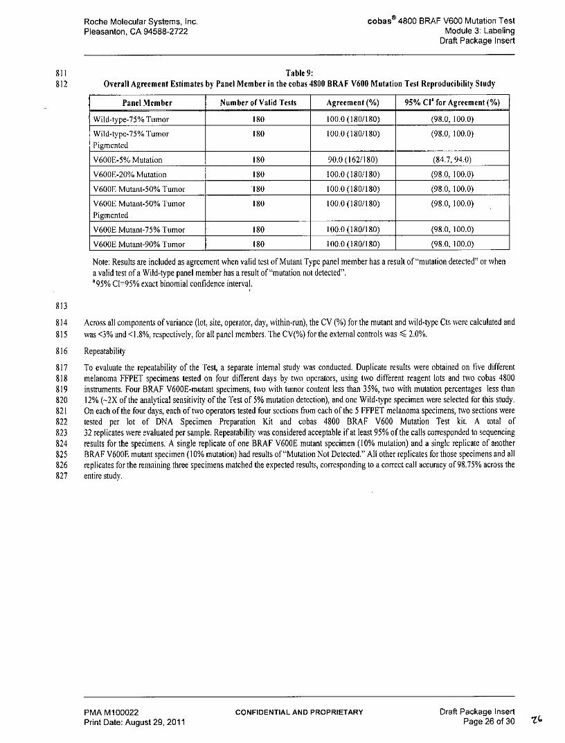
811 Table 9:812 Overall Agreement Estimates by Panel Member in the cobas 4800 BRAF V600 Mutation Test Reproducibility Study
Panel Member Number of Valid Tests Agreement (%) 95% CI' for Agreement (%)
Note: Results are included as agreement when valid test of Mutant Type panel member has a result of "mutation detected" or whena valid test of a Wild-type panel member has a result of "mutation not detected".'95% CI=95% exact binomial confidence interval.
813
814 Across all components of variance (lot, site, operator, day, within-run), the CV (%) for the mutant and wild-type Cts were calculated and815 was <3% and <1.8%, respectively, for all panel members. The CV(%) for the external controls was < 2.0%.
816 Repeatability
817 To evaluate the repeatability of the Test, a separate internal study was conducted. Duplicate results were obtained on five different818 melanoma FFPET specimens tested on four different days by two operators, using two different reagent lots and two cobas 4800819 instruments. Four BRAF V600E-mutant specimens, two with tumor content less than 35%, two with mutation percentages less than820 12% (-2X of the analytical sensitivity of the Test of 5% mutation detection), and one Wild-type specimen were selected for this study.821 On each of the four days, each of two operators tested four sections from each of the 5 FFPET melanoma specimens, two sections were822 tested per lot of DNA Specimen Preparation Kit and cobas 4800 BRAF V600 Mutation Test kit. A total of823 32 replicates were evaluated per sample. Repeatability was considered acceptable if at least 95% of the calls corresponded to sequencing824 results for the specimens. A single replicate of one BRAF V600E mutant specimen (10% mutation) and a single replicate of another825 BRAF V600E mutant specimen (10% mutation) had results of "Mutation Not Detected." All other replicates for those specimens and all826 replicates for the remaining three specimens matched the expected results, corresponding to a correct call accuracy of 98.75% across the827 entire study.
PMA M100022 CONFIDENTIAL AND PROPRIETARY Draft Package InsertPrint Date: August 29, 2011 Page 26 of 30
Roche Molecular Systems, Inc. cobasa 4800 BRAF V600 Mutation TestPleasanton, CA 94588-2722 Module 3: Labeling
Draft Package Insert
828 Table 10:829 Repeatability of the cobas 4800 BRAF V600 Mutation Test
Panel Member Total no. Total no %(% mutation) replicates tested incorrect calls accuracy
V600E (10%) 60%/55% IV 32 1 96.9
V600E (12%) 40%/35% 11113 32 0 100
V600E (10%) 30% / 30% IV 32 1 96.9
V600E (17%) 30%/35% II 32 0 100
WT 60%/60% IV 32 0 100
830 Tumor content of the specimen was assessed by examining the first and last of twelve adjacent 5-nm sections of each specimen by a831 pathologist. The tumor content of both the first and the last section is shown (for example, 95% / 95%).
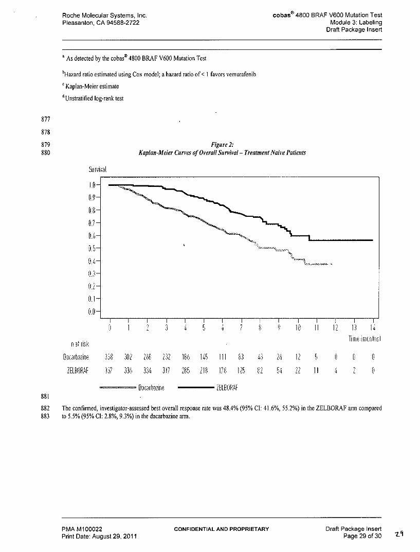
832 Correlation to Reference Method for Phase Ill Clinical Samples:
833 To evaluate the performance of the cobas@ 4800 BRAF V600 Mutation test when compared to 2X bi-directional sequencing, 596834 consecutive patients screened for the Phase III trial were identified for whom clinical, demographic, and Sanger sequencing data835 were collected. Of these cases, 94 were ineligible because of missing inclusion criteria, 4 cases were836 without pathology review, 2 cases had invalid cobas test results and 47 specimens had invalid Sanger sequencing results leaving 449837 evaluable cases. The agreement analysis between the cobas test results and Sanger sequencing results for the detection of the838 V600E mutation is shown in Table 21 below. The Positive Percent Agreement (PPA) with Sanger sequencing was 97.3% (216/222),839 and the Negative Percent Agreement (NPA) was 84.6% (192/227), with an overall Percent Agreement of 90.9%. There were a total840 of 35 mutations detected by the cobas test which were not identified as V600E mutations by Sanger sequencing. Eight (8) of these841 were wild-type, 25 were V600K and 2 were other codon 600 mutations by Sanger sequencing. Additionally, 6 specimens were842 identified as Mutation Not Detected by the cobas test but were identified as V600E by Sanger sequencing. The cross-reactivity of843 the cobas test for V600K was 66% (25/38).
844 Table 11:845 BRA F V600 Mutation Test vs. Sanger Sequencing for Specimens from Phase Ill Study*
Cobas 4800 BRAF Sanger Sequencing (Reference Method)Mutation Test Result BRAF V600E BRAF V600E Mutation Not Detected Total
MutationDetected
V600E V600K V600E2 V600R V600D** V600 Wild-Other*** type
Overall Percent Agreement 408/449 X 100% = 90.9% (87.8%, 93.2%)
847
848 *Sanger sequencing has a limit of detection of approximately 20% mutant alleles in FFPET specimens13 ,4 ,s,16 . Therefore, Sanger849 sequencing cannot be adequately used to confirm mutation status at lower percentages of mutant alleles. Consistent with this, in an850 exploratory analysis, some samples that were negative by Sanger sequencing and positive by cobas tested positive by a third method.851 The cobas test has a validated limit of detection in FFPET of 5% mutant alleles.
PMA M100022 CONFIDENTIAL AND PROPRIETARY Draft Package InsertPrint Date: August 29, 2011 Page 27 of 30
Roche Molecular Systems, Inc. cobaso 4800 BRAF V600 Mutation TestPleasanton, CA 94588-2722 Module 3: Labeling
Draft Package Insert
852 **V600D variant (GTG>GAC)853 ***Mutation not specified in the protocol (ACA insertion immediately before Codon 600)
854 The estimated prevalence of the V600E mutation in the Phase Ill clinical trial was 50 to 55% based on results with the cobas test.855 This is consistent with the prevalence of V600E in melanoma patients as reported in literature [3].
856 All of the patients in the phase Ill study N025026 (BRIM3) sponsored by Hoffmann-La Roche were selected using the cobas@857 4800 BRAF V600 Mutation Test. Enrollment in the study was limited to patients whose melanoma tissue tested positive by the858 test. Therefore it is not known whether patients who test negative by the cobas@ 4800 BRAF V600 Mutation Test will benefit from859 vemurafenib (Zelboraf TM) treatment.
860 Vemurafenib Clinical Efficacy
861 The cobas@ 4800 BRAF V600 Mutation Test was used as a companion test for selecting patients for treatment with vemurafenib862 (Zelborafrm). The clinical safety and effectiveness of vemurafenib (Zelborafrm) was demonstrated in the N025026 study (BRIM3),863 an international, randomized, open-label, controlled, multicenter, Phase III study in previously untreated patients with unresectable864 stage IIIC or stage IV melanoma with V600E BRAF mutation to evaluate the efficacy of vemurafenib (RO5185426) versus865 dacarbazine. Formalin-fixed paraffin-embedded tissue (FFPET) samples from all melanoma patients being considered for treatment866 were tested with the cobas@ 4800 BRAF V600 Mutation Test. Patients with a test positive result for the mutation were eligible for867 enrollment in the drug trials if they met other eligibility criteria. Patients with a negative test result were ineligible for drug trial868 enrollment. The study was conducted at approximately 104 sites (22 centers in the US).
869 The trial enrolled 675 patients; 337 were allocated to receive ZELBORAF and 338 to receive dacarbazine. The major efficacy outcome870 measures of the trial were overall survival (OS) and investigator-assessed progression-free survival (PFS). Other outcome measures871 included confirmed investigator-assessed best overall response rate.
872 Baseline characteristics were balanced between treatment groups. Most patients were male (56%) and Caucasian (99%), the median age873 was 54 years (24% were ? 65 years), all patients had ECOG performance status of 0 or 1, and the majority of patients had metastatic874 disease (95%).
875 Table 12:876 Efficacy of Veturafenib in Treatment-Naive Patients with BRA FV600E Mutation-Positive Melanoma
Vemurafenib Dacarbazine 1I_ (N=337) (N=338)
Overall Survival
Number of Deaths 78(23%) 121 (36%)
Hazard Ratio 0.44 0.000195% CI)' (0.33, 0.59)
Median Survival (months) Not Reached 7.995 % Cl) (9.6, Not Reached) (7.3. 9.6)
Median Follow-up (months) 6.2 4.5range) (0.4, 13.9) (<0.1, 11.7)
Progression-free survival Hazard Ratio 0.26 <0.0001(95% Cl)' (0.20, 0.33)
Median PFS (months) c I 64.9, 6.6) (1.6, 1.7)
PMA M100022 CONFIDENTIAL AND PROPRIETARY Draft Package InsertPrint Date: August 29, 2011 Page 28 of 30
Roche Molecular Systems, Inc. cobas® 4800 BRAF V600 Mutation TestPleasanton, CA 94588-2722 Module 3: Labeling
Draft Package Insert
a As detected by the cobas' 4800 BRAF V600 Mutation Test
bHazard ratio estimated using Cox model; a hazard ratio of < I favors vemurafenib
882 The confirmed, investigator-assessed best overall response rate was 48.4% (95% Cl: 41.6%, 55.2%) in the ZELBORAF arm compared883 to 5.5% (95% Cl: 2.8%, 9.3%) in the dacarbazine arm.
PMA M100022 CONFIDENTIAL AND PROPRIETARY Draft Package InsertPrint Date: Auaust 29, 2011 Pae 29 of 30
Roche Molecular Systems, Inc. cobas" 4800 BRAF V600 Mutation TestPleasanton, CA 94588-2722 Module 3: Labeling
Draft Package Insert
884 REFERENCES
885 1. Davies H, Bignell GR, Cox C, et al. Mutations of the BRAF gene in human cancer. Nature 2002; 417:949-54.
886 2. McCubrey JA, Steelman LS, Chappell WH, et al. Roles of the Raf/MEK/ERK pathway in cell growth,887 malignant transformation and drug resistance. Biochim Biophys Acta 2007; 1773:1263-1284.
888 3. Curtin JA, Fridlyand J, Kageshita T, et al. Distinct sets of genetic alterations in melanoma. N Engl I Med.889 2005;353:2135-2147.
890 4. Pollock PM, Harper UL, Hansen, KS, et al. High frequency of BRAF mutations in nevi. Nature Genetics 2003; 33:19-20.
892 6. Flaherty KT, Puzanov I, Kim KB, et al, Inhibition of mutated, activated BRAF in metastatic melanoma. N EngI J Med. 2010893 Aug 26; 363(9):809-819
894 7. Chapman PB, Hauschild A, Robert C, et al. Improved survival with vemurafenib in melanoma with BRAF V600E mutation. N895 Engl J Med. 2011;364:2507-2516.
896 8 Longo, M.C., Berninger, M.S. and Hartley, J.L. 1990. Use of uracil DNA glycosylase to control carry-over contamination in897 polymerase chain reactions. Gene. 93:125-128.
898 9. Chosewood, L.C and Wilson, D.E. Biosafety and Microbiological and Biomedical Laboratories. HHS Publication Fifth edition.899 (CDC) 21-1112. 2009
900 10. Clinical and Laboratory Standards Institute (CLSI). Protection of Laboratory Workers from Occupationally Acquired Infections.901 Approved Guideline-Third Edition. CLSI Document M29-A3 Villanova, PA:CLS1, 2005
902 11. International Air Transport Association. Dangerous Goods Regulations, 52nd Edition. 2011.
903 12. Clinical and Laboratory Standards Institute (CLSI) EP7-A2, Appendix D 2005
904 13. Querings et al. (2011) Benchmarking of Mutation Diagnostics in Clinical Lung Cancer Specimens. PLoS ONE 6(5):905 el9601.doi:10.1371/journal.pone. 0019601
906 14. Pichler et al. Evaluation of High-Resolution Melting Analysis as a Diagnostic Tool to Detect the BRAF V600E Mutation in907 Colorectal Tumors. Journal of Molecular Diagnostics, Vol. 11, No.2, March 2009
908 15. Tsiatis et al. Comparison of Sanger Sequencing, Pyrosequencing and Melting Curve Analysis for the Detection of KRAS Mutations.909 Journal of Molecular Diagnostics, Vol. 12, No. 4, July 2010
910 16. Ogino et al. (2005) Sensitive Sequencing Method for KRAS Mutation Detection by Pyrosequencing. Journal of Molecular911 Diagnostics, Vol. 7, No. 3, August 2005.
912 ROCHE, COBAS, COBAS Z, Z05 and AMPERASE are trademarks of Roche.
913 Carryover prevention technology in the AmpErase enzyme is covered by U.S. Patent 5,035,996 and foreign counterparts owned by914 Invitrogen Corporation and licensed to Roche Molecular Systems, Inc.
915 EPPENDORF is atrademarks of Eppendorf AG.
916 PIPET-AID is a trademark of Drummond Scientific.
917 NANODROP is a trademark of Thermo Fisher Scientific.
918 Copyright 2011, Roche Molecular Systems, Inc. All rights reserved.
919 7/2011 XXX-01
920 Document Revision Information
921 Doc Rev. 1.0
922 7/2011 First publishing
PMA M100022 CONFIDENTIAL AND PROPRIETARY Draft Package InsertPrint Date: August 29, 2011 Page 30 of 30 30