KITP Workshop in Physics and Mathematics of Cancer May 21-July 13, 2012 Colon Cancer Progression: Studies on Mouse Models Kyoto Univ. Grad. Sch. Medicine (Pharmacology) Makoto Mark Taketo, MD,PhD
Transcript
KITP Workshop
in Physics and Mathematics of CancerMay 21-July 13, 2012
albino mice (with white coat & red eyes)wear kimonos, whereas wild (field) mice
at the corner do not.
(From Dr. Shiroishi, T., Natl. Inst. Genet.)
0
Germ-line chimera of Apc+/716 mutant ES cells
(M.M. Taketo)
0
(M.M. Taketo)
Small intestinal polyps in Apc+/716 mouse
0
Intestinal Polyposis in Stable b-Catenin (Catnbex3) Mutants
(Harada et al., EMBO J. 18 : 5931-5942, 1999)
K19-cre
~3,000 polyps
FABP-cre
~700 polyps
0
Polyp adenoma in Apc+/716 mouse intestine
(Oshima, H. et al. Cancer Res. 57 : 1644-1649, 1997)
(H & E) (Silver Staining)
0
Mouse models of colon polyposis and cancer0
Arachidonic Acid
PGG2
PGH2
PGI2
PGF2a PGD2
EP1 EP2 EP3 EP4
TXA2
cAMP cAMPCa2+Ca2+
G Protein
Coupled Receptors
Glycerophospholipid
PGE2
Phospholipase A2
(cPLA2, sPLA2, iPLA2)
Arachidonic Acid Metabolism in Intestinal Polyposis
Cyclooxygenase
(COX-1 [Constitutive]
/COX-2 [Inducible])
PGE Synthases
(cPGES/mPGES)
Acceleration of Polyposis/Cancer
NSAIDs/Aspirin
1
COX-1 COX-2
Constitutive Expression Inducible Expression
House-Keeping Inflammation
Tumorigenesis
Cyclooxygenase Isozymes and Inhibitors
Homeostasis
(M.M. Taketo)
Inhibitors
Aspirin Coxibs
NSAIDs
1
Apc716
Polyposis
COX-2 -/- Mutant
Suppression
of intestinal
polyposis
Apc716
Polyposis
Suppression
of intestinal
polyposis
O
F
FO
S
O
O
COX-2 Inhibitor
Suppression of intestinal polyposis
by inhibition of COX-2
(M.M. Taketo)
1
300
250
200
150
100
50
0
Ptgs2 (+/+)
Ptgs2 (+/-)
Ptgs2 (-/-)
< 0.5
0.5 –1.01.0 –2.0
2.0 –3.03.0–4.0
Polyp Number / Mouse
200
100
(+/+) (+/-) (-/-)
300
400
500
600
700
Tota
l P
oly
p N
um
ber
/ M
ou
se
800
652
224
93
0
Ptgs2
Genotype
A B
Suppression of Intestinal Polyposisby Mutation in the COX-2 Gene ( Ptgs2 )
Polyp Diameter (mm)
Size Distribution
(Oshima, M. et al., Cell 87: 803-809, 1996)
1
COX-2 is expressed in tumor stroma in adenomas1
(H&E histopathology)(Immunostaining)
SMAD4 plays a key role in the TGF-b family signaling
TGF-b , activin, BMPs
TGF-b family receptors
Smad4
+
–
Loss of SMAD4
Transcriptional Activation and Inactivation
Activation
Inactivation
2
A mouse model for colon cancer invasion
cis-Apc +/716 Smad4+/-
(Apc / Smad4)
Intestinal
Polyposis
Smad4+/-
Invasive
Cancer
Apc +/716
(Gastro-duonenal
hamartomas)
(M.M. Taketo)
Compound mutant
2
Marked invasion* of intestinal
adenocarcinoma in Apc / Smad4 mouse
(M.M. Taketo)
*
*
*
*
*
*
Trabeculation
* *
2
Conclusion first!
“Cap cells” pilot cancer cell invasion
Immature myeloid cells
CCR1+
Invasion
by “Collective
Migration”
Apc–/–
Smad4–/–
TGF-b family ligands1)
CCL9
2)
MMP9
3)
Metalloproteinases
Cap cells (iMCs)
2
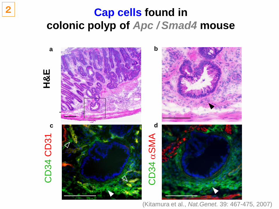
Cap cells found in
colonic polyp of Apc / Smad4 mouse
H&
EC
D3
4 C
D3
1
CD
34
aS
MA
(Kitamura et al., Nat.Genet. 39: 467-475, 2007)
2
c
a
d
b
The Cap Cells express
cognate CCL9 receptor CCR1
T
CD
34
CC
R1
aCis-Apc/Smad4Apc (+/–)
T
CD
34
CC
R1
b
(Kitamura et al., Nat.Genet. 39: 467-475, 2007)
T
T
2
cis-Apc/Smad4
cis-Apc/Smad4:Ccr1(–/–)
sempsmm
0
100
80
60
40
20Pro
po
rtio
n (%
)
***
*
Invasion depth [ shallow deep ]
Invasion depth of the polyps ( >2mm)
Cap Cells promote tumor invasionin the cis-Apc/Smad4 polyps
mp
mp
CD34 CD31
cis-Apc/Smad4 cis-A/Sm4:Ccr1(–/–)
Note that the number and size of tumors were not affected by Ccr1–/–.
mp
mp
CD34 CD31
2
ccis-Apc/Smad4:Ccr1(–/–)
a
Ep
ith
eliu
m
Mmp14
Mmp9
Gapdh
Mmp2
cis-Apc/Smad4
MM
P9
CD
34
mpmp
mpmp
cis
-Ap
c/S
ma
d4
b
DQ-gelCD34
Vill
Wt-normalApc-polypCis-polyp
Apc-polypCis-polyp
Tis
su
e
Crpt
Mmp2
Mmp14
Mmp9
Gapdh
The Cap Cells in cis-Apc/Smad4 mice
produce gelatinases (MMPs) for tumor invasion
(Kitamura et al., Nat.Genet. 39: 467-475, 2007)
2
CCR1+ cells express MMP9
at the invasion front in human CRC*
T
T
T
T
mp
mp
f
T
T
mp
T
T
Tb T
T
d
*Right-side colon cancer with TGFBRII mutation (A)10(A)9
that cannot be corrected by DNA mismatch repair in the patients
lacking the system (e.g., HNPCC).
(Kitamura et al., Nat.Genet. 39: 467-475, 2007)
2
T
mp
mp
e
T
T
mp
a
T
T
c
MMP9 aSMA CCR1 CD68 (M)H&E
Significance in Basic Research
• In contrast to previous impression thatimmune cells help protect the host byattacking cancer cells, these results showthat they can aggravate cancer by helpingcancer cells to invade through chemokine–chemokine receptor interaction.
• Because local invasion is the earliest step incancer metastasis, it is possible that thesame chemokine–chemomkine receptor axismay be involved in the metastasis of coloncancer 3.
2
Colon Cancer Metastasis and Prognosis
(5-yr Survival)
w/o Metastasis
w/ Metastasis
Localized
Regional
Distant Met5-y
ea
r su
rviv
al (%
)
100
80
60
40
20
0
Malignancy
(Modified from Cancer Statistics in Japan, 2008)
3a
Method: Colon cancer cell line injected into the
spleen of the syngeneic mice ––> Intrahepatic
dissemination ––> Metastatic expansion
Q: Do CCR1+ iMCs stimulate metastasis?
Tumor cell injection
into the spleen
Dissemination
to the liver
splenectomy Liver metastasis
3a
Colon cancer cells that disseminated to the liver
are associated with Cap Cells (iMCs)
H&E CK CD45 CD31 CD34 CK CD11b
T
L
T
L
T
L
T
L
CK F4/80
TT
LL
B2.20 CD3 CK aSMA
T
L
Metastatic foci of CMT93 mouse colon cancer cells in the liver
In vivo bioluminescent images of mice injected with luciferase-expressing CMT93 cells
Photo
n c
ounts
1 3 7 14 21
Days after tumor transplantation
Scramble
105
106
107
108
109
*
**
*P < 0.01 v.s. Scramble (student’s t-test)
The expansion of metastasis foci
(photon counts)
shCcl9
Fluoroscent intensity
(Low High)
3a
Are iMCs involved in human colon cancer
metastasis in the liver?
Can CCR1 inhibitors block metastasis?
(Cheng, J.-F. and Jack, R. Mol. Divers. 12:17, 2008)
(Berlex)
(Novartis)
(AstraZeneca)
mp
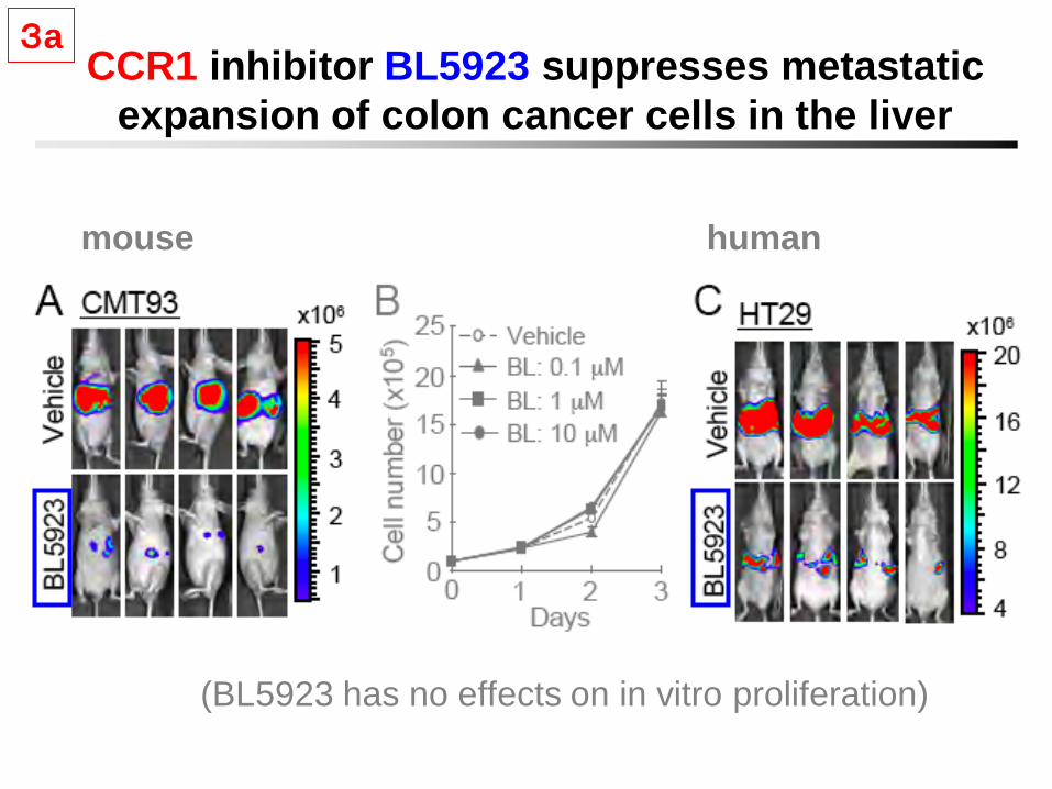
3a
CCR1 inhibitor BL5923 suppresses metastatic
expansion of colon cancer cells in the liver
(BL5923 has no effects on in vitro proliferation)
mouse human
3a
Significance of this study in clinical research
It is known that various proteases, especially metalloproteinases are involved in cancer invasion. In fact, many pharmaceutical companies developed inhibitors of MMPs, and more than ten compounds were tested in clinical trials recently. Unfortunately, however, none succeeded in the trials due to severe side effects.
Our present results suggest the possibility that we can block cancer invasion/metastasis by inhibiting the recruitment of MMP-producing iMCs with CCR1 antagonists, rather than by direct inhibition of MMPs systemically:
“Cellular Targeting Therapy”
3a
Elementary processes in cancer metastasis
Hypothesis:there must be
metastasis suppressor genes,
loss of whose activities should
help metastasis.
Method:Microarray screening of
genes with decreased levels of
expression.
3b
Model for colon cancer metastasis: rectal transplantation
(~30%)
(~100%)(~25%)
*
*Ras-activated
Primary
Tumor
3b
Identification of Aes as a metastasis suppressor
Microarray profile comparison between primary and
metastatic tumors.
Focus on the ―transcription regulator activity‖ (Gene
Ontology), because some genes in this group such as
CRSP3 and Twist were reported to regulate metastatic
potential of cancer cells.
–– Part I ––
3b
Reduced expression of Aes in colon ca met.3b
Forced expression of Aes suppresses liver met(Syngeneic mouse colon cancer cell transplantation model)
3b
Forced expression of Aes suppresses liver met
(quantified data)(Syngeneic mouse colon ca cell transplantation model)
3b
Aes inhibits Notch signaling
luciferase
pGa981-6 reporter
Over-expression Knockdown
RAMIC: Rbpj-associated molecule domain and intracellular domain
of the Notch receptor; equivalent to Notch intracellular domain NICD.
(Sonoshita, M. et al., Cancer Cell 19: 125-137, 2011)
3b
3b Canonical Notch Signaling Pathway
(Kopan & Ilagan, Cell, 2009)
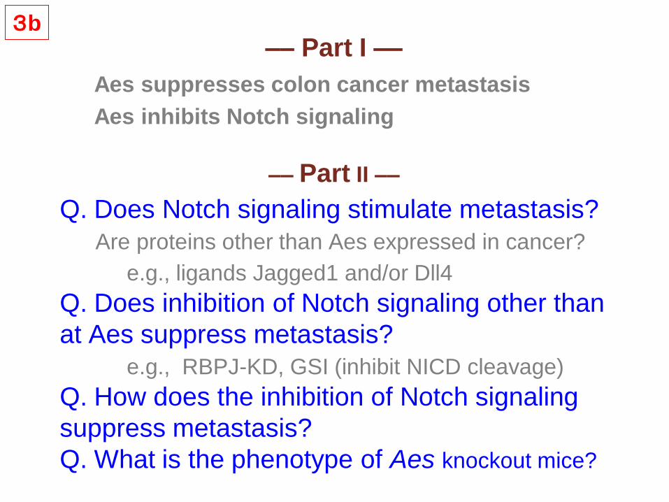
Q. Does Notch signaling stimulate metastasis?
Are proteins other than Aes expressed in cancer?
e.g., ligands Jagged1 and/or Dll4
Q. Does inhibition of Notch signaling other than
at Aes suppress metastasis?
e.g., RBPJ-KD, GSI (inhibit NICD cleavage)
Q. How does the inhibition of Notch signaling
suppress metastasis?
Q. What is the phenotype of Aes knockout mice?
Aes suppresses colon cancer metastasis
Aes inhibits Notch signaling
–– Part I ––
–– Part II ––
3b
Notch ligand Jagged1 is expressed
on vessels of primary colon cancer
Bar; 100 m Bar; 10 m
Jagged1
3b
Q. What is the mechanism of metastasis
suppression by Notch signaling inhibition?
A. Trans-endothelial migration (TEM)
relevant to intra-vasation & extra-vasation)
Transendothelial migration (TEM) assay
3b
Whole animal phenotype
of Apc/Aes mutation in the intestines
Apc / Aes
(4HT induction)
Intestinal
Polyposis
Aes-/-
Invasive Cancer
w/ Intravasation
Apc +/716
Floxed Aes allele
(M.M. Taketo)
Compound mutant
3b
Local invasion and intravasation
of Apc/Aes mouse tumors
Dotted lines: Muscularis propria
Adenoma Strong local invasion
3b
Adenoma Strong local invasion
Notch signaling inhibition
and metastasis suppression by Aes
3b
Summary
Although colon cancer arises from the mucosal epithelium, the stromal cells
play key roles in the expansion of microadenomas (e.g., COX-2 induction).
In locally invasive colon cancer, CCR1+ iMCs are recruited to the invasion
front by chemokines (CCL9/15) secreted by the tumor epithelium and
produce proteases MMP9/2. Similar iMCs play key roles in metastatic
expansion of colon cancer cells disseminated to the liver.
In another model where colon cancer cells metastasize to the liver, lungs
and lymph nodes, Notch receptors are activated by the ligands expressed on
the stromal cells such as blood vessels, smooth muscle etc. However, if the
cancer cells express Aes, transcriptional activation of Notch signaling is
inihibited. If Aes is lost in cancer cells, Notch signaling is activated and the
cancer cells actively move into (or out of) the blood vessels.
These results collectively indicate that heterotypic interactions of cancer
cells with the stroma (i.e., the microenvironment) play key roles in cancer
progression.
Acknowledgment
Graduate School of Medicine
Kyoto University
Masahiro Sonoshita
Masahiro Aoki
Hiromi Kikuchi
Koji Aoki
Takanori Kitamura
Hisahiro Hosogi
Fumihiko Kakizaki
------------------------
Takashi Kobayashi
Osamu Ogawa
Yoshiharu Sakai
Tasuku Honjo & Lab
Chiaki Takahashi
Tohoku University Graduate School
of Life Sciences
Haruhiko Fuwa
Makoto Sasaki
Kitano Hospital
Hiroki Hashida
Arimichi Takabayashi
Osaka Medical Center
for Cancer and Cardiovascular Diseases
Kazuyuki Itoh
Kiyoko Yoshioka
Kanazawa University Center for Cancer
and Stem Cell Research
Masanobu Oshima
TORAY Industries
Tetsuo Sudo
Institute of Medical Science, University of Tokyo
Toshio Kitamura
Research Institute of Microbial Diseases, Osaka Univ.
Masaru Okabe
Chiba University Graduate School of Medicine
Motoo Kitagawa
Institute of Molecular and Cell Biology, Singapore