922 ISSN 1229-9197 (print version) ISSN 1875-0052 (electronic version) Fibers and Polymers 2019, Vol.20, No.5, 922-932 Comfort and Infection Control of Chitosan-impregnated Cotton Gauze as Wound Dressing Jefferson M. Souza 1 , Mariana Henriques 2 , Pilar Teixeira 2 , Margarida M. Fernandes 3,4 , Raul Fangueiro 5 , and Andrea Zille 5 * 1 CBMDE, Design and Styling, Federal University of Piauí, Teresina - PI, 64049-550, Brazil 2 Centre of Biological Engineering, Laboratório de Investigação em Biofilmes Rosário Oliveira, University of Minho, Campus de Gualtar, 4710-057 Braga, Portugal 3 Centro de Física, Universidade do Minho, Campus de Gualtar, 4710-058 Braga, Portugal 4 Centre of Biological Engineering, University of Minho, Campus de Gualtar, 4710-057 Braga, Portugal 5 Centre for Textile Science and Technology, University of Minho, Campus de Azureḿ, 4800-058 Guimares, Portugal (Received January 16, 2019; Revised February 16, 2019; Accepted February 19, 2019) Abstract: The aim of this study was to evaluate the thermo-physiological comfort properties of surgical cotton gauze coated with chitosan (CH) and its effectiveness for the prevention of bacterial colonization. Gauze was coated with CH at mass fractions of 0.50, 0.25, 0.125, 0.10, 0.063 wt% and the friction, flexibility, thermal, moisture management and mechanical properties were evaluated. The best performing gauze in terms of comfort (0.125 wt%) was further evaluated for its ability to inhibit the growth of microorganisms such as bacteria and yeast. Results indicate that the functionalized medical gauze could induce low friction on the wound bed allowing a good degree of moisture and high absorption capacity of wound exudates. Moreover, it shows antimicrobial properties against medical-relevant pathogens. This biofunctional medical gauze demonstrates to deliver an efficient antimicrobial coating and promote the best conditions for maintenance of the wound microenvironment. Keywords: Chitosan, Cellulose, Comfort, Antimicrobial, Wound dressing Introduction A dermal wound is defined as a disruption in the integrity of the skin caused by trauma, abrasion, burns or ulcers, leading to an inadequate performance of its functions. Excessive exudates can impair wound healing and promote bacterial colonization causing difficult-to-treat infections and other complications. Thus, it is vital to restore the skin integrity and function as soon as possible [1]. One of the main roles of intact skin is to act as a barrier for the penetration in the body of the potentially harmful microbial population living on the skin surface. Indeed, when a dermal wound occurs, the skin becomes more susceptible to the colonization of bacteria and fungi [2]. For example immediately after an burn injury, Gram-positive bacteria such as Staphylococcus epidermidis and Staphylococcus aureus may rapidly colonize the wounds [2]. Later, Gram- negative organisms like Pseudomonas aeruginosa or Escherichia coli or fungi species such as Candida albicans may also be implicated [2-4]. The infections caused by these microorganisms may lead to increased mortality, morbidity, length of hospital stay and consequently costs to clinical settings, thus it is extremely important to provide strategies to prevent wound infection and/or promoting a proper wound healing [5]. Therefore, an aseptic, pathogen-free, environment is very important in helping the wound healing process and equally important a proper wound healing strategies should also include moist management. Maintaining a moist wound environment has been regarded as a key issue in order to facilitate the healing process. The moist environment prevent tissue dehydration and cell death, accelerate angiogenesis, increase the breakdown of dead tissue and fibrin and potentiate the interaction of growth factors with their target cells [6,7]. However, it is also important to note that excessive moisture, e.g. over production of exudate in the wound, may adversely affect healing. It is necessary to provide a moisture balance to obtain an optimal environment for wound healing [8]. Excessive exudate slows down or even prevents cell proliferation, interferes with growth factor availability and contains elevated levels of inflammatory mediators and activated matrix metalloproteinases (MMPs), which impair the healing process [9]. Therefore, the ideal wound dressing material should comprise properties that: i) permits a balanced moisture at the wound site, i.e. being capable of absorb excess of exudates but maintaining certain levels of moist, ii) prevent bacterial infections, iii) do not adhere to the wound bed and iv) to be soft; in order to accelerate wound healing and reduce pain and discomfort [10-12]. Nowadays, many sophisticated dressings made of a wide range of polymeric materials are available to the wound care practitioner. Polymers may be used alone or in combinations thereof, being processed in different dressing designs such as films, foams, fibrous materials, beads, hydrogels, hydrocolloids or even pharmaceutical sprays comprising nano/micro- *Corresponding author: [email protected]DOI 10.1007/s12221-019-9053-2
Transcript
922
ISSN 1229-9197 (print version)
ISSN 1875-0052 (electronic version)
Fibers and Polymers 2019, Vol.20, No.5, 922-932
Comfort and Infection Control of Chitosan-impregnated Cotton Gauze as
Wound Dressing
Jefferson M. Souza1, Mariana Henriques
2, Pilar Teixeira
2, Margarida M. Fernandes
3,4,
Raul Fangueiro5, and Andrea Zille
5*
1CBMDE, Design and Styling, Federal University of Piauí, Teresina - PI, 64049-550, Brazil2Centre of Biological Engineering, Laboratório de Investigação em Biofilmes Rosário Oliveira, University of Minho,
Campus de Gualtar, 4710-057 Braga, Portugal3Centro de Física, Universidade do Minho, Campus de Gualtar, 4710-058 Braga, Portugal
4Centre of Biological Engineering, University of Minho, Campus de Gualtar, 4710-057 Braga, Portugal5Centre for Textile Science and Technology, University of Minho, Campus de Azureḿ, 4800-058 Guimares, Portugal
(Received January 16, 2019; Revised February 16, 2019; Accepted February 19, 2019)
Abstract: The aim of this study was to evaluate the thermo-physiological comfort properties of surgical cotton gauze coatedwith chitosan (CH) and its effectiveness for the prevention of bacterial colonization. Gauze was coated with CH at massfractions of 0.50, 0.25, 0.125, 0.10, 0.063 wt% and the friction, flexibility, thermal, moisture management and mechanicalproperties were evaluated. The best performing gauze in terms of comfort (0.125 wt%) was further evaluated for its ability toinhibit the growth of microorganisms such as bacteria and yeast. Results indicate that the functionalized medical gauze couldinduce low friction on the wound bed allowing a good degree of moisture and high absorption capacity of wound exudates.Moreover, it shows antimicrobial properties against medical-relevant pathogens. This biofunctional medical gauzedemonstrates to deliver an efficient antimicrobial coating and promote the best conditions for maintenance of the woundmicroenvironment.
927 Fibers and Polymers 2019, Vol.20, No.5 Jefferson M. Souza et al.
tested directions. The coated chitosan gauze with the lowest
values of bending is the CH0.063. Its warp bending rigidity
increased 32 % and its weft bending rigidity increased 27 %
compared to control gauze. The other coated chitosan gauzes
are expected to impact negatively upon conformability of a
wound. The used bending tester was the KES-FB of the
Kawabata Evaluation System. KES-FB apparatus is a
standard tool for a thorough evaluation of textile fabric’s
deformability allowing characterization of fabric’s behaviour
under low loads and reliability of the results. Standard
parameters obtained by KES-FB method were the bending
rigidity (B) per unit width in Nm2 m-1, that is calculated as
the mean bending stiffness of two slopes and the bending
hysteresis (2HB) value in Nm m-1 that is obtained by reading
the hysteresis width at curvature ±1. Bending rigidity
represents the resistance of fabric against flexion and
bending hysteresis can be considered as a measure of fabrics
ability to recover [38]. In one hand, the increase in bending
rigidity in the warp direction is higher than in the weft
direction due to the different yarn linear densities used for
the fabric assembly. On the other hand, bending hysteresis
values in weft direction are 1.7 times lower than in warp
direction. The observed increase of hysteresis values (lower
bending recovery) in the highly dense chitosan-treated yarns
in warp direction is due to the higher resulting inter-fibre
friction [39].
Surface Friction
Chitosan coating does not only provide special functionalities
to the cotton gauze, it also imparts stiff and rough feelings
difficulting its handle or use. The surface of a textile fabric is
not uniformly flat and smooth and traditional cotton gauze
adherence of the dressing to the wound often cause frictional
trauma on removal resulting in hypertrophic scarring [40]. A
low values of coefficient of kinetic friction can be used as
acceptable indicator of a smooth fabric surface even if alone
it can be insufficient for surface characterization [41]. In
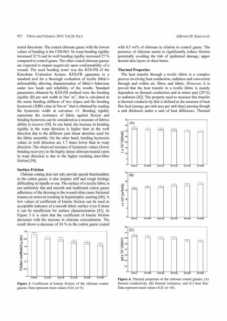
Figure 3 it is clear that the coefficient of kinetic friction
decreases with the increase in chitosan concentration. The
result shows a decrease of 24 % in the cotton gauze coated
with 0.5 wt% of chitosan in relation to control gauze. The
presence of chitosan seems to significantly reduce friction
potentially avoiding the risk of epidermal damage, upper
dermal skin layers or sheet burns.
Thermal Properties
The heat transfer through a textile fabric is a complex
process involving heat conduction, radiation and convection
through and within air, fibres and fabric. However, it is
proved that the heat transfer in a textile fabric is mainly
dependent on thermal conduction and in minor part (20 %)
to radiation [42]. The property used to measure this transfer
is thermal conductivity that is defined as the measure of heat
flux heat (energy per unit area per unit time) passing though
a unit thickness under a unit of heat difference. Thermal
Figure 3. Coefficient of kinetic friction of the chitosan coated
gauzes. Data represent mean values±S.D. (n=5).
Figure 4. Thermal properties of the chitosan coated gauzes; (A)
thermal conductivity, (B) thermal resistance, and (C) heat flux.
Data represent mean values±S.D. (n=10).
short title Fibers and Polymers 2019, Vol.20, No.5 928
conductivity is directly proportional to the heat flux, thus,
the more increase thermal conductivity, the more increase
heat flux [43]. On the other hand, thermal resistance is
inversely proportional to thermal conductivity. It represents
the temperature difference across a unit area and unit of
thickness when a unit of heat flux pass trough the fabric in a
unit of time. Thermal resistance can be used to quantitatively
evaluate the capacity of a fabric in providing an efficient
thermal barrier or in other words, to express the thermal
insulation ability of a fabric.
All the chitosan-coated gauzes display lower thermal
conductivity (Figure 4(A)) and higher thermal resistance
(Figure 4(B)) values than the control gauze due to the
enhanced fabric weight and thickness. The gauze with
0.5 wt% of chitosan shows the best results displaying the
lowest thermal conductivity and the higher thermal resistance
due to the larger thickness and the greater amount of air
entrapped in the structure of the fabric. Thus, it seems that
0.5 wt% chitosan coated gauze is able maintain a degree of
thermal insulation to provide optimum temperature for cell
proliferation. The presence of chitosan reduces the heat flow
indicating a relatively warm feeling when it touches human
skin (Figure 4(C)). Thermal insulation keeps the wound
surface warm improving the blood flow to the wound bed
and enhancing epidermal migration [44]. In dry fabrics, such
as the evaluated gauzes, thermal insulation depends
essentially on fabric thickness and, to a lesser extent, on
fabric construction and fibre conductivity [45].
Air Permeability
Air permeability is one of the most important parameters
for wound dressings. It is defined as the volume of air which
is passed in a certain period of time through a known area of
the fabric at a defined pressure difference between the two
surfaces of the fabric [46]. A medical dressing must be
permeable for gases in order to prevent maceration and gives
comfort to the patients, but an excessive air permeability
could dry out the wound and have a negative effect on
healing [47]. In fibre-based dressings air permeability is
mainly affected by the porosity since, for obvious reasons,
the passage of air through a fabric can only take place in the
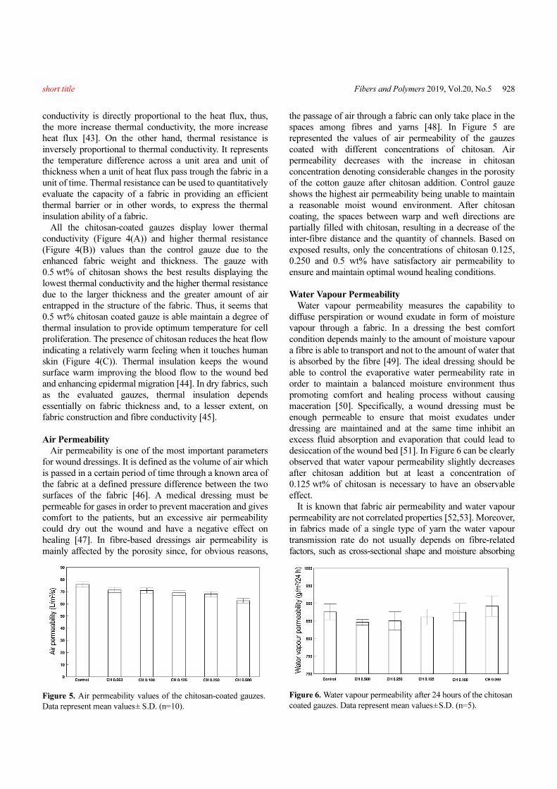
spaces among fibres and yarns [48]. In Figure 5 are
represented the values of air permeability of the gauzes
coated with different concentrations of chitosan. Air
permeability decreases with the increase in chitosan
concentration denoting considerable changes in the porosity
of the cotton gauze after chitosan addition. Control gauze
shows the highest air permeability being unable to maintain
a reasonable moist wound environment. After chitosan
coating, the spaces between warp and weft directions are
partially filled with chitosan, resulting in a decrease of the
inter-fibre distance and the quantity of channels. Based on
exposed results, only the concentrations of chitosan 0.125,
0.250 and 0.5 wt% have satisfactory air permeability to
ensure and maintain optimal wound healing conditions.
Water Vapour Permeability
Water vapour permeability measures the capability to
diffuse perspiration or wound exudate in form of moisture
vapour through a fabric. In a dressing the best comfort
condition depends mainly to the amount of moisture vapour
a fibre is able to transport and not to the amount of water that
is absorbed by the fibre [49]. The ideal dressing should be
able to control the evaporative water permeability rate in
order to maintain a balanced moisture environment thus
promoting comfort and healing process without causing
maceration [50]. Specifically, a wound dressing must be
enough permeable to ensure that moist exudates under
dressing are maintained and at the same time inhibit an
excess fluid absorption and evaporation that could lead to
desiccation of the wound bed [51]. In Figure 6 can be clearly
observed that water vapour permeability slightly decreases
after chitosan addition but at least a concentration of
0.125 wt% of chitosan is necessary to have an observable
effect.
It is known that fabric air permeability and water vapour
permeability are not correlated properties [52,53]. Moreover,
in fabrics made of a single type of yarn the water vapour
transmission rate do not usually depends on fibre-related
factors, such as cross-sectional shape and moisture absorbing
Figure 5. Air permeability values of the chitosan-coated gauzes.
Data represent mean values± S.D. (n=10).
Figure 6. Water vapour permeability after 24 hours of the chitosan
coated gauzes. Data represent mean values±S.D. (n=5).
929 Fibers and Polymers 2019, Vol.20, No.5 Jefferson M. Souza et al.
properties but is primarily a function of fabric bulk density.
In fact, in this low-density gauze, increased thickness and
weight seems to be significantly correlated to water vapour
permeability which is in turn strongly affected by the
macroporous structure of fabric [54]. The addition of
chitosan to cotton causes strong intermolecular hydrogen
interactions between the similar polysaccharidic structures
of the two polymers resulting in a decrease of the inter-fibre
distance and accessibility of the hydrophilic groups,
reducing the water vapour transmission rate [55,56].
Vertical Wicking
Liquid moisture transportation on a fabric is due to a
wetting process followed by wicking. Wetting is the initial
process of fluid spreading where the fibre-liquid interface
replaces fibre-air interface. Wicking is the flow of a liquid
through the porous media characterized by the fibre-liquid
molecular attraction at the surface. Surface tension, effective
capillary pathways and pore distribution are the main
variables responsible for the wicking ability in a textile
fabric [57]. Hygroscopic dressings based in natural fibres
such as cotton are characterised by high liquid moisture
transportation and absorption in order to allow the remove of
excess exudate from the wound. An efficient level of
absorption prevents lateral wicking that can cause maceration at
the edge of the wound and maintains a reasonable degree of
moist for wound healing [58]. However, wetting have to be
controlled since can cause the fabric to swell, changing the
geometry among capillary space positions, increasing the
weight of the dressing and ultimately affecting the vertical
wicking ability. In Figure 7 are shown the vertical wicking
heights of the coated gauzes after ten minutes in warp and
weft directions. The gauze coated with 0.5 wt% of chitosan
showed the lowest wicking heights (~1 cm). All the other
samples display better wickability and higher absorption in
weft direction as compared to warp direction. This is
because in this gauze the weft yarn diameter is larger than
the warp one. In warp direction only the 0.25 and 0.5 wt%
chitosan concentrations show different wicking height
compared to the control gauze (Figure 7(A)). On the other
hand, in weft direction all the chitosan concentrations
display a lower wicking height than control gauze (Figure
7(B)).
Overall, the presence of chitosan greatly improves the
dressing ability to retain liquid, as the fluid is entrapped
within its structure. The polymer blocks the water molecules
movement maintaining for a longer time a moist environment
for wound healing. Since, cotton gauze wickability decrease
by increasing chitosan concentration, it is important to found
an ideal chitosan concentration in order to maintain a moist
environment and at the same time avoid maceration [59].
Water Uptake
One of the most important function of a wound dressing is
its ability to absorb fluid from a highly exuding wound
maintaining a moist environment in a dry wound [60]. It is
clear that high chitosan concentrations significantly reduce
the absorptive capacity of cotton gauze. This effect is due to
the reduction in porosity and availability of hydrophilic
groups due to the hydrogen bond interactions between
cellulose and chitosan. The higher water uptake at moderate
concentration of chitosan could be attributed to the high
hydrophilicity of both cellulose and chitosan polymers
Figure 7. Vertical wicking values in (A) warp and (B) weft
directions of the chitosan coated gauzes. Data represent mean
values±S.D. (n=5).
Figure 8. Mass of absorbed water (%) in warp (grey bars) and
weft (white bars) directions of the chitosan coated gauzes. Data
represent mean values±S.D. (n=5).
short title Fibers and Polymers 2019, Vol.20, No.5 930
resulting in water diffusing very rapidly through the coated
gaze [61]. Higher chitosan concentrations did not give off
the absorbed water and limits the water access to the
cellulose fibres of cotton gaze. This limits the ability of the
dressing to preserve water that is one of the most important
issues in wound healing. Effective wound dressings must be
able to maintain a prolonged moist microenvironment to
improve the epithelialization of wound while preventing the
formation of the scab [62].
Cotton gauzes up to 0.100 wt% of coated chitosan show
an increase in water uptake compared to control gauze. The
water uptakes of the cotton gauze with 0.125 wt% show very
similar values to the control gauze. Further increase in
chitosan concentration (0.25 and 0.5 wt%) leads to significant
lower values of water uptake (Figure 8). Observed water
uptake in weft direction is the double than that in warp
direction for all tested samples. The gaze coated with
0.5 wt% of chitosan shows an impressive decrease in water
uptake of about 77 % in warp direction and 78 % in weft
direction. These results clearly showed that the water
absorption capacity of the gauzes, and consequently, their
ability to remove exudate from the wound could be tailored
by tuning chitosan content.
Antimicrobial Properties
The best performing chitosan-coated gauze in terms of
thermo-physiological comfort properties was further tested
for its antimicrobial properties in accordance with Japanese
Standard JIS L 1902:2002. Gram-negative (E. coli) and
Gram-positive (S. aureus) bacteria, as well as fungi (C.
albicans) were tested and the results presented in Table 3.
Typically, an antimicrobial agent may possess either
bacteriostatic or bactericidal properties. Bacteriostatic
activity means that it prevents the multiplication of bacteria
without destroying them while bactericidal implies the
forthright killing of the organisms. As the growth value (F)
obtained from the number of living microorganisms, after
being in contact with 0.125 wt% chitosan-impregnated
cotton gauze, is always higher than 1.5 the tests were judged
to be effective. According to the standard, a value of
bactericidal activity (L) higher than zero is an indication of
bactericidal activity, while bacteriostatic properties begin
with (S) values exceeding 2. The results has shown that
medical gauze when coated with chitosan reveals significant
bactericidal and bacteriostatic activity against both bacteria
(S>2 and L>0) but showed only a fungistatic activity (S>2
and L=0) against the fungi C. albicans.
The significant bactericidal activity against both bacteria
is a quite interesting result because of the used low
concentration of chitosan. Similar results have only been
obtained when chitosan was carboxymethylated [63] or
when it was combined with other bactericidal agents such as
zinc oxide [64] or silver nanoparticles [65]. This might be
due to the fact that higher concentrations of chitosan are
usually assumed to be needed for obtaining an antimicrobial
textile owed to its reported high values of minimum
inhibitory concentration (above 2 mg/ml in solution) [66].
The electrostatic interactions between the protonated amino
groups of chitosan -NH3
+ and the negatively charged
microbial cell membranes are known to be essential for its
antimicrobial and antifungal properties [67]. At lower
concentrations (<0.2 mg/ml), the polycationic chitosan binds
to the negatively charged bacterial surface to cause
agglutination, while at higher concentrations, the larger
number of positive charges impart a net positive charge to
the bacterial surfaces to keep them in suspension. Another
hypothesis is that chitosan interacts with the membrane of
the cell to alter cell permeability, which leads to its
disruption and subsequently leakage of proteinaceous and
other intracellular constituents [20]. Nevertheless, the actual
mechanism has not yet been fully elucidated. In this work, it
may be assumed that the chosen concentration favours the
mobility of the chitosan macromolecules and their interaction
with the membrane of bacteria impeding the occurrence of a
steric hindrance effect between chitosan and bacteria.
Regarding fungistatic activity, the results are in good
agreement with the literature, which report that chitosan
possess fungistatic rather than fungicidal properties [67].
Similarly to the effects observed in bacteria cells, chitosan
interferes directly with fungal growth by inhibiting cell wall
morphogenesis [68]. The suggested mechanism involved a
permeable chitosan film formed on the crop surface which
interfered with the fungal growth and activated several
defence processes like chitinase accumulation, proteinase
inhibitor synthesis, callus synthesis and lignification [69].
Conclusion
In this work, chitosan-coated cotton gauze with antimicrobial
properties has been developed with concomitant evaluation
of the best thermo-physiological comfort properties, providing
an added value material for wound healing purposes able to
reduce pain and discomfort to the patient. Different
concentrations of chitosan have been impregnated onto
cotton gauze and the best performing material in terms of
comfort was assessed. An exhaustive characterization of the
different functionalized cotton including the moisture control
and dressing comfort properties was carried out, which
Table 3. Antimicrobial activity of surgical cotton gauze coated
with 0.125 wt% of chitosan
Activity valueMicroorganism
E. coli S. aureus C. albicans
Microbiostatic activity value (S) 4.4±2.2 3.0±0.3 3.3±0.2
Microbiocidal activity value (L) 2.2±1.5 1.7±2.1 0.0±0.0
Growth value (F) 1.8±0.7 2.3±0.8 5.0±0.5
931 Fibers and Polymers 2019, Vol.20, No.5 Jefferson M. Souza et al.
allowed concluding that through the application of 0.125 wt%
of chitosan onto cotton gauze, a material with enhanced
flexibility, thermal properties, water and air permeability,
moist management and low adherence properties was
obtained. Despite the used low concentration of chitosan the
functionalized cotton gauze further presented bactericidal
activity against S. aureus and E. coli, and fungistatic activity
towards the fungi C. albicans. This dressing configuration
shows high potential in wound healing applications
preventing wound microbial contamination and providing
the proper healing environment at the same time promoting
conformability to the wound area and comfort to the patient.
Acknowledgements
AZ acknowledges funding from FCT - Fundação para a
Ciência e a Tecnologia within the scope of the project POCI-
01-0145-FEDER-007136 and UID/CTM/00264. A. Zille
also acknowledges financial support of the FCT through an
Investigator FCT Research contract (IF/00071/2015). JS
acknowledge CAPES Foundation, Ministry of Education of
Brazil, Proc. no 8976/13-9 and the Department of Textile
Engineering of the University of Minho, Portugal. The work
regarding the biological analysis was supported by the
Programa Operacional, Fatores de competitividade - COMPETE
and by national funds through FCT on the scope of the
projects PTDC/SAU-MIC/119069/2010, RECI/EBB-EBI/
0179/2012 and PEst-OE/EQB/LA0023/2013. PT acknowledges
SFRH/BPD/86732/2012 grant. The authors thank the Project
BioHealth - Biotechnology and Bioengineering approaches
to improve health quality, Ref. NORTE-07-0124-FEDER-
000027, co-funded by the Programa Operacional Regional
do Norte (ON.2 - O Novo Norte), QREN, FEDER.
Electronic Supplementary Material (ESM) The online
version of this article (doi: 10.1007/s12221-019-9053-2)
contains supplementary material, which is available to
authorized users.
References
1. G. S. Lazarus, D. M. Cooper, D. R. Knighton, D. J.Margolis, R. E. Pecoraro, G. Rodeheaver, and M. C.Robson, Arch. Dermatol., 130, 489 (1994).
2. D. Church, S. Elsayed, O. Reid, B. Winston, and R.Lindsay, Clin. Microbiol. Rev., 19, 403 (2006).
3. P. G. Bowler, B. I. Duerden, and D. G. Armstrong, Clin.
Microbiol. Rev., 14, 244 (2001).4. P. N. Malani, S. A. McNeil, S. F. Bradley, and C. A.
Kauffman, Clin. Infect. Dis., 35, 1316 (2002).5. E. R. M. Sydnor and T. M. Perl, Clin. Microbiol. Rev., 24,
141 (2011).6. C. K. Field and M. D. Kerstein, Am. J. Surg., 167 (1994).7. R. Wiechula, Int. J. Nurs. Pract., 9, S9 (2003).
8. D. Okan, K. Woo, E. A. Ayello, and G. Sibbald, Adv. Skin
Wound Care, 20, 39 (2007).9. S. M. McCarty and S. L. Percival, Adv. Wound Care, 2,
438 (2013).10. A. Sood, M. S. Granick, and N. L. Tomaselli, Adv. Wound
Care, 3, 511 (2014).11. T. Wang, X. K. Zhu, X. T. Xue, and D. Y. Wu, Carbohydr.
Polym., 88, 75 (2012).12. H. F. Selig, D. B. Lumenta, M. Giretzlehner, M. G.
Jeschke, D. Upton, and L. P. Kamolz, Burns, 38, 960(2012).
13. A. Francesko, M. M. Fernandes, G. Rocasalbas, S. Gautier,and T. Tzanov in “Advanced Polymers in Medicine” (F.Puoci Ed.), p.401, Springer International Publishing, 2015.
14. D. Archana, B. K. Singh, J. Dutta, and P. K. Dutta,Carbohydr. Polym., 95, 530 (2013).
15. D. Archana, B. K. Singh, J. Dutta, and P. K. Dutta, Int. J.
Biol. Macromol., 73, 49 (2015).16. S. Dhivya, V. V. Padma, and E. Santhini, Biomed., 5, 22
(2015).17. W. J. Ennis, W. Valdes, M. Gainer, and P. Meneses, Adv.
Skin Wound Care, 19, 437 (2006).18. X. Cheng, K. Ma, R. Li, X. Ren, and T. S. Huang, Appl.
Surf. Sci., 309, 138 (2014).19. F. Dinah and A. Adhikari, Ann. Royal Coll. Surg. Engl., 88,
33 (2006).20. K. Azuma, R. Izumi, T. Osaki, S. Ifuku, M. Morimoto, H.
Saimoto, S. Minami, and Y. Okamoto, J. Funct. Biomater.,6, 104 (2015).
21. V. Patrulea, V. Ostafe, G. Borchard, and O. Jordan, Eur. J.
Pharm. Biopharm., 97, 417 (2015).22. H. Ueno, T. Mori, and T. Fujinaga, Adv. Drug Delivery.
Rev., 52, 105 (2001).23. D. Alonso, M. Gimeno, R. Olayo, H. Vazquez-Torres, J. D.
Sepulveda-Sanchez, and K. Shirai, Carbohydr. Polym., 77,536 (2009).
24. X. D. Liu, N. Nishi, S. Tokura, and N. Sakairi, Carbohydr.
Polym., 44, 233 (2001).25. A. El.Shafei and A. Abou-Okeil, Carbohydr. Polym., 83,
920 (2011).26. M. Gouda and S. M. A. S. Keshk, Carbohydr. Polym., 80,
504 (2010).27. L. F. Zemljic, O. Sauperl, T. Kreze, and S. Strnad, Text.
Res. J., 83, 185 (2013).28. K. F. El-Tahlawy, M. A. El-Bendary, A. G. Elhendawy, and
S. M. Hudson, Carbohydr. Polym., 60, 421 (2005).29. M. M. G. Fouda, A. El Shafei, S. Sharaf, and A. Hebeish,
Carbohydr. Polym., 77, 651 (2009).30. J. Souza, J. Matos, M. Fernandes, A. Zille, and R.
Fangueiro, Procedia Eng., 200, 135 (2017).31. C. Chung, M. Lee, and E. K. Choe, Carbohydr. Polym., 58,
417 (2004).32. V. Goodarzi, S. H. Jafari, H. A. Khonakdar, B. Ghalei, and
M. Mortazavi, J. Membr. Sci., 445, 76 (2013).
short title Fibers and Polymers 2019, Vol.20, No.5 932
33. E. Yan, S. Fan, X. Li, C. Wang, Z. Sun, L. Ni, and D.Zhang, Mater. Sci. Eng., C, 33, 461 (2013).
34. P. Monllor, M. A. Bonet, and F. Cases, Eur. Polym. J., 43,2481 (2007).
35. W. W. Thein-Han, J. Saikhun, C. Pholpramoo, R. D. K.Misra, and Y. Kitiyanant, Acta Biomater., 5, 3453 (2009).
36. F. Fan, W. Zhang, and C. Wang, Cellulose, 22, 1427(2015).
37. R. Jayakumar, M. Prabaharan, P. T. Sudheesh Kumar, S. V.Nair, and H. Tamura, Biotechnol. Adv., 29, 322 (2011).
38. J. Peiffer, K. Kim, and M. Takatera, Text. Res. J., 87, 424(2017).
39. L. Naujokaityte, E. Strazdiene, and L. Fridrichova, Tekstil,56, 343 (2007).
40. U. Wollina, M. B. Abdel-Naser, and S. Verma, Curr. Probl.
Dermatol., 33, 1 (2006).41. M. Akgun, Fiber. Polym., 14, 1372 (2013).42. S. Mandal, G. Song, M. Ackerman, S. Paskaluk, and F.
Gholamreza, Text. Res. J., 83, 1005 (2012).43. S. Gunesoglu, Indian J. Fibre Text. Res., 31, 415 (2006).44. I. Salopek Čubrić, Z. Skenderi, A. Mihelić-Bogdanić, and
M. Andrassy, Exp. Therm Fluid Sci., 38, 223 (2012).45. S. Alay, C. Alkan, and F. Göde, J. Text. Inst., 103, 757
(2012).46. E. Onofrei, A. M. Rocha, and A. Catarino, J. Eng. Fibers
Fabr., 6, 10 (2011).47. S. B. Stanković, D. Popović, and G. B. Poparić, Polym.
Test., 27, 41 (2008).48. P. Kanakaraj and R. Ramachandran, JTATM, 9, 1 (2015).49. S. S. Ramkumar, A. Purushothaman, K. D. Hake, and D.
D. McAlister, J. Eng. Fibers Fabr., 2, 10 (2007).50. G. Schultz, D. Mozingo, M. Romanelli, and K. Claxton,
Wound Repair. Regen., 13, S1 (2005).51. U. Wollina, M. Heide, W. Müller-Litz, D. Obenauf, and J.
Ash, Curr. Probl. Dermatol., 31, 82 (2003).52. R. T. Ogulata and S. Mavruz, Fibres Text. East. Eur., 18,
71 (2010).53. B. Wilbik-Halgas, R. Danych, B. Wiecek, and K.
Kowalski, Fibres Text. East. Eur., 14, 77 (2006).54. F. Wang, S. del Ferraro, L.-Y. Lin, T. Sotto Mayor, V.
Molinaro, M. Ribeiro, C. Gao, K. Kuklane, and I. Holmér,Ergonomics, 55, 799 (2012).
55. I. Carvalho, M. Henriques, J. C. Oliveira, C. F. AlmeidaAlves, A. P. Piedade, and S. Carvalho, Sci. Technol. Adv.
Mater., 14, 035009 (2016).56. T. Rakmanee, I. Olsen, G. S. Griffiths, and N. Donos,
Analyst, 135, 182 (2010).57. M. Manshahia and A. Das, Indian J. Fibre Text. Res., 39,
441 (2014).58. M. A. Fonder, G. S. Lazarus, D. A. Cowan, B. Aronson-
Cook, A. R. Kohli, and A. J. Mamelak, J. Am. Acad.
Dermatol., 58, 185 (2008).59. J. S. Boateng, K. H. Matthews, H. N. E. Stevens, and G. M.
Eccleston, J. Pharm. Sci., 97, 2892 (2008).60. A. Nazir, T. Hussain, G. Abbas, and A. Ahmed, J. Nat.
Fibers, 12, 232 (2015).61. F. Xu, B. Weng, R. Gilkerson, L. A. Materon, and K.
Lozano, Carbohydr. Polym., 115, 16 (2015).62. H. Li, J. Yang, X. Hu, J. Liang, Y. Fan, and X. Zhang, J.
Biomed. Mater. Res., A, 98A, 31 (2011).63. B. Venkatraja, V. V. Malathy, B. Elayarajah, R. Rajendran,
and R. Rammohan, Pak. J. Biol. Sci., 16, 1438 (2013).64. P. Petkova, A. Francesko, M. M. Fernandes, E. Mendoza,
I. Perelshtein, A. Gedanken, and T. Tzanov, ACS Appl.
Mater. Interfaces, 6, 1164 (2014).65. M. Abbasipour, M. Mirjalili, R. Khajavi, and M. M.
Majidi, J. Eng. Fibers Fabr., 9, 124 (2014).66. M. M. Fernandes, A. Francesko, J. Torrent-Burgués, and T.
Tzanov, React. Funct. Polym., 73, 1384 (2013).67. D. Raafat, K. von Bargen, A. Haas, and H. G. Sahl, Appl.
Environ. Microbiol., 74, 7455 (2008).68. V. E. Tikhonov, E. A. Stepnova, V. G. Babak, I. A.
Yamskov, J. Palma-Guerrero, H.-B. Jansson, L. V. Lopez-Llorca, J. Salinas, D. V. Gerasimenko, I. D. Avdienko, andV. P. Varlamov, Carbohydr. Polym., 64, 66 (2006).
69. R. Cheung, T. Ng, J. Wong, and W. Chan, Mar. Drugs, 13,5156 (2015).
70. E. Pinho, G. Soares, M. Henriques, and M. Grootveld,AATCC J. Res., 2, 1 (2015).


![조선F 15년 1월2주 0117-최종(한유건)18173632]conjapan.pdf · HI조선/해양Flash -15년01월2주 2015. 01. 18 [281호] 최광식(02-2122-9197) gs.choie@hi-ib.com 신조선가](https://static.documents.pub/doc/80x56/6028e29714acf362dc2b3f2a/f-15e-12-0117-oeoeoee-18173632-hiflash.jpg)


