Comparative reproductive anatomy in the South African giant land snails (Gastropoda: Pulmonata: Achatinidae) A.R. Mead Mead, A.R. Comparative reproductive anatomy in the South African giant land snails (Gastropoda: Pulmonata: Achatinidae). Zool. Med. Leiden 78 (25), 31.xii.2004: 417-449, figs 1-39.— ISSN 0024-0672. A.R. Mead, Department of Ecology and Evolutionary Biology, University of Arizona, Tucson, Ari- zona 85721, U.S.A. (e-mail: [email protected]). Key words: Mollusca; Gastropoda; Pulmonata; Achatinidae; biogeography; taxonomy; genital anatomy; Southern Africa; East Africa. The history and current taxonomic status of 62 nominal taxa are revised that have been associated in the literature with the subgenus Tholachatina Bequaert, 1950, of genus Archachatina Albers, 1850, and the genus Cochlitoma Férussac, 1821, in the land snail family Achatinidae Swainson, 1840. Tangible, reliable characters have been found in the detailed features of the reproductive anatomy in this family. The results of comparative anatomical study convincingly reflect phylogeny in contrast to the comparative study of only the shell characters. This latter more strongly reflects the effects of the intrinsically variable environment over time. In the present study, both sets of characters are needed to refine identification. Change, and therefore speciation, is shown in the reproductive system through anatomical differ- ences that may develop in the functional interrelationships of the two integral reproductive systems of hermaphroditism. Limited adjustment to anatomical change over time has established for each genus a typical, characteristic reproductive anatomical pattern or configuration. Because this pat- tern has a basically high degree of physical stability within a population, it becomes an identifying character for the genus, and more restrictedly so for the species. Two new genera (Bruggenina and Brownisca) and two new species (Cochlitoma kilburni and C. wigleyi) are described on the basis of distinctive anatomical characters. The genus Cochlitoma sensu Pilsbry (1904) is resurrected and redescribed. It contains most of the southern African achatinid species. Bequaert’s subgenus Tholachatina (1950) of West African genus Archachatina is invalid. The genus Archachatina Albers, 1850, has no endemic species in southern Africa. Contents Introduction ..................................................................................................................................................................................... 418 The nomen Cochlitoma ............................................................................................................................................................ 418 Genus Cochlitoma Férussac, 1821 ................................................................................................................................. 424 Cochlitoma kilburni spec. nov. ........................................................................................................................................... 425 Cochlitoma wigleyi spec. nov. ............................................................................................................................................. 439 Tholachatina species not transferred to Cochlitoma ...................................................................................... 443 Bruggenina gen. nov. ................................................................................................................................................................. 443 Brownisca gen. nov. .................................................................................................................................................................... 445 Anatomical abbreviations .................................................................................................................................................. 446 Institutional acronyms ........................................................................................................................................................... 447 Acknowledgements ................................................................................................................................................................. 447 References .......................................................................................................................................................................................... 448
Transcript
Comparative reproductive anatomy in the South African giant land snails (Gastropoda: Pulmonata: Achatinidae)
A.R. Mead
Mead, A.R. Comparative reproductive anatomy in the South African giant land snails (Gastropoda: Pulmonata: Achatinidae).Zool. Med. Leiden 78 (25), 31.xii.2004: 417-449, figs 1-39.— ISSN 0024-0672.A.R. Mead, Department of Ecology and Evolutionary Biology, University of Arizona, Tucson, Ari-zona 85721, U.S.A. (e-mail: [email protected]).
Key words: Mollusca; Gastropoda; Pulmonata; Achatinidae; biogeography; taxonomy; genital anatomy; Southern Africa; East Africa.The history and current taxonomic status of 62 nominal taxa are revised that have been associated in the literature with the subgenus Tholachatina Bequaert, 1950, of genus Archachatina Albers, 1850, and the genus Cochlitoma Férussac, 1821, in the land snail family Achatinidae Swainson, 1840. Tangible, reliable characters have been found in the detailed features of the reproductive anatomy in this family. The results of comparative anatomical study convincingly reflect phylogeny in contrast to the comparative study of only the shell characters. This latter more strongly reflects the effects of the intrinsically variable environment over time. In the present study, both sets of characters are needed to refine identification. Change, and therefore speciation, is shown in the reproductive system through anatomical differ-ences that may develop in the functional interrelationships of the two integral reproductive systems of hermaphroditism. Limited adjustment to anatomical change over time has established for each genus a typical, characteristic reproductive anatomical pattern or configuration. Because this pat-tern has a basically high degree of physical stability within a population, it becomes an identifying character for the genus, and more restrictedly so for the species. Two new genera (Bruggenina and Brownisca) and two new species (Cochlitoma kilburni and C. wigleyi) are described on the basis of distinctive anatomical characters. The genus Cochlitoma sensu Pilsbry (1904) is resurrected and redescribed. It contains most of the southern African achatinid species. Bequaert’s subgenus Tholachatina (1950) of West African genus Archachatina is invalid. The genus Archachatina Albers, 1850, has no endemic species in southern Africa.
Contents
Introduction ..................................................................................................................................................................................... 418The nomen Cochlitoma ............................................................................................................................................................ 418Genus Cochlitoma Férussac, 1821 ................................................................................................................................. 424Cochlitoma kilburni spec. nov. ........................................................................................................................................... 425 Cochlitoma wigleyi spec. nov. ............................................................................................................................................. 439Tholachatina species not transferred to Cochlitoma ...................................................................................... 443Bruggenina gen. nov. ................................................................................................................................................................. 443Brownisca gen. nov. .................................................................................................................................................................... 445Anatomical abbreviations .................................................................................................................................................. 446Institutional acronyms ........................................................................................................................................................... 447Acknowledgements ................................................................................................................................................................. 447References .......................................................................................................................................................................................... 448
418 Mead. South African Achatinidae. Zool. Med. Leiden 78 (2004)
Introduction
This report establishes, on the basis of comparative genital anatomy, a proper generic framework for the approximately 62 species and subspecies of mostly South African Achatinidae, which for over a half a century have remained in the single polyphyletic subgenus Tholachatina Bequaert, 1950, of the genus Archachatina Albers, 1850 (Bequaert, 1950). Over the past thirty years, the dissection and comparative examination of the soft anatomies of a considerable number of Southern African species have been made. These studies have demonstrated vast, consistent differences in the reproductive ana-tomical pattern between the Archachatina of West Africa, and the African frontiers of achatinid distribution and concurrent more derived character states in the Southern African species. At this stage of research, it is clear that nomenclatural and taxonomic changes must be made in order to create a better understanding of order and phylogeny in this com-plex group. In essence: Bequaert’s subgenus Tholachatina is clearly polyphyletic; its generic nomen is nomenclaturally invalid; and its type species has been preempted, i.e., the type species of Bequaert’s Tholachatina is congeneric with Pilsbry’s type species of Cochlitoma Férussac (1821). Research results prove that the nomen Cochlitoma sensu Pils-bry (1904) should be used for all species with the same basic genital pattern as its type species, Cochlitoma zebra (Bruguière, 1789). Two other divergent, anatomically distinct groups of species, which were originally included in Bequaert’s Tholachatina, should be given their own generic status and nomina. Finally, on the basis of distinct, differen-tiating anatomical features in its reproductive tract, the genus Archachatina proves to be limited to the West African faunal district, without endemic representatives in the southern African faunal district. All anatomical figures of reproductive systems (del. A.R. Mead) are oriented in ventral view.
The nomen Cochlitoma
In his Manual of Conchology, Pilsbry (1904: 76-104) set out with a definite purpose to create a separate and distinct group for the South African achatinid species. He pointed out that earlier removals of genera from Férussac’s list (1821) had “left only no. 354, Helix zebra Fér., Achatina zebra of authors, the sole unassigned species of this list.” He emphasized, “I propose to restrict the name Cochlitoma to species of this type.” He redefined the genus Cochlitoma, designated (p. 77) Bulimus zebra Bruguière, 1789, as type species, and stated, “Almost all of the South African Achatinae belong to Cochlitoma, and agree in having the apex rather large and rounded.” Thirty-five nomina from the genus Achatina were selected by him for inclusion in this newly redefined genus (table 1). Connolly (1939: 297) avoided adopting Pilsbry’s classification of Cochlitoma by synonymizing Cochlitoma with Achatina and selecting instead a broader and more con-servative scheme of using and retaining as much as possible the generic nomen Acha-tina for his treatment of the achatinid species in his monographic survey of the South African non-marine mollusca. As a result, over the decades, Connolly’s authoritative work gained broad, general use as the premier reference for that region of Africa, but
Mead. South African Achatinidae. Zool. Med. Leiden 78 (2004) 419
Pilsbry’s selected nomen Cochlitoma slipped further into obscurity and disuse. Earlier, Pilsbry & Cockerell (1933) stated, “Major Connolly, whose knowledge of these shells [“Achatina (Cochlitoma) graueri Thiele”] is very extensive, is not disposed to allow Cochlitoma generic rank, but the fact remains that these shells are significantly differentiated from the tropical Achatina.” The latter part of this quote reflects early recognition of the differences between the genus Achatina and the recently described new genus Bequaertina Mead (1994). However, it was Pilsbry with Cockerell (1933) who stated prophetically, “The scanty observations on the anatomy of South African achati-nids show that the group is somewhat complex and deserves much more extended investigation.” The present project has addressed specifically this problem. J.C. Bequaert of Harvard University (1950) was the next to explore in depth the relationships of the achatinid species in southern Africa. Before the Second World War, he had started his “section at a time” revision of the Achatinidae with its two most conspicuous genera, viz. Achatina and Archachatina (Mead, 1995: 257). After the war, this project was augmented considerably by U.S. governmental agencies because they requested and funded him to try to discover the source in Africa of a pestiferous giant land snail, which during the war had spread alarmingly into many areas of the Pacific. Anatomical studies (Mead, 1950) confirmed for certain that the pest was Acha-tina fulica Bowdich, 1822. Bequaert completed this first section of his revision with the achatinid fauna of southern Africa. He, like Pilsbry, recognized the importance of set-ting this assemblage of species aside as a separate, natural group. At that time, he firmly believed that Achatina and Pilsbry’s Cochlitoma were synonymous. According-ly, he assigned 19 (table 2) of Pilsbry’s 35 Cochlitoma nominal taxa (table 1) to his own new subgenus Tholachatina of Archachatina. He designated as “subgenotype”: “Acha-tina zebra var. granulata Krauss, 1848 = Achatina granulata Pfeiffer, 1854” [sic = Achatina granulata (Krauss, 1848)]. Finally, he selected 12 additional nomina from the literature
Table 1. Thirty-five species nomina selected from genus Achatina and placed in genus Cochlitoma by Pils-bry (1904: 79-104). Numbers are his numbers.
420 Mead. South African Achatinidae. Zool. Med. Leiden 78 (2004)
and from his own research (table 3) to complete the initial full complement of nomina in his new subgenus. Bequaert’s most conspicuous omission was that of Achatina zebra, a species which long has characterized the Southern African fauna. The reasons for this are found in his several early expeditions from Belgium to West Africa, where the very large Archacha-tina species with their dome shaped apex and extraordinarily large eggs, and the still larger Achatina achatina L. malacologically dominated the environment. Added to this was the fact that Bequaert, in species identification, was most strongly influenced by similarities in shell characters, especially the presence or absence of sculpture on the nepionic whorls. This latter character often proved exasperating to him in separating his subgenus Achatina, of West and Central Africa, from his subgenus Lissachatina of East Africa. These facts explain his decisive statement (Bequaert, 1950: 10 footnote 2), “I do not regard the South African A. zebra as generically or subgenerically separable from Achatina achatina (Linné)” of West Africa. The broad shell apices and the correspond-ingly large eggs in some of the southern African achatinid species further seemed to draw a conspicuous parallel to an apparently similar situation that Bequaert had observed many times in West Africa. As a result, Bequaert also excluded from his subgenus Tholachatina other nominal taxa associated with the southern African fauna, viz. bi-sculpta, natalensis, smithii, and varicosa. These, along with the nomen zebra were left in Bequaert’s subgenus Achatina. Bequaert was aware of Pilsbry’s and Mead’s probes into molluscan anatomy and he showed great interest in any demonstration of the subject. But fifty years ago, regret-tably, the subject had not yet progressed far enough as a discipline to influence his thinking in the Achatinidae (Mead, 1950 et seq.). The present studies, however, demonstrate that the genus Achatina and Pilsbry’s Cochlitoma are indeed contrastingly different anatomically. Further, they prove that Achatina zebra and Achatina granulata are congeneric. They are conchologically distinct
Table 2. Nineteen nomina selected from Pilsbry’s genus Cochlitoma (1904) by Bequaert and placed in Be-quaert’s new subgenus Tholachatina of Archachatina (1950). Bequaert designated some nomina as subspe-cies or synonyms.
Table 3. Twelve species selected by Bequaert (1950) from the literature and from his own research to complete the initial full complement of nomina in his subgenus Tholachatina of Archachatina.
Mead. South African Achatinidae. Zool. Med. Leiden 78 (2004) 421
Table 4. Transitional changes between Tholachatina and Cochlitoma, 1950 to date. Unless otherwise indi-cated, the name Bequaert refers to his 1950 work and the name Pilsbry refers to his 1904 work. The generic nomen following the initial cited species nomen, author, and date, is the proper genus to which it belongs, in the light of the present studies. * = Species dissected by author.
aenigmatica van Bruggen, 1977. Incertae sedis; soft anatomy unknown. Here provisionally retained in Cochlitoma. See below.
*afromontana Bequaert & Clench, 1934. Achatina. Bequaert relegated this species to a subspecies of Pils-bry’s Achatina osborni (1919) and placed this latter species in Bequaert’s subgenus Tholachatina of Archachatina. See osborni below.
*albopicta E.A. Smith, 1878. Lissachatina. Bequaert transferred from Pilsbry’s Cochlitoma to Bequaert’s subgenus Lissachatina. See Mead, 1995a.
altitudinaria Crowley & Pain, 1961. Here transferred to Bruggenina new genus. See below.aurora Pfeiffer, 1854. Archachatina. Bequaert transferred as a junior subjective synonym of Archachatina
papyracea (Pfeiffer, 1845). The present author considers the greatly worn holotype is closer to an immature Archachatina purpurea Gmelin, 1790.
*bequaerti Crowley & Pain, 1961. Here transferred to Bruggenina new genus. See below.*bisculpta E.A. Smith, 1878. Achatina. Bequaert correctly transferred from Pilsbry’s Cochlitoma to Bequaert’s
subgenus Achatina. Soft anatomy: Sirgel, 2000.burnupi E.A. Smith, 1890. Cochlitoma. Sirgel (2000) concludes that it is a junior subjective synonym of
Archachatina dimidiata.churchilliana Melvill & Ponsonby, 1895. Cochlitoma. Unicolourous shell needs to be distinguished from
those of C. zuluensis and C. natalensis. Soft anatomy: Sirgel, 2000.cinnamomea Melvill & Ponsonby, 1894. Cochlitoma.*connollyi Preston, 1912. Achatina. Bequaert placed in Lissachatina; but on the basis of soft anatomy (Mead,
1995a: 258), it belongs in Bequaert’s subgenus Achatina. See van Bruggen, 1967: 17.*crawfordi Morelet, 1889. Cochlitoma. Bequaert reduced to a subspecies of Tholachatina simplex (E.A. Smith).
Mead (1950) accepted Bequaert’s identification, but here considers it anatomically distinct from C. simplex.
dacostana Preston, 1909. Incertae sedis. Bequaert included it in his Tholachatina without comment. “Hab: East Africa.” Here retained in Cochlitoma for the record.
delorioli Bonnet, 1864. Lissachatina. Bequaert transferred from Pilsbry’s Cochlitoma to Bequaert’s Lissacha-tina. Mead (1995a) showed this to be a junior subjective synonym of Lissachatina allisa Reeve, 1849.
*dimidiata E.A. Smith, 1878. Cochlitoma. Both van Bruggen (1972) and Sirgel (2000) discuss this species in terms of C. burnupi and C. schencki and have illustrated the soft anatomy. Also, see Mead in van Bruggen & Appleton, 1977: 27.
*drakensburgensis Melvill & Ponsonby, 1897. Cochlitoma. See C. semidecussata. fulgurata Pfeiffer, 1851. Cochlitoma.Bequaert transferred from Pilsbry’s Cochlitoma to Bequaert’s subgenus
Achatina as a subspecies of A. zebra.gebhardti Knipper, 1956. Here transferred to Bruggenina new genus. See below.*granulata Krauss, 1848. Cochlitoma. Bequaert invalidly selected it as the type species of his subgenus
Tholachatina of Archachatina. See below.*graueri Thiele, 1911. Bequaertina. Pilsbry & Cockerell (1933) felt that this species was “an intrusion of a
South African type into the Central African region” and placed it in subgenus Cochlitoma of genus Achatina. Soft anatomy: Mead, 1994.
indotata Reeve, 1849. Cochlitoma. Bequaert transferred from Pilsbry’s Cochlitoma to Bequaert’s subgenus Achatina as a junior subjective synonym of A. zebra.
insularis Crowley & Pain, 1961. Achatina. From an endemic population of pygmy specimens of Achatina tincta Reeve, 1842, on Matadi Island near the mouth of the Congo River. It is a junior subjective synonym of Achatina tincta.
kraussi Reeve, 1842. Cochlitoma. Bequaert transferred from Pilsbry’s Cochlitoma to Bequaert’s subgenus Achatina as a subspecies of A. zebra.
limitanea van Bruggen, 1984. Cochlitoma. Subspecies of C. ustulata. The soft anatomy may determine that it should be of species rank.
422 Mead. South African Achatinidae. Zool. Med. Leiden 78 (2004)
linterae G.B. Sowerby, 1889. Cochlitoma. Bequaert transferred from Pilsbry’s Cochlitoma to Bequaert’s sub-genus Achatina as a subspecies of A. zebra. Van Bruggen (1965) considered it a synonym of A. zebra.
livingstonei Melvill & Ponsonby, 1897. Cochlitoma. Van Bruggen’s illustration, under the title of this spe-cies (1968: 55) is that of an unidentified species of Bequaert’s subgenus Achatina. Sirgel’s interpreta-tion and illustrations are here endorsed (2000).
*machachensis E.A. Smith, 1902. Cochlitoma. Soft anatomy: van Bruggen (1970: 465); Mead in van Bruggen (1985: 281).
*marinae Sirgel, 1989. Cochlitoma. Soft anatomy.*meadi Bequaert, 1950. Here transferred to Bruggenina new genus. See below. The rehydrated specimen
in Mead (1950, fig. 22) may not appear normal.*montistempli van Bruggen, 1965. Cochlitoma. Needs to be differentiated anatomically from van Bruggen’s
“Archachatina (Tholachatina) spec. indet.” (1989).*natalensis Pfeiffer, 1854. Cochlitoma, as Pilsbry indicated; not in genus Achatina as Bequaert suggested.
Bequaert correctly recognized Pilsbry’s A. occidentalis as a junior subjective synonym. Soft anato-my: see below.
*neumanni Thiele, 1933. Here transferred to Brownisca new genus. See below. Soft anatomy: see below.obtusa Connolly, 1931. Here transferred to Brownisca new genus. See below. Verdcourt (1966) figures this
rare species.occidentalis Pilsbry, 1904. See natalensis. oedigyra Melvill & Ponsonby, 1894. Cochlitoma. Bequaert listed it as a junior subjective synonym of Thola-
chatina simplex.omissa van Bruggen, 1965. Cochlitoma. Sympatric with C. montistempli. Needs to be adequately distin-
guished anatomically from that species.*osborni Pilsbry, 1919. Achatina. This species and its subspecies afromontana are anatomically closely re-
lated to Achatina stuhlmanni v. Martens, 1892 and therefore not “most nearly related to A. linterae Sowerby, of the Cape Province, South Africa” as Bequaert & Clench, 1934 state. Soft anatomy: Mead, 1950, figs 23, 24.
*parthenia Melvill & Ponsonby, 1903. Cochlitoma. Bequaert transferred to Bequaert’s subgenus Pintoa of Achatina. Van Bruggen & Appleton, 1977, urged that it be returned to Bequaert’s Tholachatina. Soft anatomy immature, inconclusive (Mead ms).
*penestes Melvill & Ponsonby, 1893. Cochlitoma. Bequaert transferred to Bequaert’s subgenus Pintoa of Achatina. Soft anatomy is distinctly that of Cochlitoma (Mead ms).
*pentheri Sturany, 1898. Cochlitoma. Soft anatomy: see below. Small species.rhabdota Melvill & Ponsonby, 1898. Cochlitoma. Bequaert listed as a junior subjective synonym of Thola-
chatina ustulata (Lamarck, 1822).sanctaeluciae van Bruggen, 1989. Cochlitoma. Needs to be more clearly distinguished anatomically from
A. zuluensis, 1939. Soft anatomy: Sirgel, 2000.sandgroundi Bequaert, 1950. Here transferred to Bruggenina new genus. See below. Soft anatomy: van
Bruggen, 1972. A penis papilla is not present in the genus; an invaginated penial atrium is shown in his fig. 4.
*saskai Knipper, 1956. Here transferred to Bruggenina new genus. See below. Soft anatomy: Knipper, 1956.
scaevola Melvill & Ponsonby, 1893. Achatina. Bequaert transferred this sinistral specimen from Pilsbry’s Cochlitoma to Bequaert’s subgenus Achatina as a junior subjective synonym of A. smithii.
schencki von Martens, 1889. Cochlitoma. Bequaert placed it as a subspecies of Archachatina dimidiata E.A. Smith, 1878. Van Bruggen, 1972 declared it a synonym of that species.
*semidecussata Pfeiffer, 1846. Cochlitoma. Dissection of long, fresh series of mature C. semidecussata, C. drakensbergensis and C. semigranosa will separate these closely related, confusing species. Contrast-ingly, C. pentheri is a small species.
*semigranosa Pfeiffer, 1861. Cochlitoma. See C. semidecussata.*simplex E.A. Smith, 1878. Cochlitoma. See C. zebrula and C. oedigyra.
Table 4. cont.
Mead. South African Achatinidae. Zool. Med. Leiden 78 (2004) 423
*smithii Craven, 1880. Achatina. Bequaert correctly transferred from Pilsbry’s Cochlitoma to Bequaert’s subgenus Achatina. Soft anatomy: Sirgel, 2000.
stefaninii Connolly, 1925. Here transferred to Brownisca new genus. See below.subcylindrica Preston, 1909. Cochlitoma. equaert incorrectly placed it as a subspecies of Tholachatina pen-
theri Sturany, 1898. Van Bruggen, 1966b: 375, 1967: 20, identified it as a junior subjective synonym of Tholachatina transvaalensis E.A. Smith, 1878.
transvaalensis E.A. Smith, 1878. Cochlitoma. See van Bruggen 1966b, 1967. Soft anatomy: Sirgel, 2000.*ustulata Lamarck, 1822. Cochlitoma. Soft anatomy: Mead, 1988; van Bruggen, 1967; Sirgel, 1989.*varicosa Pfeiffer, 1861. Cochlitoma.Mead (1991) correctly predicted that dissection would prove this to
belong to Tholachatina, not in Achatina as Bequaert suggested. *vestita Pfeiffer, 1854. Cochlitoma. Soft anatomy: see below; van Bruggen, 1966a; van Bruggen & Apple-
ton, 1977.weberi Bequaert, 1950. Here transferred to Brownisca new genus. See below.*zebra Bruguière, 1789. Cochlitoma, as Pilsbry indicated; not in subgenus Achatina as Bequaert suggested.
The soft anatomy decisively supports this conclusion. See C. zebroides.zebroides E.A. Smith, 1878. Cochlitoma. Bequaert transferred from Pilsbry’s Cochlitoma to Bequaert’s sub-
genus Achatina as a subspecies of A. zebra. zebrula von Martens, 1900. Cochlitoma. Bequaert listed as a junior subjective synonym of Tholachatina
simplex, E.A. Smith, 1878.*zuluensis Connolly, 1939. Cochlitoma. Soft anatomy: Mead in van Bruggen & Appleton, 1977: 26.
Table 4. cont.
species in the valid genus Cochlitoma, and have the same basic reproductive anatomical pattern, but with tangible, specific differences. Also, both independently have been des-ignated “type species.” When Bequaert (1950: 201) selected Achatina granulata as type species of his Tholachatina, he unknowingly made Tholachatina the nomenclatural equiva-lent of a junior subjective synonym of Cochlitoma. Hence Cochlitoma of Pilsbry (1904) stands as the valid generic name for all the southern African achatinid species that are anatomically congeneric with Pilsbry’s type species Cochlitoma zebra. Since the publication of Bequaert’s work (1950), the nomina listed in table 4 were added to or removed from Bequaert’s Tholachatina, or were questioned, as shown in the table annotations. In this table 4, an attempt has been made to record succinctly the taxonomic and nomenclatural effects of verifying the validity of the generic nomen Cochlitoma on the basis of tangible differences in the soft anatomies of its species. The 62 nomina, which have been associated in the literature with Cochlitoma, are here assigned, as shown, to the following genera in the following numbers: Cochlitoma, 41 (including the two new species); Achatina, seven; Lissachatina, two; Archachatina, one; Bequaertina, one; and the two new genera, ten. Items with an asterisk (*) list the 30 species dissected by the author. Where others have contributed relevant anatomical information, special mention is made in the individual annotations. In 1966, Verdcourt made use of Be-quaert’s classification system for his valuable report on the East African fauna. It is clear that the identifying characters of a fair share of the anatomically known species of Cochlitoma could be better interpreted through additional careful, compara-tive dissections of fresh or well-preserved specimens from a variety of localities. This would bring into perspective the demonstrably influential factors of climate, environ-ment, distributional limits, ontogenetic development, stage of reproductive cycle, and even preservative methodology. In this way, the true proportions of key distinguishing characters, and the minor but consistent differences in some of the closely related
424 Mead. South African Achatinidae. Zool. Med. Leiden 78 (2004)
species, will be better understood. This is especially the case because the reproductive system that has evolved in this southern-most achatinid genus is clearly the most com-plex and the most difficult to interpret in the whole family. A consistent, small anatomical difference assumes proportionately greater importance in a complex genital system. For example, the simple, vertical penial groove of C. pentheri is easily discernible upon dissection of that species. In contrast, its nearest relative (new species) to the south has an obscure, hidden left lateral penial cleft, which deeply underlies its penial groove and thereby greatly increases the capacity of the penial lumen (see below). Recent reports on earlier studies (Mead, 1995b, 2001) have demonstrated in the Achatinidae a high correlativity of distributional progression and a phylogenetically significant increasing complexity of the hermaphroditic anatomy.
Genus Cochlitoma Férussac, 1821
Shells small to moderately large (38-144 mm), mostly ovate-elongate, with the long axis usually emphasized by a more slender spire and body whorl. Few are turriform or ovate. The most prominent identifying character is the broad to dome-shaped apex, which has confused these species with those of genus Archachatina. In general, the sculp-ture is finely and uniformly granulate on the early whorls. The caliber increases gradu-ally in the succeeding whorls. Vertical striae begin to emerge and blend with the granulae, leaving a smoother last whorl. Two species have a conspicuous, raised, rough costellate periostracum. Many species with conspicuous stripes and spots, some are unicolorous. Several closely related species often manifest a developmental inadequacy in the process of forming the columella, which produces either a permanently partially open umbilicus or an irregular, partially closed umbilicus. Although both the West African Archachatina and the endemic southern African Cochlitoma have obtuse shell apices and lay large eggs, their comparative genital anatomies demonstrate that they are only distantly related. The basic genital anatomical pattern in Cochlitoma is unique. Dorsal and ventral lobes, or flat surfaces of the penis, flatten further and elevate laterally to form a diagnos-tic boat-like trough, or penial groove, into which the basal vas deferens normally lies. The penis may remain basically simple, or enlarge and develop secondary clefts, folds or lobes. The origin of the penial retractor muscle at the junction of the apical portions of the penis and basal vas deferens may generate strands, masses or sheets of muscle which variously obscure, penetrate, cover, or bind together the apical genital structures of the male conduit. These developments, all taking place within the confines of the penial sheath, crowd, compress and often distort the genital features, making both dis-section and interpretation difficult. Comparatively little structural modification takes place in the basal female conduit. The spermathecal duct is long and its apically placed spermatheca is attached by muscle and connective tissue strands to the spermoviduct, i.e. it is dolichothecal. Eggs are often inordinately large. This reproductive anatomical pattern is in strong contrast to that found in the ana-tomically known species of the West African genus Archachatina, of which Bequaert made Tholachatina a subgenus. In the genus Archachatina, the penis is simple, gross and well extended beyond the penis sheath, whereas in Cochlitoma, it is small, complex and usually confined completely to the penis sheath (Mead, 1979, figs 7, 8; 1991, figs 5, 13, 14; 1998).
Mead. South African Achatinidae. Zool. Med. Leiden 78 (2004) 425
The genital system of Cochlitoma granulata (Krauss, 1848) is shown in figs 23 and 24 to demonstrate the relatively simple form that is found in the plesiomorphic species of the genus. The diagnostic penial groove typically provides precisely for the retention of the basal vas deferens. The genus Cochlitoma is endemic to southern Africa, south of the Tropic of Capricorn.
Cochlitoma kilburni spec. nov.
Shell.— Shell slender, turriform; thin, brittle (figs 1, 2). Whorls 8-8 ½, moderately convex, expanding slowly and regularly, descending at a greater rate. Nepionic whorls strongly convex, robust, coarsely and evenly beaded. Whorls 2 and 3 tend to be narrowly restricted in size, emphasizing a blunt, rounded apex. Spire elevated, much longer than aperture; nearly straight-sided. Sutures shallow, minutely irregu-lar, slight tendency to shoulder below suture. Last whorl uniformly expanding, but somewhat elongate. Aperture acuminate-ovate. Outer lip very thin, fragile, translu-cent, gently and nearly evenly arcuate. Columella pale, slender, nearly straight, or more commonly slightly concave; squarely or diagonally truncate. Callus obscure to nearly absent. Anomphalous. No colour pattern is apparent in the shell of this species. A colour spectrum of me-dium brown to dusty black is limited to the periostracum. Where the periostracum has worn off on the last whorl and on the nepionic whorls, the exposed shell is uniformly dull white, blue white or tan white. Scattered areas, where residual periostracum per-sists, superficially suggest a slight vertical pattern in the upper whorls. Similarly, short, ghost arcs of brown colour may seem to appear on the larger whorls where areas of darker brown periostracum are spirally contiguous. In only two of the 16 lots in the type series, specimens were found in which these contiguous areas formed a slightly darker peripheral line, but without any corresponding trace on the barren shell. It is the differential between the rates of growth of the periostracum and the shelly layers that produces the distinctive texture in this species. As new growth is initiated, it is covered with a thin, translucent, shiny layer of pale fulvous periostracum. Soon the leading edge of the periostracum grows so fast that it extends as a thin, horny shelf above and beyond the underlying, more slowly growing shell layers. With this in-creased growth, the periostracum independently curls upward, then back upon itself to form a hollow, costella of fuscous periostracum. Closely alternating in parallel with these rolls are extremely narrow (1-2 mm), vertical, recessed strips, where shell and periostracum were growing contiguously and were initially fused into a single flat, shiny surface, conforming to the curvature of the shell. With increasing age, these frag-ile rolls variously fracture or break off, sometimes completely, producing, for example, a naked, periostracum-free crawling surface just apical to the aperture (fig. 2). This ap-propriately clears the way at the apertural apex for laying down new shell on a smooth surface. In general, however, most of these costellae remain nearly whole and intact. Because of the roughness of the broken rolls, soil and plant debris readily adhere to the periostracum, imparting an advantageous degree of camouflage. This is particularly the case because specimens have been reported as being found buried in coastal forest soil. One has only to attempt removing cotton fibres from museum specimens to ob-serve how effective this type of sculpture is in snagging debris.
426 Mead. South African Achatinidae. Zool. Med. Leiden 78 (2004)
Figs 1-6. Shells of Cochlitoma species. 1-2, C. kilburni new species, holotype, Natal Mus. 6782/T1918, Eastern Cape, Pondoland, Mgazi River mouth, in forest, leg. R.N. Kilburn, viii.1969. 3, Same, higher magnification of the dorsal surface area between last and penultimate whorls, where the deep suture shields the thick, dark-coloured periostracum. 4-5, C. vestita (Pfeiffer), Natal Mus. 2095, Zululand, Kosi Bay, leg. F. Toppin, 1906. 6, Same, higher magnification of the dorsal surface area below periphery showing the delicate, smaller, translucent, scrolled periostracum.
1
654
3210
mm
10 m
m
Mead. South African Achatinidae. Zool. Med. Leiden 78 (2004) 427
The three strongly convex, robust nepionic whorls are coarsely and evenly beaded, with the spiral rows slightly dominant and the periostracum thin and obscure. In the fourth whorl the beaded pattern becomes disrupted with an emerging greater vertical orientation, along with the initial appearance of minute flaps of tattered periostracum. In the fifth and following whorls, the periostracum density increases and the granules become subdued and linearly oriented vertically. A lirate-granulate sculpture soon emerges along the sharp vertical junction between the attached surface periostracum and the elevated periostracal rolls. Eventually, in some specimens, large areas where all the periostracum has been lost, only the thin, obscure lirae remain on the otherwise smooth, colourless barren shell. C. kilburni is most closely related to C. vestita (figs 4, 5) and shares with that species a scrolled periostracum. Because of the extraordinary nature of this character and be-cause both species were found in southeast Africa, it was assumed by early authors that the character was unique and that therefore only a single variable species was involved. In C. kilburni the scrolls are more resilient environmentally and tend to persist (fig. 3). In C. vestita (fig. 6) the scrolls are thinner, smaller, lighter colored, more brittle, and more narrowly aligned. In a high percentage of the examined specimens of this latter species, nearly all of the scrolls, except those at the leading edge of the shell, have
Table 5. Selected comparative measurements of Cochlitoma kilburni type series. * = 2-specimen lot, the larger is the holotype. All others are paratypes. + = 4-specimen lot from the Con-nolly collection, illustrated in Connolly, 1939, plate X. Abbreviations: L = length of shell; LW = length of last whorl; W = greatest width of shell. Shell measurements, seriatim: no. of whorls; shell length greatest width; aperture length x width; length of last whorl. Measurements are in millimeters and ra-tios are in percentages.
428 Mead. South African Achatinidae. Zool. Med. Leiden 78 (2004)
Figs 7-13. Shells of Cochlitoma species. 7-8, C. pentheri (Sturany), Natal Mus. V6752, Kwazulu Natal, North Coast, Umhlanga Lagoon, in coastal dune forest, leaf litter, leg. R.N. Kilburn & L. Davis, 10.xii.1998. 9-10, C. wigleyi new species, holotype, Natal Mus. V7312/T1928, Eastern Cape, Glen Eden, Bulura in dune forest on seaward side of dunes, in depressions among leaf litter, burrowing shallowly, leg. R.N. Kilburn, 14.viii.1999. 11, C. natalensis (Pfr.), immature, Natal Mus. V6410, Zululand, Kosi Bay area, Bhan-ga Nek, 15 mi. S of Mozambique border, dune forest, leg. O. Bourquin, xii.1964-i.1965. 12, C. vestita (Pfeiffer), immature, Natal Mus. A6972, Zululand, Kosi Bay. 13, C. zuluensis (Conn.), immature, Natal Mus. B123, Zululand, Kosi Bay, leg. Toppin, 24.iii.1906.
7
10 m
m
12
109
11
8
13
10 m
m
10 m
m
Mead. South African Achatinidae. Zool. Med. Leiden 78 (2004) 429
been broken off, leaving the shell with a pattern of only closely parallel vertical lirae, separated by smooth, slightly concave strips (0.5-1.0 mm in width) where shell and periostracum were initially fused before each scroll was generated. Cortie & Aiken (1997, plate viii) show specimens of these two species side-by-side. In contrast to the grossly beaded nepionic whorls of C. kilburni, the three nepionic whorls of C. vestita are delicately and evenly granular. Below the periphery in the fourth and fifth whorls, the sculpture is sharply reduced. In the following whorls, a granuloli-rate sculpture evolves over the entire surface of the lower whorls along with the full development of the fragile scrolled periostracum. Van Bruggen (1966: 102) has pointed out that this type of sculpture may be classified as a “luxury development without adaptive value.” Now, it is believed that in the light of this new species, the scrolled periostracum may assist in camouflage, in addition to acting as a deterrent against predators. There is no intrinsic colour pattern in the shell of C. kilburni. In C. vestita, a shell pat-tern emerges as diffuse vertical streaks in the third whorl and increases with greater growth, producing a conspicuous pattern of long, thin, vertical castaneous bands, which usually fail to reach the suture above, but readily show through the translucent perios-tracum. The spire is conspicuously longer than the aperture in C. kilburni, but is about equal to the aperture length in C. vestita. In C. kilburni the whorls descend more rapidly than they expand, whereas in C. vestita the whorls expand and descend at a regular rate, producing a more ample shell. Soft anatomy.— The genital system of C. kilburni (fig. 25) reflects the basic Cochlito-ma pattern. The penial retractor (pr) attaches to the right tentacular retractor (rtr) of the columellar muscle system. Apically the vagina (v) bifurcates to form the free oviduct (fo) and the spermathecal duct (sd), which latter apically gives rise to the sacculate spermatheca (s) at the level of the spermoviduct (so). Apically, the free oviduct joins the apical vas deferens (avd) to form the spermoviduct. The penis sheath (ps) at the base of
Table 6. Selected comparative measurements of Cochlitoma vestita. * = F. Paetel specimens. For table ex-planations, see table 5.
430 Mead. South African Achatinidae. Zool. Med. Leiden 78 (2004)
10 m
m10
mm
10 m
m
14 1615
17 1918
20 2221
Mead. South African Achatinidae. Zool. Med. Leiden 78 (2004) 431
the male conduit, completely surrounds and obscures the penis and its apical attach-ment to the penial retractor. Basally, the penis sheath joins the vagina to form the genital atrium (ga). Cutting longitudinally and spreading the penis sheath (fig. 26) exposes the cotyledon-like clavate penis, which contains a conspicuous, deep penial groove (pg) into which the basal vas deferens (bvd) rests. The basally tapered penis contains the pe-nial atrium (pa), which everts to initiate copulation. This morphological interrelation-ship between penis, penial groove and basal vas deferens is diagnostic for the genus Cochlitoma. After the basal vas deferens penetrates the penis sheath and essentially dou-bles in size with its greater circular musculature, it emerges as the apical vas deferens (avd), which manifestly functions as an ejaculatory duct, in addition to providing inter-nal physical support for the intromittent organ, consisting of the everted penis and the everted penial atrium. Remarks.— Van Bruggen (1966: 106) was the first to dissect and illustrate the genital system of C. vestita. His illustration is quite comparable in its general proportions to the present fig. 27 of this species. Both specimens were from Chimonzo, Mozambique, and both figures show that, in each case, the penis sheath was not dissected. Whereas in fig. 25 of C. kilburni, and fig. 27 of C. vestita, the general aspects of the genital system of the two species appear to be very similar, figs 26 and 28 of the dissected penis sheath of each species show tangible anatomical differences. In C. vestita, the penial retractor enters the penis sheath as a fairly broad muscle band, abruptly bulks into a gross nodular mass of muscle tissue, fans out, and superficially penetrates and binds together into a cocoon-like mass all of the basal male conduit structures that are embraced by the penis sheath. In the process of dissection, considerable muscle tissue had to be broken to expose and determine the nature of these compacted structures. The penis is large and bulky. Its larger ventral lobe is sacculate, with an apex that is nearly free as an arcuate margin, to which are attached a series of slender, shiny, separate muscle strands arising indepen-dently from the penial retractor. Basal to this, a centre of muscle tissue elaborates a carpet of muscle fibrils which essentially completely covers the smaller dorsal lobe, the basal vas deferens, and the penial groove. This greater anatomical complexity of the penis in C. vestita sets this species apart from the sharply defined simplicity of C. kilburni. The distinctive, enlarged muscular mass at the base of the penial retractor (bpr) in C. vestita is also apparent in emaciated, overwintering specimens (fig. 29) from Cape Vidal (NM V7218). In the present study, the single available alcohol-preserved mature specimen (NM V4239/T1920) and the three juvenile specimens of C. kilburni (NM V7847/T1934, V7876/T1925) have a black, coarse, conspicuous reticulate pattern on the exposed parts of the body wall, in contrast to the pale, fine delicate network of grooves and lines in adult and juvenile specimens of C. vestita (NM 4213, L4708, V2219, V7218). Additional fresh specimens may prove this to be a dependable, distinguishing character.
Figs 14-22. Shells (and original label) of Cochlitoma species. 14-16, C. natalensis (Pfeiffer); 14, Nat. Hist. Mus. London 19991515, South Africa, J.J. Macandrew Colln., flamed; 15, same as fig. 14, unicolourous; 16, same specimen as fig. 15, at high magnification showing umbilical ridge formed by imperfectly closed umbilical groove. 17-19, Achatina occidentalis Pilsbry, 1904, holotype; 19, the original museum specimen label of Pilsbry’s controversial Achatina. 20-22, C. zuluensis (Conn); 20, Natal Mus. V6731, Zululand, St. Lucia; 21, same as fig. 20, unicolourous; 22, same specimen as fig. 20, at high magnification, showing retained open umbilical groove.
432 Mead. South African Achatinidae. Zool. Med. Leiden 78 (2004)
Because of its great similarity to C. vestita, C. natalensis (Pfr. 1854) enters as a second species into the problem of distinguishing C. kilburni (figs 14, 15, 16). This species is endemic in the same general district of northeast South Africa and southeast Mozam-bique where C. vestita is found. Pilsbry (1904: 102) included Achatina natalensis in his Manual of Conchology, but assigned it to the genus Cochlitoma. In the same volume (p. 23) he described and illus-trated (pl. 45, figs 1, 2) a new species, Achatina occidentalis (figs 17, 18) labeled to be from
23 24
2625
Mead. South African Achatinidae. Zool. Med. Leiden 78 (2004) 433
West Africa, Corisco Island (fig. 19). Bequaert (1950: 15) questioned the type locality, concluded correctly that the unique type “could not be separated from Achatina natalen-sis”, but mistakenly concluded that it was a West African species, “closely related to welwitschi.” He thus retained A. natalensis in the genus Achatina rather than to include it in his new subgenus Tholachatina of Archachatina. However, well before this, Connolly (1912, 1925) had listed A. natalensis among the endemic achatinids of South Africa and Mozambique and figured it in the literature for the first time (1939 pl. XI, fig. 3). C. na-talensis is here recognized as the senior subjective synonym of Achatina occidentalis and a distinct species, so closely allied to C. vestita that their shells of less than five whorls are easily confused (figs 11, 12). Table 8 contrasts, in parallel, five principal shell charac-teristics that distinguish between C. natalensis and C. vestita. The soft anatomy of these two ecological and phylogenetic neighbours reflects their close relationship through the fact that in both species the basal portion of the penial retractor has undergone myopachynsis or muscle hypertrophy. This development has produced a bulky, sprawling mass of muscle tissue which surrounds and obscures the apical portions of the penis and basal van deferens. In effect, it has created de novo what is essentially an additional ancillary reproductive organ. Its special function is to provide the required physical support, from within, for the thin-walled penis during the evagination of the basal male conduit to form the complex intromittent organ. A more exaggerated example of this phenomenon is seen in the anatomy of the nearby Cochlitoma machachensis (E.A. Smith, 1902) (Mead, in van Bruggen, 1985). It is phyloge-netically significant that the homologue of this muscular structure is also present in C. ustulata (Lamarck, 1822) as demonstrated by Mead (1988). Its close relationship to C. vestita was predicted at that time. Only a single lot of three medium size alcohol preserved specimens of C. natalensis was found for the study of the reproductive anatomy. This lot had the following data:
Figs 23-26. Reproductive systems of Cochlitoma species. 23-24, C. granulata (Pfeiffer); 23, reproductive system NM V823, Natal, Botha’s Hill, South Africa. Plesiomorphic form. Abbreviations: avd = apical vas deferens, bvd = basal vas deferens, fo = free oviduct, ga = genital atrium, p = penis, pr = penial retractor, ps = penis sheath, rtr = right tentacular retractor, s = spermatheca, sd = spermathecal duct, so = spermovi-duct, v = vagina. This is the only Cochlitoma species known so far in which the penis projects well beyond the penis sheath, but it does have the diagnostic penial groove. 24, Same, with penis sheath (ps) cut longi-tudinally and the basal vas deferens (bvd) detached from and lifted out of the penial groove (pg). Muscle strands originating in the penial retractor (pr) descend and attach to the inner wall of the penis sheath, insuring at eversion during copulation that the entire assemblage of male reproductive organs will be pulled into the lumen of the evaginating and assembled intromittent organ. This latter, on the basis of earlier studies (Mead, 1950, figs. 41, 50-53), is probably composed externally of both the everted penis (p) and the everted penial atrium portion of the genital atrium (ga). Internally, it would be physically sup-ported by the rest of the male organs including the evaginated penial sheath, the basal vas deferens and the basal portion of the more muscular apical vas deferens (avd). 25-26, C. kilburni new species; 25, repro-ductive system, NM V4239/1920, Eastern Cape, Port St. Johns, South Africa. Typical of the genus, the penis is reduced in size and completely surrounded by the penis sheath. 26, same specimen, penis sheath cut longitudinally to expose the dicotyledonoid penis. A thin coating of muscle fibrils over the surface of the penis gives it a slick appearance. The basal vas deferens (bvd) remains in situ among the basally at-tached, long muscle fibrils of the penial groove (pg). The slender basal portion of the penis forms the pe-nial atrium (pa), which initiates the eversion process to form the intromittent organ during copulation. All bar scales 10 mm.
434 Mead. South African Achatinidae. Zool. Med. Leiden 78 (2004)
Figs 27-29. Reproductive systems of Cochlitoma vestita (Pfeiffer). 27, Reproductive system, NM 4213, Chimonzo near João Belo, Mozambique. Bar scale 10 mm. 28, NM L4708, Chimonzo, Mozambique. The base of the penial retractor (pr) generates copious muscle tissue that forms a thick coating over the sur-face of the penis, greatly obscuring both the ventral penial lobe (vl) and the dorsal penial lobe (dl) and completely obscuring the penial groove. Apically, the muscle tissue forms a large interlaced nodular mass that essentially completely surrounds the basal vas deferens (bvd). Bar scale 10 mm. 29, NM V7218, Cape Vidal, Zululand, South Africa, leg, Herbert, Seddon & Tattersfield, 27.xi.1998. This emaciated specimen shows more clearly the reproductive organelles located at the base of the male conduit. The basal-most portion of the male conduit remains within the opened penis sheath (ps) as the penial atrium. The slender penial retractor (pr) abruptly enlarges toward its base and then greatly expands, with added muscle tissue, into the basal penial retractor (bpr), a de novo supporting organelle, which completely enshrouds the penial groove and the swollen apical portion of the basal vas deferens. Bar scale 1.0 mm.
29
28
27
Mead. South African Achatinidae. Zool. Med. Leiden 78 (2004) 435
NM V6410, Zululand, Kosi Bay area, Bhanga Nek, 15 mi. S of Mozambique border, dune forest, leg. O. Bourquin, xii.1964-i.1995. The spreading, penetrating muscle mass on the combined apices of the penis and basal vas deferens is bold and conspicuous in C. vestita (fig. 28). But in C. natalensis (figs 30, 31) it is thinner and more sparse. Although two of the three specimens in this lot were gravid with as many as eight disproportionately large eggs (up to 8.25.4 mm), there was a noticeable element of immaturity in the colour and form of the reproductive system and in the viscera. This suggests that even though the specimens were repro-ductively mature, they were not yet fully developed ontogenetically. Protandry has been reported in the family (Mead, 1950: 280). Now, the terms protogyny and paedo-genesis come to mind. Regrettably, not a single full grown, mature, preserved specimen of C. natalensis has been found so far. Only until then will we be able to determine the extent of the anatomical differences between these two closely related species. Mean-while, the contrasting sculpture of the larger shell specimens will be determinative. Museum lots identified as C. natalensis by the author were listed from Natal, St. Lucia, and Transvaal in South Africa and from Delagoa Bay and Praia Sepulveda (Xai Xai) in Mozambique. It should be emphasized that C. zuluensis (Connolly, 1939) has the greatest affinity within the genus for retaining an open umbilicus, even at the stage of full growth (figs 20-22). Hence, its albinotic forms easily can be mistaken for albinotic C. natalensis (fig. 15). However, the more slender, elevated spire of the latter species, with its more uni-form, delicate, engraved granulations, sets it apart. In both species, however, granula-tions enlarge, elongate, and blend into growth ridges, which give away to a nearly smooth surface on the last whorl. The fact that both species are endemic in the same general geographic region of northeast South Africa and southeast Mozambique, adds further confusion. In contrast, the small, tightly spiraled, immature specimens of C. zuluensis (fig. 13) are readily distinguished from those of its neighbouring species (figs 11, 12). The uniformly pale yellowish, immaculate C. churchilliana M. & P., 1895, which also usually has a partially closed umbilicus, forms the more robust, more coarsely sculptured third member in a triumvirate of these closely related species that produce pale, unicolourous forms. But its conspicuously broader apex and spire, and its more
Table 7. Selected comparative measurements of Cochlitoma natalensis. For table explanations, see table 5.
436 Mead. South African Achatinidae. Zool. Med. Leiden 78 (2004)
Figs 30-33. Reproductive systems of Cochlitoma species. 30-31, C. natalensis (Pfeiffer); 30, reproductive system revealing gravid female conduit with large eggs, NM V6410, Kosi Bay, Bhanga Nek, South Af-rica, xii.1964- i.1965. Bar scale 10 mm. 31, in the V6410 lot of three gravid specimens, all basal male conduits revealed anatomically underdeveloped genitalia, as shown here. Protogyny may be present. dl, vl = dorsal, ventral penial lobes. Bar scale 1.0 mm. 32-33, C. wigleyi new species; 32, reproductive system, ELM W2468, Bosbokstrand, South Africa. Apex of basal vas deferens projects from penis sheath. Bar scale 10 mm. 33, basal male conduit, NM V7027, Glen Eden, Bulura, South Africa. The development in the penis of a deep ventro-left lateral cleft (vlc) physically emphasizes the division between the dorsal penial lobe (dl) and the ventral penial lobe (vl), resulting in a greatly increased capacity of the penial lumen. Bar scale 1.0 mm.
3331
3230
Mead. South African Achatinidae. Zool. Med. Leiden 78 (2004) 437
course growth ridges, are in contrast to the refined lines of the other two species. Con-nolly (1939, pls X, XI) includes photographs of the type specimens of all three of these species. Recently, Sirgel (2000) has amply demonstrated the immense problems that are inherent in some of these closely related achatinid species groups in northeastern South Africa, especially where the available specimen material is both limited and variable. Distribution.— The known distribution of C. kilburni covers a c. 200 km stretch of coastal Pondoland forest in the Transkei north of East London between Dwesa Nature Reserve (32°17’S; 28°50.5’E) in the south and Umtamvuna Nature Reserve (31°03’S; 30°10.5’E) in the north. Recorded collection sites within this area are Hluleka Nat. Res., Nggeleni, Mgazi River Mouth-Port St. Johns, Lusikisiki and Umtentu River Mouth. All of the known populations of C. vestita are located ca 360 km further north than Umtam-
Table 8. Contrasts, in parallel, five principal shell characteristics that distinguish between C. natalensis and C. vestita.
Cochlitoma natalensisShell shiny, acuminate-ovate, thin but substantial; apex subacute.
Spire with noticeably convex whorls and rather deep sutures; penultimate and last whorls clearly delineated.
Sculpture: Whorls 1-5 very finely and evenly granulose; in the following whorls, granulae be-come flattened and more obscure; variable verti-cal lines and growth ridges emerge; some areas becoming nearly smooth below periphery. Verti-cally oriented, subdued granulae dominate.
Colour pattern: Typically whip-like, narrow dis-crete strongly vertical irregular castaneous stripes and bands that originate near the columella, 1/4-1/3 the width of the space between, and termi-nate in a pen line well below the upper suture, leaving conspicuous pattern-free zones in the young forms. In older forms, the slender stripes tend to reach the suture, become fractionate and irregular. Shell may be pale unicolourous.
Columella: The callus makes a narrowly incom-plete juncture with the columella, occasionally forming a small gap, or usually forming what looks like a poorly repaired, fine linear cleft. This extends curvilinearly and basally to form an acute point with the abruptly angular columellar trun-cation, often elevated in older specimens. Virtually all specimens manifest this developmental defi-ciency (fig. 16).
Spire with nearly straight sides and quite shallow sutures; penultimate and last whorls blend to-gether.
Sculpture: Whorls 1-4 distinctly granulose; gran-ules enlarge in fifth whorl and lineolate to costu-late ridges emerge in the following whorls; hol-low, translucent closely parallel costulae develop from ridges; these may break and peel open, leav-ing sharp, fine elevated linear ridges. Vertical costellae and lirae dominate.
Colour pattern: Confusingly similar to that of C. natalensis, especially in young forms. In older forms, pattern tends to be more bold, less fila-mentous, more angular, abrupt, irregular, occa-sionally fusing. May appear smudged or disorga-nized.
Columella: Before the fifth whorl is completed, the slower developing callus may form an open umbilicus. Soon after, it closes naturally and com-pletely with no irregularity manifested in the older specimens.
438 Mead. South African Achatinidae. Zool. Med. Leiden 78 (2004)
Figs 34-37. Reproductive systems of Cochlitoma species. 34, C. wigleyi new species, same specimen as in fig. 33 with the flat, ear-like ventral penial lobe folded back over the penial groove. This exposes the depth of the cleft, the greatly distended dorsal penial lobe (dl), and the inner flattened surface of the ventral penial lobe (vl), with its outer, double thick thinly rolled lip. Bar scale 1.0 mm. 35, C. pentheri (Sturany), reproductive system, NM 2085, Mtamvuna Gorge, South Africa, indigenous forest, in leaf littler, leg. D. Herbert & R.N. Kilburn, 21-23.vi.1995. Bar scale 10 mm. 36, Same, basal male conduit with the basal vas deferens (bvd) lifted out of the penial groove (pg). Bar scale 10 mm. 37, Same, basal male conduit, with the two adhering penial lobes teased apart and separated, exposing both the penial groove (pg) and the penial cleft (pc) and dividing the two halves of the basal penial retractor (pr). Slender muscle strands insert on the basal penis for initiating the penial eversion process. Bar scale 1.0 mm.
3736
35
34
Mead. South African Achatinidae. Zool. Med. Leiden 78 (2004) 439
vuna. The most southern known population is in Cape Vidal (28°07’S; 32°33’E) in north-eastern South Africa. From there, they extend c. 140 km to the recorded locality of Kosi Bay (26°57.5’S; 32°50’E), near the South Africa/Mozambique border and 225 km still further north into southeastern Mozambique to Maputo (25°57’S; 32°35’E) and Chi-monzo (24°56’S; 33°17’E). Type locality.— Mgazi River mouth (= Mngazi, = Umgazi), in forest, Port St. Johns district, Pondoland, Eastern Cape (Transkei), South Africa. Type series.— See table 5. Paratype specimens were found in: ELM, NHML, NM, RMCA, ZMB, ZSM. Paratypes were deposited in: ANSP, MCZ, MNHN, RBINS, SMF. Holotype.— Natal Museum, Pietermaritzburg, NM 6782/T1918. Dedication.— Because of the similarity in the scrolled nature of their periostracal layers, two apparently closely related species of Southern African achatinids have long been considered to be the “variable” Achatina vestita Pfeiffer, 1854. The growing suspi-cions of Dr R.N. Kilburn of the Natal Museum in Pietermaritzburg, South Africa, and the resultant current comparative anatomical studies now establish the fact that a north-ern group of populations and an anatomically distinct southern group of populations do indeed represent two different species. The patronym of the new species appropri-ately reflects this history. Other similar North-South relationships are emerging in continued studies.
Cochlitoma wigleyi spec. nov.
Shell.— Shell conic-ovate, thick, substantial (figs 9, 10). Whorls 7½-8, convex to strongly convex, descending more rapidly than expanding. First nepionic whorl is well formed, inflated, prominent, and elevated at half way. The second descends strongly,
Table 9. Selected comparative measurements of Cochlitoma wigleyi type series. For table explanations, see table 5. * = A 2-specimen NM lot, the larger is the holotype. All others are paratypes.
440 Mead. South African Achatinidae. Zool. Med. Leiden 78 (2004)
forming almost vertical walls, and producing a collar-like elevation on the apex of the broad-conical spire. This is emphasized usually by a very deep suture between second and third whorls, almost as if the second whorl had been physically forced into the apex of the third whorl. The following whorls are remarkably uniform in appearance, accen-tuated by deep sutures and convex to bulging whorls, forming an elongate-conic spire with fluted margins. Last whorl expanding barely proportionately. Aperture relatively small, semiacute ovate, considerably shorter than the spire. Columella pale, slightly concave; curved truncation. Callus white, translucent, conspicuous. Small individual fuscous spots appear basally in the fourth whorl; soon these ex-tend apically to the suture, forming irregular vertical tapering bands. These are black, faintly fringed with brown and are about as wide as the space between them. With in-creased growth, the bands become wider, more irregular and closer together. This along with the very dark periostracum produces, especially in the fresh specimen, an unnatu-rally dark, dingy, smoky appearance. No unicolourous specimens have been found in the 62 specimens examined, although a few individuals appear pale or faded. The first nepionic whorl is smooth, with faint vertical striae. In the second whorl, minute beads begin to appear vertically, but are more strongly oriented spirally. In the two following whorls these uniformly small beads are evenly spaced, but with a dominating spiral orientation. The beads become larger and more elevated with greater growth. With wear and loss of colour, they stand out in a striking geometric pattern of pale pinpoints, or minute spirally oriented bars, on the dark ground co-lour. In the lower whorls, the spiral rows begin to drift apart, but they often remain crowded immediately below the suture. They tend to spread wider apart at the shoulder, producing relatively flat areas between the spiral rows. At the periphery, the prominent beaded sculpture is reduced fifty percent in caliber to a strikingly sharp, shiny, near-uniform, beaded-granulate sculpture, which persists only slightly di-minished to the columella and over the perietal. The parietal is heavily and uni-formly covered by a mat of tightly packed, small caliber beads. As the growth rate slows in the last whorl, the beads at the shell lip become tightly compressed into vertical rows, still with their strong spiral orientation. The overall sculptural pat-tern is dominantly spiral. This species is most likely to be confused with the highly variable Cochlitoma pen-theri (Sturany, 1898), which is of similar proportions, but has a thinner, more fragile ovate-turriform shell (figs 7, 8). Its smooth first nepionic whorl is flat and small. The following whorls expand and descend evenly to produce a slender, straight-sided, tapering spire with a smaller, evenly rounded dome-shaped apex. Moderately convex whorls descend regularly, but expand slowly. Last whorl slightly enlarged. Columella tends to be straight, slender and parallel to the shell axis. The colour pattern is highly variable, with a full range of stripes, bands and spots, to pale yellowish unicolourous. Individuals of some populations may be almost uniformly similar to each other. The sculpture of C. pentheri consists of subdued, uniformly small, closely packed granules, abruptly changing below the periphery to half reduction in caliber or, through intergrades, gradually changing to a smooth, shiny surface on the last whorl. The pari-etal is smooth or nearly so, with faint diminishing granulate sculpture. The overall sculptural pattern is strongly vertical. Soft anatomy.— The genital system of C. wigleyi (fig. 32), in its basic dimensions, is
Mead. South African Achatinidae. Zool. Med. Leiden 78 (2004) 441
most similar to that of C. pentheri (fig. 35). Major differences in the soft anatomy are found at the base of the male conduit. In each species, the intact penis sheath com-pletely obscures these differences. When the penis sheath is cut longitudinally and spread open, the anatomical differences are revealed. In anatomically plesiomorphic C. pentheri (fig. 36) the penis has the form of a smooth, rounded cotyledon, curving to the right in ventral view and tapering basally to a slen-der basal penis, which functions as a penial atrium. Probing this, lifts the basal vas deferens (bvd) out of the penial groove (pg) into which it normally rests. Apically, the basal vas deferens is swollen and blends smoothly into the contour of the penis. Basally, it tapers strongly, penetrates the wall of the penis sheath (ps), and emerges as the more robust apical vas deferens (avd). Within the penial groove is a conspicuous tuft of long, coarse muscle fibrils which has arisen from the base of the penial retractor (pr). The simple, smooth curved lines of the penis are misleading, for microdissection reveals an almost imperceptible layer of muscle fibrils and connective tissue over much of the penial surface. Its source is clearly that of the penial retractor. With extreme care the encrusting muscle fibrils can be removed from the penis to reveal its unapparent bi-lobed nature (fig. 37). The slender basal penis gives rise apically to the elongate ventral lobe (vl), with its centrally placed penial groove and the ventral portion of the penial retractor. The more discoid dorsal lobe (dl) is broadly connected with the ventral lobe along the left lateral surface. A dorsolateral penial cleft (pc) appears as a V-shaped di-vide between the two lobes. The lumina of the two lobes are broadly confluent, forming a single large collapsed chamber, which is thickly carpeted with villi and crowded with internally projecting, elevated rolls of secretory tissue. These features provide internal capacity for the muscular penis sheath to be drawn into the everted penis for physical support during the eversion process. Apically the dorsal lobe is confluent with the bul-bous portion of the basal vas deferens and is the primary site of the penial retractor muscle system. Trailing muscle strands attach for support on the basal penis. In many aspects, this genital configuration represents the basic penial pattern in Cochlitoma. In apomorphic C. wigleyi, instead of the two penial lobes and their lumina being fused along the left margin of the penis, an elongate, thin, flat ventral cleft, with a
Table 10. Selected comparative measurements of Cochlitoma pentheri. For table explanations, see table 5.
442 Mead. South African Achatinidae. Zool. Med. Leiden 78 (2004)
straight, sharp, palpable lip, develops in the same locus. This ventro-left lateral cleft (vlc) penetrates the penis broadly and deeply from the extreme left in ventral view, separating the two lobes by a deep fold of penial tissue. This places the two penial lobes in an apparent side-by-side position (fig. 33). Upon closer examination, however, it was discovered that the cleft was not equally situated between the dorsal and ventral penial lobes. Instead, the ventral lobe was found to be completely collapsed, with contiguous inner surfaces of its lumen. It was compressed and tightly wrapped over the contour of the contiguous, inflated dorsal lobe. In essence, what initially appeared to be the ventral lobe, indeed was the ventral lobe, but only on its double thick surface. Its apparent bulk was being provided by the dorsal lobe, which it was closely covering. With great care, this flat, doubly thick, ear-like ventral lobe was lifted off the bulging portion of the dorsal lobe to reveal the depth of the ventro-left lateral cleft (fig. 34). The lumen capac-ity of only the dorsal lobe is apparent upon dissection, whereas the potential lumen capacity in the normally flat ventral lobe is released apparently only during the forma-tion of the intromittent organ. Probing demonstrated that at its greatest depth the cleft was in direct contact with the tissue immediately behind the penial groove, located in the near-centre of the opposite side of the ventral lobe. A development of this nature has increased considerably the volume capacity of the penial lumen. The more expansive penial lumen seems to correlate with the fact that the larger, vertically fluted penis sheath of C. wigleyi demands greater accommodation when it is drawn seriatim into the evaginated penis at eversion when the intromittent organ is formed during copulation (Mead, 1950). Remarks.— After dissection and prolonged examination under water and upon be-ing returned to the alcohol preservative, the detached flat ventral penial lobe in each of the dissected specimens ultimately became distended with the preservative fluid to nearly the size of the normally distended dorsal lobe. This fact strongly suggests that the ontogenetic development of the penial lobes in this species should be studied. All five dissected specimens of C. wigleyi, including representatives from all three known colonies, showed these same complex anatomical relationships, which so far are the most complex in the genus, and in the nineteen anatomically known genera in the family Achatinidae. Each of the two lots of preserved specimens collected on the north bank of the Bu-lura River in April 1999, contained a single gravid specimen. In lot NM V7027, the specimen had 22 eggs of about equal numbers of large, medium and small size, varying from 6.5 5.3 mm to 3.8 3.0 mm. In lot NM V7146, the specimen had 18 large eggs, the largest being 7.5 5.4 mm. Distribution.— C. wigleyi was discovered in the coastal dune forests 15-40 km north-east of East London, South Africa. Populations of C. pentheri are found 400-500 km fur-ther north in the greater Durban coastal areas. Type locality.— Glen Eden, Bulura, E. of East London, in dune forest on seaward side of dunes, in depressions among leaf-litter, burrowing shallowly (32°53’S; 28°06’E). Type series.— See table 9. Paratypes were found in: ELM, NM. Paratypes were de-posited in: ANSP, MCZ, MNHN, NHML, RBINS, RMCA, SMF, ZMB, ZSM. Holotype.— Natal Museum, Pietermaritzburg, NM V7312/T1928 Dedication.— This species is named in recognition of Michael J. Wigley, a member of the Border Shell Club, affiliated with the Conchological Society of Southern Africa
Mead. South African Achatinidae. Zool. Med. Leiden 78 (2004) 443
and the East London Museum in East London, South Africa. He first collected speci-mens of this new species in Bulura (32°53’S; 28°6’E) in 1995, although collectors had deposited specimens in the museum in 1970, after which a brush fire burned over the collecting area. He collected his first live specimens in April 1999, during a night-col-lecting expedition in Bulura. At an altitude of 7 m above sea level, he discovered crawl-ing individuals actively feeding on the ground cover vegetation at the base of the dunes, where water collects. He personally promptly set up special measures to protect and conserve this first living colony. Since then, he and his colleagues have found other es-tablished colonies in the coastal forest as far northeast as Bosbokstrand (32°46.737’S; 28°10.985’E). Remarks.— The diagnostic anatomical features of the reproductive system, the dis-tinctively gross sculpture, and the extremely dark colour pattern in the younger shells appear to be consistent in the three major colonies discovered so far. However, minor conchological differences have emerged and seem to persist in each of the colonies. Shells from Cefane River (32°48.113’S; 28°7.307’E) tend to be smaller, more compact and have flat-sided whorls, which form a shorter, more conic spire. Shells from Bulura have a longer, more slender, tapered spire with moderately convex whorls. Speci-mens from Bosbokstrand produce the largest shells, with deeper sutures and more convex whorls, giving some specimens a robust appearance. These colonies are geo-graphically close and so far present no convincing evidence that would support trinominal taxonomic status; however, environmental studies should be strongly encouraged.
Tholachatina species not transferred to Cochlitoma
For anatomical, conchological, distributional or nomenclatural reasons, ten species, formerly assigned to subgenus Tholachatina, are not eligible for inclusion in the genus Cochlitoma. They are all rare species with exceedingly limited extant museum speci-mens and with still less recorded information. A study of the available anatomies shows that they separate into two groups, based on either a long spermathecal duct, with the spermatheca attaching by muscle fibrils and connective tissue to the spermoviduct (dolichothecal), or a short, or even sessile spermathecal duct, with the spermatheca attaching similarly to the more basal free oviduct (brachythecal). On this basis, two new genera are here proposed. In 1977, van Bruggen described a new achatinid species from Victoria Falls, Zim-babwe, which defied all efforts to place it satisfactorily in an extant achatinid genus. It was given the specific name “aenigmatica” and provisionally assigned to the subgenus Tholachatina. Because it does not seem to fit into either of the here proposed new genera, this species should be retained in the genus Cochlitoma until adequately preserved material becomes available.
Bruggenina gen. nov.
Shell small to medium, ovate-elongate to elongate; apex small, short and obtuse; early whorls descend and expand slowly; spire nearly straight sided. The most charac-teristic feature of the shell is the striking degree of homogeneity in the sculpture. Small,
444 Mead. South African Achatinidae. Zool. Med. Leiden 78 (2004)
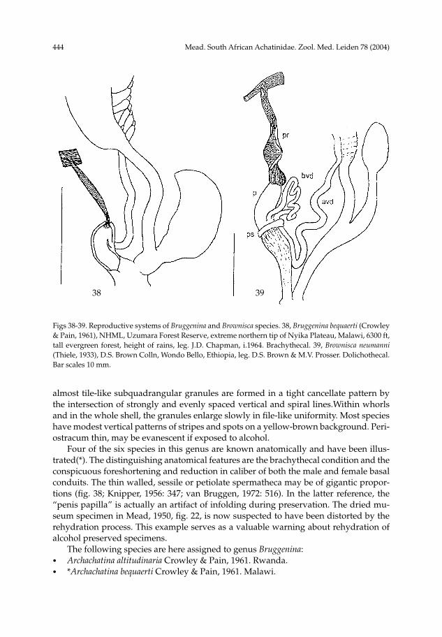
almost tile-like subquadrangular granules are formed in a tight cancellate pattern by the intersection of strongly and evenly spaced vertical and spiral lines.Within whorls and in the whole shell, the granules enlarge slowly in file-like uniformity. Most species have modest vertical patterns of stripes and spots on a yellow-brown background. Peri-ostracum thin, may be evanescent if exposed to alcohol. Four of the six species in this genus are known anatomically and have been illus-trated(*). The distinguishing anatomical features are the brachythecal condition and the conspicuous foreshortening and reduction in caliber of both the male and female basal conduits. The thin walled, sessile or petiolate spermatheca may be of gigantic propor-tions (fig. 38; Knipper, 1956: 347; van Bruggen, 1972: 516). In the latter reference, the “penis papilla” is actually an artifact of infolding during preservation. The dried mu-seum specimen in Mead, 1950, fig. 22, is now suspected to have been distorted by the rehydration process. This example serves as a valuable warning about rehydration of alcohol preserved specimens. The following species are here assigned to genus Bruggenina:• Archachatina altitudinaria Crowley & Pain, 1961. Rwanda.• *Archachatina bequaerti Crowley & Pain, 1961. Malawi.
Figs 38-39. Reproductive systems of Bruggenina and Brownisca species. 38, Bruggenina bequaerti (Crowley & Pain, 1961), NHML, Uzumara Forest Reserve, extreme northern tip of Nyika Plateau, Malawi, 6300 ft, tall evergreen forest, height of rains, leg. J.D. Chapman, i.1964. Brachythecal. 39, Brownisca neumanni (Thiele, 1933), D.S. Brown Colln, Wondo Bello, Ethiopia, leg. D.S. Brown & M.V. Prosser. Dolichothecal. Bar scales 10 mm.
3938
Mead. South African Achatinidae. Zool. Med. Leiden 78 (2004) 445
• Archachatina gebhardti Knipper, 1956. Tanzania.• *Archachatina meadi Bequaert, 1950; Mead, 1950, fig. 22. Tanzania. • *Archachatina sandgroundi Bequaert, 1950; van Bruggen, 1972, figs 1-4. Zimbabwe.• *Archachatina saskai Knipper, 1956, fig. 3. Tanzania. A. sandgroundi has been selected as type species of the genus because it has the most information recorded in the literature. Van Bruggen’s report (1972) on Archachatina sandgroundi and Knipper’s earlier re-port (1956) on A. saskai and A. gebhardti constitute the most valuable available sources of information that relate to this new genus. Distribution.— The species in this Rift Valley genus centre in Tanzania, with out-posts in Rwanda and Zimbabwe. The recent intensive collecting in this region by Drs M. Seddon and P. Tattersfield of the National Museum, Cardiff, has revealed valuable new material and information relevant to this new genus. Dedication.— It is a privilege to dedicate this genus to Dr Dolf van Bruggen of Leiden, Netherlands, who has published richly over many years on the malacology of Africa, particularly southern Africa, and is considered an authority in the Achatinidae, among other groups.
Brownisca gen. nov.
The transfer to Bruggenina of six of the ten species still remaining in Bequaert’s Archachatina, subgenus Tholachatina, leaves a group of four conspicuously similar appearing species, all of which are from northeast Africa: • A. neumanni Thiele, 1933. Ethiopia.• A. obtusa (Connolly, 1931). Uganda.• A. stefaninii (Connolly, 1925). Somalia.• A. weberi Bequaert, 1950. Sudan. Coincidentally, Bequaert anticipated this exact grouping of species within his Thola-chatina (1950: 206). In the shell of Brownisca, the apex is large and dome shaped; the early whorls descend and expand quickly, producing a broad, long spire that is longer than the aperture; and the sculpture is fine, uniform and depressed. It was this latter character that probably persuaded Bequaert to include them along with those species now in Bruggenina when he established subgenus Tholachatina of Archachatina. Verd-court (1966: 109) illustrates a paratype of “Archachatina obtusa” which Bequaert (1950) had moved to Bequaert’s new name, Euaethiopina from Clench & Archer’s (1930) junior primary homonym Euaethiops. The multiple taxonomic confusion is explained in Mead (1995: 263). In this genus, only the soft anatomy of dolichothecal A. neumanni is known (fig. 39). On this basis, this species is here designated type species of the new genus Brownisca. Under the circumstances of the present great paucity of information, but given their fair conchological similarity, their relatively near geographic proximity, and their long his-toric association in Tholachatina, it is wise to retain provisionally the present group in Brownisca. The soft anatomy proves to be of extraordinary proportions and unique in the family with respect to its reproductive pattern (fig. 39). Paradoxically, the basal vas deferens (bvd) leaves the penis not apically and adjacent to the origin of the penial re-
446 Mead. South African Achatinidae. Zool. Med. Leiden 78 (2004)
tractor (pr), as is normal in the family. Instead, it leaves at the base of what has become a greatly elevated, featureless cul-de-sac type of penial tube (p). At its base it is sharply delineated by an exceedingly short, collar-like penis sheath (ps), only one-fifth as long as it is wide. Adjacently at this base, the basal vas deferens exits the penial tube, imme-diately forms a tightly curled, elevated loop, and then slips under the collar of the penis sheath, only to emerge within 3-4 mm as the apical vas deferens (avd). A distinctive feature of the penis sheath in this species is the rapid diminution and attenuation of the muscle tissue immediately below the penis sheath collar, so much so that the basal lim-its of the penis sheath can scarcely be determined. And even the penial retractor is ex-traordinary in that it is longitudinally folded and transversally laminated. A second specimen in the same lot revealed essentially the same genital configuration, eliminating the initial impression that the first specimen was strangely malformed. The dolichothecal basal female conduit showed no corresponding departure from a generalized achatinid configuration. Although the species of Cochlitoma also are doli-chothecal, the deviations in the male conduit of A. neumanni warrant a generically dis-tinct status. Nor can the brachythecal Bruggenina be considered an acceptable genus for this species. Only until additional soft anatomies in this genus can be examined will the true taxonomic status of Brownisca be known. Dedication.— The two known preserved soft anatomies and the three dry speci-mens were collected in Wondo Bello Forest, 20 km SE of Shashamanne (Sashamene), Ethiopia, by the late Dr David S. Brown of The Natural History Museum, London. Since he ranks as a world authority on the freshwater snails of Africa, the genus suitably bears his name. Two additional good dry specimens of A. neumanni were more recently discovered in the Leiden Museum (RMNH) bearing the following data: Reg. No. 2013. Ethiopia, ca. 30 km SSE of Sashamene, ca. 250 km S of Addis Ababa, in montane gallery forest, Mrs B.E.E. de Wilde-Duyfjes, 16.vi.1965.
Mead. South African Achatinidae. Zool. Med. Leiden 78 (2004) 447
v vaginavl ventral penial lobevlc ventro-left lateral cleft
Institutional acronyms
ANSP Academy of Natural Science, Philadelphia (C. Fricker)ELM East London Museum, South Africa (M. Bursey)LNK Landessammlungen für Naturkunde, Karlsruhe (H.W. Mittmann)MCZ Museum of Comparative Zoology, Harvard (K.J. Boss)MNHN Muséum National d’Histoire Naturelle, Paris (S. & A. Tillier)NHML The Natural History Museum, London (P. Mordan, F. Naggs, D.S. Brown)NM Natal Museum, Pietermaritzburg, South Africa (D.N. Kilburn, D.G. Herbert)NMC National Museum of Wales, Cardiff (M. Seddon, P. Tattersfield)NMW Naturhistorisches Museum, Wien (E. Edlinger)RMNH Nationaal Natuurhistorisch Museum, Leiden (E. Gittenberger, A.C. van
Bruggen)RBINS Royal Belgian Institute of Natural Sciences, Brussels (J. van Goethem)RMCA Royal Museum for Central Africa, Tervuren (R. Jocque, D. van den Spiegel)SMF Forschungsinstitut und Naturmuseum Senckenberg, Frankfurt (R. Janssen)UOS University of Stellenbosch, South Africa (W.F. Sirgel)ZMB Museum für Naturkunde der Humboldt-Universität, Berlin (M. Glaubrecht)ZSM Zoologische Staatssammlung, München (B. Ruthensteiner)
Acknowledgements
Richard N. Kilburn and Dai G. Herbert (NM) and Mary Bursey (Senior Museum Naturalist, ELM) have generously and repeatedly provided what in sum has proved to be an extremely valuable base for assessing the phylogeny of the southern African Acha-tinidae. All have initiated special expeditions to make extensive searches in suspect lo-calities. Both A.C. van Bruggen (RMNH) and W.F. Sirgel (UOS) are specialists in the Achatinidae and are experienced in the soft anatomy of some of its species. Their pub-lications and council, for a long time, have influenced the conclusions reached in the present work. Sincere thanks are given to these and to others who also have contributed substan-tially to the completion of this project. The personnel listed in the accompanying In-stitutional acronyms, who provided and generously extended loans of revelant speci-mens; Peter Mordan, Fred Naggs, David Brown, Joan Pickering, and Kathy Way (NHML) who, over the years, have willingly provided assistance in special problems in the literature and in the collections; Margaret G. Kidwell of our Department of Ecology and Evolutionary Biology, who volunteered critical administrative assistance; C.A. Hedgcock, Senior Research Specialist of our Gould-Simpson research laboratories, who took charge of all aspects of the photography; Emilee M. Mead of Desert Archaeology, Inc., Tucson, who labeled the line drawings and prepared their layout; Mark Brewer and Joseph A. Romero, of our department, who did the word processing; and finally my wife Eleanor, who has given over sixty-one years of enthusiastic assistance in my
448 Mead. South African Achatinidae. Zool. Med. Leiden 78 (2004)
malacological endeavors. This project could not possibly have been completed in its necessarily limited time frame without the help of these generous people.
References
Bequaert, J.C., 1950. Studies in the Achatininae, a group of African land snails.— Bulletin of the Museum of Comparative Zoology at Harvard College 105: 1-216.
Bequaert, J.C., & W.J. Clench, 1934. Studies on African land and fresh-water mollusks. IV Descriptions of four presumably new Achatinidae; with notes on the genus Leptocala.— Revue de Zoologie et de Botanique Africaines 24: 269-276.
Bruggen, A.C. van, 1965. Two new species of Achatinidae (Mollusca, Gastropoda Pulmonata) from the Drakensberg Range, with general remarks of Southern African Achatinidae.— Revue de Zoologie et de Botanique Africaines 71: 79-91.
Bruggen, A.C. van, 1966a. Notes on non-marine molluscs from Mozambique and Bechuanaland, with a checklist of Bechuanaland species.— Annals of the Transvaal Museum 25: 99-112.
Bruggen, A.C. van, 1966b. The terrestrial Mollusca of the Kruger National Park: a contribution to the malacology of the Eastern Transvaal.— Annals of the Natal Museum 18: 315-399.
Bruggen, A.C. van, 1967. Miscellaneous notes on Southern African Gastropoda Euthyneura (Mollusca).— Zoologische Verhandelingen Leiden 91: 1-34.
Bruggen, A.C. van, 1968. Additional data on the terrestrial molluscs of the Kruger National Park.— Annals of the Natal Museum 20: 47-58.
Bruggen, A.C. van, 1970. Non-Marine Mollusca.— South African Animal Life (Results of the Lund University Expedition in 1950-51) 14: 444-476.
Bruggen, A.C. van, 1972. New data on Southern African Achatinidae (Mollusca, Gastropoda Pulmona-ta).— Zoologische Mededelingen Leiden 47: 513-529.
Bruggen, A.C. van 1977. Some Southern African Achatinidae (Mollusca, Gastropoda Pulmonata) in American museums, with the description of a new species.— Proceedings Koninklijke Nederlandse Akademie van Wetenschappen (C) 80: 245-256.
Bruggen, A.C. van, 1984. Size and distribution of Archachatina ustulata, a South African achatinid land snail, with the description of a new subspecies.— Basteria 48: 31-36.
Bruggen, A.C. van, 1985. The terrestrial molluscs of Lesotho (Southern Africa), a first contribution, with detailed notes on Archachatina machachensis (Mollusca, Gastropoda).— Proceedings Koninklijke Nederlandse Akademie van Wetenschappen (C) 88: 267-296.
Bruggen, A.C. van, 1989. Notes on three species of Archachatina (Mollusca, Gastropoda Pulmonata: Achatinidae) from eastern South Africa.— Proceedings Koninklijke Nederlandse Akademie van Wetenschappen (C) 92: 165-177.
Bruggen, A.C. van, & C.C. Appleton, 1977. Studies on the ecology and systematics of the terrestrial molluscs of the Lake Sibaya area of Zululand, South Africa.— Zoologische Verhandelingen Leiden 154: 1-44.
Clench, W.J., & A.F. Archer, 1930. New land snails from Tanganyika Territory.— Occasional Papers of the Boston Society of Natural History 5: 295-300.
Connolly, M., 1912. A revised reference list of South African non-marine Mollusca; with descriptions of new species in the South African Museum.— Annals of the South African Museum 11: 59-306.
Connolly, M., 1925. The non-marine Mollusca of Portuguese East Africa.– Transactions of the Royal Society of South Africa 12: 105-220.
Connolly, M., 1931. Descriptions of new non-marine Mollusca from North, South, and Central Africa, with notes on other species.— Annals & Magazine of Natural History (10) 8: 305-338.
Connolly, M., 1939. A monographic survey of South African non-marine Mollusca.— Annals of the South African Museum 33: 1-660.
Cortie, M.B., & D. Aiken, 1997. The Achatinid landsnails of South Africa.— Special Publication The Conchological Society of Southern Africa 7: 1-15.
Férussac, Baron A.E. de, 1821-1822. Tableaux systématiques des animaux mollusques: i-xlviii, 1-114.— Paris.
Mead. South African Achatinidae. Zool. Med. Leiden 78 (2004) 449
Knipper, H., 1956. Neue Achatiniden aus Ostafrika (Moll. Gastrop. Stylomm.).— Veröffentlichungen aus dem Überseemuseum Bremen (A) 2: 341-356.
Mead, A.R., 1950. Comparative genital anatomy of some African Achatinidae (Pulmonata).— Bulletin of the Museum of Comparative Zoology at Harvard College 105: 219-291.
Mead, A.R., 1979. Anatomical studies in the African Achatinidae - a preliminary report (Proc. Sixth Eur. Malac. Congr. Amsterdam, 1977).— Malacologia 18: 133-138.
Mead, A.R., 1988. Anatomy of the South African Archachatina ustulata (Lamarck) (Pulmonata: Achatini-dae).— Journal of Molluscan Studies 54: 363-365.
Mead, A.R., 1991. Anatomical criteria in the systematics of the Achatinidae (Pulmonata).— Proceedings of the Tenth International Malacological Congress (Tübingen, 1989) (2): 549-553.
Mead, A.R., 1994. A new subfamily and genus in Achatinidae (Pulmonata: Sigmurethra).— Bulletin of the Natural History Museum, London (Zoology) 60(1): 1-37.
Mead, A.R., 1995a. Anatomical studies reveal new phylogenetic interpretations in Lissachatina (Pulmo-nata: Achatinidae).— Journal of Molluscan Studies 61: 257-273.
Mead, A.R., 1995b. Anatomy, phylogeny, and zoogeography in the African land snail family Achatinidae. Unitas Malacologica.— Abstracts Twelfth International Malacological Congress, Vigo, Spain, 1995: 422-423.
Mead, A.R. 1998. A new species of Archachatina in the Dahomey Gap of West Africa and its implications in phylogeny (Pulmonata: Achatinidae).— Journal of African Zoology 112: 123-145.
Mead, A.R. 2001. Reproductive anatomy defines phylogeny and interprets distribution in the African giant land snails.— Abstracts Unitas Malacologica, World Congress of Malacology 2001, Vienna, Austria: 215.
Pilsbry, H.A., 1904-1905. African Achatinidae.— Manual of Conchology (2) 17: i-xviii, 1-232 (pp. 76-104). Academy of Natural Sciences, Philadelphia.
Pilsbry, H.A., & T.D.A. Cockerell, 1933. African Mollusca, chiefly from the Belgian Congo.— Proceedings of the Zoological Society of London 1933: 365-375.
Sirgel, W.F., 1989. A new species of Achatinidae from southern Africa (Mollusca: Gastropoda: Pulmo-nata).— Annals of the Natal Museum 30: 197-210.
Sirgel, W.F., 2000. Comparative genital anatomy of some South African Achatinidae (Pulmonata).— Annals of the Natal Museum 41: 209-235.
Verdcourt, B., 1966. The identity of Achatina bloyeti Bourguignat (Mollusca: Achatinidae) with some notes on other species of the genus occurring in East Africa.— Revue de Zoologie et de Botanique Africaines 74: 97-120.
Received: 26.vii.2004Accepted: 11.x.2004Edited: A.C. van Bruggen
450 Mead. South African Achatinidae. Zool. Med. Leiden 78 (2004)