The small animal surgeon routinely creates wounds in the gastrointestinal (GI) tract forbiopsy, for foreign body or neoplasm removal for correction of gastric dilatationvolvulus, or to relieve intestinal and colonic obstruction. Unlike dehiscence of a skinwound, which is often easily remedied with appropriate local wound treatment,dehiscence of a wound of the GI tract often leads to generalized bacterial peritonitisand potentially death. Consequently, technical failures and factors that negativelyaffect GI healing are of great clinical significance to the surgeon. Surgery of the GItract must be considered clean-contaminated at best, and as one progresses aborallydown the GI tract, the bacterial population increases. Therefore, intraoperativespillage, wound dehiscence, or perforations that occur in the lower small intestine orcolon tend to be associated with a higher mortality rate than those of the stomach orupper small intestine.
WOUND HEALING OF THE GI TRACT
Basic understanding of GI tract healing is essential to the surgeon since it dictatesproper clinical approach in those cases in which GI complications develop. Immedi-ately after wounding, platelets aggregate, the coagulation mechanism is activated,and fibrin clots are deposited to control hemorrhage.1 The fibrin clot offers minimalwound strength on the first postoperative day, but the main wound support during thelag phase of healing comes from the sutures.2 Fibrin also has adhesive properties andmay increase the risk of secondary obstruction since these fibrinous adhesions mayultimately be converted to fibrous adhesions. Enterocyte regeneration begins almostimmediately after wounding; however, the epithelium offers little biomechanical
The author has nothing to disclose.Department of Small Animal Clinical Sciences, College of Veterinary Medicine, PO Box 100126,Health Science Center, University of Florida, Gainesville, FL 32610-0126, USAE-mail address: [email protected]
support.2 This lag or inflammatory phase is the most critical period during GI woundhealing, and most dehiscences take place within 72 to 96 hours.2
The proliferative or logarithmic phase of GI wound healing lasts from days 3 through14.1 Fibroplasia occurs logarithmically during this period. The fibroblasts producelarge amounts of immature collagen, resulting in rapid gains in wound strength, butthis is a dynamic process in which collagen synthesis takes place in the presence ofcollagenolysis. In the stomach and small intestine, collagenase activity at the woundedge is minimal and rapid gains in tensile and bursting strength occur. At the end of14 days, gastric and small intestinal wound bursting strength is approximately 75%that of normal tissue.1 Conversely, the colon heals much more slowly due to markedcollagenase activity at the wound edge and regains only about 50% of its normaltensile strength 14 days post wounding.1 Factors such as traumatic suturing, fecalcontamination, and infection all increase the amount of local collagenase produced atthe wound and hence can increase the risk of infection.1
The maturation phase of wound healing is characterized by reorganization andcross-linking of collagen fibers. This phase extends from day 14 through day 180 inthe gastrointestinal tract of the dog.1 Similar to skin wounds, the size and thicknessof the scar decrease during this time without weakening the wound. The maturationphase is relatively unimportant clinically in GI wound healing, except in those caseswhere significant adhesions are present or in cases of sclerosing encapsulating orfibrosing peritonitis.3,4
COMPLICATIONS ASSOCIATED WITH GASTRIC DILATATION VOLVULUSPredictors of Mortality and Gastric Necrosis
Mortality continues to occur in all published reports of gastric dilatation volvulus(GDV). However, over the past four decades, survival rates have improved due to earlyrecognition, rapid gastric decompression, earlier cardiovascular resuscitation, andavailability of better medical and surgical care. In older studies from the 1970s andearly 1980s, GDV-related mortality rates ranged from 43% to 60%.5,6 However, in alarge epidemiologic study of over 1900 dogs with GVD, the mortality rate had reducedto 33% in cases evaluated during the late 1980s and early 1990s7 and was evenreported to be as low as 15% in the mid 1990s.8 In a recent study the postoperativemortality rate of 306 dogs with GDV was 10%. Those dogs receiving gastropexy alonehad mortality of only 3%, while dogs receiving partial gastrectomy with gastropexyhad a rate of 9% and those receiving partial gastrectomy, splenectomy, andgastropexy had a rate of 20%.9 That study also showed a reduction of mortality indogs if the clinical signs occurred less than 6 hours prior to presentation. While onestudy suggested that reduced duration of clinical signs and shorter time frompresentation to the surgery table also decreased mortality rate in 166 dogs withGDV,10 a second study contradicted these findings.11 Severity of clinical presentationhas also been related to outcome, with recumbency at presentation increasing risk ofdeath by 4.4 times and dogs with varying degrees of obtundation having mortality thatranged between 3% and 36%.7–9,12
The presence of preoperative cardiac ventricular dysrhythmias has been evaluatedas a predictive indicator for survival in dogs with GDV since they may act as a sentinelfor gastric or splenic ischemia. In one study, dogs having intermittent ventriculararrhythmia on admission had a significantly higher mortality rate than did dogs withventricular tachycardia. Additionally, 48% of those dogs presenting with preoperativecardiac arrhythmias underwent splenectomy or partial gastrectomy, whereas just27% of the dogs without arrhythmias required those procedures.9 However, in
916 Ellison
another study of 295 dogs with GDV, 40% of the animals developed arrhythmiasperioperatively and survival rate was unaffected by their presence.8
Approximately 40% of dogs with GDV also develop concurrent disseiminatedintravascular coagulopathy (DIC). Reported abnormal hemostatic profiles with GDVinclude prolonged prothrombin time (PT), prolonged activated partial thromboplastintime (aPTT), reduced fibrinogen concentration, increased fibrin degradation product(FDP) concentration, reduced platelet counts, and reduced antithrombin III (ATIII)activity, and these may be useful in estimating gastric ischemia/necrosis and DIC. Ina study of 20 dogs with GDV, thrombocytopenia occurred in 9 of 20 dogs, followedby decreased ATIII (8 of 20), elevated FDP (6 of 20), prolonged PT (6 of 20),hypofibrinogenemia (6 of 20), hyperfibrinogenemia (5 of 20), prolonged aPTT (5 of 20),and shortened aPTT (4 of 20). Approximately 70% of dogs with more than oneabnormal hemostatic test result had gastric necrosis, whereas dogs with one or noabnormality rarely had gastric necrosis.13
Considerable attention has turned to serum lactate as predictor of gastric necrosisand increased mortality. In an original retrospective study of 102 dogs with GDV, dogswith serum lactate values less than 6 mM had a survival rate of 99%, whereas dogswith initial lactate concentrations of greater than 6 mM were associated with only a58% survival rate.13 A more recent study showed a positive predictive correlationbetween lactate values before and after intravenous fluid resuscitation. In that study,even animals with an elevated initial serum lactate less than 9 mM were likely tosurvive, as long as the post resuscitation value dropped to 5.6 mM or less.14 Otherfactors reported to increase postoperative mortality include hypotension, concurrentperitonitis, DIC, and blood plasma transfusions.10
Although the timing of surgery varies between studies, what is clear is that shocktherapy and gastric decompression need to be followed by definitive surgicalmanagement of GDV. Definitive management of GDV involves (1) repositioning of thestomach with resection or involution of any devitalized gastric wall and (2) aprophylactic gastropexy technique to prevent recurrence. In studies using shocktherapy and gastric decompression without gastropexy, the recurrence rate of GDVwas 56% to 76%, with many recurrences within 3 months after initial presenta-tion.15,16 Because of this we now advocate laparotomy as soon as the patient is areasonable anesthetic risk. This allows for early assessment of gastric wall viabilityand gastric derotation, which increases gastric circulation and helps with cardiovas-cular resuscitation.
DIAGNOSIS AND MANAGEMENT OF GASTRIC NECROSIS
Intraoperative clinical criteria for determining gastric wall viability include assessmentof color, peristalsis, and tissue thickness. Viable gastric wall may be discolored andblue or dark red in appearance but is normal thickness upon palpation and will oftencontract when pinched. Ischemic or necrotic gastric wall, on the other hand, is black,gray, or green and is thin on palpation and lacks peristalsis.17 Fluorescein dye is notan accurate indicator of ischemia since the gastric wall is too thick to be affected byvital dyes. However, surface laser Doppler flowmetry of the gastric serosal surfacehas been shown to be a relatively good predictor of viability.18
Standard methods for gastrectomy involve ligation of branches of the left gastro-epiploic arteries and veins, allowing areas along the greater curvature of the stomachto be resected. The stomach is resected back to areas of healthy bleeding. Spillageis prevented through the use of Babcock forceps or stay sutures. The mucosa andsubmucosa are closed with a continuous inverted Cushing pattern. The serosa andmuscularis are then closed with a similar pattern. We have also used utostapling
917Complications of Gastrointestinal Surgery
equipment for rapid gastrectomy procedures with minimum risk of spillage. The TA 90autostapler is used with the green (4.8 mm) or blue (3.5 mm) cartridge. Often thesurgeon needs to overlap the staple lines by a few mm to prevent leakage betweenthe staples. The author has seen one clinical case in which perforation and leakageoccurred between the overlapping staple lines several months after the originalresection. Partial invagination of the stomach has been a useful technique for thoseanimals with small areas of gastric necrosis (Fig. 1A). With this technique, ligation ofsome of the short gastric vessels may be necessary to allow transection of thegastrosplenic ligament.19 The area of gastric necrosis is then invaginated using acontinuous Lembert suture pattern (see Fig. 1B). Since the invaginated tissue willultimately slough, an H2 antagonist such as cimetidine or mucosal protectant such assucralfate should be given for 5 to 7 days postoperatively. While the development ofa bleeding ulcer in a single dog 21 days after gastric resection prompted arecommendation for prolonged administration of gastric protectants after this proce-dure,20 the author has used the invagination technique successfully on many dogsthat failed to develop overt signs of gastric ulceration after surgery.
RATIONAL FOR PROPHYLACTIC GASTROPEXY
There are reports documenting the occurrence of GDV following splenectomy forlarge splenic tumors or splenic torsion in dogs.21,22 Because of this, many veterinary
Fig. 1. (A) Intraoperative image of the stomach of a 3-year-old Chinese sharpei presenting forGDV. A 3 � 3 cm area of full-thickness necrosis exists along the ventral gastric fundus. (B) Thearea of gastric necrosis has been invaginated using a continuous Lembert suture pattern.
918 Ellison
surgeons in North America advocate the practice of performing a prophylacticgastropexy after a splenic mass is removed in large or giant breeds of dogs. A recentreport from the United Kingdom controverts these earlier studies, suggesting thatthere was no increased risk of dogs with splenectomy developing GDV compared tothe risk incidence for the general population of dogs.23 The contradictory conclusionsof these studies may be related to differences in breed distribution, leaving the issueof prophylactic gastropexy after splenectomy somewhat controversial.
While gastropexy is clearly indicated as a therapeutic procedure in cases ofnaturally occurring GDV,11 there is now growing evidence that it may be indicated asa prophylactic procedure in dogs at risk for developing the syndrome. In 136 dogswith GDV, there was only a 4.3% recurrence rate in dogs and a 547-day mediansurvival time in dogs receiving a gastropexy procedure. Conversely, there was arecurrence rate of 54.5% with a median survival time of 188 days in those dogs notreceiving a GDV. The mortality associated with GDV recurrence was reported to be83% in those dogs not receiving gastropexy.11
Breed incidence of GDV has been closely evaluated, with giant and large breedshaving a lifetime probability of 22% to 24% in 1914 dogs as determined by studies usingthe Veterinary Medical Data Base.7 Also, great Danes with moderate and high abdominalheight-to-width ratios were approximately 5½ to 8 times as likely to develop GDV as werethose with low abdominal height-to-width ratios. Breeds such as the great Dane andbloodhound had reported lifetime incidences of 53% and 39%, respectively.24 Adecision-tree analysis for prophylactic gastropexy using lifetime probability of death fromGDV and expected cost savings for veterinary services as outcome measures wasundertaken to determine the preferred course of action in several dog breeds.25
Prophylactic gastropexy was the preferred choice of action for all breeds examined, withthe reduction in mortality (vs no gastropexy) ranging from 2.2-fold (Rottweiler) to 29.6-fold(great Dane). Assuming a prophylactic gastropexy costs US$400, the procedure was costeffective when the lifetime risk of GDV was 34% or greater.25
Complications associated with gastropexyMost North American surgeons use an antral gastropexy procedure to affix thestomach to the right abdominal wall and prevent GDV. Common antral gastropexytechniques used in North America include the tube gastrostomy,26 the incisionalgastropexy,27 the circumcostal gastropexy,28 the belt-loop gastropexy,29 and theventral midline gastropexy.16 Recurrence rates are typically reported 5% to 29% forthe tube gastrostomy and between 3% and 8% with the other techniques.30 In mostreports, recurrence is typically defined to include the occurrence of gastric dilatationwithout volvulus and does not necessarily indicate failure of the gastropexy site.Prophylactic gastropexy techniques may also be performed using minimally invasivetechniques such as laparoscopy or endoscopy.31
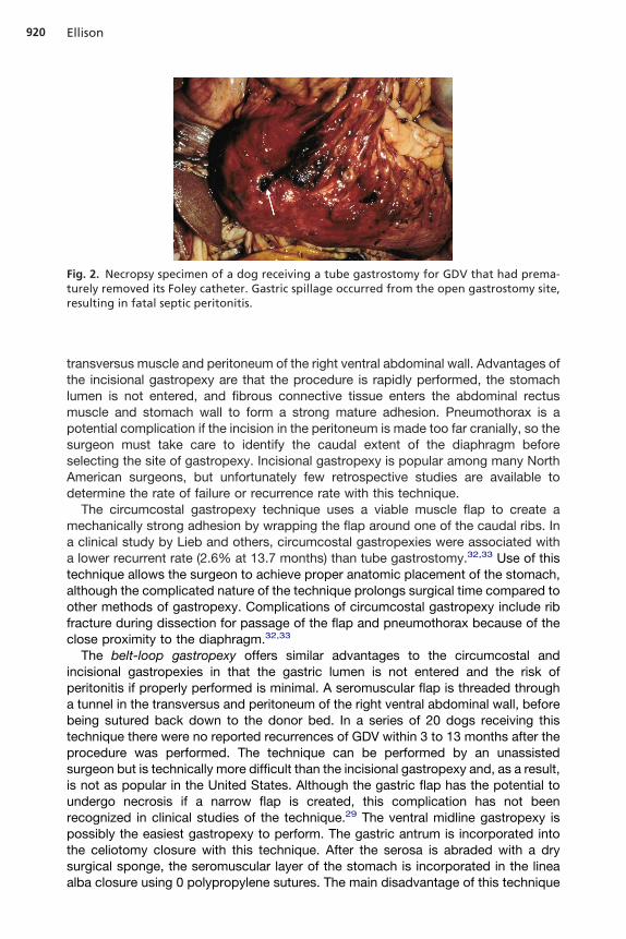
Potential advantages of the tube gastropexy are that the tube not only creates apermanent adhesion of the gastric antrum to the abdominal wall but also allows forcontinued gastric decompression in the early postoperative period. In addition, slurriedfood or medications can be offered through the tube. The main disadvantages of thetechnique are the nursing care and long hospital period required for tube managementand the potential for fatal peritonitis secondary to leakage around the tube or earlyremoval by the dog (Fig. 2). In a study by Fox and others, complication rate of tubegastropexy was approximately 18%, with local cellulitis around the tube site being themost common complication and 2 of 24 dogs developing fatal peritonitis.32
Incisional gatropexy is performed by making a 3- to 4-cm longitudinal incision in theseromuscular layer of the antrum, suturing this to a similar length incision in the
919Complications of Gastrointestinal Surgery
transversus muscle and peritoneum of the right ventral abdominal wall. Advantages ofthe incisional gastropexy are that the procedure is rapidly performed, the stomachlumen is not entered, and fibrous connective tissue enters the abdominal rectusmuscle and stomach wall to form a strong mature adhesion. Pneumothorax is apotential complication if the incision in the peritoneum is made too far cranially, so thesurgeon must take care to identify the caudal extent of the diaphragm beforeselecting the site of gastropexy. Incisional gastropexy is popular among many NorthAmerican surgeons, but unfortunately few retrospective studies are available todetermine the rate of failure or recurrence rate with this technique.
The circumcostal gastropexy technique uses a viable muscle flap to create amechanically strong adhesion by wrapping the flap around one of the caudal ribs. Ina clinical study by Lieb and others, circumcostal gastropexies were associated witha lower recurrent rate (2.6% at 13.7 months) than tube gastrostomy.32,33 Use of thistechnique allows the surgeon to achieve proper anatomic placement of the stomach,although the complicated nature of the technique prolongs surgical time compared toother methods of gastropexy. Complications of circumcostal gastropexy include ribfracture during dissection for passage of the flap and pneumothorax because of theclose proximity to the diaphragm.32,33
The belt-loop gastropexy offers similar advantages to the circumcostal andincisional gastropexies in that the gastric lumen is not entered and the risk ofperitonitis if properly performed is minimal. A seromuscular flap is threaded througha tunnel in the transversus and peritoneum of the right ventral abdominal wall, beforebeing sutured back down to the donor bed. In a series of 20 dogs receiving thistechnique there were no reported recurrences of GDV within 3 to 13 months after theprocedure was performed. The technique can be performed by an unassistedsurgeon but is technically more difficult than the incisional gastropexy and, as a result,is not as popular in the United States. Although the gastric flap has the potential toundergo necrosis if a narrow flap is created, this complication has not beenrecognized in clinical studies of the technique.29 The ventral midline gastropexy ispossibly the easiest gastropexy to perform. The gastric antrum is incorporated intothe celiotomy closure with this technique. After the serosa is abraded with a drysurgical sponge, the seromuscular layer of the stomach is incorporated in the lineaalba closure using 0 polypropylene sutures. The main disadvantage of this technique
Fig. 2. Necropsy specimen of a dog receiving a tube gastrostomy for GDV that had prema-turely removed its Foley catheter. Gastric spillage occurred from the open gastrostomy site,resulting in fatal septic peritonitis.
920 Ellison
is that if a future laparotomy is performed, there is risk that an inadvertent gastrotomymight occur with secondary gastric spillage.16
Laparoscopy-assisted or extracorporeal gastropexy procedures have been usedas elective procedures in breeds at risk for developing GDV. These elective proce-dures are commonly done on an outpatient basis and consistently are reported ascreating less pain for the patient and good efficacy for creation of a permanentadhesion. Potential complications include perforation of the gastric lumen or anylaparoscopy-related complication such as splenic laceration during insufflation. In aseries of 25 client-owned dogs, 23 received laparoscopy-assisted gastropexy as anelective procedure and 2 as a treatment for GDV. None of the dogs developed GDVthe year after surgery and 20 evaluated with ultrasonography were found to haveintact gastropexy attachments.31
In summary, with the exception of the tube gastrostomy a variety of open andminimally invasivegastropexy techniques are associated with a low failure rate andminimal complications if properly performed. There are inherent advantages and disad-vantages with each technique, making the choice of the procedure a matter of surgeon’spreference.
COMPLICATIONS FOLLOWING PARTIAL GASTRECTOMY
Gastric resection for GDV related necrosis usually has few long-term side effectssince the gastrectomy site is typically fundic in origin. Unfortunately, since mostmalignant neoplasms involve the pyloric antral area, resection of the pylorus isrequired. Pyloric resection may necessitate an end-to-side anastomosis of theduodenum to the stomach also known as a Billroth I gastroduodenostomy procedure.Alternatively, creation of a blind duodenal loop with end-to-side-to-side anastomosisof the stomach to the jejunum is known as a Billroth II gastrojejunostomy. Of these 2procedures, there is less postoperative vomiting and morbidity associated with thegastoduodenostomy procedure.34 The tumor mass may also invade the proximalduodenum and common bile duct, forcing the surgeon to anastomose the gallbladderto the duodenum (cholecystoduodenostomy) to reestablish bile flow back into theintestine. Ascending cholecystitis has been reported as a common sequelae to acholecystoenterostomy. The pathogenesis is thought to be due to a reflux ofduodenal contents into the gallbladder. Clinical signs include fever, abdominal pain,vomiting, neutrophilia, and elevation of liver enzymes. Patients usually respond to oralantibiotic therapy, but recurrent episodes are common. Creation of a stoma at least2.5 cm in length may decrease gallbladder retention of ingesta and minimize theoccurrence of postoperative cholecystitis (see Complications of Biliary Surgery).Other reported complications of cholecstoenterostmy in dogs include hepatic ab-scess, acquired portosytemic shunts, pancreatitis, and vomiting.35
Gastric bypass procedures are technically difficult to perform and have numerouscomplications associated with them, such as the dumping syndrome, marginal ulcersat the anastomosis site, and cholecystitis.34 After gastric resection and especiallygastojejunostomy procedures, rapid gastric emptying (dumping) occurs, which maylead to abdominal bloating, pain, vomiting, and diarrhea as well as vasomotorsymptoms, which also cause tachycardia.36 Since the pyloric antrum contains themucus-secreting cells of the stomach and is often removed during the resection,marginal ulceration of the anastomosis site may also occur.34 Damage to thepancreatic duct may cause resultant pancreatitis after any of the aforementionedgastrectomy techniques.35 Sporadic vomiting is usually seen during the first 24 to 72hours after pyloric surgery and is possibly due to bilious duodenal reflux into thecausing secondary gastritis. We therefore recommend treating the animal with gastric
921Complications of Gastrointestinal Surgery
protectants such as sucralfate or using H2 blockers such as ranitidine or famotidinefor 5 to 7 days postoperatively.
COMPLICATIONS ASSOCIATED WITH INTESTINAL SURGERYIntestinal Wound Dehiscence
Wound dehiscence of intestinal biopsy, enterotomy, or intestinal resection andanastomosis sites often leads to generalized bacterial peritonitis and subsequentdeath. Risk of intestinal anastomotic leakage can be affected by the cause of theobstruction, failure to adequately identify ischemic tissue, improper suturing orstapling technique, and a variety of factors that negatively affect wound healing suchas sepsis, malnutrition, and antineoplastic therapy. In a retrospective study of 115cases of intestinal anastomosis in dogs and cats, leakage occurred in 13 of 90 dogsbut in none of 25 cats.37 The incidence of leakage and postoperative complicationswas directly related to the cause of the problem, and mortality was higher in dogs thatneeded intestinal surgery because of foreign body obstruction compared to thosewith intestinal neoplasia.37 In this study, discriminate analysis indicated that dogs withpreoperative peritonitis, intestinal foreign body, and serum albumin concentration of2.5 g/dL or less were also most likely to have leakage from the intestinal wound.37 Asimple method of checking the anastomotic wound site for leakage is to inject thesutures site with saline to check for an adequate seal. In a recent study, 38 jejunalbiopsies in dogs were performed and closed using 3 or 4 full-thickness simpleinterrupted sutures. Saline volumes needed to achieve intraluminal pressures of 20and 34 cm H2O in a 10-cm canine jejunal segment containing a closed biopsy siteusing 2 methods of luminal occlusion were recorded. The 95% confidence intervalsfor the volume of saline needed to achieve 20 and 34 cm H2O intraluminal pressurewith digital occlusion were 10.9 to 13.6 mL and 16.3 to 19.0 mL, respectively, and withDoyen occlusion, 8.5 to 11.1 mL and 12.1 to 14.8 mL, respectively. Therefore,intestinal surgical sites can be checked intraoperatively for leakage by intraluminalinjection of 10 to 12 mL of saline if the loop of intestine is sealed with Doyen forcepsplaced 10 cm apart.38
FACTORS INCREASING THE RISK OF INTESTINAL LEAKAGEInadequate removal of ischemic tissueIschemic intestine is often black or gray and easily discernible from normal bowel.Determining viability in cyanotic appearing bowel is sometimes difficult. The intestinefirst should be decompressed with a needle and suction apparatus to relievevenous congestion. Intraoperative criteria for establishing intestinal viability arecolor, arterial pulsations, and the presence of peristalsis. Questionable areas ofbowel should be pinched to determine whether smooth muscle contraction andperistalsis are present. If clinical criteria are inadequate to determine viability,intravenous fluorescein dye or surface oximetry can be used. A 10% fluoresceinsolution is given at a dosage of 1 mL/5 kg intravenously through any peripheral vein.39
After 2 minutes, the tissues are subjected to long-wave ultraviolet light (Wood’slamp). Areas of bowel are considered viable if they have a bright green glow. Areasof bowel are not viable if they have a patchy density with areas of nonfluorescenceexceeding 3 mm, have only perivascular fluorescence, or are completely nonfluo-rescent.39 Oxygen saturation may also be a reliable method of determiningintestinal wall viability. A sterile probe is placed on the surface of the bowel andan oxygen saturation level reading will occur. According to published reports inrabbits, saturation levels above 81% typically indicate that the bowel is viable,whereas values below 76% were consistent with mucosal necrosis and thosebelow 64% indicated transmural intestinal necrosis.40
922 Ellison
Poor wound appositionDirect approximation of the wound edge allows for optimum rapid healing character-ized by primary intestinal wound healing. Accurate apposition allows for rapidmucosal reepithelialization, and early formation occurs of young well-vascularizedcollagen between the submucosa, muscularis, and serosa. Other advantages ofapproximating patterns for intestinal anastomosis are that (1) the lumen diameter isnot compromised, (2) the wound strength meets or exceeds everting or invertingwound strengths after 24 hours, and (3) the adhesions are minimal.2 Sutures shouldnot be tied too tight since crushing of tissue has been shown to cause more tissueischemia directly at the suture line and is discouraged. Mucosal eversion or tissueoverlap retards healing and should be avoided. Mucosal eversion results in delayedfibrin seal formation, delayed mucosal reepithelialization, increased mucocele forma-tion, prolonged inflammatory response, and marked adhesion formation. Eversionmay initially widen the intestinal lumen diameter, but the prolonged inflammatoryresponse eventually narrows the lumen, contributing to the risk of stenosis. Evertinganastomoses also have an increased tendency for leakage, especially in the face ofseptic peritonitis, and should never be used in the colon. Inversion of the wound edgecreates an internal cuff of tissue that reduces lumen diameter. Hemodynamiccompromise of the inverted submucosa occurs, resulting in mucosal edema andnecrosis. After 5 days the internal cuff usually sloughs. Inverting anastomoses arecharacterized by a rapid serosa-to-serosa seal and minimal adhesion formation. Yetbecause of their safety against leakage, inverting patterns may be the preferredtechnique for the colon.
As an alternative to hand suturing, autostapling may be used for intestinalanastomosis. The GIA and TA auto staplers lay an overlapping double row of staplesfor security and, when used in combination, create a functional “end-to-end anasto-mosis.” The GIA portion of the anastomosis is inverted, whereas the TA portion of theanastomosis is everted. Recent studies in human have shown that leakage rates aresimilar to hand-sewn techniques but autostapler use significantly reduces surgicaltime.41 In veterinary surgery, direct comparisons between hand-sewn and stapledanastomosis are poorly documented, but in a recent study, nonspecialists were ableto achieve very good results with stapling devices in 25 of 30 dogs, with anastomoticproblems occurring in only 2 dogs.42 An alternative to sutured anastomosis is the useof an AutoSuture Premium 35 skin stapler with stainless skin staples (Covidien,Norwalk, CT, USA). After triangulating the intestine with 3 stay sutures, the skinstapler is used to place staples every 2 to 3 mm around the perimeter of the wound.These closures are more rapidly done than hand-sewn anastomosis and have similarbursting strengths. However, mucosal eversion may occur between staples.43
Improper selection of suture materialFor hand-sewn anastomoses, either continuous or interrupted patterns can be usedwith equal efficacy. Although both absorbable and nonabsorbable suture materialshave been used successfully for anastomosis, the braided nonabsorbable suturematerials such as silk may harbor bacteria and create granulomatous inflammatoryreaction or draining suture sinus. Monofilament nonabsorbable sutures such as Nylonand polypropylene are safe in contaminated environments. However, polypropylenehas been associated with foreign body adherence in one case series.44 Absorbablesuture materials reported in the veterinary literature for intestinal suturing include chromicgut, polyglycolic acid (Dexon), polygalactin 910 (Vicryl), polydioxanone (PDS), polyg-lyconate (Maxon), and poliglecaprone (Monocryl). Of these, surgical gut is notrecommended for anastomosis because it is rapidly broken down by collagenase.
923Complications of Gastrointestinal Surgery
Polygalactin 910 and polyglycolic acid are braided and retain good tensile strength forup to 28 days. Vicryl is commonly used for intestinal anastomosis in humans withgood published success and is popular in Europe for veterinary use. In North America,monofilament sutures such as polydioxanone (PDS) and polyglyconate (Maxon) aremore commonly used. These polyester monofilament suture materials are absorbedby hydrolysis and therefore are unaffected by contaminated environment. Theymaintain up to 40% of their original tensile strength after 3 weeks. Many surgeons arealso starting to use shorter acting monofilament suture such as Monocryl or Biosynfor intestinal anastomosis. They have similar handling properties to PDS but aredegraded more quickly. The newer “Plus” sutures are impregnated with the antibac-terial agent Triclosan. Their efficacy in reducing infection in contaminated dermalwounds may foster an increased use in intestinal anastomosis.
OmentalizationAll anastomoses should be covered with a vascularized omental flap that is tacked inplace. Omentum is useful in (1) restoring blood supply to a devascularized area, (2)facilitating lymphatic drainage, and (3) minimizing mucosal leakage and secondaryperitonitis. The role of omentum is significant when one considers that in one study90% mortality rates were seen with intestinal anastomoses after omental resectionwas performed in dogs.45 Free omental flaps are not as effective as pedicle omentalflaps and may in fact lead to anastomosis failure.45
OTHER FACTORS AFFECTING DEHISCENCE
Healing of visceral wounds is negatively affected by a number of other factors.Chronic weight loss of 15% to 20% due to cancer cachexia or other reasons has anegative effect on visceral wound healing. Correction of cachexia as well as earlypostoperative enteral feeding appears to increase collagen deposition and burstingwound strength.46 Glucocorticoids have a negative effect on wound healing whengiven in large doses prior to the third day after wounding.1 NSAIDs appear to affectthe early inflammatory phase of wound healing but do not appear to interfere with theproliferative phase of wound healing or have a significant negative effect on visceralhealing strength.47 Radiation therapies interfere with fibroblast mobilization, replica-tion, and collagen synthesis as well as causing sclerosis of microvasculature, therebyreducing oxygenation at the wound site. Whenever possible, radiation therapy shouldbe initiated after visceral wound healing is complete. The negative effects of canceron wound healing appear to be secondary to nutritional deficiencies rather than directtumor impairment on wound healing. Visceral wound healing may actually be mildlyaugmented, owing to release growth factors by the neoplasm. Effects of chemother-apeutic agents on visceral wound healing are variable. Drugs such as vincristine,vinblastine, and azathioprine seem to be safe when used in therapeutic doses. Drugssuch as cyclophosphamide, methotrexate, 5-FU, and doxorubicin have been shownto delay wound healing in both experimental and clinical studies.46,48 Cisplatinappears to significantly impair intestinal wound healing in rats and should be usedwith caution after intestinal surgery.49
EFFECT OF EARLY POSTOPERATIVE ENTERAL FEEDING ON VISCERAL HEALING
Malnutrition induces intestinal mucosal atrophy, reduced motility, increased inci-dence of ileus, and the potential for bacterial translocation through the bowel wall,with resultant sepsis.50 Impaired wound healing due to nutritional causes may bereversed by feeding an enteral or a parenteral diet that supplies energy needs in the
924 Ellison
form of fatty acids and sugars and provides essential amino acids. Feedings of highprotein meals after injury can optimize conditions for normal visceral wound healing.Amino acids provided through enteral nutrition are utilized for the synthesis ofstructural proteins such as actin, myosin, collagen, and elastin. Early, if not immedi-ate, postoperative enteral feeding has been shown to have a positive influence on thehealing rate of intestinal anastomosis in dogs.51 Bursting pressures and collagenlevels of ileal and colorectal anastomosis were compared in beagles fed elementaldiets versus those fed only electrolytes and water for 4 days.51 The dogs fed orallyhad nearly twice the bursting strengths of the control group and nearly double theamount of both immature and mature collagen at the wound site.51 Total parenteralnutrition (TPN) does not appear to ameliorate the mucosal atrophy or increasecollagen deposition as did enteral nutrition.50 In human studies, the incidence ofseptic complications is significantly lower in people fed between 8 and 24 hours aftersurgery versus those maintained on TPN. Additionally, early-fed patients had areduced incidence of postoperative ileus and reduced hospital stay.50
EFFECTS OF MASSIVE SMALL INTESTINAL RESECTION
The propensity for short-bowel syndrome after massive intestinal resection dependson the amount of tissue excised, the location of the resection, and the time allowedfor adaptation. Resection of up to 80% of the small intestine in puppies may allow fornormal weight gain, whereas resection of 90% produces morbidity and mortality indogs.52 After resection of large portions of small intestine, maldigestion, malabsorp-tion, diarrhea induced by fatty acids or bile salts, bacterial overgrowth, and gastrichypersecretion may occur. Location of the resection is important in people. Highresection of the duodenum and upper jejunum may decrease pancreatic enzymesecretion because pancreatic-stimulating hormones such as secretin and cholecysto-kinin are produced in the mucosa of these sections. These reductions in release ofpancreatic enzymes contribute to maldigestion. Maldigestion of protein, carbohy-drate, and fat leads to catabolism, negative nitrogen balance, and steatorrhea.Unabsorbed sugars also may cause osmotic diarrhea. If the ileocecal valve isresected, bacteria may ascend, overgrow in the small bowel, and contribute todiarrhea. After massive resection, the remaining small intestine adapts by increasinglumen diameter, enlarging microvilli, and increasing mucosal cell number. Thesecompensatory changes may take several weeks; during this period, parenteral fluids,electrolytes, and hyperalimentation may be necessary for the survival of the animal.With proper supportive care, the animal will be able to maintain weight even withdiarrhea. Medical treatments for unresponsive diarrhea after massive resection indogs include frequent small meals, low-fat diets such as intestinal diet (I/D Hills,Topeka, KS), elemental diet supplements, medium-chain triglyceride oils, pancreaticenzyme supplements, B vitamins, kaolin antidiarrheals, and poorly absorbed oralantibiotics such as neomycin.
A recent retrospective study determined outcome in 13 dogs and 7 cats thatunderwent extensive (ie, �50%) resection of the small intestine.53 In this study, in all7 cats and 8 of the 13 dogs, extensive intestinal resection was performed because ofa foreign body. Mean � SD estimated percentage of intestine that was removed was68 � 14% (range, 50%–90%). Two dogs were euthanized 3 days after surgerybecause of dehiscence of the surgical site and development of septic peritonitis; 1dog died of acute respiratory distress syndrome 5 days after surgery. The remaining10 dogs and 7 cats were discharged from the hospital, and follow-up informationwas available for 15 of the 17. Median survival time was 828 days, and 12 of the15 animals for which long-term follow-up information was available had good
925Complications of Gastrointestinal Surgery
outcomes. However, none of the factors examined, including percentage ofintestine resected, were significantly associated with outcome. In summary, mostdogs and cats that underwent extensive resection of the small intestine had agood outcome after a variable period of intestinal adaptation. The amount ofintestine resected was not always associated with outcome.
SURGICAL MANAGEMENT OF INTESTINAL LEAKAGE
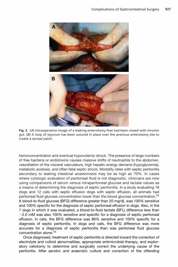
Revision of the primary surgical site, copious abdominal lavage, and broad-spectrumintravenous antibiotic therapy are the mainstays of addressing intestinal anastomoticleakage and are addressed separately in this chapter (Diagnosis and DrainageOptions for Septic Abdomen). After revision of the primary anastomotic site, mechan-ical reinforcement if the anastomosis is typically performed. Use of an omental pediclegraft (described earliere) is sufficient in most animals; however, when the omentum isdevitalized or when more substantial mechanical reinforcement is desired, intestinalserosal patching is recommended. The serosal patch technique uses the antimesen-teric surface of the small bowel to cover or buttress an adjacent area of questionabletissue viability or an area that cannot be reliably sutured. Jejunum is commonly usedbecause its freely movable mesentery allows it to be mobile. The serosal patchprovides mechanical stability and will help to induce and localize a fibrin seal over thequestionable area. A section of jejunum free of mesenteric tension is transposed overthe perforation or area to be buttressed. It is important not to stretch, kink, or twist itsmesenteric root or the vascular supply may be disrupted. The bowel chosen for thepatch is then gently looped to prevent luminal bowel obstruction. Multiple perfora-tions sometimes require patching using a back-and-forth looping of the entiresegment of bowel. The lateral aspects of the bowel wall or antimesenteric border areused for the patch (Fig. 3A). The patch is not sutured directly to the edges of thedefect but rather 3 to 4 mm beyond its margins. Simple interrupted sutures of 4-0nylon or polypropylene are placed 3 to 4 mm from the wound edges and 3 to 4 mmapart (see Fig. 3B). The sutures grasp the submucosa of the patch and bowel wall butdo not penetrate the lumen.
Externalized intestinal anastomosis has been used in humans in the management ofleaking colonic anastomosis, and this novel approach was successfully used tomanage a leaking ileocolic anastomosis in a dog.54 A 6-year-old, spayed femaleLabrador retriever was presented 48 hours after an intestinal resection and anasto-mosis for management of a small intestinal foreign body. Abdominal ultrasoundconfirmed the presence of peritoneal effusion. Cytology of fluid collected by abdom-inocentesis revealed a large number of degenerate neutrophils with intracellular cocci.A diagnosis of septic peritonitis was made, presumably because of dehiscence of theanastomosis. Upon repeat exploratory celiotomy, the intestinal anastomosis wasfound to be leaking intestinal contents into the abdomen. An end-to-end, ileocolicanastomosis was performed and subsequently exteriorized into the subcutaneousspace via a paramedian incision through the abdominal wall. The anastomosis wasinspected daily for 4 days before it was returned to the abdomen and the subcuta-neous defect was closed. Serial cytology of the peritoneal fluid, which was performedduring this 4-day postoperative period, confirmed progressive resolution of theperitonitis and the dog was discharged from the hospital with a successful recovery.
DIAGNOSIS AND DRAINAGE OPTIONS FOR SEPTIC ABDOMEN
With generalized septic peritonitis, massive fluid and protein movement to theperitoneal cavity result in a shift of fluid away from the intravascular space, causing
926 Ellison
hemoconcentration and eventual hypovolemic shock. The presence of large numbersof free bacteria or endotoxins causes massive shifts of neutrophils to the abdomen,vasodilation of the visceral vasculature, high hepatic energy demand (hypoglycemia),metabolic acidosis, and often fatal septic shock. Mortality rates with septic peritonitissecondary to leaking intestinal anastomosis may be as high as 70%. In caseswhere cytologic evaluation of peritoneal fluid is not diagnostic, clinicians are nowusing comparisons of serum versus intraperitoneal glucose and lactate values asa means of determining the diagnosis of septic peritonitis. In a study evaluating 18dogs and 12 cats with septic effusion dogs with septic effusion, all animals hadperitoneal fluid glucose concentration lower than the blood glucose concentration.55
A blood-to-fluid glucose (BFG) difference greater than 20 mg/dL was 100% sensitiveand 100% specific for the diagnosis of septic peritoneal effusion in dogs. Also, in the7 dogs in which it was evaluated, a blood-to-fluid lactate (BFL) difference less than�2.0 mM was also 100% sensitive and specific for a diagnosis of septic peritonealeffusion. In cats, the BFG difference was 86% sensitive and 100% specific for adiagnosis of septic peritonitis. In dogs and cats, the BFG difference was moreaccurate for a diagnosis of septic peritonitis than was peritoneal fluid glucoseconcentration alone.55
Once diagnosed, treatment of septic peritonitis is directed toward the correction ofelectrolyte and colloid abnormalities, appropriate antimicrobial therapy, and explor-atory celiotomy to determine and surgically correct the underlying cause of theperitonitis. After aerobic and anaerobic culture and correction of the offending
Fig. 3. (A) Intraoperative image of a leaking enterotomy that had been closed with chromicgut. (B) A loop of jejunum has been sutured in place over the previous enterotomy site tocreate a serosal patch.
927Complications of Gastrointestinal Surgery
problem, aggressive peritoneal lavage is performed; either the abdomen is closedprimarily or closed peritoneal drainage techniques are used. Thorough peritoneallavage of the abdominal cavity is performed with body-temperature 0.9% NaCl orlactated Ringer’s solution. Lavaging a cat with 500 to 750 mL and a large dog with 3to 5 L of fluid will help remove the bacteria and foreign debris, which are the initiatorsof the peritoneal inflammation. Although lavage of the peritoneum may have thetheoretical disadvantage of spreading bacterial contamination, peritoneal lavage iswell established as a means of reducing morbidity and mortality due to septicperitonitis. All lavage fluid should be aspirated because when bacteria are suspendedin residual lavage fluid, phagocytosis is impaired. Three cycles of lavage andaspiration are recommended.
Peritoneal drains, when used, vary in their effectiveness with single-lumen passivedrains such as Penrose tubes, recovering much less fluid than that collected bydouble-lumen active drains in most studies.56 Gravity drains have several otherdisadvantages, including early occlusion of the drain by the omentum, fibrin, orexudate and mechanical irritation of the peritoneum. Last, passive drains mayfacilitate migration of bacteria into the peritoneal cavity. Active suction drains, such asthe Jackson-Pratt drain, improve efficacy of fluid removal and maintain a closedsystem that minimizes bacterial contamination of the peritoneum, making them theclear choice if closed abdominal drainage is elected for following surgery. As anadded benefit to the use of closed suction drains, the clinician is able to monitorchanges in gross and cytologic features of peritoneal fluid on a daily basis until drainremoval is performed. Intra-abdominal drains may also be used to perform atechnique called intermittent peritoneal lavage. Isothermic, sterile fluids are adminis-tered via a peritoneal catheter or fenestrated tube. The fluids are then removed bygravity flow back through a separate outflow tube. Our clinical impressions are thatthis technique has been helpful in reducing mortality associated with diffuse perito-nitis. However, experimental results suggest that good lavage of the entire abdomenis not provided.57 The development of hypoproteinemia and hypokalemia is common,although these complications are less severe than those noted during open peritonealdrainage.
Open peritoneal drainage is a process by which the linea alba and skin are leftpartially or completely open and covered with sterile dressings, which are changed atfrequent intervals. The main advantages of open peritoneal drainage are that it allowsunimpeded drainage of fluid and exudate from the peritoneal cavity and at the sametime alters the anaerobic environment of the peritoneum. Many surgeons advocateopen peritoneal drainage as the optimal treatment for generalized septic peritoni-tis.58–60 The mortality rate associated with open peritoneal drainage for managementof septic peritonitis in dogs and cats ranges between 11% and 48%.56–58 Moststudies document that mortality is increased if sepsis is secondary to GI leakagecompared to leakage from the reproductive tract.58,59 The author facilitates openperitoneal lavage by placing continuous or interrupted Nylon or polypropylene suturesloosely in the linea alba, allowing it to gap 1 to 2 cm. The skin and subcutaneoustissues are not closed and are covered with antibiotic ointment and nonadherentgauze dressing before applying a large cotton padded bandage (Fig. 4). The animalis continuously observed and the bandage aseptically changed at 12- to 24-hourintervals under general anesthesia and strict aseptic technique. The abdominalwound is closed primarily in 1 to 5 days depending on when the peritonealinflammation has resided. Common complications associated with open peritonealdrainage are patient dehydration and hypoproteinemia secondary to massive fluidand protein loss into the bandage.
928 Ellison
It is unclear whether open or closed peritoneal drainage is superior in the treatmentof septic peritonitis in dogs and cats. Open peritoneal drainage is a more effectivetechnique for achieving peritoneal drainage than is tube drainage56 and is generallyrecommended if contamination is diffuse and not readily removed during the initialexploratory celiotomy. With open drainage, large volumes of abdominal fluid andexudate can be removed from the abdomen and the bacterial environment can befavorably altered, therefore decreasing the number of anaerobic microorganisms. Inhumans it has been reported that open abdominal drainage can improve a patient’smetabolic condition, reduce abdominal adhesion formation, and leave access forrepeated exploration and inspection of the abdomen.61 Yet that same study actuallyindicated a higher mortality rate associated with patients managed with open peritonealdrainage compared to patients managed with closed abdomens, largely due to theacquisition of nosocomial infections. Other reported complications included massive fluidand protein loss, increased nursing care, increased cost, additional anesthetic, entericfistula formation, incisional herniation, evisceration, small bowel obstruction secondaryto adhesion formation, and nosocomial infection.61 Information provided by a limitednumber of retrospective studies in veterinary medicine offer guidelines as to whenopen peritoneal drainage may be indicated.58–60,62,63 In general, open peritonealdrainage is considered in cases where the source of contamination cannot be
Fig. 4. (A) Image from a cat with multiple intestinal perforations secondary to an arrowtraversing the abdomen. After repair of all intestinal wounds, the abdomen was thoroughlylavaged and open abdominal drainage was performed. The abdominal wall has been partiallyclosed with a loosely placed polypropylene sutures. (B) The abdomen is covered with a steriledressing that is changed under general anesthesia or deep sedation on a daily basis. Theabdominal is typically closed within 1 to 5 days depending on the degree of inflammation.
929Complications of Gastrointestinal Surgery
identified, the contamination is severe or longstanding, or the virulence of organismsis considered high, such as that seen with fecal contamination or anaerobicinfections. In these situations, open peritoneal drainage offers the possibility ofreexploration and subsequent lavage. On the other hand, if the source of contami-nation is successfully eliminated and lavaging the abdomen is effective at removingresidual debris, the abdomen may be closed with good results in a majority of thecases.62
Vacuum-assisted closure (VAC) (V.A.C., Kinetic Concepts, Inc, San Antonio, TX,USA) is a relatively new concept in veterinary medicine that can be adopted for usein cases of septic peritonitis. Vacuum-assisted therapy consists of placing a dressingsponge made of open-cell polyurethane ether foam over the partially closed abdom-inal incision. Embedded in this sponge is a noncollapsible, side-ported evacuationtube that is connected to an adjustable pump. The vacuum applied may be usedintermittently or continuously. All the cells in the open cell foam communicate so thevacuum is evenly applied to all wound surfaces in contact with the sponge. A plasticadhesive drape (Ioban) is placed around the abdomen to create an air-tight seal. Theexcellent drainage characteristics provided by vacuum therapy appear to be atherapeutic option for the treatment of septic peritonitis. One human study compareddifferent bandaging techniques for the management of patients with open abdomensand found a 40% reduction in mortality in patients treated with VAC therapy.64 Thereare limited studies in the human literature supporting the use of VAC therapy inpatients with septic peritonitis, but the positive results from these studies areencouraging.64,65
COMPLICATIONS AFTER SUBTOTAL COLECTOMY
Subtotal colectomy is a salvage technique for treatment of recurrent megacolon. InNorth America, dogs have traditionally been treated medically because of theirpropensity for postoperative diarrhea after removal of large segments of the colon.However, in a recent UK study, 8 dogs with acquired megacolon underwent subtotalcolectomy with preservation of the ileocolic junction.66 The diagnosis was confirmedin all animals by abdominal palpation, plain radiography, and postoperative histo-pathologic findings. There were no intraoperative complications, although 1 dog diedpostoperatively as a result of septic peritonitis. Long-term follow-up was obtained byclinical records and telephone interviews with the owners of the 7 surviving dogs.Resolution of obstipation and improved stool consistency of the remaining dogs wereimproved at discharge and all surviving animals eventually returned to normaldefecation in 5 to 10 weeks and were alive 11 to 48 months (mean 40.5 months) aftersurgery. These results emphasized the long-term effectiveness of subtotal colectomywith preservation of the ileocolic junction in dogs with idiopathic megacolon.
In cats, subtotal colectomy has been a successful salvage procedure for idiopathicmegacolon for more than 25 years.67–70 Some controversy exists as to whether thececum should be preserved and a colon-to-colon anastomosis (colocolostomy) doneor whether it can be sacrificed and an ileum-to-colon anastomosis (ileocolostomy)performed. Ileocolostomy has the inherent luminal disparity to deal with but ligation ofthe ileocolic artery and removal of the cecum allow easy transposition of the mobileileum down to the colonic stump. With colocolostomy, luminal disparity is kept tominimum, but the technique may be technically demanding because the relativelyimmobile mesocolon places considerable tension on the suture line. Studies indicatethat removal of the cecum does not lead to ascending bacterial enteritis, and catswith ileocolostomies do as well clinically as those with the cecum preserved.69,70
930 Ellison
Colocolostomy can also be performed using an end-to-end anastomosis autostapler,but it must be done via a typhlotomy incision (EEA; Covidien Inc).
Cats are often somewhat depressed and anorectic for 48 hours following subtotalcolectomy. They will sometimes have a moderate fever of 103° to 103.5°F in theabsence of leukocytosis.68 Dark tarry liquid feces are usually noted for about 3 to 4days. The presence of abdominal tenderness, vomiting, detection of intracellularbacteria on peritoneal fluid cytology, or significant glucose differential betweenperitoneal fluid and serum warrants early reexploration of the abdomen. Normally,feces remain liquid and poorly formed for 2 to 6 weeks after surgery, at which timethey usually become soft and poorly formed for the remainder of the cat’s life.Long-term follow-up studies indicate that most cats seem to maintain their normalbody weight or even gain weight after the procedure.67,68 After surgery, cats generallyuse the litter box 2 or 3 times a day, but the total amount of water loss in the fecesequals that of normal cats.63 The ileum increases it absorptive capacity by increasingvillous height. Bacterial overgrowth, folic acid deficiency, and anemia are notuncommon, despite initial concerns.70 The major complaint of cat owners is chronicperineal soiling caused by the loose feces. If this becomes a problem, it can often bemanaged by clipping hair in the perineal area. Occasionally cats become reconsti-pated and must be treated medically with lactulose and cisapride for a period of time.In recurrent cases, reexploration and removal of additional residual colon aresometimes necessary.
SUMMARY
A large number of naturally occurring disease conditions are treated by GI surgery insmall animals. The GI tract is rich in blood supply and has the potential to heal in rapidfashion but GI effluent is contaminated and surgery is fraught with many pitfalls, themost notable of which is leakage of the surgical site due to technical error, patientcachexia, chemotherapy, metabolic disorders, or preexisting septic peritonitis. Oncepresent, septic peritonitis requires reoperation, patching techniques for the leakagesites, aggressive fluid resuscitation, and appropriate antibiotic therapy based onculture and sensitivity. Innovative surgical techniques such as the VAC drainage orextra abdominal anastomosis placement may increase the survival rates in thesecritical patients but need further investigation.
REFERENCES
1. Peacock EE. The gastrointestinal tract. In: Surgery and biology of wound repair. 3rdedition. Philadelphia: WB Saunders; 1984. p. 78–85.
2. Ellison GW. Wound healing in the gastrointestinal tract. Semin Vet Med Surg (SmAnim) 1989;4:287–98.
3. Boothe HW, Lay JC, Moreland KJ. Sclerosing encapsulating peritonitis in three dogs.J Am Vet Med Assoc 1991;198:267–70.
4. Hardie EM, Rottman JB, Levy JK. Sclerosing encapsulating peritonitis in four dogsand a cat. Vet Surg 1994;23:107–14.
5. Muir WW. Gastric dilatation-volvulus in the dog, with emphasis on cardiac arrhyth-mias. J Am Vet Med Assoc 1982;180:739–42.
6. Betts CW, Wingfield WE, Green RW. A retrospective study of gastric dilatation-torsionin the dog. J Sm Anim Pract 1974;15:727–34.
7. Glickman LT, Glickman NW, Pérez CM, et al. Analysis of risk factors for gastricdilatation and dilatation-volvulus in dogs. J Am Vet Med Assoc 1994;204:1465–71.
8. Brockman DJ, Washabau RJ. Canine gastric dilatation/volvulus syndrome in a veter-inary critical care unit: 295 cases. J Am Vet Med Assoc 1995;207(4):460–4.
931Complications of Gastrointestinal Surgery
9. Mackenzie G, Barnhart M, Kennedy S, et al. A retrospective study of factors influenc-ing survival following surgery for gastric dilatation-volvulus syndrome in 306 dogs. JAm Anim Hosp Assoc 2010;46:97–102.
10. Beck JJ, Staatz AJ, Pelsue DH, et al. Risk factors associated with short-term outcomeand development of perioperative complications in dogs undergoing surgery becauseof gastric dilatation-volvulus: 166 cases (1992-2003). J Am Vet Med Assoc 2006;229:1934–9.
11. Glickman LT, Lantz GC, Schellenberg DB, et al. A prospective study of survival andrecurrence following the acute gastric dilatation-volvulus syndrome in 136 dogs. J AmAnim Hosp Assoc 1998 34:253–9.
12. de Papp E, Drobatz KJ, Hughes D. Plasma lactate concentration as a predictor ofgastric necrosis and survival among dogs with gastric dilatation-volvulus: 102 cases(1995-1998). J Am Vet Med Assoc 1999;215:49–52.
13. Millis DL, Hauptman JG, Fulton RB Jr. Abnormal hemostatic profiles and gastricnecrosis in canine gastric dilatation-volvulus. Vet Surg 1993;22:93–7.
14. Zacher LA, Berg J, Shaw SP, et al. Association between outcome and changes inplasma lactate concentration during presurgical treatment in dogs with gastric dila-tation-volvulus: 64 cases (2002-2008) J Am Vet Med Assoc 2010;236:892–7.
15. Eggertsdóttir AV, Moe L. A retrospective study of conservative treatment of gastricdilatation-volvulus in the dog. Acta Vet Scand 1995;36:175–84.
16. Meyer-Lindenberg A, Harder A, Fehr M, et al. Treatment of gastric dilatation-volvulusand a rapid method for prevention of relapse in dogs: 134 cases (1988-1991). J AmVet Med Assoc 1993;203:1303–7.
17. Mattheisen DT. Partial gastrectomy as treatment of gastric volvulus in 30 dogs. VetSurg 1985;14:185–94.
18. Monnet E, Pelsue D, MacPhail C. Evaluation of laser Doppler flowmetry for measure-ment of capillary blood flow in the stomach wall of dogs during gastric dilatation-volvulus. Vet Surg 2006;35:198–205.
19. MacCoy DM, Kneller SF, Sundberg JP. Partial invagination of the canine stomach fortreatment of infarction of the gastric wall. Vet Surg 1989;15:237–45.
20. Parton AT, Volk SW, Weisse C. Gastric ulceration subsequent to partial invagination ofthe stomach in a dog with gastric dilatation-volvulus. J Am Vet Med Assoc 2006;228:1895–900.
21. Marconato L. Gastric dilatation-volvulus as complication after surgical removal of asplenic haemangiosarcoma in a dog. J Vet Med A Physiol Pathol Clin Med 2006;53:371–4.
22. Millis DL, Nemzek J, Riggs C, et al. Walshaw R. Gastric dilatation-volvulus aftersplenic torsion in two dogs. J Am Vet Med Assoc 1995;207:314–5.
23. Goldhammer MA, Haining H, Milne EM, et al. Assessment of the incidence of GDVfollowing splenectomy in dogs. J Small Anim Pract 2010;51:23–8.
24. Glickman LT, Glickman NW, Schellenberg DB, et al. Non-dietary risk factors forgastric dilatation-volvulus in large and giant breed dogs. J Am Vet Med Assoc2000;217:1492–9.
25. Ward MP, Patronek GJ, Glickman LT. Benefits of prophylactic gastropexy for dogs atrisk of gastric dilatation-volvulus. Prev Vet Med 2003;60:319–29.
26. Parks J. Surgical management of gastric torsion. Vet Clin North Am Small Anim Pract1979;9(2):259–67.
27. MacCoy DM, Sykes GP, Hoffer RE. A gastropexy technique for permanent fixation ofthe pyloric antrum. J Am Anim Hosp Assoc 1982;18:763–8.
28. Fallah AM, Lumb WV, Nelson AW. Circumcostal gastropexy in the dog: a preliminarystudy. Vet Surg 1982;11:19–22.
932 Ellison
29. Whitney WO, Scavelli TD, Matthiesen DT. Belt loop gastropexy: technique andsurgical results in 20 dogs. J Am Anim Hosp Assoc 1989;25:75–9.
30. Hosgood G. Clinical update: gastric dilatation-volvulus in dogs. J Am Vet Med Assoc1994;204:1742–7.
31. Rawlings CA, Mahaffey MB, Bement S, et al. Prospective evaluation of laparoscopic-assisted gastropexy in dogs susceptible to gastric dilatation. J Am Vet Med Assoc2002;221:1576–81.
32. Fox SM. Gastric dilatation volvulus: results from 31 surgical cases of circumcostal vstube gastrostomy. Calif Vet 1985;8:8–11.
33. Leib MS, Konde LJ, Wingfield WE, et al. Circummcostal gastropexy for preventingrecurrence of gastric dilatation-volvulus in the dog: an evaluation of 30 cases. J AmVet Med Assoc 1985;187:245–8.
34. Papageorges M, Breton L, Bonneau NH. Gastric drainage procedures: effects innormal dogs II. Clinical observations and gastric emptying. Vet Surg 1987;16:332– 40.
35. Papazoglou LG, Mann FA, Wagner-Man C, et al. Long-term survival after chole-cystoenterostomy: a retrospective study of 15 cases. J Am Anim Hosp Assoc2008;44:67–74.
36. Feikes HL, Syphers CE, Hinshaw DB, et al. Coronary blood flow in experimentaldumping syndrome in the dog. J Am Med Assoc 1961;178:1012–3.
37. Ralphs SC, Jessen CR, Lipowitz AJ. Risk factors for leakage following intestinalanastomosis in dogs and cats: 115 cases (1991-2000). J Am Vet Med Assoc2003;223(1):73–7.
38. Saile K, Boothe HW, Boothe DM. Saline volume necessary to achieve predeterminedintraluminal pressures during leak testing of small intestinal biopsy sites in the dog. VetSurg 2010;39(7):900–3.
39. Ellison GW, Jokinen MC, Park RD. End-to-end intestinal anastomosis in the dog: acomparative fluorescein dye, angiographic and histopathologic evaluation. J Am AnimHosp Assoc 1982;18:729–36.
40. Erikoglu M, Kaynak A, Beyatli EA, et al. Intraoperative determination of intestinalviability: a comparison with transserosal pulse oximetry and histopathological exam-ination. J Surg Res 2005;128(1):66.
41. Golub R, Golab RW, Cantu R, et al. A multivariate analysis of factors contributing toleakage of intestinal anastomosis. J Am Coll Surg 1999;184:364–72.
42. Jardel N, Hidalgo A, Leperlier D, et al. One stage functional end-to-end stapledintestinal anastomosis and resection performed by nonexpert surgeons for thetreatment of small intestinal obstruction in 30 dogs. Vet Surg 2011;40(2):216–22.
43. Coolman BR, Ehrhart N, Pijanowski G, et al. Comparison of skin staples with suturesfor anastomosis of the small intestine of dogs. Vet Surg 2000;29:392.
44. Milovancev M, Weisman DL, Palmisano MP. Foreign body attachment to polypropyl-ene suture material extruded into the small intestinal lumen after enteric closure inthree dogs. J Am Vet Med Assoc 2005;225:1701,1713–5.
45. McLackin AD. Omental protection of intestinal anastomosis. Am J Surg 1973;125:134.
46. McCaw DL. The effects of cancer and cancer therapies on wound healing. Semin VetMed Surg 1989;4:2817.
47. Donner GS, Ellison GW, Peyton LC. Effect of flunixin meglumine on surgical woundstrength and healing in the rat. Am J Vet Res 1986;47:2247–51.
48. Laing EJ. The effect of antineoplastic agents on wound healing. Compend ContinEduc Sm Anim Pract 1989;11:136.
933Complications of Gastrointestinal Surgery
49. Moore AS, Kitchell BE. New chemotherapy agents in veterinary medicine. Vet ClinicsSm An Pract 2003;37:629–49.
50. Braga M. Early postoperative enteral nutrition improves oxygenation and reducescosts compared with total parenteral nutrition. Clin Nutr 2001;29:242–8.
51. Moss G, Greenstein A, Lew S, et al. Maintenance of GI function after bowel surgeryand immediate full nutrition. 1. Doubling of canine colorectal anastomotic burstingpressure and intestinal wound mature collagen content. J Parenter Enteral Nutr1980;4:535–8.
52. Chatworthy HW, Saleby R, Lovingood C. Extensive small bowel resection in youngdogs: its effect on growth and development. Vet Surg 1952;32:341–6.
53. Gorman SC, Freeman LM, Mitchell SL, et al. Extensive small bowel resection in dogsand cats: 20 cases (1998-2004). J Am Vet Med Assoc 2006;228:403–7.
54. Simcock J, Kuntz CA, Newman R. Externalized ileocolic anastomosis: case report. JAm Anim Hosp Assoc 2010;46:274–80.
55. Bonczynski JJ, Ludwig LL, Barton LJ, et al. Comparison of peritoneal fluid andperipheral blood pH, bicarbonate, glucose, and lactate concentration as a diagnostictool for septic peritonitis in dogs and cats. Vet Surg 2003;32(2):161–6.
56. Donner GS, Ellison GW. The use and misuse of abdominal drains in small animalsurgery. Cont Educ Pract Vet 1986;8:705–15.
57. Hosgood G, Salisbury SK, Cantwell HD, et al. Intraperitoneal circulation and drainagein the dog. Vet Surg 1989;18:261–8.
58. Greenfield CL, Walshaw R. Open peritoneal drainage for treatment of contaminatedperitoneal cavity and septic peritonitis in dogs and cats: 24 cases. J Am Vet MedAssoc 1987;191:100–5.
59. Woolfson JM, Dulisch ML. Open peritoneal drainage in the treatment of generalizedperitonitis in 25 dogs and cats. Vet Surg 1986;15:27–32.
60. Orsher RJ, Rosin E. Open peritoneal drainage in experimental peritonitis in dogs. VetSurg 1984;13:222–6.
61. Bosscha K, Hulstaert PF, Visser MR, et al. Open management of the abdomen andplanned reoperations in severe bacterial peritonitis. Eur J Surg 2000;166:44–9.
62. Lanz OI, Ellison GW, Bellah JR, et al. Surgical treatment of septic peritonitis withoutabdominal drainage in 28 dogs. J Am Anim Hosp Assoc 2001;37:87.
63. Staatz AJ, Monnet E, Seim HB III. Open peritoneal drainage versus primary closure forthe treatment of septic peritonitis in dogs and cats: 42 cases (1993-1999). Vet Surg2002;31:174–80.
64. Grutzner KU. Role of vacuum therapy in the management of the septic abdomen.Zentralbl Chir 2006;131(Suppl 1):S115–9.
65. Hinck D, Struve R, Gatzka F. Vacuum-assisted fascial closure in the management ofdiffuse peritonitis. Zentralbl Chir 2006;131(Suppl 1):S108–10.
66. Nemeth T, Solymosi N, Balka G. Long-term results of subtotal colectomy for acquiredhypertrophic megacolon in eight dogs. J Small Anim Pract 2008;49:618–24.
67. Bright RM, Burrows CF, Goring R, et al. Subtotal colectomy for treatment of acquiredmegacolon in the dog and cat. J Am Vet Med Assoc 1986;188:1412–5.
68. Rosin E, Walshaw R, Malhaff C, et al. Subtotal colectomy for treatment of chronicconstipation associated with idiopathic megacolon in cats: 38 cases. J Am Vet MedAssoc 1988;193:850–3.
69. Bertoy RW, MacCoy DM, Wheaton LG, et al. Total colectomy with ileorectal anasto-mosis in the cat. Vet Surg 1989;18:204–10.
70. Gregory CR, Guilford WG, Berry CR, et al. Enteric function in cats after subtotalcolectomy for treatment of megacolon. Vet Surg 1990;19:216–20.