CONTINUING EDUCATION Patterns of Lymphatic Drainage from the Skin in Patients with Melanoma* Roger F. Uren, MD 1–3 ; Robert Howman-Giles, MD 1–3 ; and John F. Thompson, MD 3,4 1 Nuclear Medicine and Diagnostic Ultrasound, RPAH Medical Centre, Sydney, New South Wales, Australia; 2 Department of Medicine, University of Sydney, Sydney, New South Wales, Australia; 3 Sydney Melanoma Unit, Royal Prince Alfred Hospital, Camperdown, New South Wales, Australia; and 4 Department of Surgery, University of Sydney, Sydney, New South Wales, Australia An essential prerequisite for a successful sentinel lymph node biopsy (SLNB) procedure is an accurate map of the pattern of lymphatic drainage from the primary tumor site in each patient. In melanoma patients, mapping requires high-quality lympho- scintigraphy, which can identify the actual lymphatic collecting vessels as they drain into the sentinel lymph nodes. Small- particle radiocolloids are needed to achieve this goal, and im- aging protocols must be adapted to ensure that all true sentinel nodes, including those in unexpected locations, are found in every patient. Clinical prediction of lymphatic drainage from the skin is not possible. The old clinical guidelines based on Sappey’s lines therefore should be abandoned. Patterns of lymphatic drainage from the skin are highly variable from patient to patient, even from the same area of the skin. Unexpected lymphatic drainage from the skin of the back to sentinel nodes in the triangular intermuscular space and, in some patients, through the posterior body wall to sentinel nodes in the para- aortic, paravertebral, and retroperitoneal areas has been found. Lymphatic drainage from the head and neck frequently involves sentinel nodes in multiple node fields and can occur from the base of the neck up to nodes in the occipital or upper cervical areas or from the scalp down to nodes at the neck base, bypassing many node groups. The sentinel node is not always found in the nearest node field and is best defined as “any lymph node receiving direct lymphatic drainage from a primary tumor site.” Lymphatic drainage can occur from the upper limb to sentinel nodes above the axilla. Drainage to the epitrochlear region from the hand and arm as well as to the popliteal region from the foot and leg is more common than was previously thought. Interval nodes, which lie along the course of a lym- phatic vessel between a lesion site and a recognized node field, are not uncommon, especially in the trunk. Drainage across the midline of the body is quite common in the trunk and in the head and neck. Micrometastatic disease can be present in any sen- tinel node regardless of its location, and for the SLNB technique to be accurate, all true sentinel nodes must be biopsied in every patient. Key Words: lymphatic drainage; skin melanoma J Nucl Med 2003; 44:570 –582 T his article has been prepared to complement the review of sentinel lymph node biopsy (SLNB) in melanoma written by Mariani et al. (1) and published in 2002. That review provided a detailed account of the technical aspects of SLNB in melanoma. In this article, we concentrate on the common and less common patterns of lymphatic drainage that are seen in melanoma patients. It is critically important for any unexpected drainage pattern to be detected in every such patient for the SLNB method to be accurate. LYMPHATIC MAPPING OF THE SKIN Lymphatic mapping of the skin has been studied for several centuries. When Sappey published an elegant and comprehensive atlas in 1874, many believed that there was little more to discover on this topic (2). Sappey defined demarcation lines that passed down the midline front and back, along a horizontal line around the waist at the level of the umbilicus anteriorly, and to the level of the L2 vertebra posteriorly. It was Sappey’s firm view that lymph channels did not cross these lines and that prediction of the direction of lymphatic drainage from the skin was quite simple if these rules were followed. Most clinicians were comfortable with this system, and it was followed in clinical practice for almost 100 y. After the development of lymphoscintigraphy in the 1950s (3), however, interest in studying patterns of lym- phatic drainage in patients with melanomas was rekindled. Researchers observed that Sappey’s rules did not always prove to be correct (4,5). They found that there were “zones of ambiguity” close to Sappey’s lines at which prediction of the direction of lymphatic drainage was not possible. This finding led to the concept that within a 10-cm region strad- dling Sappey’s lines, lymphatic drainage was uncertain. With this knowledge, clinicians began to use lymphoscin- tigraphy in patients with melanomas located in these am- biguous areas to identify lymph node fields that received Received May 20, 2002; revision accepted Sep. 25, 2002. For correspondence or reprints contact: Roger F. Uren, MD, Suite 206, RPAH Medical Centre, 100 Carillon Ave., Newtown, New South Wales 2042, Australia. E-mail: [email protected]*NOTE: FOR CE CREDIT, YOU CAN ACCESS THIS ACTIVITY THROUGH THE SNM WEB SITE (http://www.snm.org/education/ce_online.html) THROUGH APRIL 2004. 570 THE JOURNAL OF NUCLEAR MEDICINE • Vol. 44 • No. 4 • April 2003 by on May 21, 2018. For personal use only. jnm.snmjournals.org Downloaded from
Transcript
CONTINUING EDUCATION
Patterns of Lymphatic Drainage from the Skinin Patients with Melanoma*Roger F. Uren, MD1–3; Robert Howman-Giles, MD1–3; and John F. Thompson, MD3,4
1Nuclear Medicine and Diagnostic Ultrasound, RPAH Medical Centre, Sydney, New South Wales, Australia; 2Department ofMedicine, University of Sydney, Sydney, New South Wales, Australia; 3Sydney Melanoma Unit, Royal Prince Alfred Hospital,Camperdown, New South Wales, Australia; and 4Department of Surgery, University of Sydney, Sydney, New South Wales, Australia
An essential prerequisite for a successful sentinel lymph nodebiopsy (SLNB) procedure is an accurate map of the pattern oflymphatic drainage from the primary tumor site in each patient.In melanoma patients, mapping requires high-quality lympho-scintigraphy, which can identify the actual lymphatic collectingvessels as they drain into the sentinel lymph nodes. Small-particle radiocolloids are needed to achieve this goal, and im-aging protocols must be adapted to ensure that all true sentinelnodes, including those in unexpected locations, are found inevery patient. Clinical prediction of lymphatic drainage from theskin is not possible. The old clinical guidelines based onSappey’s lines therefore should be abandoned. Patterns oflymphatic drainage from the skin are highly variable from patientto patient, even from the same area of the skin. Unexpectedlymphatic drainage from the skin of the back to sentinel nodesin the triangular intermuscular space and, in some patients,through the posterior body wall to sentinel nodes in the para-aortic, paravertebral, and retroperitoneal areas has been found.Lymphatic drainage from the head and neck frequently involvessentinel nodes in multiple node fields and can occur from thebase of the neck up to nodes in the occipital or upper cervicalareas or from the scalp down to nodes at the neck base,bypassing many node groups. The sentinel node is not alwaysfound in the nearest node field and is best defined as “anylymph node receiving direct lymphatic drainage from a primarytumor site.” Lymphatic drainage can occur from the upper limbto sentinel nodes above the axilla. Drainage to the epitrochlearregion from the hand and arm as well as to the popliteal regionfrom the foot and leg is more common than was previouslythought. Interval nodes, which lie along the course of a lym-phatic vessel between a lesion site and a recognized node field,are not uncommon, especially in the trunk. Drainage across themidline of the body is quite common in the trunk and in the headand neck. Micrometastatic disease can be present in any sen-tinel node regardless of its location, and for the SLNB techniqueto be accurate, all true sentinel nodes must be biopsied in everypatient.
Key Words: lymphatic drainage; skin melanoma
J Nucl Med 2003; 44:570–582
This article has been prepared to complement the reviewof sentinel lymph node biopsy (SLNB) in melanoma writtenby Mariani et al. (1) and published in 2002. That reviewprovided a detailed account of the technical aspects ofSLNB in melanoma. In this article, we concentrate on thecommon and less common patterns of lymphatic drainagethat are seen in melanoma patients. It is critically importantfor any unexpected drainage pattern to be detected in everysuch patient for the SLNB method to be accurate.
LYMPHATIC MAPPING OF THE SKIN
Lymphatic mapping of the skin has been studied forseveral centuries. When Sappey published an elegant andcomprehensive atlas in 1874, many believed that there waslittle more to discover on this topic (2). Sappey defineddemarcation lines that passed down the midline front andback, along a horizontal line around the waist at the level ofthe umbilicus anteriorly, and to the level of the L2 vertebraposteriorly. It was Sappey’s firm view that lymph channelsdid not cross these lines and that prediction of the directionof lymphatic drainage from the skin was quite simple ifthese rules were followed. Most clinicians were comfortablewith this system, and it was followed in clinical practice foralmost 100 y.
After the development of lymphoscintigraphy in the1950s (3), however, interest in studying patterns of lym-phatic drainage in patients with melanomas was rekindled.Researchers observed that Sappey’s rules did not alwaysprove to be correct (4,5). They found that there were “zonesof ambiguity” close to Sappey’s lines at which prediction ofthe direction of lymphatic drainage was not possible. Thisfinding led to the concept that within a 10-cm region strad-dling Sappey’s lines, lymphatic drainage was uncertain.
With this knowledge, clinicians began to use lymphoscin-tigraphy in patients with melanomas located in these am-biguous areas to identify lymph node fields that received
Received May 20, 2002; revision accepted Sep. 25, 2002.For correspondence or reprints contact: Roger F. Uren, MD, Suite 206,
RPAH Medical Centre, 100 Carillon Ave., Newtown, New South Wales 2042,Australia.
E-mail: [email protected]*NOTE: FOR CE CREDIT, YOU CAN ACCESS THIS ACTIVITY THROUGH
THE SNM WEB SITE (http://www.snm.org/education/ce_online.html)THROUGH APRIL 2004.
570 THE JOURNAL OF NUCLEAR MEDICINE • Vol. 44 • No. 4 • April 2003
by on May 21, 2018. For personal use only. jnm.snmjournals.org Downloaded from
lymphatic drainage before elective dissection (6–10). Thesewere patients with melanomas near the midline, around thewaist, and in the head and neck. The method proved veryaccurate in this role, and nodal recurrences rarely were seenoutside the fields identified by lymphatic mapping.
The description by Morton and colleagues of the SLNBtechnique with blue dye injections for patients with mela-nomas (11) prompted others to search for simpler alterna-tive approaches. Alex et al. (12) and Krag et al. (13) adaptedthe technique of Morton et al. by using a radiocolloid tolabel the sentinel node so that it could be found with a�-detection probe. Lymphoscintigraphy was also quicklyadapted to locate the sentinel node and thus became animportant and integral part of the procedure (14). Atpresent, preoperative lymphoscintigraphy is a routine partof the SLNB method practiced in most major centers. It iscombined with blue dye injection before surgery and a�-detection probe intraoperatively.
There is general agreement that this combination is themost accurate way to identify all true sentinel nodes inevery patient. If the sentinel node is located accurately, thenthe benefits of SLNB, such as minimal surgery with lowmorbidity, will follow.
This approach, when combined with a more detailedhistologic examination of sentinel nodes (15), will have asignificant impact on staging patients with melanomas andultimately may aid in the development of better therapiesfor patients who are truly node positive or node negative. Itis quite possible that, in the past, many patients thought tobe node negative were in fact node positive but that the truesentinel node was missed.
SENTINEL NODE
“A sentinel lymph node is any lymph node which re-ceives lymph drainage directly from a tumor site” (16).
A sentinel node is not just the first node seen on dynamicimaging, because there may be multiple separate lymphchannels that have different rates of lymph flow. If thesechannels drain to different nodes, then all of these nodes aresentinel nodes, regardless of the time taken for the lymphcontaining the radiocolloid to reach them. A sentinel node isalso not necessarily the node closest to the primary site.Lymphatic vessels can bypass many nodes before reachingthe sentinel node (Fig. 1).
The best way to identify a sentinel node on lymphoscin-tigraphy is therefore to visualize the lymphatic collectingvessel on dynamic imaging as it drains directly into thesentinel node (Fig. 2). In order to achieve this goal, theremust be adequate numbers of radiocolloid particles in thelymph fluid during the early dynamic phase; small-particleradiocolloids therefore must be used. This lymphatic col-lecting vessel is the same one that the surgeon sees stainingblue in the operative field during sentinel node surgery.
LYMPHOSCINTIGRAPHY METHODS
Lymphoscintigraphy to locate sentinel lymph nodes inpatients with melanomas involves the intradermal injectionof a radiocolloid near the melanoma site or excision biopsysite (1,14). Injections of 5–10 MBq in a volume of 0.05–0.1mL are used, and typically 4 injections are required, al-though the number of injections depends on the primarymelanoma size. After tracer injection, dynamic imaging isperformed to follow the course of the lymphatic collectingvessels until they reach the draining sentinel nodes. Animage should be acquired as the vessels reach the node fieldso that sentinel nodes directly receiving the channels can beidentified and distinguished from any second-tier nodes thatmay be seen. This phase of the study usually takes 10–20min.
Delayed scans are performed 2–2.5 h later, at which timeall regions that could possibly drain the primary melanomasite are examined with static images of 5–10 min. Appro-priate lateral, posterior, oblique, or vertex views are alsoacquired as necessary to define the exact locations of allsentinel nodes. We routinely use a transmission source onall delayed images to highlight the body outline, and theseimages are especially useful for retrospective review of theimages. We often repeat delayed scans without the trans-mission source, however, as in some patients a faint sentinelnode in a new node field is obscured by the scatteredactivity from the source. Most of the images shown in thisarticle were acquired without a transmission source for thisreason, and the body outline was added later.
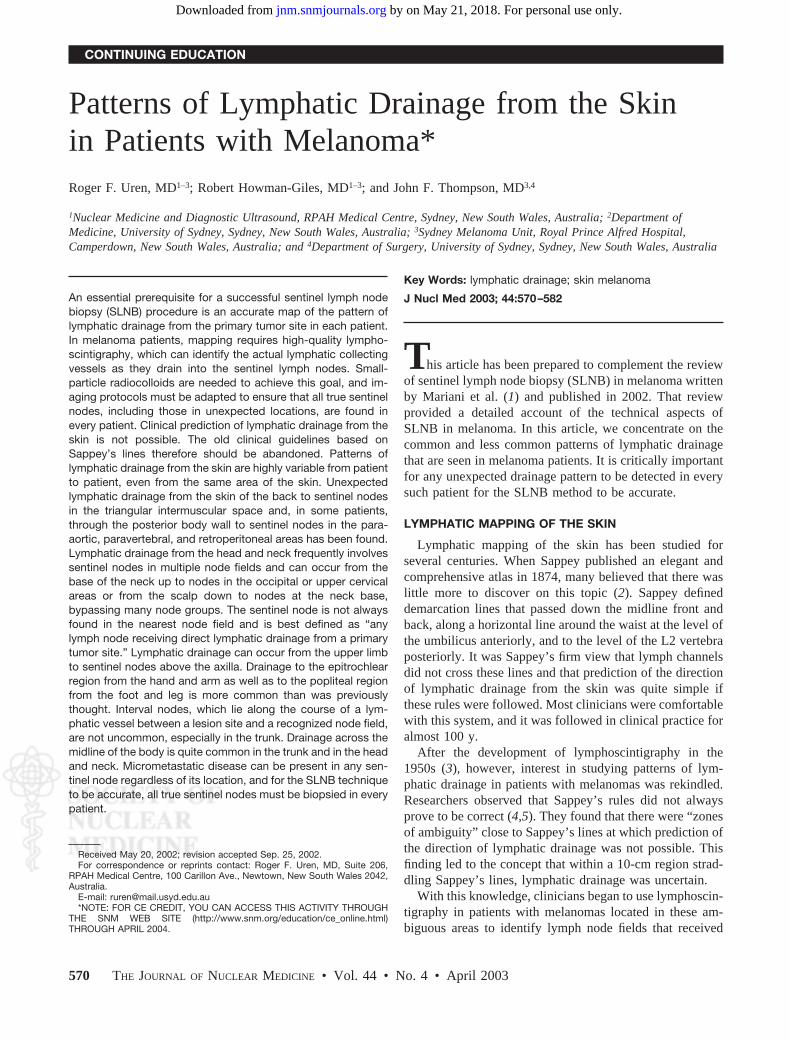
The surface locations of all sentinel nodes are marked onthe overlying skin with an “X” of indelible ink; a permanentpoint tattoo of carbon black (Fig. 1) can also be applied andis a useful guide for clinical or ultrasound follow-up oversubsequent years. The depth of the sentinel node from theskin mark is measured in an orthogonal view with a radio-active marker placed on the skin mark. The depth can then
FIGURE 1. Patient with melanoma on vertex of scalp just toleft of midline and lymphatic drainage down to left level V nodeat base of neck. (A) Lymphoscintigraphy findings on delayedimaging 2 h after injection of 7 MBq of 99mTc-antimony sulfidecolloid intradermally at 4 points around excision biopsy site.Anterior and left lateral views are shown, and lymphatic vesselcan be faintly seen passing directly to sentinel node in left lateralview. Lt � left; Rt � right. (B) Patient at end of study. Sentinelnode (SN) location is marked on skin with “X.” Injection site onscalp is indicated by thick arrow.
LYMPHATIC DRAINAGE IN SKIN MELANOMA • Uren et al. 571
by on May 21, 2018. For personal use only. jnm.snmjournals.org Downloaded from
be measured from the film directly or by using electroniccalipers. Some centers use a �-probe in a nuclear medicinesuite to further aid in the localization of sentinel nodes, butwe have not found this procedure necessary. Regardless ofhow imaging data for a patient are presented to the surgeon,it is essential that the surgeon completely understands thepresentation. The surgeon must be familiar with the appear-ance of the images in order to refer to them while searchingfor sentinel nodes during surgery. This very close commu-nication with surgical colleagues is vital for the accuracy ofthe SLNB method.
We have successfully used this protocol for over 3,000patients with cutaneous melanomas. More detailed descrip-tions of our technique and imaging protocol can be foundelsewhere (14,16).
If possible, lymphatic mapping should be done beforewide local excision of the primary melanoma, as the latterdisrupts lymph drainage pathways and may cause a lack ofmigration of the tracer or the identification of lymph nodesthat are not true sentinel nodes.
A radiocolloid must gain access to the lumen of the initiallymphatic vessels under physiologic conditions to allowaccurate mapping of lymphatic drainage. A brief consider-ation of the microanatomy of the lymphatic system is there-fore relevant here.
Physiology and Microanatomy of CutaneousLymphatics
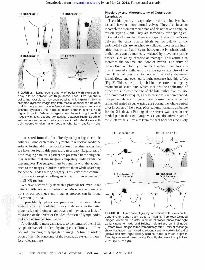
The initial lymphatic capillaries are the terminal lymphat-ics and have no intraluminal valves. They also have anincomplete basement membrane and do not have a completemuscle layer (17,18). They are formed by overlapping en-dothelial cells, so that there are gaps of about 10–25 nmbetween the cells. Elastin fibrils on the outside of theendothelial cells are attached to collagen fibers in the inter-stitial matrix, so that the gaps between the lymphatic endo-thelial cells can be markedly widened by movement of thetissues, such as by exercise or massage. This action alsoincreases the volume and flow of lymph. The entry ofradiocolloid or blue dye into the lymphatic capillaries isthus increased significantly by massage or exercise of thepart. External pressure, in contrast, markedly decreaseslymph flow, and even quite light pressure has this effect(Fig. 3). This is the principle behind the current emergencytreatment of snake bite, which includes the application ofdirect pressure over the site of the bite, rather than the useof a proximal tourniquet, as was previously recommended.The patient shown in Figure 3 was unusual because he hadremained seated in our waiting area during the whole periodafter injection of the tracer. (Our patients normally ambulatefor the 2-h delay.) Pooling of the tracer was seen in themedial part of the right lymph vessel and the inferior part ofthe 2 left vessels. Pressure from the seat back was the likely
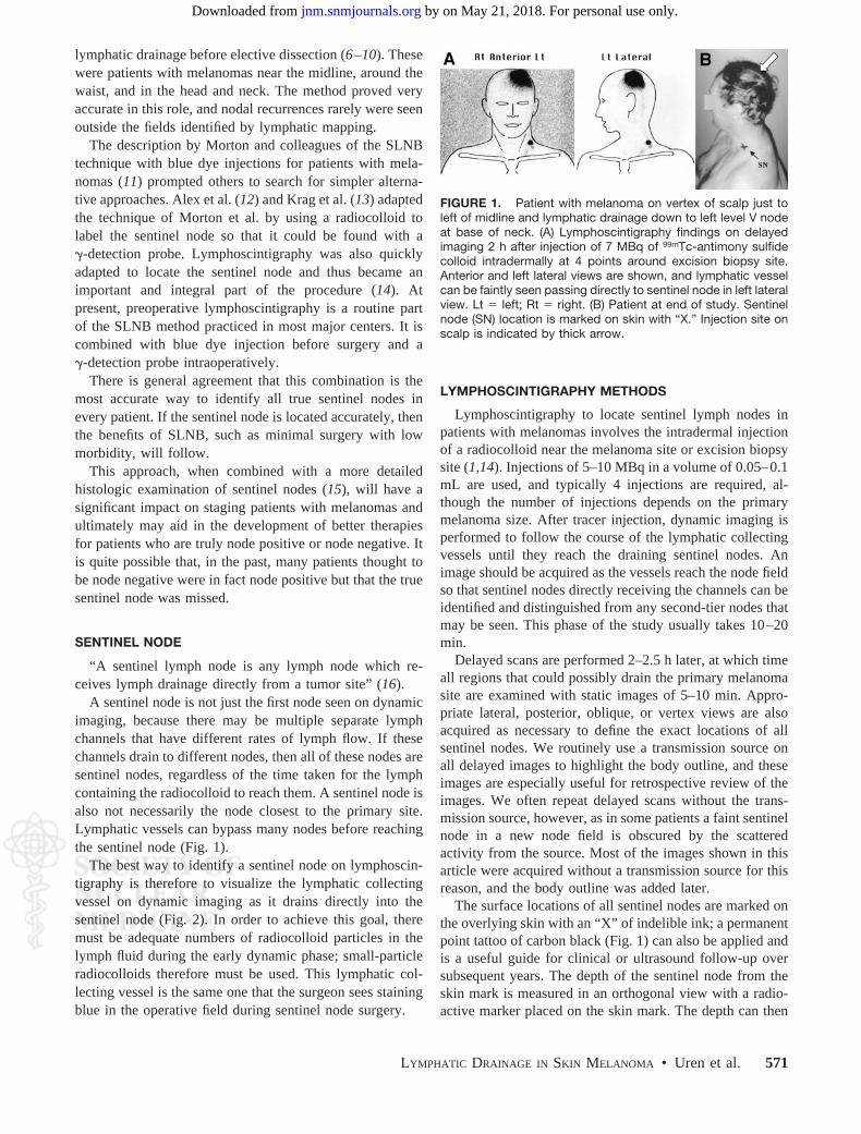
FIGURE 2. Lymphoscintigraphy of patient with excision bi-opsy site on anterior left thigh above knee. Two lymphaticcollecting vessels can be seen passing to left groin in 10-minsummed dynamic image (top left). Medial channel can be seendraining to sentinel node in femoral area, whereas more lateralchannel bypasses this node to reach another sentinel nodehigher in groin. Delayed images show these 2 bright sentinelnodes with faint second-tier activity between them. Depth ofsentinel nodes beneath skin is shown in left lateral view withpoint source on skin marks (bottom right). Lt � left; Rt � right.
FIGURE 3. Lymphoscintigraphy of patient with excision bi-opsy site on upper back close to midline. (Top row) Delayedimages, obtained 2 h after injection of tracer, show faint rightaxillary sentinel node and brighter left axillary sentinel node.(Bottom row) Images taken immediately after 2 min of massageshow that tracer has moved to second sentinel node in left axilla(arrow) and that right axillary sentinel node is much brighter.Even light external pressure significantly decreased lymph flow.Lt � left; Rt � right.
572 THE JOURNAL OF NUCLEAR MEDICINE • Vol. 44 • No. 4 • April 2003
by on May 21, 2018. For personal use only. jnm.snmjournals.org Downloaded from
cause, and massage with a medial to lateral stroke wasperformed over both channels, causing the sentinel nodes ineach axilla to brighten and a second sentinel node to appearin the left axilla. Lymph flow is also decreased by lowtemperatures, and the scanning room should be kept at anambient temperature of at least 21°C. The lymphatic capil-laries follow a tortuous course and frequently anastomosewith each other but continue to have no intraluminal valves.They join together eventually to form lymphatic collectingvessels that have a 3-layer wall and that do have intralumi-nal valves.
The rates of lymph flow within lymphatic collectingvessels vary in different parts of the body (Table 1) (19).The most rapid flow occurs from the legs and feet, followedby that from the arms and hands. Flow from sites in thetrunk is 3 to 4 cm/min on average, while the slowest flowoccurs from the head, neck, and shoulder regions. Thelymphatic vessels have an intrinsic pump mechanism main-taining steady lymph flow (20), but this mechanism re-sponds to an increase in hydrostatic pressure by signifi-cantly increasing lymph flow (such as that which occurs inthe legs during standing). Lymph flow is also increased byheat and inflammation, and although gravity affects thespeed of flow through hydrostatic pressure, it does notinfluence the direction of flow. The intraluminal valvespresent in the lymphatic collecting vessels ensure thatlymph flow is unidirectional toward the draining lymphnodes (17).
The paths taken by collecting vessels on their way todraining node fields vary from patient to patient and fromskin site to skin site. These paths can sometimes be ex-tremely complex and tortuous (Fig. 4) (16). Lymphaticvessels can converge to form fewer larger vessels (Fig. 5)but sometimes divide into multiple vessels, most commonlyin the upper thigh. The collecting vessels usually passthrough the subcutaneous fat layer and generally do notpenetrate the deep fascia until a node field such as the groinor axilla is reached.
Lymph NodesLymph nodes trap radiocolloids by a complex physio-
logic process and do not act as simple mechanical filters.This process first involves opsonization, the mechanism by
which the particles are recognized as foreign (1). Opsoniza-tion can occur in the lymph fluid or in the node itself andaids in later phagocytosis of the particles. A matrix ofreticulin fibrils forms a complex lattice in the sinuses of
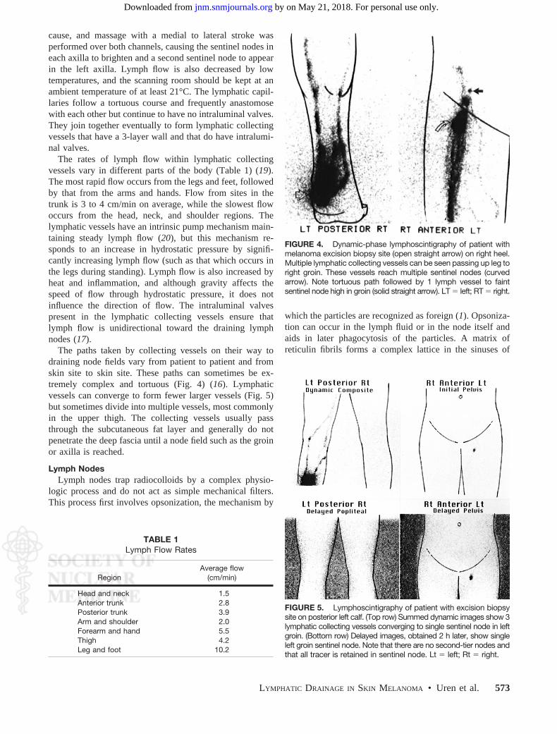
FIGURE 4. Dynamic-phase lymphoscintigraphy of patient withmelanoma excision biopsy site (open straight arrow) on right heel.Multiple lymphatic collecting vessels can be seen passing up leg toright groin. These vessels reach multiple sentinel nodes (curvedarrow). Note tortuous path followed by 1 lymph vessel to faintsentinel node high in groin (solid straight arrow). LT � left; RT � right.
TABLE 1Lymph Flow Rates
RegionAverage flow
(cm/min)
Head and neck 1.5Anterior trunk 2.8Posterior trunk 3.9Arm and shoulder 2.0Forearm and hand 5.5Thigh 4.2Leg and foot 10.2
FIGURE 5. Lymphoscintigraphy of patient with excision biopsysite on posterior left calf. (Top row) Summed dynamic images show 3lymphatic collecting vessels converging to single sentinel node in leftgroin. (Bottom row) Delayed images, obtained 2 h later, show singleleft groin sentinel node. Note that there are no second-tier nodes andthat all tracer is retained in sentinel node. Lt � left; Rt � right.
LYMPHATIC DRAINAGE IN SKIN MELANOMA • Uren et al. 573
by on May 21, 2018. For personal use only. jnm.snmjournals.org Downloaded from
lymph nodes and slows the movement of particles, such asradiocolloids, so that they can be phagocytosed by themacrophages and tissue histiocytes that line the sinuses(21). These phagocytic cells are most abundant in the sub-capsular sinus. Most of the tracer therefore is retained in thislocation.
Most of the radiocolloid that reaches a lymph node willbe retained in the node by this process, regardless of theparticle size, so that even when small-particle colloids, suchas 99mTc-antimony sulfide colloid, are used, the sentinelnode is often the only radiolabelled node on delayed 2-himages (Figs. 2 and 5–11). A small percentage of the tracercan pass to second-tier nodes, regardless of the particle size,and we have found this characteristic to correlate directlywith the speed of lymph flow in lymphatic collecting vessels(22). The higher the flow rate, the greater the incidence of
radiocolloid passing to second-tier nodes. This observationsuggests that the physiologic process of phagocytosis thatretains radiocolloid in the sentinel node can be over-whelmed if too many particles reach the node over a shorttime.
RadiocolloidsThe radiocolloids that best display lymphatic vessels and
thus allow the identification of sentinel nodes are those that
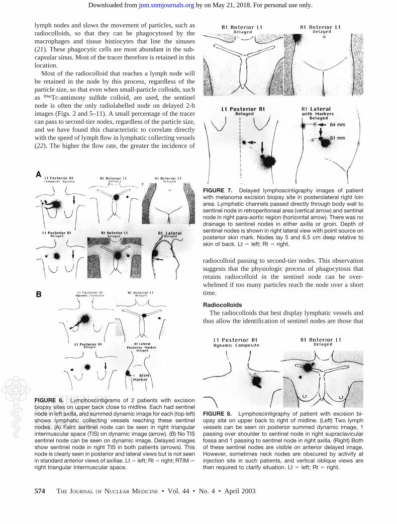
FIGURE 6. Lymphoscintigrams of 2 patients with excisionbiopsy sites on upper back close to midline. Each had sentinelnode in left axilla, and summed dynamic image for each (top left)shows lymphatic collecting vessels reaching these sentinelnodes. (A) Faint sentinel node can be seen in right triangularintermuscular space (TIS) on dynamic image (arrow). (B) No TISsentinel node can be seen on dynamic image. Delayed imagesshow sentinel node in right TIS in both patients (arrows). Thisnode is clearly seen in posterior and lateral views but is not seenin standard anterior views of axillae. Lt � left; Rt � right; RTIM �right triangular intermuscular space.
FIGURE 7. Delayed lymphoscintigraphy images of patientwith melanoma excision biopsy site in posterolateral right loinarea. Lymphatic channels passed directly through body wall tosentinel node in retroperitoneal area (vertical arrow) and sentinelnode in right para-aortic region (horizontal arrow). There was nodrainage to sentinel nodes in either axilla or groin. Depth ofsentinel nodes is shown in right lateral view with point source onposterior skin mark. Nodes lay 5 and 6.5 cm deep relative toskin of back. Lt � left; Rt � right.
FIGURE 8. Lymphoscintigraphy of patient with excision bi-opsy site on upper back to right of midline. (Left) Two lymphvessels can be seen on posterior summed dynamic image, 1passing over shoulder to sentinel node in right supraclavicularfossa and 1 passing to sentinel node in right axilla. (Right) Bothof these sentinel nodes are visible on anterior delayed image.However, sometimes neck nodes are obscured by activity atinjection site in such patients, and vertical oblique views arethen required to clarify situation. Lt � left; Rt � right.
574 THE JOURNAL OF NUCLEAR MEDICINE • Vol. 44 • No. 4 • April 2003
by on May 21, 2018. For personal use only. jnm.snmjournals.org Downloaded from
readily enter the lymphatic capillaries; these are radiocol-loids with particle sizes in the range of 5–50 nm (23,24).These particles easily enter the initial lymphatics underphysiologic conditions, and their entry is enhanced by ex-ercise or massage if movement is slow. With such small-particle radiocolloids, about 5%–8% of the injected dosewill migrate from the injection site to the sentinel node ornodes (24). Appropriate small-particle radiocolloids are ul-trafiltered 99mTc-sulfur colloid (passed through a 100-nmfilter), 99mT-nanocolloid of albumin, and 99mTc-antimonysulfide colloid.
Large-particle radiocolloids (with particles of �200 nmin diameter), such as unfiltered 99mTc-sulfur colloid, havedifficulty moving through the interstitial matrix and enterthe lymphatic capillaries only in small numbers, even withexercise or massage. Lymphatic collecting vessels thus areusually not seen on dynamic imaging when large-particlecolloids are used. Most of the injected dose remains at theinjection site, despite exercise or massage, with only about0.5% of the dose reaching the sentinel nodes (23). Identifi-cation of the sentinel nodes then becomes problematic;definitions based on count ratios relative to the backgroundare relied upon. The problem with this approach is thatsometimes one sentinel node has very low activity com-pared with another sentinel node and may not be identifiedas a sentinel node without dynamic imaging. These faintnodes are sometimes the only positive sentinel nodes in thenode field (Fig. 12).
Lymphatic Mapping in Cutaneous Melanomas toLocate Sentinel Nodes
Since 1984, the Sydney Melanoma Unit has been per-forming lymphatic mapping with 99mTc-antimony sulfidecolloid to locate draining node fields in patients with inter-mediate-thickness melanomas located in the so-called am-biguous zones before elective dissection of the relevantnode field. Over a 6-y period, we had performed about 200studies (14).
As soon as Morton and colleagues described successfulSLNB in melanoma patients by injection of blue dye (11),we began to apply the method described above to locatesentinel nodes by using lymphoscintigraphy on the day
before surgery. This meant that all patients with intermedi-ate-thickness melanomas were studied regardless of thesites of the lesions on the skin. Since our examinationrequired us to locate every sentinel node and not just toimage in standard positions, we began to observe drainageto lymph nodes in completely unexpected places (16,25).Some were in new node fields not previously known todrain the skin. We quickly began to appreciate that therewas unambiguous drainage from very few sites on the skinand that, without preoperative lymphoscintigraphy, accurateSLNB was simply not possible in many patients. Thisvariability in lymph drainage and drainage to sentinel nodesin unexpected places has also been observed by others(9,26,27).
We have now performed lymphatic mapping for over3,000 patients with cutaneous melanomas and have accu-mulated a large body of data relating to common anduncommon cutaneous lymphatic drainage pathways. All ofthese studies were performed by a small group of nuclearmedicine physician consultants and were not done by train-ees. The surgical correlation and SLNB procedures were allperformed by a group of specialists in melanoma surgery.The following is a detailed description of the patterns oflymphatic drainage that we have observed.
PATTERNS OF LYMPHATIC DRAINAGE FROM SKIN
In the studied group of 3,059 patients, 7 showed nomovement of tracer from the injection site over a 2.5-hperiod. These were older patients; 5 patients had melanomasites on the head and neck, and 2 patients had melanomasites on the trunk. Lymphatic drainage to sentinel nodesoccurred in a single node field in 1,963 patients (64%), 2node fields in 803 (26%), 3 node fields in 207 (7%), 4 nodefields in 62 (2%), and 5 node fields in 7. The majority ofskin sites thus drained to a single node field.
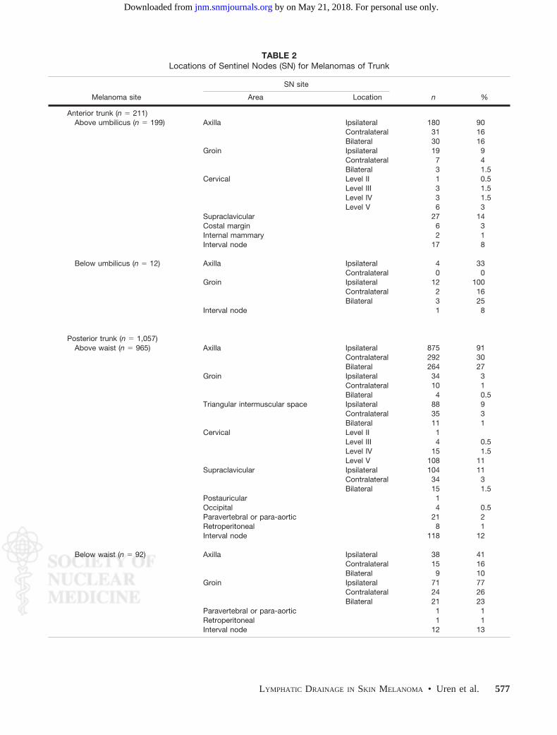
Posterior TrunkThe locations of sentinel nodes draining the posterior
trunk are summarized in Table 2. This group includes 2
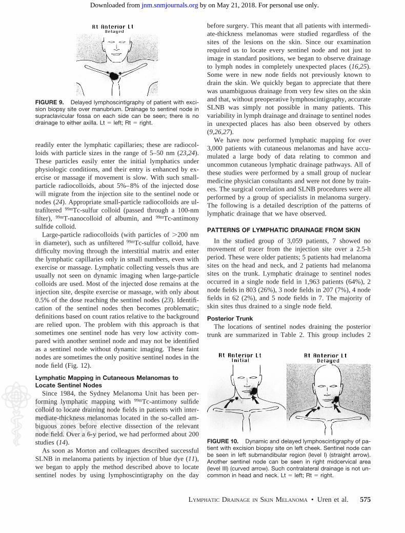
FIGURE 10. Dynamic and delayed lymphoscintigraphy of pa-tient with excision biopsy site on left cheek. Sentinel node canbe seen in left submandibular region (level I) (straight arrow).Another sentinel node can be seen in right midcervical area(level III) (curved arrow). Such contralateral drainage is not un-common in head and neck. Lt � left; Rt � right.
FIGURE 9. Delayed lymphoscintigraphy of patient with exci-sion biopsy site over manubrium. Drainage to sentinel node insupraclavicular fossa on each side can be seen; there is nodrainage to either axilla. Lt � left; Rt � right.
LYMPHATIC DRAINAGE IN SKIN MELANOMA • Uren et al. 575
by on May 21, 2018. For personal use only. jnm.snmjournals.org Downloaded from
lymphatic drainage pathways that were completely unex-pected and that, before our description, were not known toreceive direct lymphatic drainage from the skin of the back.These lymphatic pathways drain to the triangular intermus-cular space lateral to the scapula, behind the axilla (28), andpass through the posterior body wall directly to sentinelnodes in the retroperitoneal and paravertebral areas (29).
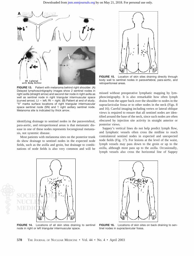
The more common of these 2 pathways is drainage fromthe skin of the back to the triangular intermuscular space(Figs. 6 and 13). We have observed this drainage pathway in12% of our patients with back melanomas. Skin sites thatwe have found to drain to the triangular intermuscular spaceare shown in Figure 14. Sometimes 2 nodes in this space areseen one above the other, both lying just deep to the deepfascia, and the pathway then passes anteriorly, following thecourse of the circumflex scapular vessels into the posteriorpart of the axilla. Therefore, in some patients, the tracer willpass from a sentinel node in the triangular intermuscularspace to a second-tier node in the axilla. We have seen thisphenomenon occur in several patients. Without accuratelymphatic mapping by lymphoscintigraphy, this phenome-non could lead to a radiolabelled second-tier node beingmistakenly identified as the sentinel node and removed fromthe axilla, while the true sentinel node in the triangularintermuscular space remains in the patient. Histologic ex-amination of this radiolabelled axillary node will yield afalse report of the lymph node status in the patient. Thissituation would occur if only a �-probe were used to findand remove radiolabelled nodes from the axilla or if thelymphoscintigraphy imaging protocol were inadequate.
Older protocols called only for anterior views of the axilla,but posterior and lateral views are required to identify thesentinel nodes in this unexpected location, because attenu-ation of the photons as they pass through the patient’s bodymeans that nodes in the triangular intermuscular space maynot be seen at all in an anterior view (Fig. 6). Drainage to asentinel node in the triangular intermuscular space oftenoccurs along with drainage to a sentinel node in anothernode field, but we have encountered 8 patients with exclu-sive drainage to a sentinel node in this unexpected location.
The second unexpected lymphatic drainage pathway thatwe have observed draining the skin of the back is one thatinvolves direct passage through the posterior body wall tosentinel nodes in the paravertebral, para-aortic, or retroper-itoneal areas. This drainage pattern usually involves intra-abdominal sites, but we have also seen paravertebral nodesin the thorax as sentinel nodes draining the skin of the back.The skin sites that may drain through this unexpected path-way are concentrated mainly in the posterior loin area (Fig.15). We have observed this pathway in 4% of patients withback melanomas, making it much less common than thepathway draining to the triangular intermuscular space. Ifwe consider only the posterior loin area, however, we finddrainage through this pathway in 24% of patients. Again,drainage to sentinel nodes in these unexpected areas isusually accompanied by drainage to sentinel nodes in ex-pected node fields (the axilla and groin); however, we haveencountered 4 patients with exclusive drainage to sentinelnodes in these areas but with no drainage whatsoever tonodes in the axilla or groin (Fig. 7) (30). The importance of
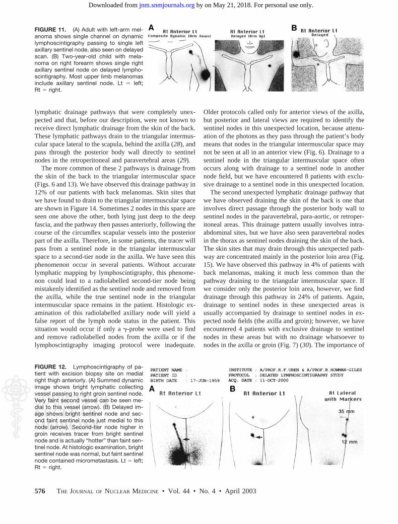
FIGURE 11. (A) Adult with left-arm mel-anoma shows single channel on dynamiclymphoscintigraphy passing to single leftaxillary sentinel node, also seen on delayedscan. (B) Two-year-old child with mela-noma on right forearm shows single rightaxillary sentinel node on delayed lympho-scintigraphy. Most upper limb melanomasinclude axillary sentinel node. Lt � left;Rt � right.
FIGURE 12. Lymphoscintigraphy of pa-tient with excision biopsy site on medialright thigh anteriorly. (A) Summed dynamicimage shows bright lymphatic collectingvessel passing to right groin sentinel node.Very faint second vessel can be seen me-dial to this vessel (arrow). (B) Delayed im-age shows bright sentinel node and sec-ond faint sentinel node just medial to thisnode (arrow). Second-tier node higher ingroin receives tracer from bright sentinelnode and is actually “hotter” than faint sen-tinel node. At histologic examination, brightsentinel node was normal, but faint sentinelnode contained micrometastasis. Lt � left;Rt � right.
576 THE JOURNAL OF NUCLEAR MEDICINE • Vol. 44 • No. 4 • April 2003
by on May 21, 2018. For personal use only. jnm.snmjournals.org Downloaded from
identifying drainage to sentinel nodes in the paravertebral,para-aortic, and retroperitoneal areas is that metastatic dis-ease in one of these nodes represents locoregional metasta-sis, not systemic disease.
Most patients with melanoma sites on the posterior trunkdo show drainage to sentinel nodes in the expected nodefields, such as the axilla and groin, but drainage to combi-nations of node fields is also very common and will be
missed without preoperative lymphatic mapping by lym-phoscintigraphy. It is also remarkable how often lymphdrains from the upper back over the shoulder to nodes in thesupraclavicular fossa or to other nodes in the neck (Figs. 8and 16). Careful imaging including vertex or lateral obliqueviews is required to ensure that all sentinel nodes are iden-tified around the base of the neck, since such nodes are oftenobscured by injection site activity in straight anterior orposterior views.
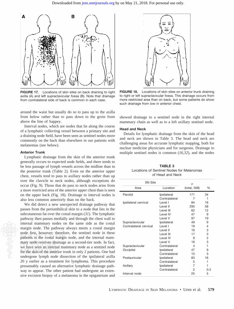
Sappey’s vertical lines do not help predict lymph flow,and lymphatic vessels often cross the midline to reachcontralateral sentinel nodes in expected and unexpectednode fields (Fig. 17). For lesions at the level of the waist,lymph vessels may pass down to the groin or up to theaxilla, although most pass up to the axilla. Occasionally,lymph vessels also cross the horizontal line of Sappey
FIGURE 13. Patient with melanoma behind right shoulder. (A)Delayed lymphoscintigraphy images show 2 sentinel nodes inright axilla (straight arrow) and second-tier node in right axilla aswell as sentinel node in right triangular intermuscular space(curved arrow). Lt � left; Rt � right. (B) Patient at end of study.“X” marks surface locations of right triangular intermuscularspace sentinel node (SN) and 1 right axillary sentinel node.Melanoma site is indicated by thick arrow.
FIGURE 14. Locations of all skin sites draining to sentinelnode in right or left triangular intermuscular space.
FIGURE 15. Location of skin sites draining directly throughbody wall to sentinel nodes in paravertebral, para-aortic, andretroperitoneal areas.
FIGURE 16. Locations of skin sites on back draining to sen-tinel nodes in supraclavicular fossa.
578 THE JOURNAL OF NUCLEAR MEDICINE • Vol. 44 • No. 4 • April 2003
by on May 21, 2018. For personal use only. jnm.snmjournals.org Downloaded from
around the waist but usually do so to pass up to the axillafrom below rather than to pass down to the groin fromabove the line of Sappey.
Interval nodes, which are nodes that lie along the courseof a lymphatic collecting vessel between a primary site anda draining node field, have been seen as sentinel nodes morecommonly on the back than elsewhere in our patients withmelanomas (see below).
Anterior TrunkLymphatic drainage from the skin of the anterior trunk
generally occurs to expected node fields, and there tends tobe less passage of lymph vessels across the midline than inthe posterior trunk (Table 2). Even on the anterior upperchest, vessels tend to pass to axillary nodes rather than upover the clavicle to neck nodes, although exceptions dooccur (Fig. 9). Those that do pass to neck nodes arise froma more restricted area of the anterior upper chest than is seenon the upper back (Fig. 18). Drainage to interval nodes isalso less common anteriorly than on the back.
We did detect a new unexpected drainage pathway thatpasses from the periumbilical skin to a node that lies in thesubcutaneous fat over the costal margin (31). The lymphaticpathway then passes medially and through the chest wall tointernal mammary nodes on the same side as the costalmargin node. The pathway always meets a costal marginnode first, however; therefore, the sentinel node in thesepatients is the costal margin node, and the internal mam-mary node receives drainage as a second-tier node. In fact,we have seen an internal mammary node as a sentinel nodefor the skin of the anterior trunk in only 2 patients. One hadundergone lymph node dissection of the ipsilateral axilla20 y earlier as a treatment for lymphoma. This procedurepresumably caused an alternative lymphatic drainage path-way to appear. The other patient had undergone an exten-sive excision biopsy of a melanoma in the epigastrium and
showed drainage to a sentinel node in the right internalmammary chain as well as to a left axillary sentinel node.
Head and NeckDetails for lymphatic drainage from the skin of the head
and neck are shown in Table 3. The head and neck arechallenging areas for accurate lymphatic mapping, both fornuclear medicine physicians and for surgeons. Drainage tomultiple sentinel nodes is common (16,32), and the nodes
FIGURE 17. Locations of skin sites on back draining to rightaxilla (A) and left supraclavicular fossa (B). Note that drainagefrom contralateral side of back is common in each case.
FIGURE 18. Locations of skin sites on anterior trunk drainingto right or left supraclavicular fossa. This drainage occurs frommore restricted area than on back, but some patients do showsuch drainage from low in anterior chest.
TABLE 3Locations of Sentinel Nodes for Melanomas
of Head and Neck
SN Site n(total, 508) %Area Location
Parotid Ipsilateral 171 34Contralateral 7 1
Ipsilateral cervical Level I 84 16Level II 295 58Level III 62 12Level IV 47 9Level V 97 19
Supraclavicular Ipsilateral 49 9Contralateral cervical Level I 19 4
Level II 16 3Level III 17 3Level IV 9 2Level V 16 3
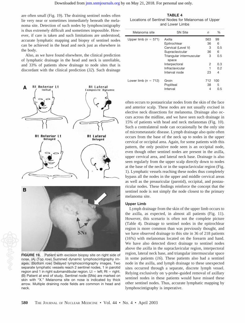
are often small (Fig. 19). The draining sentinel nodes oftenlie very near or sometimes immediately beneath the mela-noma site. Detection of such nodes by lymphoscintigraphyis thus extremely difficult and sometimes impossible. How-ever, if care is taken and such limitations are understood,accurate lymphatic mapping and biopsy of sentinel nodescan be achieved in the head and neck just as elsewhere inthe body.
Also, as we have found elsewhere, the clinical predictionof lymphatic drainage in the head and neck is unreliable,and 33% of patients show drainage to node sites that isdiscordant with the clinical prediction (32). Such drainage
often occurs to postauricular nodes from the skin of the faceand anterior scalp. These nodes are not usually excised inelective neck dissections for melanoma. Drainage also oc-curs across the midline, and we have seen such drainage in15% of patients with head and neck melanomas (Fig. 10).Such a contralateral node can occasionally be the only siteof micrometastatic disease. Lymph drainage also quite oftenoccurs from the base of the neck up to nodes in the uppercervical or occipital area. Again, for some patients with thispattern, the only positive node seen is an occipital node,even though other sentinel nodes are present in the axilla,upper cervical area, and lateral neck base. Drainage is alsoseen regularly from the upper scalp directly down to nodesat the base of the neck or in the supraclavicular region (Fig.1). Lymphatic vessels reaching these nodes thus completelybypass all the nodes in the upper and middle cervical areasas well as the preauricular (parotid), occipital, and postau-ricular nodes. These findings reinforce the concept that thesentinel node is not simply the node closest to the primarymelanoma site.
Upper LimbLymph drainage from the skin of the upper limb occurs to
the axilla, as expected, in almost all patients (Fig. 11).However, this scenario is often not the complete picture(Table 4). Drainage to sentinel nodes in the epitrochlearregion is more common than was previously thought, andwe have observed drainage to this site in 36 of 218 patients(16%) with melanomas located on the forearm and hand.We have also detected direct drainage to sentinel nodesabove the axilla in the supraclavicular region, interpectoralregion, lateral neck base, and triangular intermuscular spacein some patients (16). These patients also had a sentinelnode in the axilla, and lymph drainage to these unexpectedsites occurred through a separate, discrete lymph vessel.Relying exclusively on �-probe–guided removal of axillarysentinel nodes in these patients would have missed theseother sentinel nodes. Thus, accurate lymphatic mapping bylymphoscintigraphy is imperative.
FIGURE 19. Patient with excision biopsy site on right side ofnose. (A) (Top row) Summed dynamic lymphoscintigraphy im-ages. (Bottom row) Delayed lymphoscintigraphy images. Twoseparate lymphatic vessels reach 2 sentinel nodes, 1 in parotidregion and 1 in right submandibular region. Lt � left; Rt � right.(B) Patient at end of study. Sentinel node (SNs) are marked onskin with “X.” Melanoma site on nose is indicated by thickarrow. Multiple draining node fields are common in head andneck.
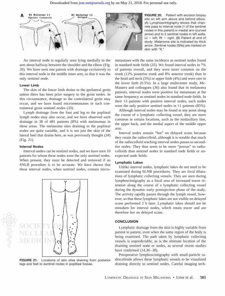
TABLE 4Locations of Sentinel Nodes for Melanomas of Upper
An interval node is regularly seen lying medially in thearm about halfway between the shoulder and the elbow (Fig.20). We have seen one patient with drainage exclusively tothis interval node in the middle inner arm, so that it was theonly sentinel node.
Lower LimbThe skin of the lower limb drains to the ipsilateral groin
unless there has been prior surgery to the groin nodes. Inthis circumstance, drainage to the contralateral groin mayoccur, and we have found micrometastases in such con-tralateral groin sentinel nodes (33).
Lymph drainage from the foot and leg to the popliteallymph nodes may also occur, and we have observed suchdrainage in 38 of 481 patients (8%) with melanomas inthese areas. The melanoma sites draining to the poplitealnodes are quite variable, and it is not just the skin of thelateral heel that drains here, as was previously thought (34)(Fig. 21).
Interval NodesInterval nodes can be sentinel nodes, and we have seen 10
patients for whom these nodes were the only sentinel nodes.When present, they must be detected and removed if anSNLB procedure is to be accurate. We have shown thatthese interval nodes, when sentinel nodes, contain micro-
metastases with the same incidence as sentinel nodes foundin standard node fields (35). We found interval nodes in 7%of patients overall, and they were more common on thetrunk (12% posterior trunk and 8% anterior trunk) than inthe head and neck (5%) or upper limb (4%) and were rare inthe lower limb (0.5%). In a large multicenter study, Mc-Masters and colleagues (36) also found that in melanomapatients, interval nodes were positive for metastases at thesame frequency as sentinel nodes in standard node fields. Intheir 13 patients with positive interval nodes, such nodeswere the only positive sentinel nodes in 11 patients (85%).
Although interval nodes may be found at any point alongthe course of a lymphatic collecting vessel, they are morecommon in certain locations, such as the midaxillary line,the upper back, and the medial aspect of the middle upperarm.
Interval nodes remain “hot” on delayed scans becausethey retain the radiocolloid, although it is notable that muchof the radiocolloid reaching interval nodes passes to second-tier nodes. They thus seem to be more “porous” to radio-colloids than sentinel nodes in standard node fields or un-expected node fields.
Lymphatic LakesUnlike interval nodes, lymphatic lakes do not need to be
examined during SLNB procedures. They are focal dilata-tions of lymphatic collecting vessels. They are seen duringlymphoscintigraphy as a focal area of increased tracer re-tention along the course of a lymphatic collecting vesselduring the dynamic early postinjection phase of the study.The activity rapidly passes through the lymph vessel, how-ever, so that these lymphatic lakes are not visible on delayedscans performed 2 h later. Lymphatic lakes should not bemistaken for interval nodes, which retain tracer and aretherefore hot on delayed scans.
CONCLUSION
Lymphatic drainage from the skin is highly variable frompatient to patient, even when the same region of the body isbeing examined. The path taken by lymphatic collectingvessels is unpredictable, as is the ultimate location of thedraining sentinel node or nodes, as several recent studieshave confirmed (14,36 –38).
Preoperative lymphoscintigraphy with small-particle ra-diocolloids allows these lymphatic vessels to be visualizeddraining directly to sentinel nodes. Careful imaging tech-
FIGURE 20. Patient with excision biopsysite on left arm above and behind elbow.(A) Lymphoscintigraphy shows that chan-nels pass to interval node (1 of the sentinelnodes in this patient) in medial arm (curvedarrow) and to 2 sentinel nodes in left axilla.Lt � left; Rt � right. (B) Patient at end ofstudy. Melanoma site is indicated by thickarrow. Sentinel nodes (SNs) are marked onskin with “X.”
FIGURE 21. Locations of skin sites draining from posteriorlegs and feet to sentinel nodes in popliteal fossae.
LYMPHATIC DRAINAGE IN SKIN MELANOMA • Uren et al. 581
by on May 21, 2018. For personal use only. jnm.snmjournals.org Downloaded from
niques will thus allow all true sentinel nodes to be identifiedin each patient, even if these nodes lie outside standard nodefields or are interval nodes lying between the primary siteand a node field. This information is an important contri-bution to the management of patients with melanomas, as itwill lead to more accurate nodal staging of patients withhigh-risk melanomas.
We now know that the clinical prediction of the pattern oflymphatic drainage in an individual patient is unreliable andinaccurate. We also now know that we have an accuratemethod of mapping lymphatic drainage in every patient,making the difficulties associated with clinical predictionirrelevant. This technique, which provides an accurate mapof lymphatic drainage in each patient, can thus have a directand important impact on the clinical management of pa-tients with melanomas.
ACKNOWLEDGMENTS
We thank all of the excellent nuclear medicine technol-ogists whose dedication has been so important to the suc-cess of our lymphatic mapping work, including Kim Ioan-nou, Nicholas Trpezanovski, Angelique Nguyen, TraceySmith, Sally Raymond, and Ian Dyer. We thank the othernuclear medicine physicians who have added their enthusi-asm to our work, including Drs. David Chung, RobertMansberg, John Roberts, Reginald Hutcherson, and Eliza-beth Bernard. We also thank the surgeons of the SydneyMelanoma Unit for referring their patients and providing thesurgical correlation and validation of the lymphatic drainagepatterns that we have documented, including Professor Wil-liam McCarthy, Dr. Michael Quinn, Dr. Kirwin Shannon,Professor Christopher O’Brien, Dr. Andrew Spillane, Dr.Robyn Saw, and Dr. Jonathon Stretch.
REFERENCES
1. Mariani G, Gipponi M, Moresco L, et al. Radioguided sentinel lymph nodebiopsy in malignant cutaneous melanoma. J Nucl Med. 2002;43:811–827.
2. Sappey MPC. Anatomy, Physiology and Pathology of the Lymphatic Vessels inMan and Vertebrates [in French]. DeLahaye A, Lecrosnier E, trans-eds. Paris,France; 1874.
3. Sherman AI, Ter-Pogossian M, Tocus EC. Lymph node concentration of radio-active colloidal gold following interstitial injection. Cancer. 1953;6:1238–1240.
4. Haagansen CD, Feind CR, Herter FP, et al. Lymphatics of the trunk. In: Haa-gansen CD, ed. The Lymphatics in Cancer. Philadelphia, PA: WB Saunders;1972:437–458.
5. Sugarbaker EV, McBride CM. Melanoma of the trunk: the results of surgicalexcision and anatomic guidelines for predicting nodal metastasis. Surgery. 1976;80:22–30.
6. Bergqvist L, Strand S, Hafstrom L, et al. Lymphoscintigraphy in patients withmalignant melanoma: a quantitative and qualitative evaluation of its usefulness.Eur J Nucl Med. 1984;9:129–135.
7. Fee HJ, Robinson DS, Sample WF, et al. The determination of lymph shed bycolloidal gold scanning in patients with malignant melanoma: a preliminarystudy. Surgery. 1978;84:626–632.
8. Meyer CM, Lecklitner ML, Logic JR, et al. Technetium-99m sulfur-colloidcutaneous lymphoscintigraphy in the management of truncal melanoma. Radiol-ogy. 1979;131:205–209.
9. Norman J, Cruse W, Espinosa C, et al. Redefinition of cutaneous lymphaticdrainage with the use of lymphoscintigraphy for malignant melanoma. Am J Surg.1991;162:432–437.
11. Morton DL, Wen D-R, Wong JH, et al. Technical details of intraoperativelymphatic mapping for early stage melanoma. Arch Surg. 1992;127:392–399.
12. Alex JC, Weaver DL, Fairbank JT, et al. Gamma-probe-guided lymph nodelocalization in malignant melanoma. Surg Oncol. 1993;2:303–308.
13. Krag DN, Meijer SJ, Weaver DL, et al. Minimal-access surgery for staging ofmalignant melanoma. Arch Surg. 1995;130:654–658.
14. Uren RF, Howman-Giles RB, Shaw HM, et al. Lymphoscintigraphy in high riskmelanoma of the trunk: predicting draining node groups, defining lymphaticchannels and locating the sentinel node. J Nucl Med. 1993;34:1435–1440.
15. Cochran AJ. The pathologist’s role in sentinel lymph node evaluation. SeminNucl Med. 2000;30:11–17.
16. Uren RF, Thompson JF, Howman-Giles RB. Lymphatic Drainage of the Skin andBreast: Locating the Sentinel Nodes. Amsterdam, The Netherlands: HarwoodAcademic Publishers; 1999.
17. Leak LV. Lymphatic vessels. In: Johannessen JV, ed. Cardiovascular System,Lymphoreticular and Hematopoietic System. New York, NY: McGraw-Hill;1980:159–183.
18. Ryan TJ. The lymphatics of the skin. In: Jarrett A, ed. The Physiology andPathophysiology of the Skin. London, England: Academic Press; 1978:1755–1780.
19. Uren RF, Howman-Giles RB, Thompson JF. Variation in cutaneous lymphaticflow rates. Ann Surg Oncol. 1997;4:279–280.
20. Sjoberg T, Steen S. Contractile properties of lymphatics from the human lowerleg. Lymphology. 1991;24:16–21.
21. Nichols WS, Chisari FV. Structure and function of the lymphoid tissues. In:Williams WJ, Beutler E, Erslev AJ, et al., eds. Hematology. New York, NY:McGraw-Hill; 1990:49–50.
22. Uren RF, Howman-Giles RB, Thompson JF. Demonstration of second tier lymphnodes during preoperative lymphoscintigraphy for melanoma: incidence varieswith primary tumour site. Ann Surg Oncol. 1998;5:517–521.
23. Bergqvist L, Strand S-E, Persson BRR. Particle sizing and biokinetics of inter-stitial lymphoscintigraphic agents. Semin Nucl Med. 1983;8:9–19.
24. Strand SE, Persson BRR. Quantitative lymphoscintigraphy I: basic concepts foroptimal uptake of radiocolloids in the parasternal lymph nodes of rabbits. J NuclMed. 1979;20:1038–1046.
25. Thompson JF, Uren RF, Shaw HM, et al. The location of sentinel lymph nodesin patients with cutaneous melanoma: new insights into lymphatic anatomy. J AmColl Surg. 1999;189:195–206.
27. Leong SP, Achtem TA, Habib FA, et al. Discordancy between clinical predictionsvs lymphoscintigraphic and intraoperative mapping of sentinel lymph nodedrainage of primary melanoma. Arch Dermatol. 1999;135:1472–1476.
28. Uren RF, Howman-Giles RB, Thompson JF, et al. Lymphatic drainage totriangular intermuscular space lymph nodes in melanoma on the back. J NuclMed. 1996;37:964–966.
29. Uren RF, Howman-Giles RB, Thompson JF. Lymphatic drainage from the skinof the back to intra-abdominal lymph nodes in melanoma patients. Ann SurgOncol. 1998;5:384–387.
30. Uren RF, Howman-Giles RB, Thompson JF, et al. Exclusive lymphatic drainagefrom a melanoma on the back to intraabdominal lymph nodes. Clin Nucl Med.1998;23:71–73.
32. O’Brien CJ, Uren RF, Thompson JF, et al. Prediction of potential metastatic sitesin cutaneous head and neck melanoma using lymphoscintigraphy. Am J Surg.1995;170:461–466.
2003;44:570-582.J Nucl Med. Roger F. Uren, Robert Howman-Giles and John F. Thompson Patterns of Lymphatic Drainage from the Skin in Patients with Melanoma
http://jnm.snmjournals.org/content/44/4/570This article and updated information are available at:
Information about subscriptions to JNM can be found at:
http://jnm.snmjournals.org/site/misc/permission.xhtmlInformation about reproducing figures, tables, or other portions of this article can be found online at:
(Print ISSN: 0161-5505, Online ISSN: 2159-662X)1850 Samuel Morse Drive, Reston, VA 20190.SNMMI | Society of Nuclear Medicine and Molecular Imaging
is published monthly.The Journal of Nuclear Medicine