Page 1
Universidade de Lisboa
Faculdade de Motricidade Humana
MANUAL LYMPHATIC DRAINAGE IN
CHRONIC VENOUS DISEASE
Tese elaborada com vista à obtenção do Grau de Doutor em
Motricidade Humana na especialidade de Reabilitação
Orientador: Professor Doutor Paulo Alexandre Silva Armada da Silva
Júri: Presidente
Reitor da Universidade de Lisboa
Vogais: Doutor Carlos Fernando Pereira Alves Professor Associado com Agregação Convidado e Jubilado Faculdade de Ciências Médicas da Universidade Nova de Lisboa
Doutora Maria Margarida Marques Rebelo Espanha Professora Associada Faculdade de Motricidade Humana da Universidade de Lisboa
Doutor Paulo Alexandre Silva Armada da Silva Professor Auxiliar Faculdade de Motricidade Humana da Universidade Nova de Lisboa
Doutor Carlos Manuel da Costa Almeida Professor Auxiliar Convidado Faculdade de Medicina da Universidade de Coimbra
Doutor Eduardo Manuel Brazete Carvalho Cruz Professor Coordenador Escola Superior de Saúde do Instituto Politécnico de Setúbal
Rute Sofia dos Santos Crisóstomo
Setembro 2014
Page 3
Universidade de Lisboa
Faculdade de Motricidade Humana
MANUAL LYMPHATIC DRAINAGE IN
CHRONIC VENOUS DISEASE
Tese elaborada com vista à obtenção do Grau de Doutor em
Motricidade Humana na especialidade de Reabilitação
Orientador: Professor Doutor Paulo Alexandre Silva Armada da Silva
Júri: Presidente
Reitor da Universidade de Lisboa
Vogais: Doutor Carlos Fernando Pereira Alves Professor Associado com Agregação Convidado e Jubilado Faculdade de Ciências Médicas da Universidade Nova de Lisboa
Doutora Maria Margarida Marques Rebelo Espanha Professora Associada Faculdade de Motricidade Humana da Universidade de Lisboa
Doutor Paulo Alexandre Silva Armada da Silva Professor Auxiliar Faculdade de Motricidade Humana da Universidade Nova de Lisboa
Doutor Carlos Manuel da Costa Almeida Professor Auxiliar Convidado Faculdade de Medicina da Universidade de Coimbra
Doutor Eduardo Manuel Brazete Carvalho Cruz Professor Coordenador Escola Superior de Saúde do Instituto Politécnico de Setúbal
Rute Sofia dos Santos Crisóstomo
This work was partially supported by PhD grant by the Portuguese Fundação para a
Ciência e Tecnologia, number SFRH/BD/62673/2009.
Setembro 2014
Page 5
iii
NOTE:
This thesis is based on the following studies:
Crisóstomo, R. S., Candeias, M. S., & Armada-da-Silva, P. A. (2014). The
use of ultrasound in the evaluation of the efficacy of calf muscle pump function in
primary chronic venous disease. Phlebology, 29(4), 247-256. doi:
10.1177/0268355512471757 (Annex 1).
Crisóstomo RS, Candeias MS, Ribeiro AM, Martins CD, Armada-da-Silva
PA. (2013). Manual lymphatic drainage in chronic venous disease: A duplex
ultrasound study. Phlebology. doi: http://dx.doi.org/10.1177/026835551350278
(Annex 2).
Crisóstomo RS, Candeias MS, Armada-da-Silva PA. Venous flow during
manual lymphatic drainage applied to different regions of the lower extremity.
Physiotherapy, (submitted).
Crisóstomo RSS, Costa DS, Martins CL, Fernandes TIR, Armada-da-Silva
PAS. Influence of manual lymphatic drainage on health-related quality of life and
symptoms of chronic venous insufficiency: a randomized controlled trial. Archives
of Physical Medicine and Rehabilitation, (accepted).
Page 7
v
DEDICATORY
This work is dedicated to my family: Bárbara, António, Pedro, Liliana, Paulo,
Ema, Joaquina and Joaquim.
Thank you...
Page 9
vii
ACKNOWLEDGMENTS
I would like to thank Physiotherapist Diana Arraia and Cardiopneumologist
Miguel Candeias (also a co-author in three of the studies in this thesis), for helping in
data collection, for staying with me from the moment when this research was only a
dream, and for helping me to make it true.
Also, I would like to give a very special thanks to participants in Lisbon and
in Castelo Branco that kindly volunteered to participate in the studies making up this
thesis.
I would like to thank Aida Paulino, M.D. in General Surgery, and Nurses
Maria Conceição Branco and Manuel Machado for referring patients to this study
from Unidade Local de Saúde de Castelo Branco.
I would like to thank Professor Isabel Salavessa for reviewing the English of
some of the studies and Mrs. Edite Santos for helping me with the formatting of this
thesis. Also thanks to those from Escola Superior de Saúde Dr. Lopes Dias, Instituto
Politécnico de Castelo Branco (where three studies were carried out), who always
pleased me with nice and supportive words, and helped me to make this work come
true.
I would like to thank Faculdade de Motricidade Humana from Universidade
de Lisboa and Laboratório de Biomecânica e Morfologia Funcional, for all suport.
I would like to thank my students and ex-students and colleagues, who also
shared with me the enthusiasm for studying manual lymphatic drainage and chronic
venous disease, and especially to Physiotherapist Margarida Ribeiro, a co-author in
one study, and Physiotherapist Mylène Martins.
I want to thank Physiotherapist Daniela Costa e Catarina Martins, co-authors
in some of the studies, for all their support as colleague and friends, and for treating
all patients in the last study. Also thanks to Cardiopneumologist Tânia Fernandes
(co-author in the last study), for helping in data collection.
I want to thank Physiotherapist Nuno Duarte for transmitting to me the
enthusiasm about studying manual lymphatic drainage.
Page 10
viii
To my best friends that stay, talk, laugh and cry or not with me in the right
moments, and to those that for some moments in the last four years were important to
me, but the life let me apart from them, thank you. João Costa, Veronika Kozlova,
Stela Frazão and Pedro André a special thanks to you.
And finally a very special thanks to Paulo Armada da Silva, my scientific
adviser for this PhD, for accepting to be a part of this work, for all orientation, for
being patient with my difficulties and help me to overcome them, and for all advises,
this work wouldn't be possible without you. Thank you, from my heart.
Page 11
ix
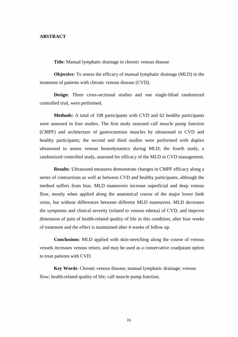
ABSTRACT
Title: Manual lymphatic drainage in chronic venous disease
Objective: To assess the efficacy of manual lymphatic drainage (MLD) in the
treatment of patients with chronic venous disease (CVD).
Design: Three cross-sectional studies and one single-blind randomized
controlled trial, were performed.
Methods: A total of 108 participants with CVD and 62 healthy participants
were assessed in four studies. The first study assessed calf muscle pump function
(CMPF) and architecture of gastrocnemius muscles by ultrasound in CVD and
healthy participants; the second and third studies were performed with duplex
ultrasound to assess venous hemodynamics during MLD; the fourth study, a
randomized controlled study, assessed for efficacy of the MLD in CVD management.
Results: Ultrasound measures demonstrate changes in CMPF efficacy along a
series of contractions as well as between CVD and healthy participants, although the
method suffers from bias. MLD maneuvers increase superficial and deep venous
flow, mostly when applied along the anatomical course of the major lower limb
veins, but without differences between different MLD maneuvers. MLD decreases
the symptoms and clinical severity (related to venous edema) of CVD, and improve
dimension of pain of health-related quality of life in this condition, after four weeks
of treatment and the effect is maintained after 4 weeks of follow up.
Conclusions: MLD applied with skin-stretching along the course of venous
vessels increases venous return, and may be used as a conservative coadjutant option
to treat patients with CVD.
Key Words: Chronic venous disease; manual lymphatic drainage; venous
flow; health-related quality of life; calf muscle pump function.
Page 13
xi
RESUMO
Título: Drenagem linfática manual na doença venosa crónica
Objetivo: Avaliar a eficácia da drenagem linfática manual (DLM) no
tratamento de utentes com doença venosa crónica (DVC).
Desenho do estudo: Foram realizados três estudos transversais e um estudo
prospetivo, controlado e com ocultação simples.
Metodologia: Nos 4 estudos foram avaliados 108 participantes com DVC e
62 participantes saudáveis. No primeiro estudo foi avaliada a bomba muscular
venosa da perna (BMVP) e a arquitetura dos músculos gémeos por ultrassonografia,
em participantes com DVC e saudáveis. No segundo e terceiro estudos foram
avaliadas por ultrassonografia vascular as variações hemodinâmicas venosas durante
a DLM. No quarto estudo, o estudo prospetivo, avaliou-se a eficácia da DLM no
tratamento de doentes com DVC.
Resultados: A avaliação por ultrassonografia identificou alterações na
eficácia da BMVP durante uma série de 10 contrações nos participantes com DVC e
nos participantes saudáveis, contudo, este método apresentou uma fiabilidade pobre.
As manobras de DLM aumentaram o fluxo venoso, sobretudo quando aplicadas na
localização anatómica das principais veias do membro inferior. A DLM aliviou
sintomas e reduziu a dimensão da dor da qualidade de vida relacionada com a saúde
e severidade clinica da DVC (sobretudo edema) no fim de 4 semanas de tratamento.
Parte destes resultados mantiveram-se após 4 semanas de follow-up.
Conclusão: Tracionar a pele durante a DLM, ao longo do percurso das veias,
aumenta o retorno venoso e revela-se como uma potencial estratégia conservadora e
coadjuvante no tratamento de doentes com DVC.
Palavras-chave: Doença venosa crónica; drenagem linfática manual; fluxo
venoso; qualidade de vida relacionada com a saúde; bomba muscular venosa da
perna.
Page 15
xiii
INDEX
TABLE INDEX ...................................................................................................... xvii
FIGURE INDEX ......................................................................................................xix
LIST OF ABBREVIATORS ...................................................................................xxi
1 INTRODUCTION ............................................................................................... 1
2 REVIEW OF LITERATURE ............................................................................. 5
2.1 Health and social impact of CVD ................................................................ 5
2.1.1 Epidemiology ...................................................................................... 5
2.1.2 Functional and HRQL implications of CVD ....................................... 6
2.1.3 Socio-economic impact of CVD .......................................................... 7
2.2 Pathology of CVD ......................................................................................... 9
2.2.1 Etiology and anatomical location of CVD ........................................ 10
2.2.2 Physiopathology hypothesis for CVD ............................................... 12
2.2.3 Symptoms in CVD ............................................................................. 16
2.2.4 Signs of CVD ..................................................................................... 20
2.3 CVD diagnosis ............................................................................................. 23
2.4 Calf muscle pump function ........................................................................ 25
2.4.1 The three venous muscle pumps of the lower limb ........................... 26
2.4.2 Impairment of calf muscle pump and functional capacity ................ 28
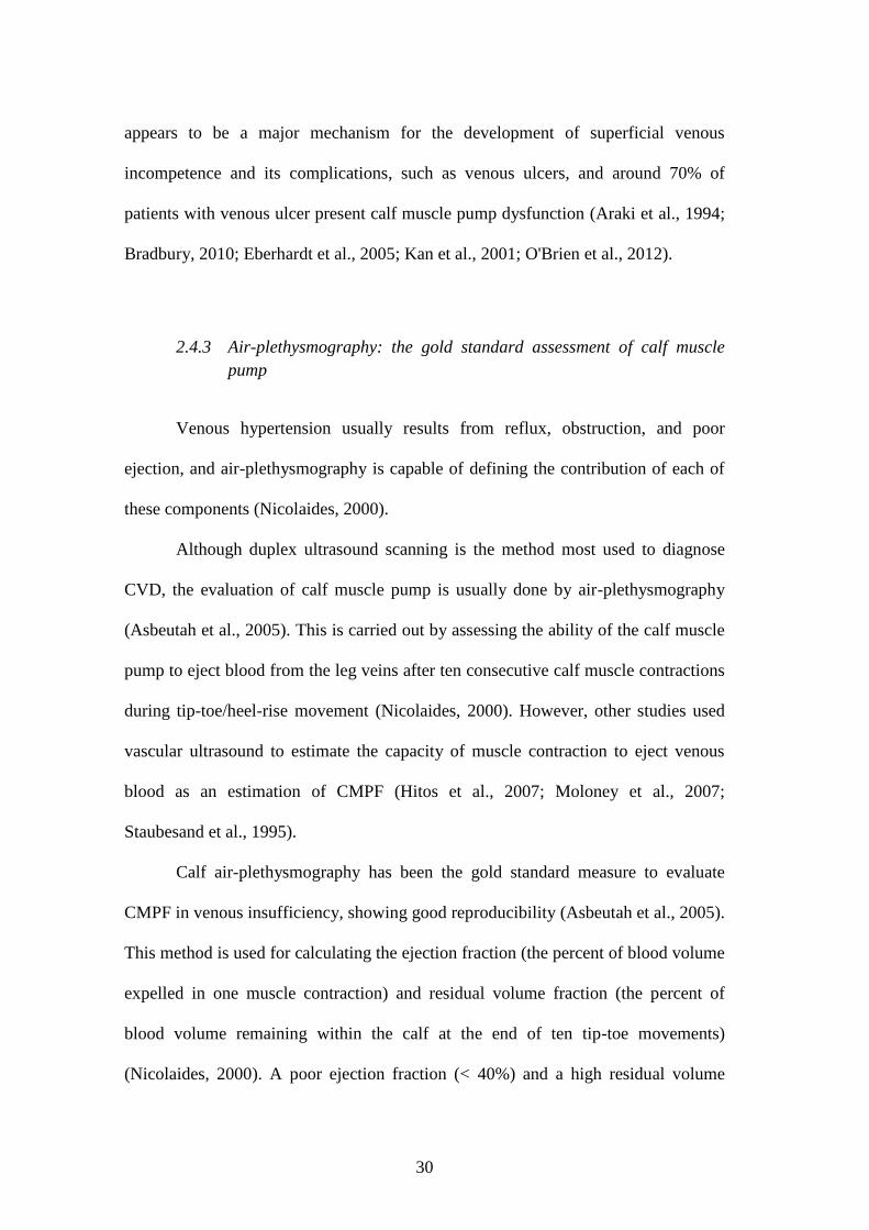
2.4.3 Air-plethysmography: the gold standard assessment of calf muscle
pump .......................................................................................................... 30
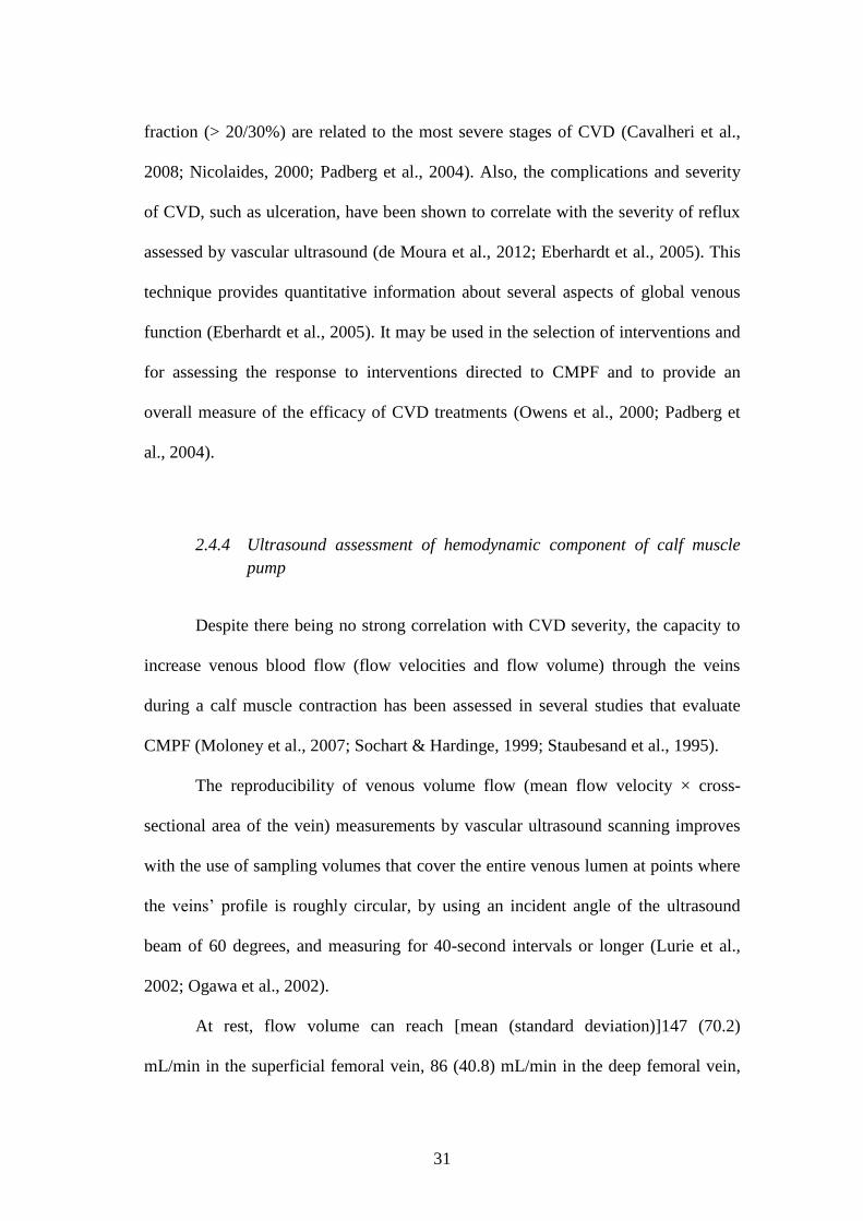
2.4.4 Ultrasound assessment of hemodynamic component of calf muscle
pump .......................................................................................................... 31
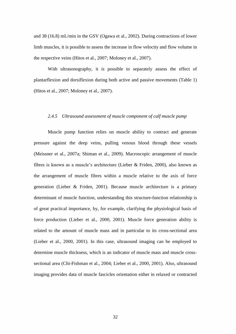
2.4.5 Ultrasound assessment of muscle component of calf muscle pump .. 32
2.5 Manual lymphatic drainage ....................................................................... 35
2.5.1 Brief history of manual lymphatic drainage ..................................... 35
2.5.2 Manual lymphatic drainage - Leduc method .................................... 37
2.5.3 Therapeutic efficacy of manual lymphatic drainage ........................ 38
2.5.4 Decongestive lymphatic therapy ....................................................... 40
2.5.5 Contraindications/Precautions ......................................................... 42
2.5.6 Manual lymphatic drainage in CVD ................................................. 43
2.6 The role of conservative treatments of CVD ............................................ 44
3 SCOPE OF THE THESIS AND HYPOTHESIS ............................................ 47
Page 16
xiv
3.1 Objectives of the study ............................................................................... 48
3.2 Hypothesis ................................................................................................... 49
4 INSTRUMENTS AND METHODS ................................................................. 51
4.1 Studies outline ............................................................................................. 51
4.2 Ethics ........................................................................................................... 52
4.3 Participants ................................................................................................. 54
4.4 Diagnose and Severity of disease ............................................................... 56
4.5 Duplex scanning: diagnose of CVD .......................................................... 57
4.6 CEAP classification .................................................................................... 58
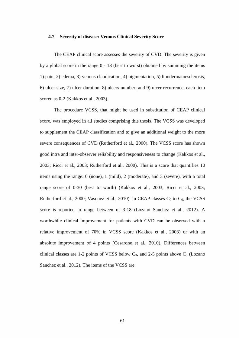
4.7 Severity of disease: Venous Clinical Severity Score ................................ 61
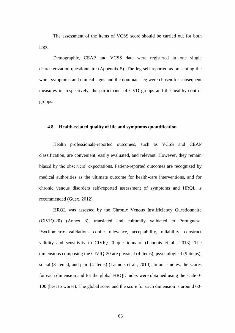
4.8 Health-related quality of life and symptoms quantification ................... 63
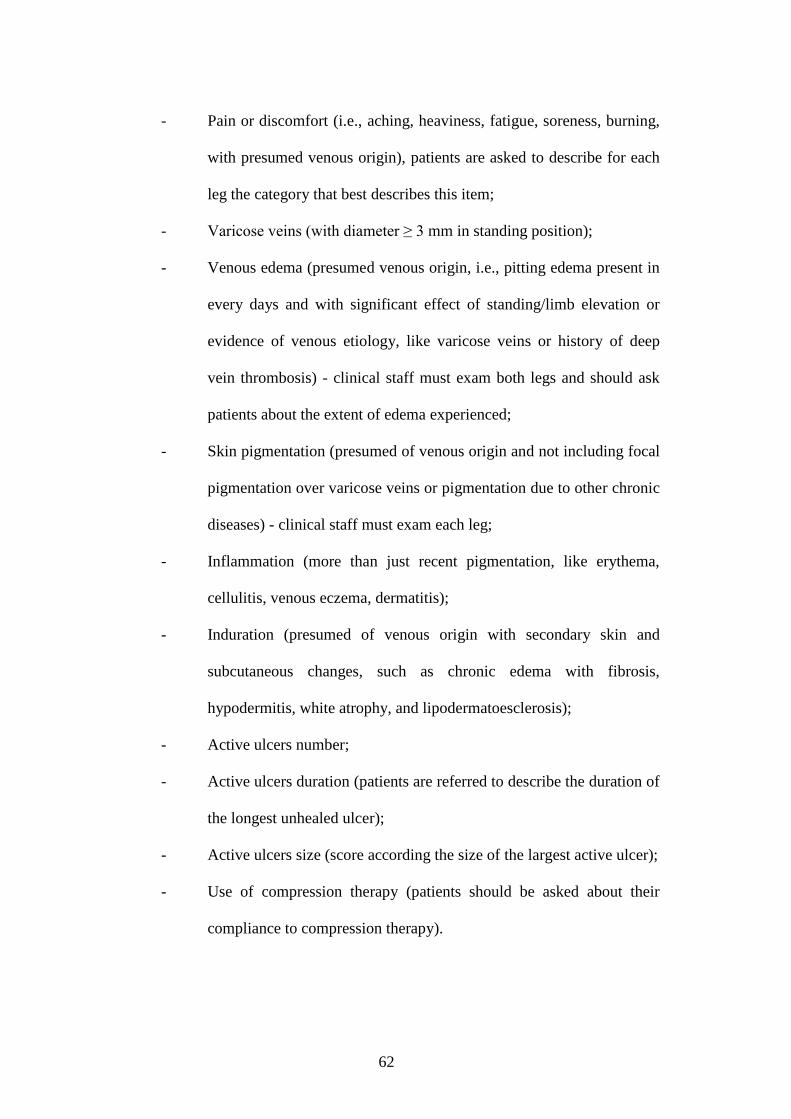
4.9 Symptoms .................................................................................................... 64
4.10 Vascular ultrasound assessments for venous hemodynamic .................. 65
4.10.1 Popliteal vein assessment for calf muscle pump function ................ 65
4.10.2 Venous hemodynamics during manual lymphatic drainage ............. 68
4.11 Calf muscles architecture .......................................................................... 75
4.12 Leg volume .................................................................................................. 76
4.13 Ankle isokinetic dynamometer assessment .............................................. 78
4.14 Educational session..................................................................................... 79
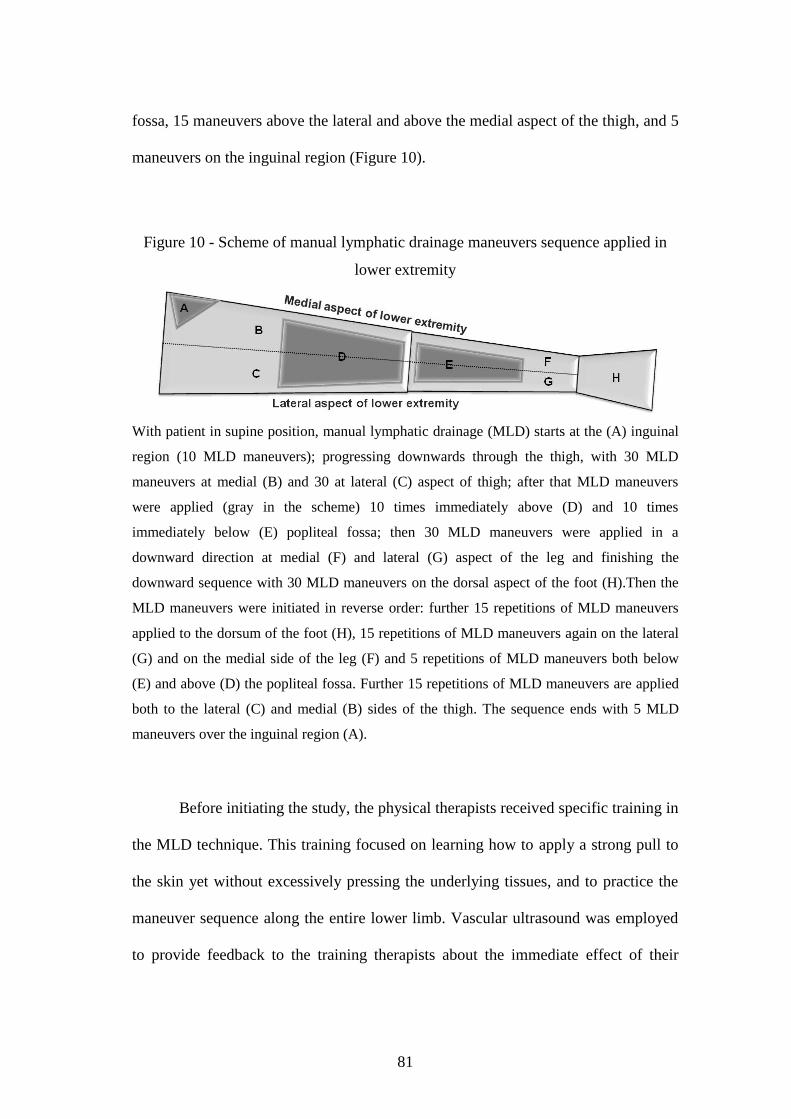
4.15 Manual lymphatic drainage sequence and method ................................. 80
4.16 Statistical analysis....................................................................................... 82
5 RESULTS ........................................................................................................... 85
5.1 The use of ultrasound in the evaluation of the efficacy of calf muscle
pump function in primary chronic venous disease.................................. 92
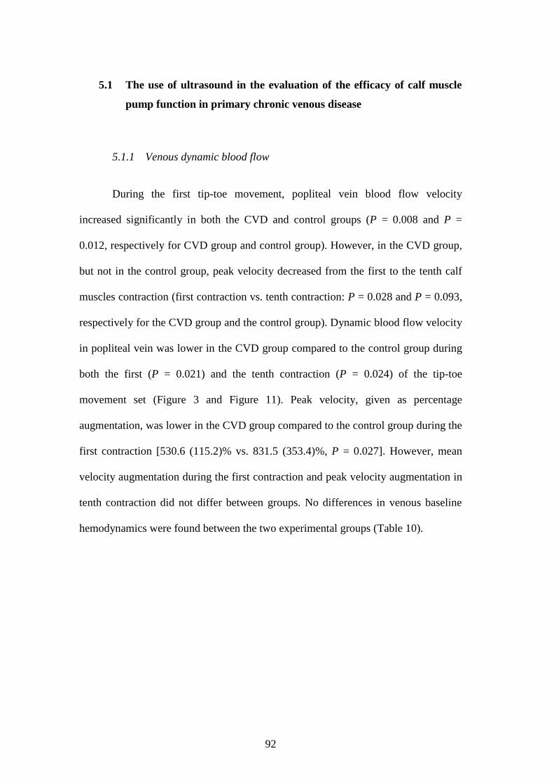
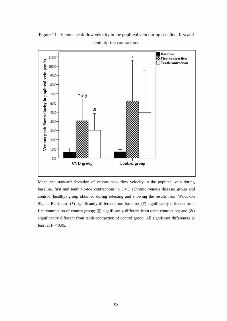
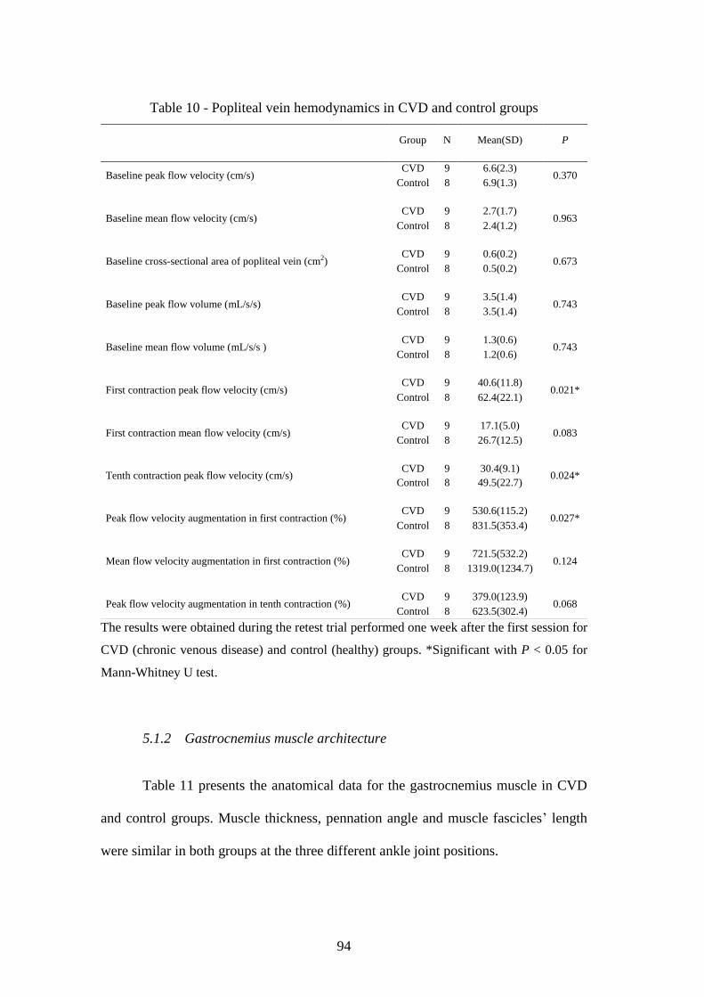
5.1.1 Venous dynamic blood flow .............................................................. 92
5.1.2 Gastrocnemius muscle architecture ................................................. 94
5.1.3 Test-retest reliability of calf muscle pump and gastrocnemius
architecture ultrasound measures ................................................................ 95
5.2 Venous flow at superficial and deep venous system during manual
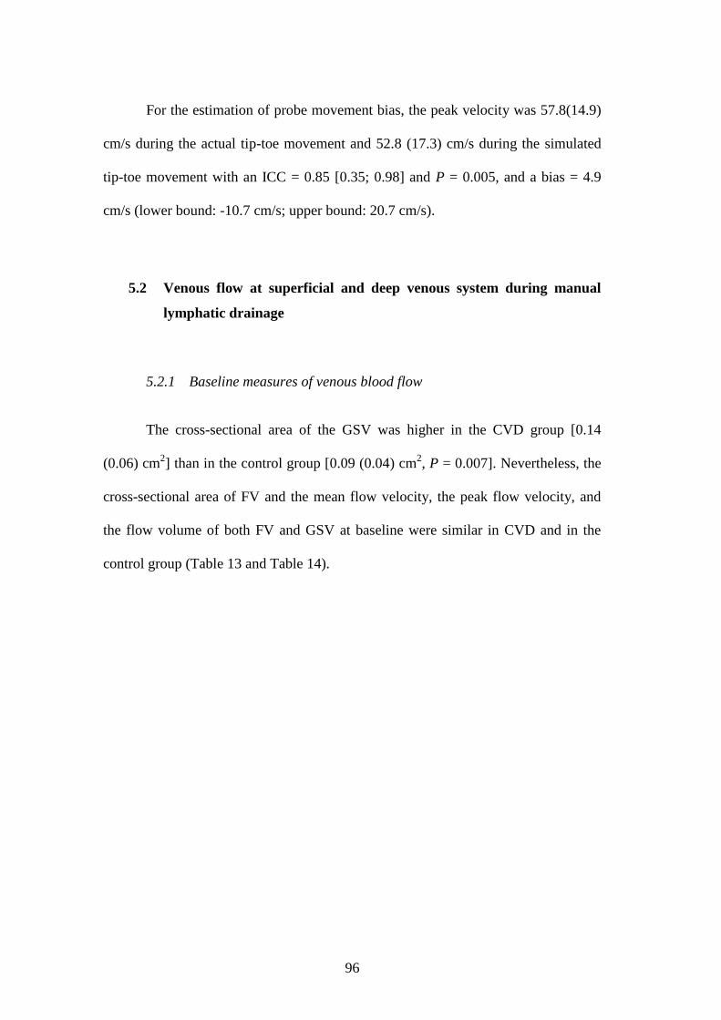
lymphatic drainage ..................................................................................... 96
5.2.1 Baseline measures of venous blood flow .......................................... 96
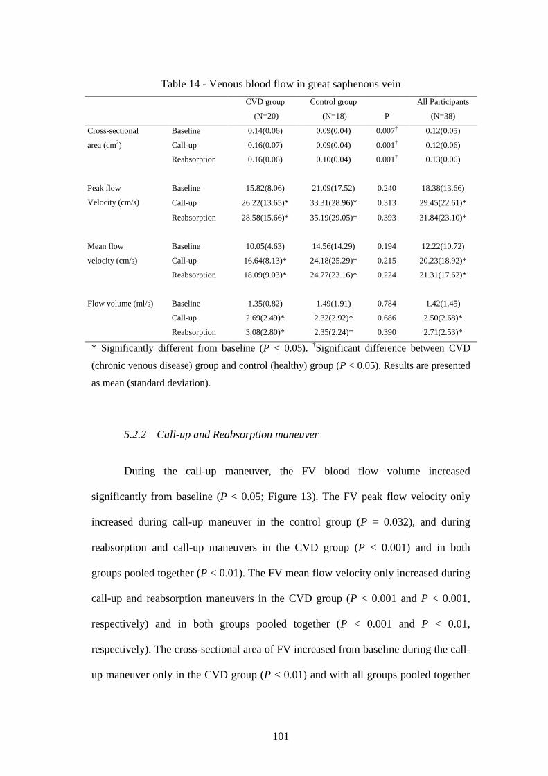
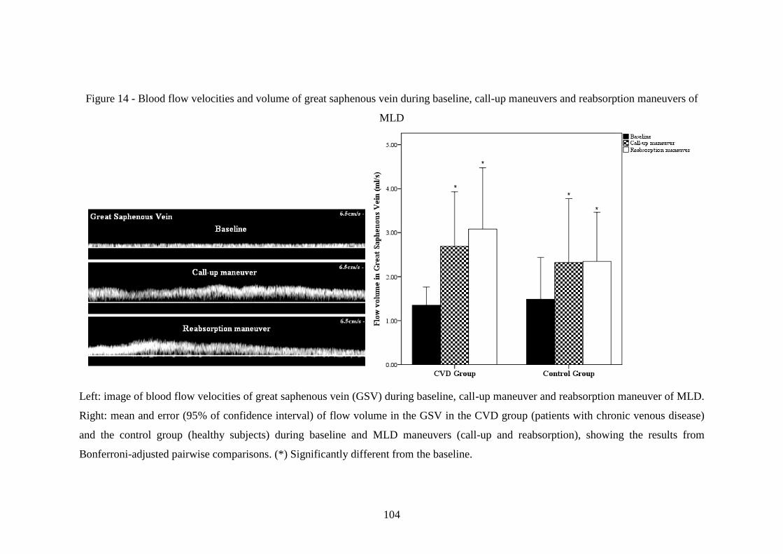
5.2.2 Call-up and Reabsorption maneuver .............................................. 101
5.2.3 Severity of CVD and MLD maneuvers ........................................... 105
5.3 Venous flow during manual lymphatic drainage applied to different
regions of the lower extremity. ................................................................ 105
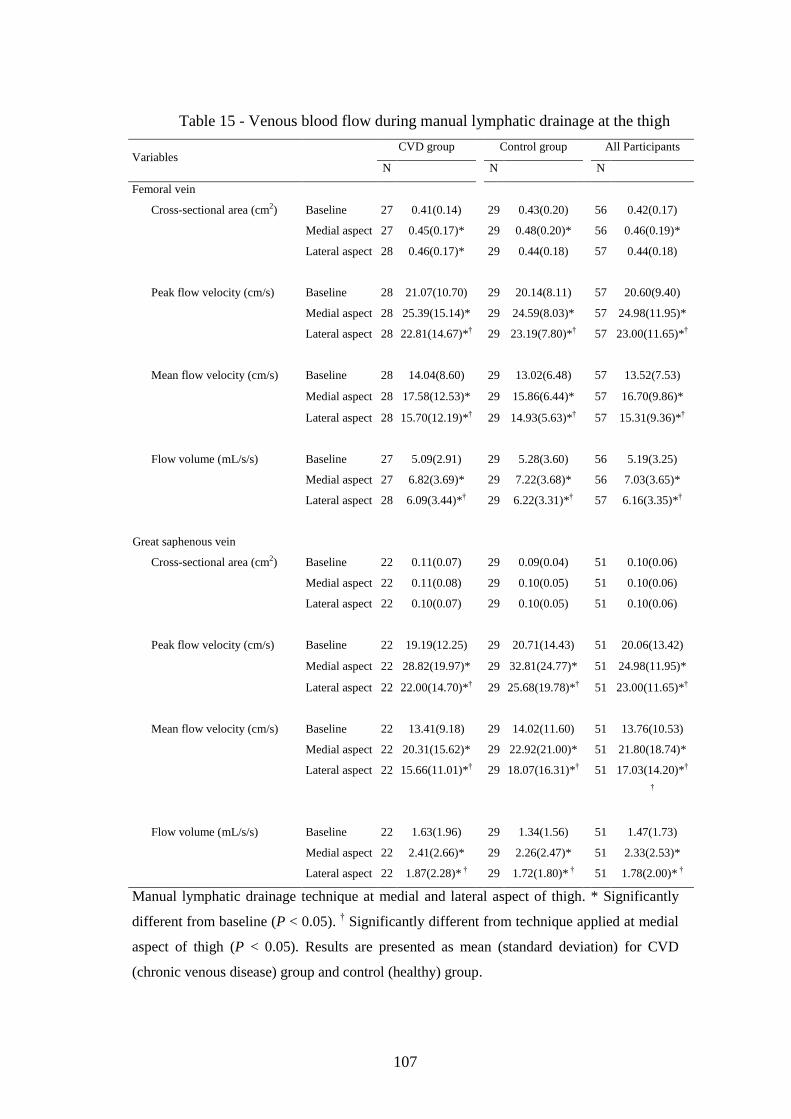
5.3.1 Manual lymphatic drainage at the thigh ........................................ 105
Page 17
xv
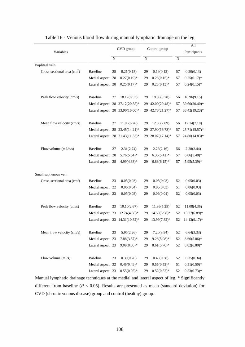
5.3.2 Manual lymphatic drainage at the leg ............................................ 106
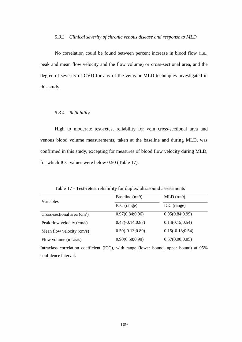
5.3.3 Clinical severity of chronic venous disease and response to MLD 109
5.3.4 Reliability ........................................................................................ 109
5.4 Efficacy of manual lymphatic drainage in chronic venous disease
patients ....................................................................................................... 110
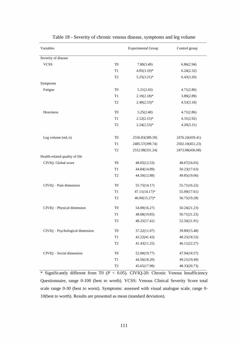
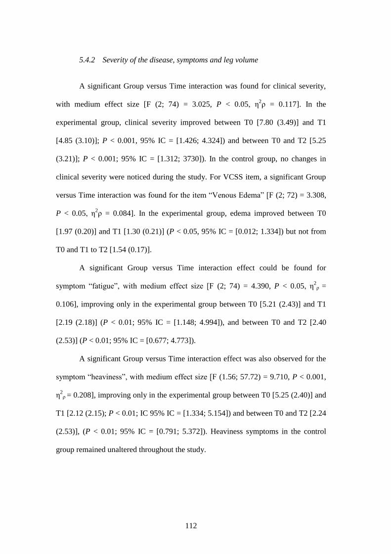
5.4.1 Health-related quality of life ........................................................... 110
5.4.2 Severity of the disease, symptoms and leg volume .......................... 112
5.4.3 Ankle muscles strength .................................................................... 113
6 DISCUSSION ................................................................................................... 117
6.1 Ultrasound assessment of calf muscle pump function ........................... 117
6.2 Hemodynamic effects of manual lymphatic drainage ........................... 120
6.2.1 Call-up versus Reabsorption maneuver .......................................... 121
6.2.2 The effect of manual lymphatic drainage when applied to different
regions of the lower limb ............................................................................ 123
6.2.3 The effect of chronic venous disease severity on manual lymphatic
drainage efficacy in venous return ............................................................. 125
6.3 Therapeutic efficacy of manual lymphatic drainage for treatment of
patients with chronic venous disease ...................................................... 128
7 CONCLUSION ................................................................................................ 133
7.1 Clinical notes ............................................................................................. 134
7.2 Limitations ................................................................................................ 135
7.3 Future directions ....................................................................................... 136
8 REFERENCES ................................................................................................ 137
9 APPENDIXES .................................................................................................. 153
9.1 Appendix 1 -Informed Consent of Study I ............................................. 155
9.2 Appendix 2 -Informed Consent of Study II ............................................ 159
9.3 Appendix 3 -Informed Consent of Study III .......................................... 163
9.4 Appendix 4 -Informed Consent of Study IV .......................................... 167
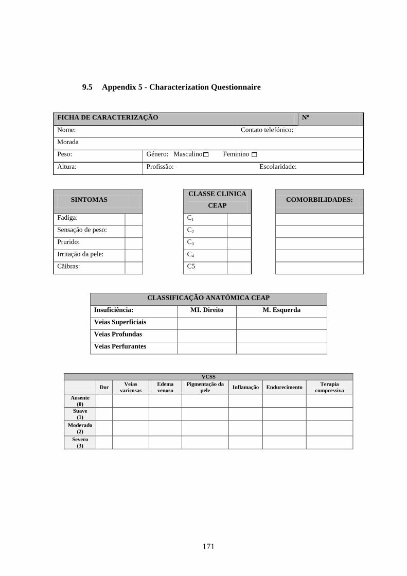
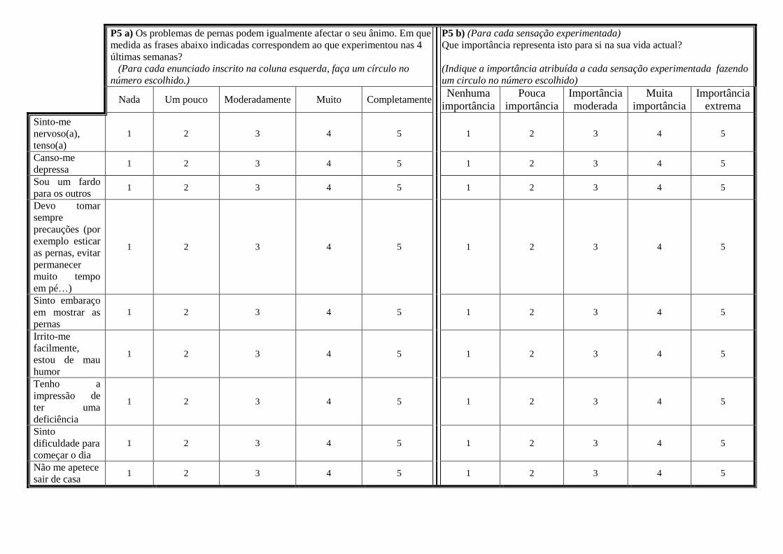
9.5 Appendix 5 - Characterization Questionnaire ....................................... 171
10 ANNEXES ........................................................................................................ 173
10.1 Annex 1 - Article: The use of ultrasound in the evaluation of the
efficacy of calf muscle pump function in primary chronic venous
disease ........................................................................................................ 175
10.2 Annex 2 - Article: Manual lymphatic drainage in chronic venous
disease: A duplex ultrasound study ........................................................ 187
10.3 Annex 3 - Chronic Venous Disease Quality of Life Questionnaire
(CIVIQ-20) ................................................................................................ 199
Page 19
xvii
TABLE INDEX
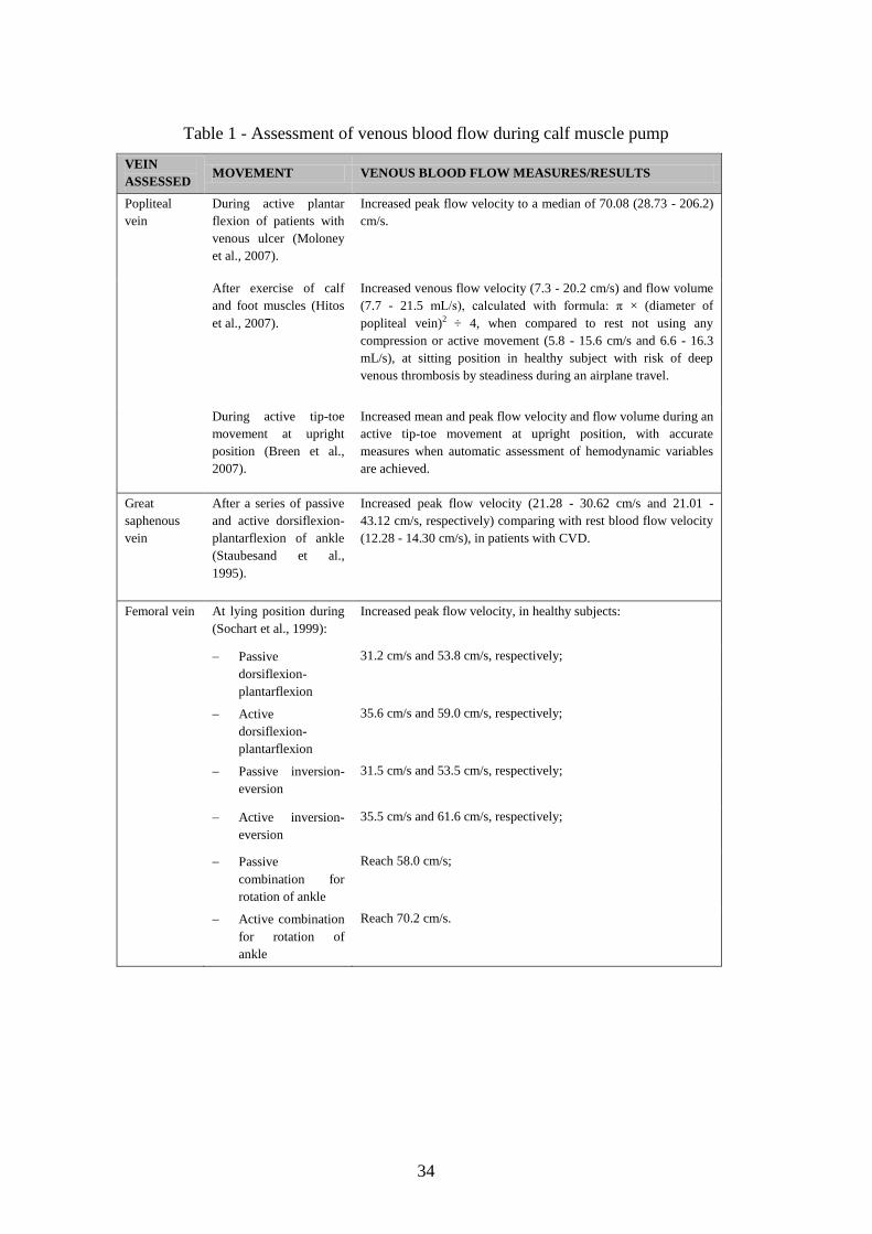
Table 1 - Assessment of venous blood flow during calf muscle pump ..................... 34
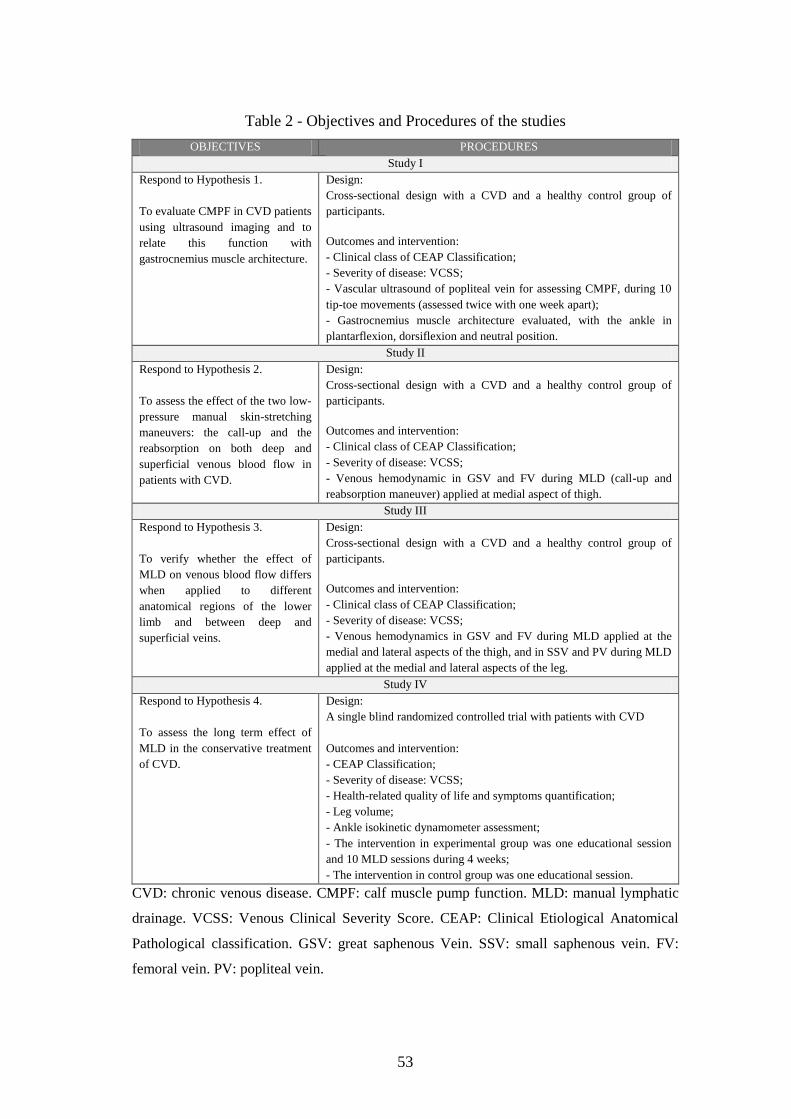
Table 2 - Objectives and Procedures of the studies ................................................... 53
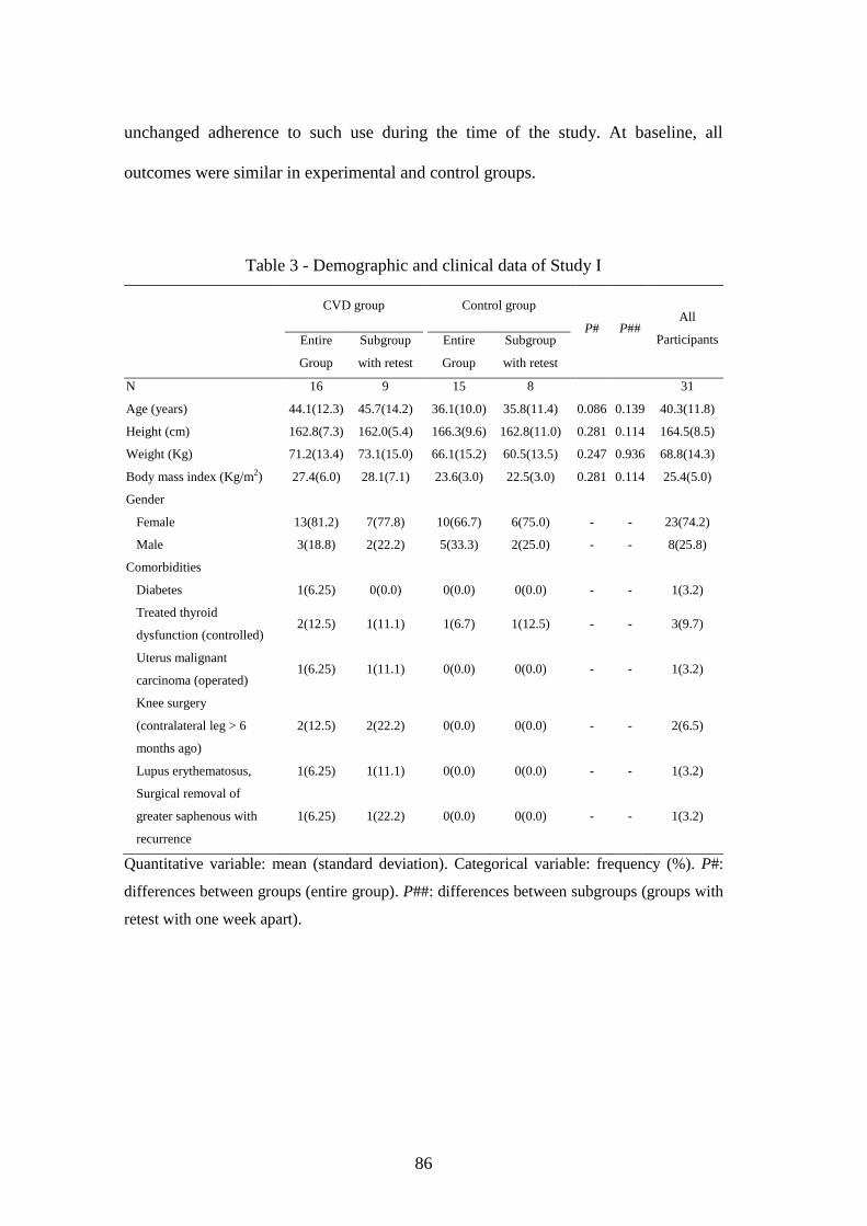
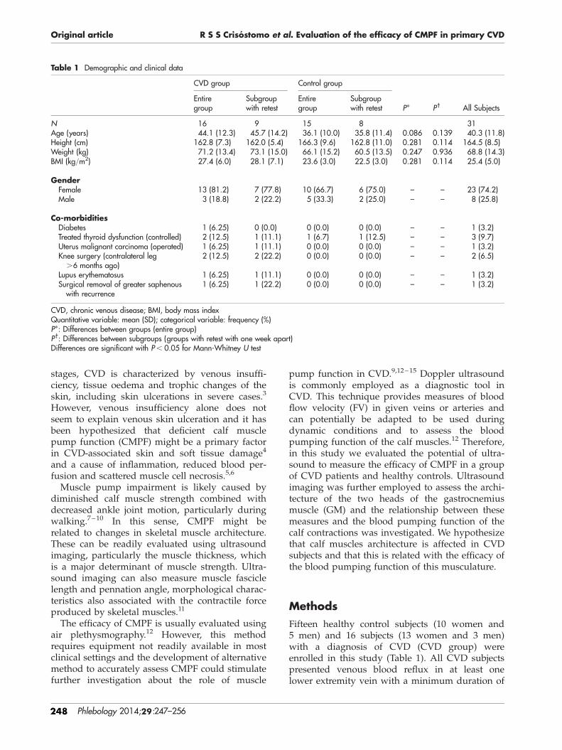
Table 3 - Demographic and clinical data of Study I .................................................. 86
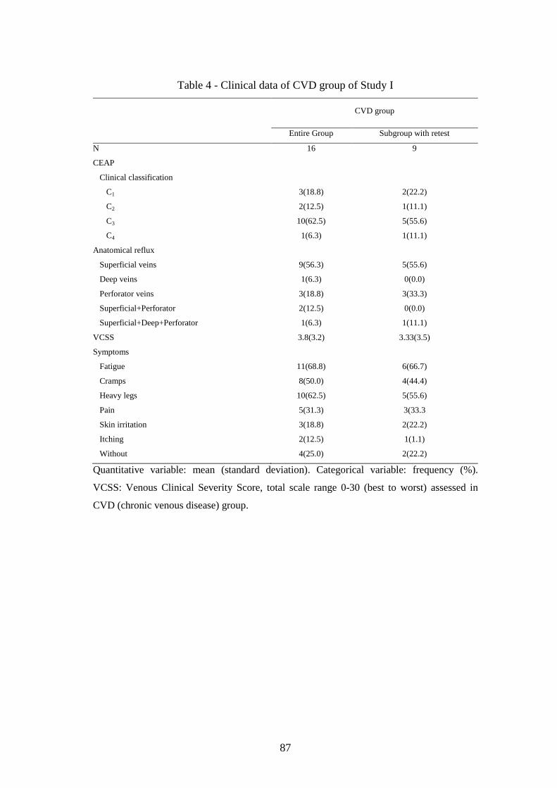
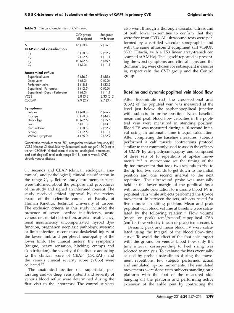
Table 4 - Clinical data of CVD group of Study I ....................................................... 87
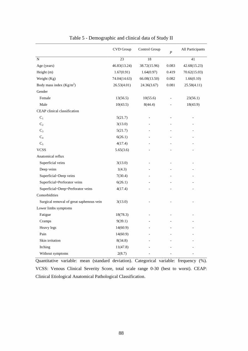
Table 5 - Demographic and clinical data of Study II ................................................. 88
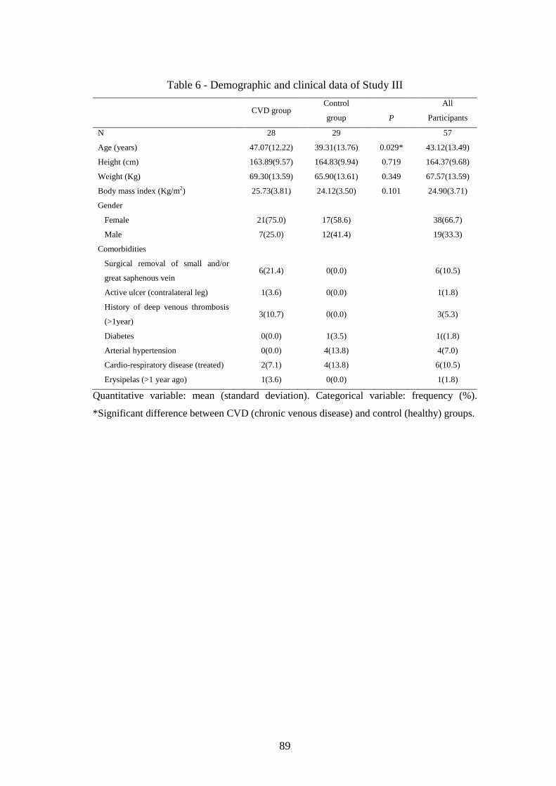
Table 6 - Demographic and clinical data of Study III ............................................... 89
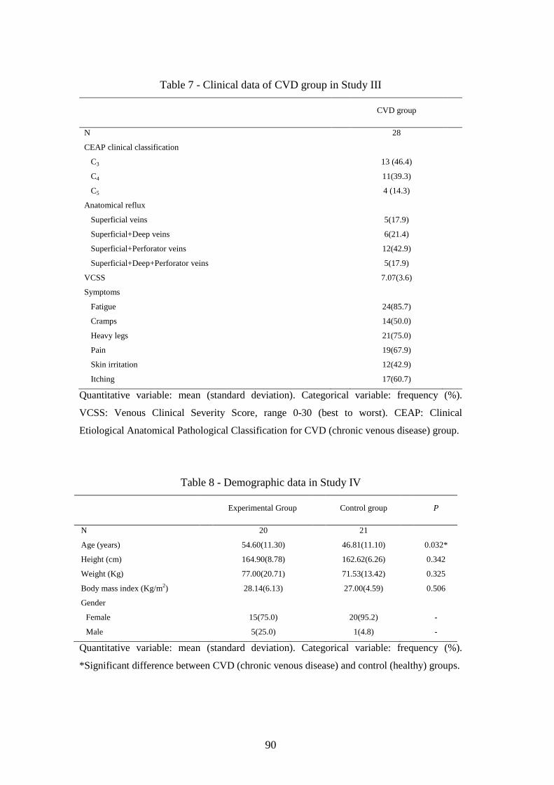
Table 7 - Clinical data of CVD group in Study III .................................................... 90
Table 8 - Demographic data in Study IV ................................................................... 90
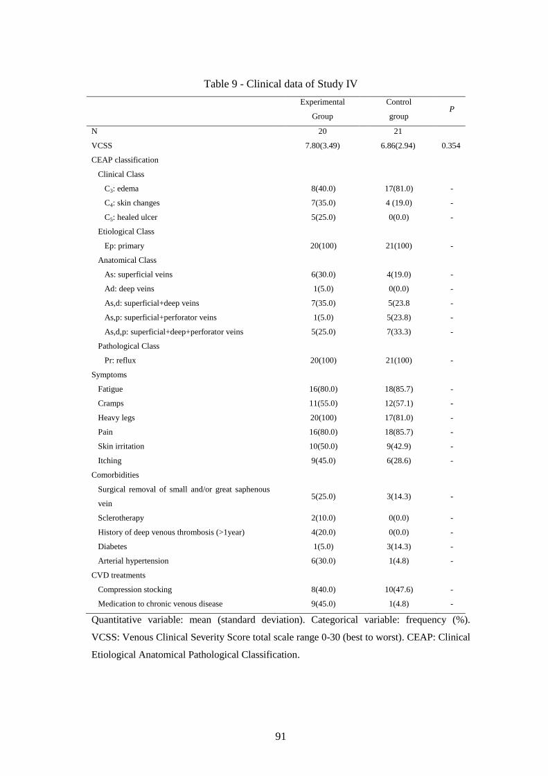
Table 9 - Clinical data of Study IV ............................................................................ 91
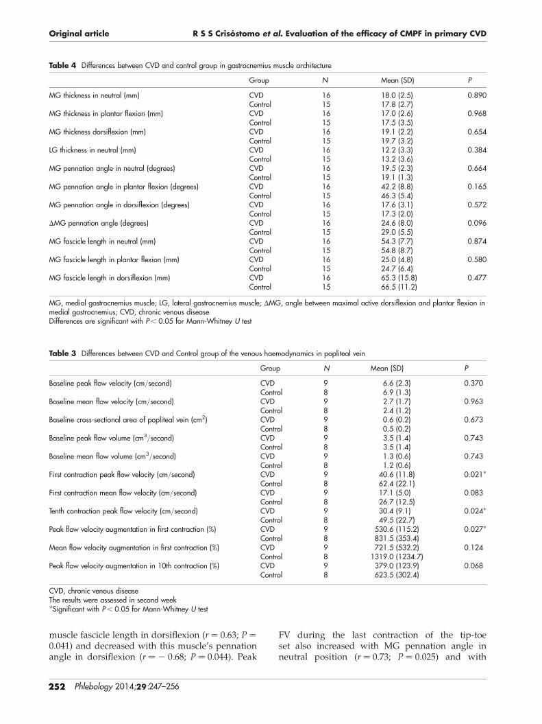
Table 10 - Popliteal vein hemodynamics in CVD and control groups ...................... 94
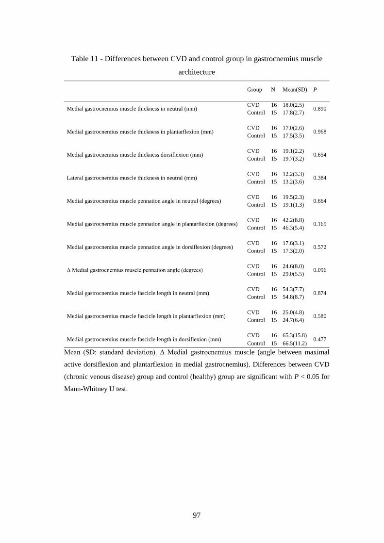
Table 11 - Differences between CVD and control group in gastrocnemius muscle
architecture ................................................................................................................. 97
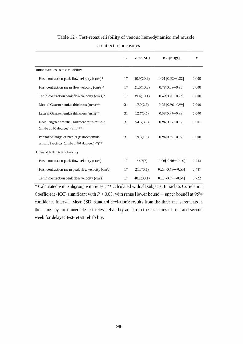
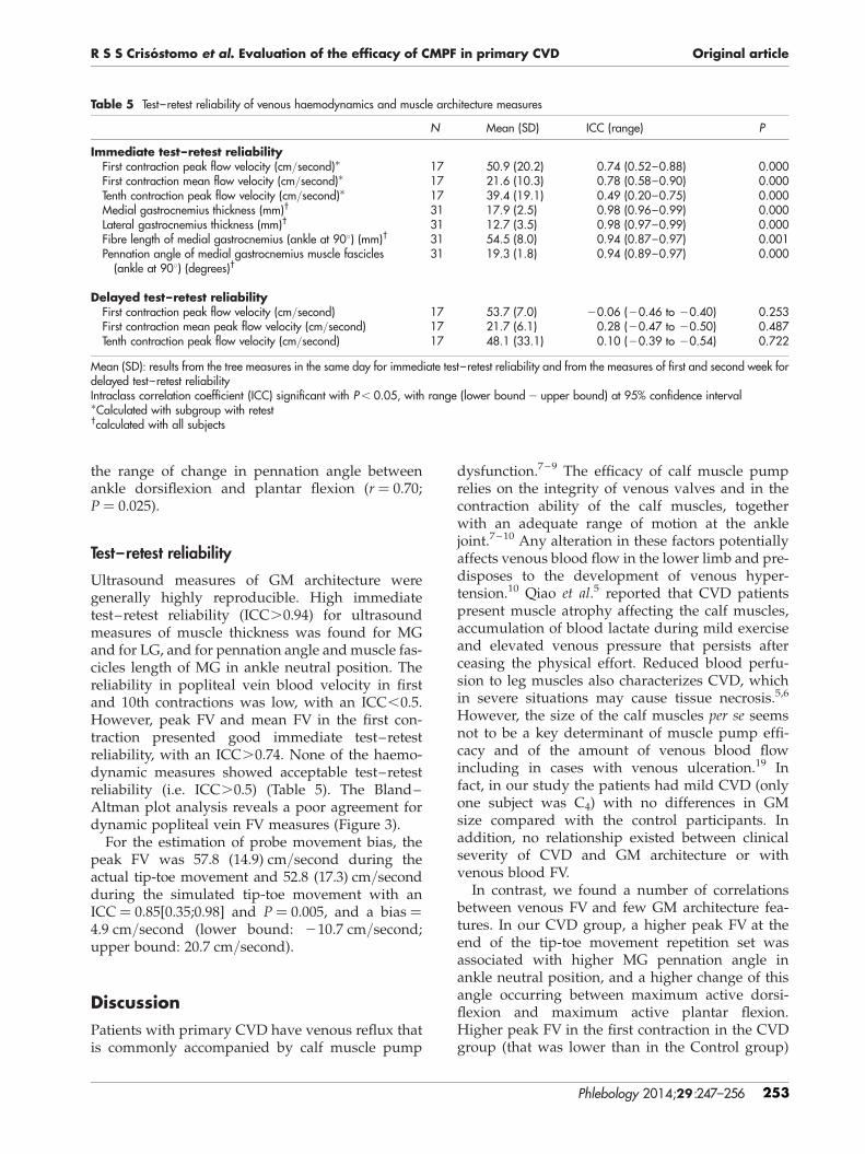
Table 12 - Test-retest reliability of venous hemodynamics and muscle architecture
measures ..................................................................................................................... 98
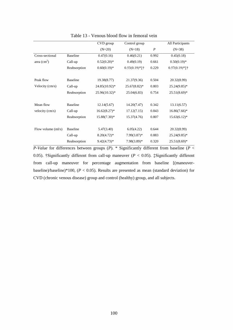
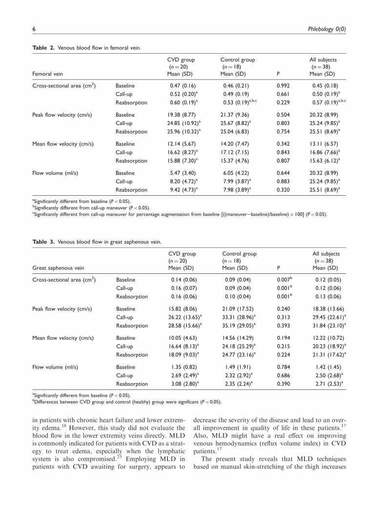
Table 13 - Venous blood flow in femoral vein ........................................................ 100
Table 14 - Venous blood flow in great saphenous vein ........................................... 101
Table 15 - Venous blood flow during manual lymphatic drainage at the thigh ...... 107
Table 16 - Venous blood flow during manual lymphatic drainage on the leg ......... 108
Table 17 - Test-retest reliability for duplex ultrasound assessments ....................... 109
Table 18 - Severity of chronic venous disease, symptoms and leg volume ............ 111
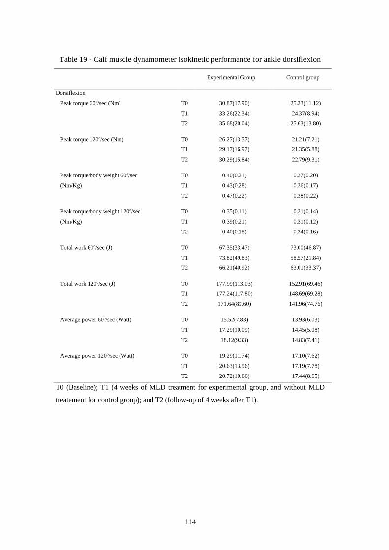
Table 19 - Calf muscle dynamometer isokinetic performance for ankle dorsiflexion
................................................................................................................................. 114
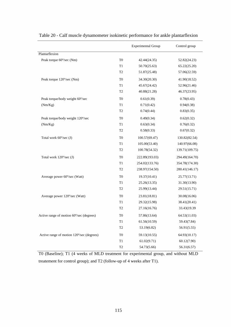
Table 20 - Calf muscle dynamometer isokinetic performance for ankle plantarflexion
................................................................................................................................. 115
Page 21
xix
FIGURE INDEX
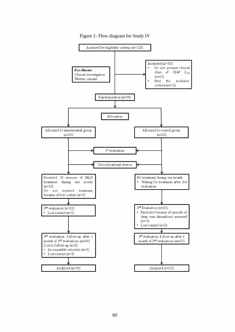
Figure 1- Flow diagram for Study IV ........................................................................ 60
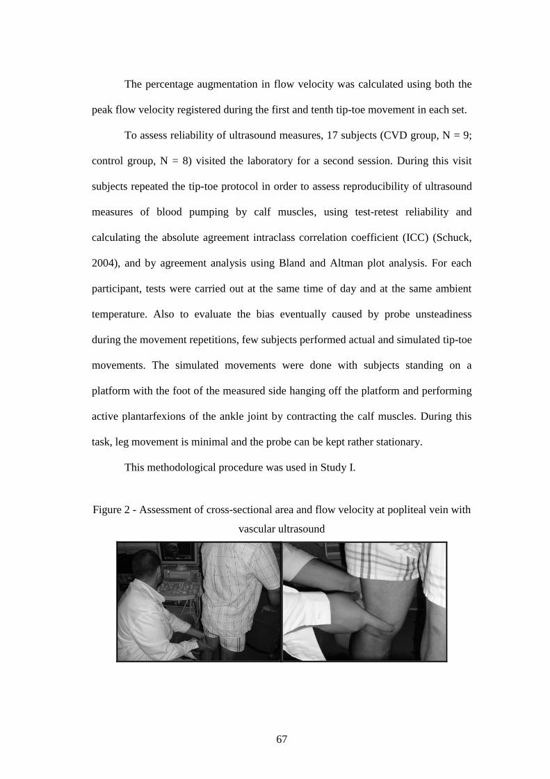
Figure 2 - Assessment of cross-sectional area and flow velocity at popliteal vein with
vascular ultrasound .................................................................................................... 67
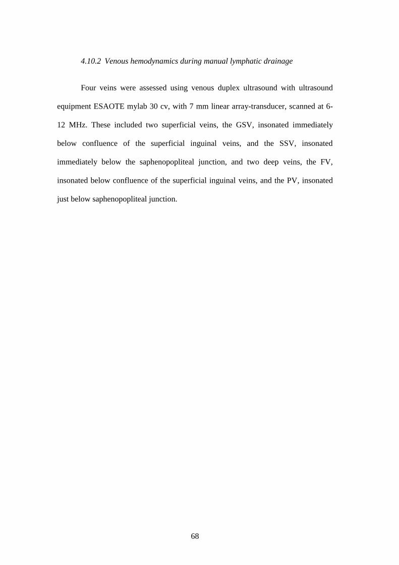
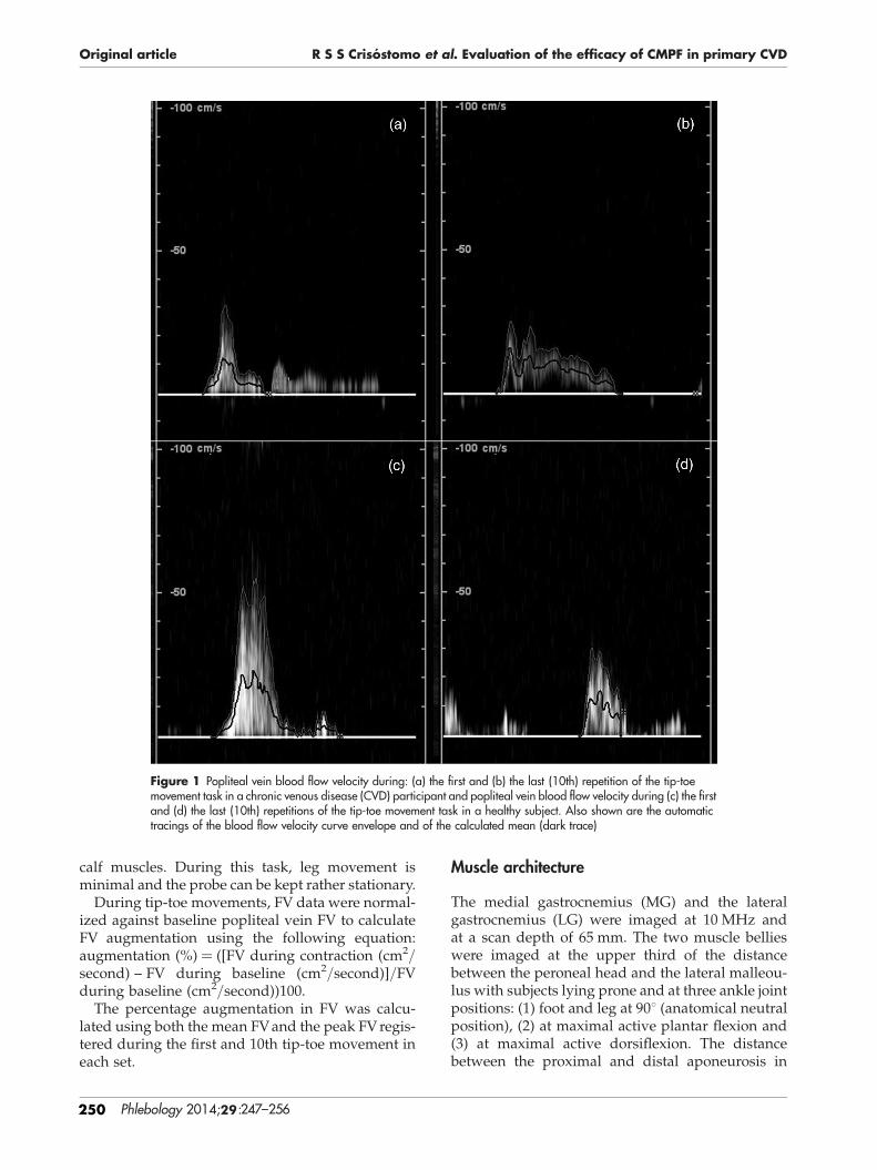
Figure 3 - Venous ultrasound assessment at popliteal vein during first and tenth calf
contraction, during tip-toe movement for both control and CVD group ................... 69
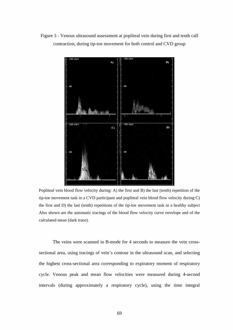
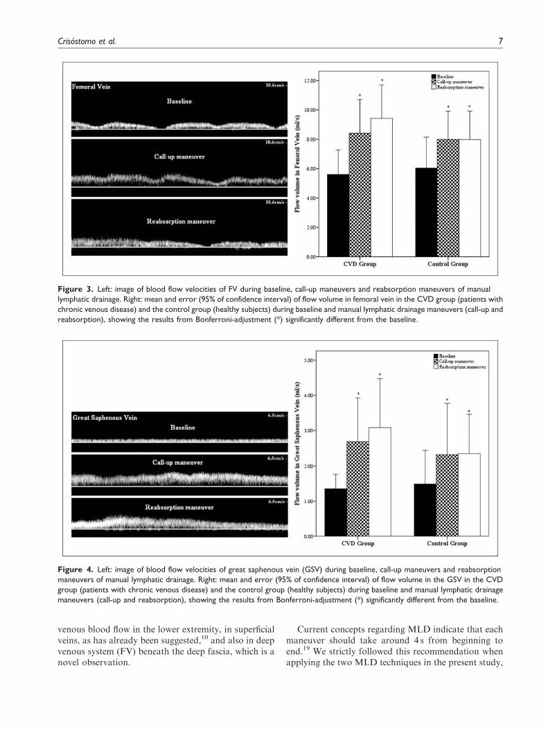
Figure 4 - Representative ultrasound images showing the effect of MLD ................ 70
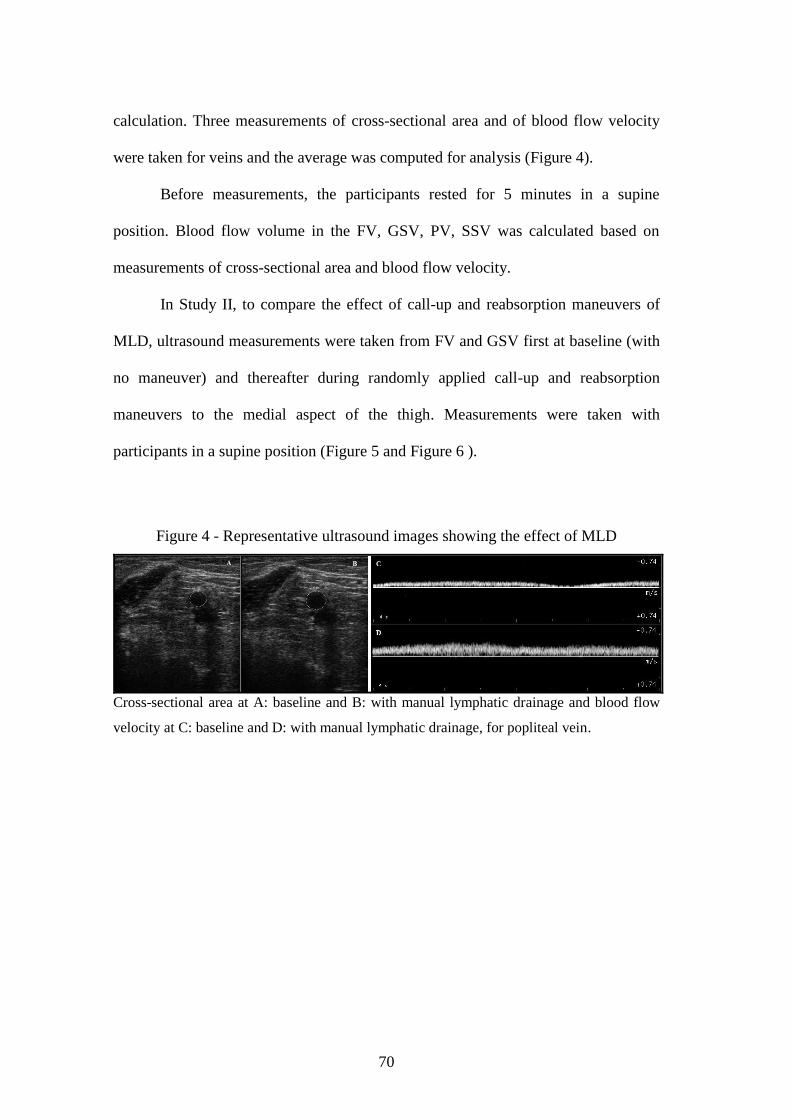
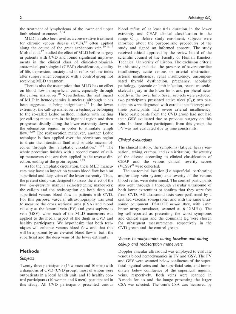
Figure 5 - Randomization for duplex ultrasound assessment .................................... 71
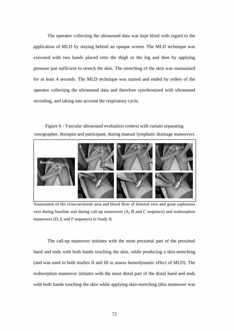
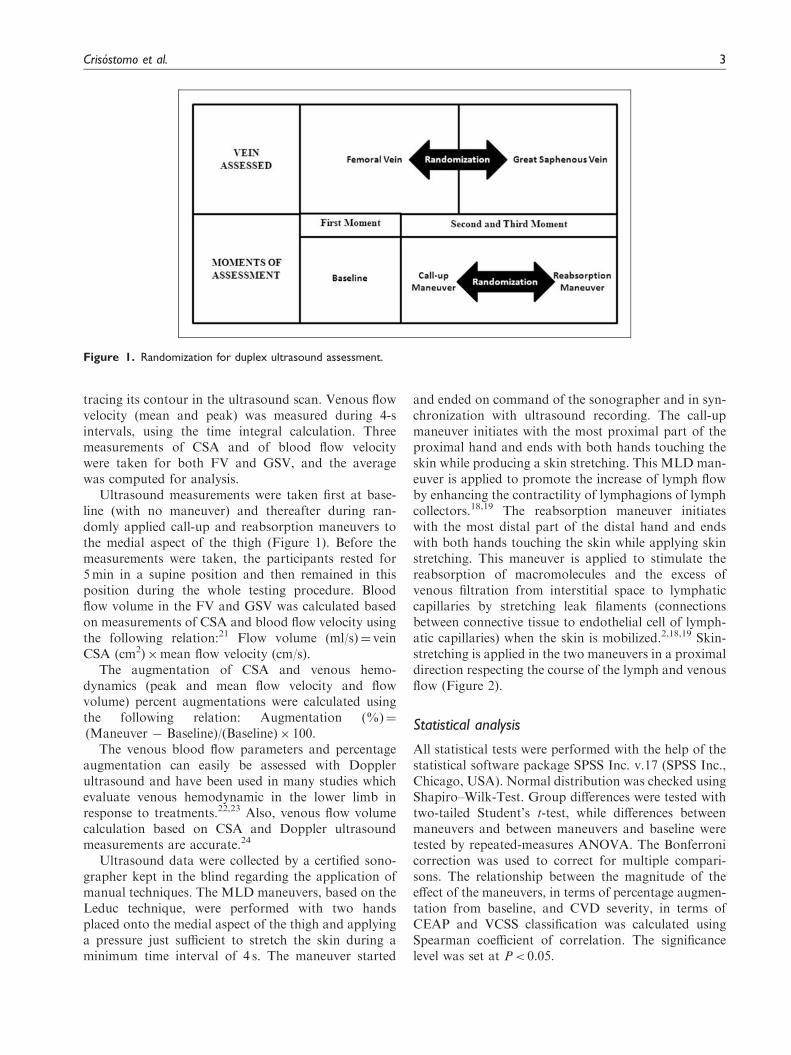
Figure 6 - Vascular ultrasound evaluation context with curtain separating
sonographer, therapist and participant, during manual lymphatic drainage maneuvers
................................................................................................................................... 72
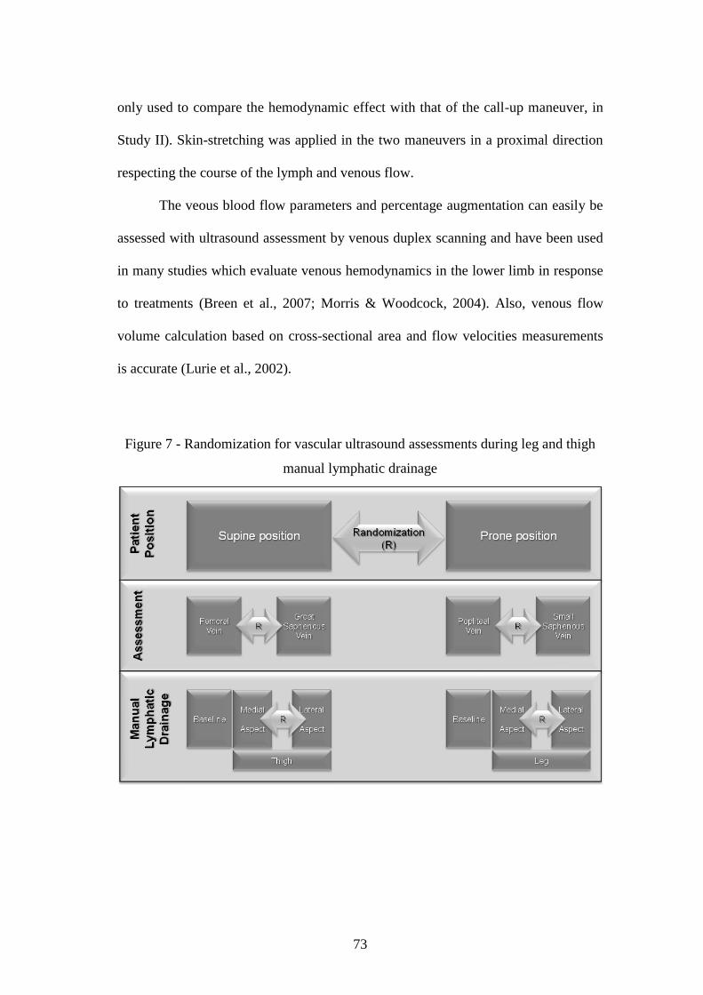
Figure 7 - Randomization for vascular ultrasound assessments during leg and thigh
manual lymphatic drainage ........................................................................................ 73
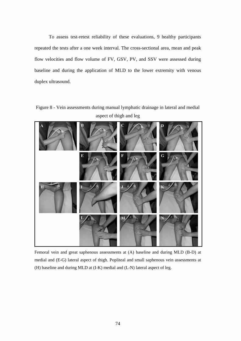
Figure 8 - Vein assessments during manual lymphatic drainage in lateral and medial
aspect of thigh and leg ............................................................................................... 74
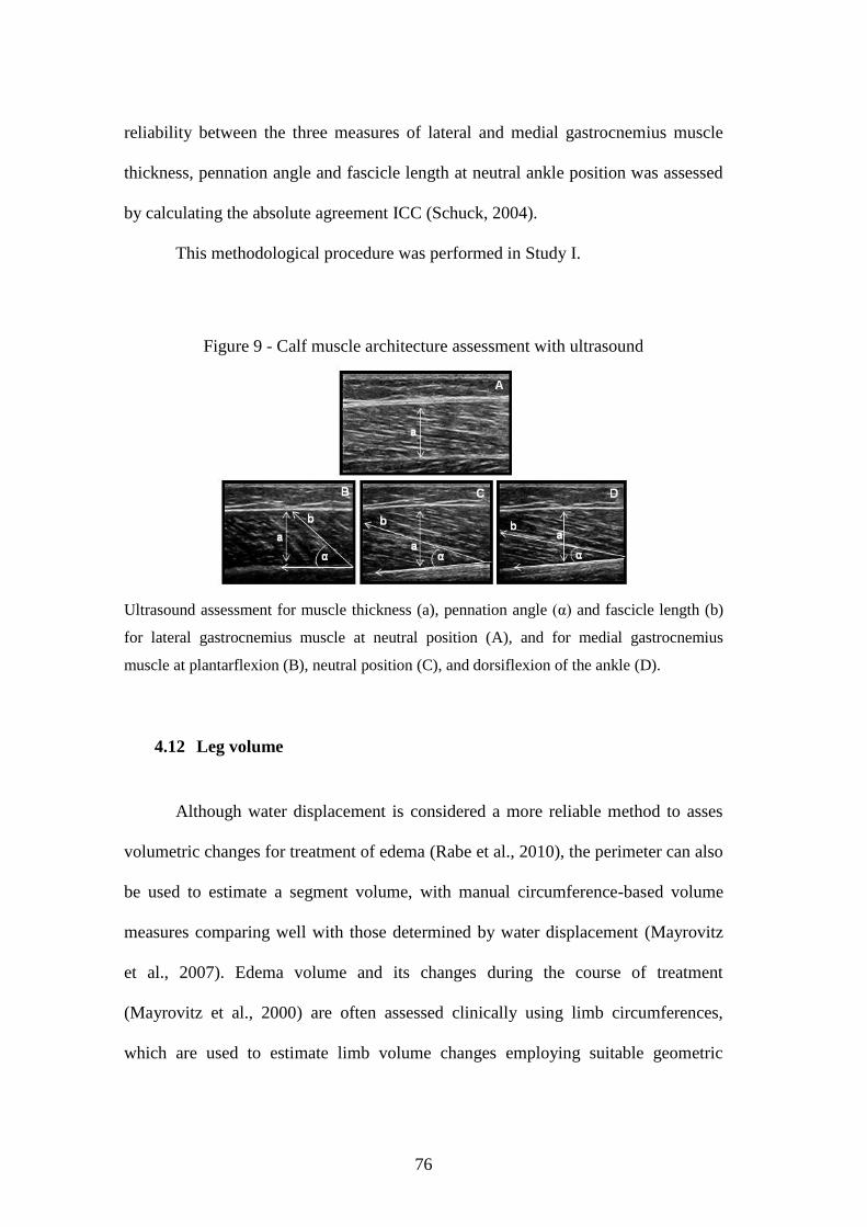
Figure 9 - Calf muscle architecture assessment with ultrasound ............................... 76
Figure 10 - Scheme of manual lymphatic drainage maneuvers sequence applied in
lower extremity .......................................................................................................... 81
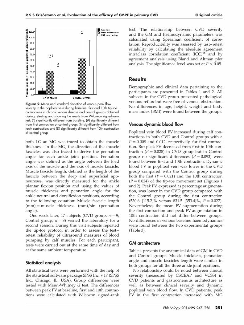
Figure 11 - Venous peak flow velocity in the popliteal vein during baseline, first and
tenth tip-toe contractions ........................................................................................... 93
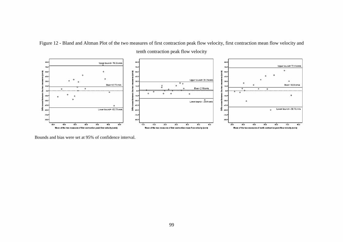
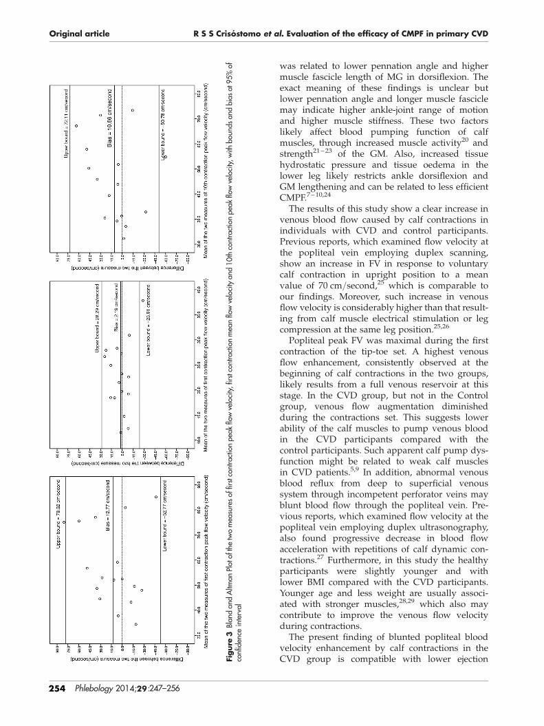
Figure 12 - Bland and Altman Plot of the two measures of first contraction peak flow
velocity, first contraction mean flow velocity and tenth contraction peak flow
velocity ....................................................................................................................... 99
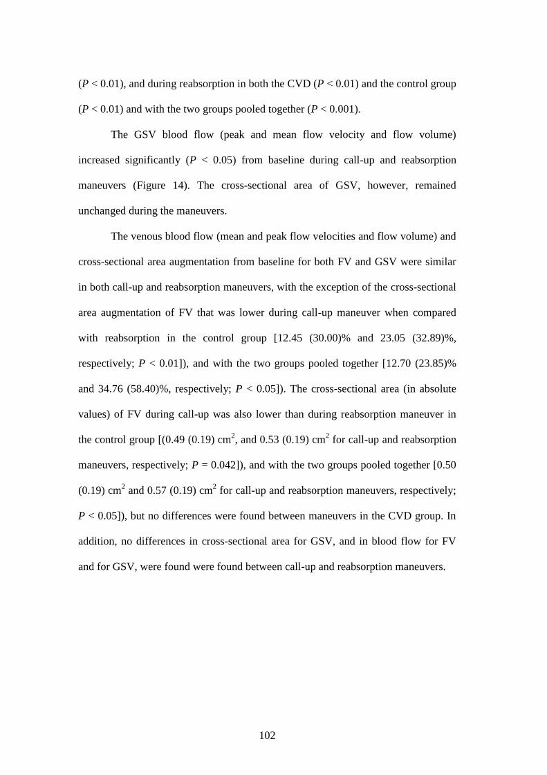
Figure 13 - Blood flow velocities and volume of femoral vein during baseline, call-
up maneuvers and reabsorption maneuvers of MLD ............................................... 103
Figure 14 - Blood flow velocities and volume of great saphenous vein during
baseline, call-up maneuvers and reabsorption maneuvers of MLD ......................... 104
Page 23
xxi
LIST OF ABBREVIATORS
CEAP - Clinical Etiological Anatomical Classification
CIVIQ - Chronic Venous Insufficiency Questionnaire
CMPF - Calf Muscle Pump Function
CVD - Chronic Venous Disease
FV - Femoral Vein
GSV - Great Saphenous Vein
HRQL - Health-related quality of life
IC - Interval of Confidence
ICC - Intraclass Correlation Coefficient
MLD - Manual Lymphatic Drainage
PV - Popliteal Vein
SSV - Small Saphenous Vein
VCSS - Venous Clinical Severity Score
Page 25
1
1 INTRODUCTION
In Portugal the prevalence of chronic venous disease (CVD) in males is
17.8% and in females is 34.1%, i.e., around 2.5 million cases, with around 70.000
new cases every year (Capitão et al., 1995).
Chronic venous insufficiency represents the most severe cases of CVD, and is
related to a physiopathology that associates venous hypertension, vein valve damage,
venous obstruction, calf muscle pump impairment, inflammations of tissues (skin,
subcutaneous tissue and muscle) and veins, alterations of morphology and function
of veins, with abnormal venous reflux, venous edema, changes of the skin and
subcutaneous tissues, with ulcer representing the more severe stage of this condition
(Eklof et al., 2004; Meissner et al., 2007b; Panny et al., 2009; Qiao et al., 2005;
Rowland, 2001; Simka, 2007; Yamaki et al., 2010). It is estimated that 100.000
Portuguese citizens present an open (or active) venous ulcer (Oliveira et al., 2003),
and 280.000 an open or healed venous ulcer, with 10% of healed ulcer at risk for
recurrence (Bradbury, 2010; Pina et al., 2005).
The first problem referred from patients are the symptoms that, together with
varicose veins, is present form mild to severe cases of CVD, strongly contributing to
a the negative self-esteem that also characterizes this disease (Boisseau, 2007;
Bradbury et al., 1999; Campbell et al., 2007; Cesarone et al., 2010; Darvall et al.,
2012; Eklof et al., 2009; Koupidis et al., 2008). The diminished health-related quality
of life (HRQL) of this condition in these patients, with functional impairments,
reflected in their daily activities are well studied (Andreozzi et al., 2005; Darvall et
al., 2012; Furtado et al., 2008; Koupidis et al., 2008). This negative impact is so
Page 26
2
important that the previous view of this condition as an aesthetic problem has been
abandoned for years.
The socioeconomic burden of this condition is very high. The indirect costs
are substantial and are associated with the symptoms, functional impairment,
emotional disturbances and negative impact in HRQL (Lamping et al., 2003). The
direct costs of CVD treatment are almost entirely related to its high prevalence,
morbidity, and chronicity (Labropoulos et al., 2009; Lamping et al., 2003). In
developed countries, around 1-3% of the heath costs are due to CVD (Eklof et al.,
2004). However, when patients with less severe stages of the disease are diagnosed
and treated early, the physiopathology course of the disease can be prevented or even
receded (Nullen, 2010).
Manual lymphatic drainage (MLD) is a low pressure form of skin-stretching
massage, described as a conservative treatment option for the treatment of patients
with CVD (Steins & Jünger, 2000) as a coadjutant of other treatments, like stockings
and surgery for example (Molski et al., 2013; Molski et al., 2009). It is assumed that
this technique may have an effect on superficial venous flow, when a special
maneuver the so-called call-up maneuver from the Leduc method (Leduc & Leduc,
2000) is applied, although to a limited extent (Leduc et al., 2011).
This technique has been proposed as an option in the treatment of venous
lymphedema associated to CVD (Mortimer, 2000; Raju et al., 2012). It is suggested
by the literature that this technique should be applied in the course of the great
saphenous vein (GSV) to treat patients with CVD, but the capacity of MLD to
improve venous flow has been poorly studied (Leduc et al., 2000; Molski et al.,
2009; Peyre & Robert, 2000). Despite controversial evidence regarding the ability to
Page 27
3
reduce edema or lymphedema, MLD associated with other treatments, the so-called
lymphatic decongestive therapy, may have an important role in improving health and
the functional status in patients with edema associated to sport injury or related to
breast cancer surgery, just to mention two common situations (Ebert et al., 2013;
Huang et al., 2013; Vairo et al., 2009). However, the real efficacy of MLD to treat
patients with CVD remains unknown.
The importance of calf muscle pump function (CMPF) in CVD development
is well established (Araki et al., 1994; Kan & Delis, 2001; Moloney et al., 2007;
O'Brien et al., 2012; Panny et al., 2009; Recek, 2013; Shiman et al., 2009; Simka,
2007; Yang et al., 1999). Whether MLD has any role in improving calf muscle pump
function in CVD patients has not been explored before. Also, it seems very important
that low cost and reliable methods for dynamic evaluation muscle pump function in
CVD are developed, taking into account its critical importance.
The major aim of the work reported in this thesis was to evaluate the efficacy
of MLD in the treatment of patients with CVD. To this end, the efficacy of MLD
maneuvers (call-up and reabsorption) in increasing venous flow in both superficial
and deep venous system of the lower limbs was evaluated. Also, the reproducibility
of ultrasound evaluation of the CMPF was assessed in order to ascertain its potential
role in testing the efficacy of MLD.
For this purpose, after this brief introduction where an overall view of the
work is presented we proceed to the "Review of the Literature" section, where major
concepts on which our investigation relied are explored, like the social and health
impact of CVD, its diagnose and major physiopathology, the importance and the role
of calf muscle pump in this disease and the possibility of assessments of this
Page 28
4
function, the principles of MLD and the evidence supporting the use of this therapy
as a conservative treatment of CVD, and the role of the conservative treatments in
the intervention in this disease. In the section "Scope of the Problem and
Hypothesis", the principles and objectives of this thesis are presented and our four
basic hypotheses are established. The fourth section, "Instruments and Methods",
describes the design for each of the four studies comprising this thesis, as well as the
detail related with the participants, instruments and methodologies, and statistical
analysis. The section "Results" presents the results and outcomes of the four studies
in a descriptive manner and guided by the major objectives highlighted in previous
sections. In the "Discussion" section offers an interpretation of the results based on
existing evidence and taking into account the hypotheses that were formulated and
divided in three subsections: "Ultrasound assessments of calf muscle pump
function", "Hemodynamic effects of manual lymphatic drainage" and "Therapeutic
efficacy of manual lymphatic drainage for treatment of patients with chronic venous
disease". The "Conclusion" section presents the synthesis of the results of the thesis
and reflects about clinical implications of the results and limitation of this work.
Page 29
5
2 REVIEW OF LITERATURE
2.1 Health and social impact of CVD
2.1.1 Epidemiology
Chronic venous insufficiency represents the most severe stages of CVD and
is characterized by the presence of edema, alterations of the skin and venous ulcer,
either healed or active, and comprises the C3-6 of clinical etiological anatomical
pathological (CEAP) classification (Eklof et al., 2009). Despite its frequency in the
population, the prevalence of CVD is still underestimated. Epidemiological data
estimate that this condition affects 1-17% of men and 1-40% of the women
(Robertson et al., 2008), accounting to around 2.5 million people in the USA
(Koupidis et al., 2008). The estimated prevalence of CVD varies according to its
severity, being around 10%, 9%, 1.5% and 0.5% for CEAP clinical levels C3, C4, C5
and C6, respectively (Gloviczki et al., 2011). In Portugal, approximately 20.7-36.8%,
of men and 40.8-62.4% of women aged 24 years or over suffer from CVD (Capitão
et al., 1995), and around 50% of the total number of skin ulcers is of venous origin
(Pina et al., 2005). In Europe, the prevalence of varicose veins (C2, from clinical
CEAP classification) vary in the range 7-40% in men and 25-32% in women
(Robertson et al., 2008). Variation in estimations of CVD prevalence are likely
explained by differences in gender, age, ethnic group, and variations in diagnostic
criteria and methods (Robertson et al., 2008).
Page 30
6
2.1.2 Functional and HRQL implications of CVD
Patients with CVD display impaired functional capacity (de Moura et al.,
2012; Koupidis et al., 2008) and diminished HRQL (Andreozzi et al., 2005; Beebe-
Dimmer et al., 2005; Furtado et al., 2008; Vasquez & Munschauer, 2008). The
severity of CVD, HRQL scores, the clinical signs, and venous ultrasound findings
are usually correlated (Passman et al., 2011).
The impact of CVD on HRQL is primarily seen in the physical items and in
the emotional role, but in advance stages (presence of venous ulcers) the mental
dimension might also become involved (Andreozzi et al., 2005). In its most severe
stages, the impact of CVD in HRQL is similar to that of other chronic diseases, such
as diabetes, cancer, chronic pulmonary disease, or other like heart failure (Andreozzi
et al., 2005).
Patients with venous ulcer present severe pain, which is in relationship with
impaired tissue healing ability, diminished HRQL, and lowered self-esteem and
social interactions (Koupidis et al., 2008). The psychological effects of CVD may not
be strictly related to ulceration itself but rather with the symptoms (80.5-69.4%),
altered appearance and esthetical concerns (66.7%), lack of sleep (66.6%), functional
impairment (58.3%), and disappointment with treatment outcomes (50%) (Koupidis
et al., 2008). Also, patients with uncomplicated varicose veins often have severe
symptoms that adversely affect their HRQL, irrespectively of the severity of the
disease, refuting the view that this disease is mostly an aesthetic problem (Darvall et
al., 2012). In this disease, 49% of men and 62% of women have symptoms related to
CVD (Gloviczki et al., 2011), like pain, itching, tingling, cramps, restless legs,
Page 31
7
swelling, heaviness, and fatigue (Darvall et al., 2012; Eklof et al., 2009). A recent
survey reveals that 14.9% of the general Greek population refer symptoms and/or
present signs related to CVD (Dimakakos et al., 2013).
Concerning the lower extremities, one-third of people with CVD report health
or cosmetic problems that turn going out of home and to participate in social
activities a burden, and they avoid wearing clothes exposing their legs or going on
vacations to very warm places (Dimakakos et al., 2013). According to self-reports,
functional status is diminished in these patients (de Moura et al., 2012; Passman et
al., 2011) also because of some physical dysfunctions, like abnormalities in gait (van
Uden et al., 2005), impaired balance, peripheral neuropathy (Newland et al., 2009;
Shiman et al., 2009), weak calf muscles (plantar and dorsiflexors muscles) (Panny et
al., 2009; Qiao et al., 2005; van Uden et al., 2005), or diminished ankle range of
motion (Dix et al., 2003; Furtado et al., 2008; Panny et al., 2009). These dysfunctions
are also associated with impaired muscle pump function in the lower extremity
(Shiman et al., 2009), an important risk factor for venous ulceration (Kan et al.,
2001).
2.1.3 Socio-economic impact of CVD
Severe CVD has a significant economic impact, mostly due to raised
morbidity. During the last decade neither CVD-associated and inflation-adjusted
mean hospital charge, nor length of hospital stay decreased, possibly as a result of
poor advancements in prevention and treatment of this disease (Tsai et al., 2005). In
advanced stages, venous ulcers require wound care, compression, chemical and
Page 32
8
mechanical debridement and, in frequent cases, antibiotic therapy (Tsai et al., 2005).
Western European countries spend 1.5-2% of their annual health budget in the
treatment of this disease (Sandor, 2004). The economic burden is not just associated
with clinical visits and outpatient treatments, but also with travel time, loss of work
hours for patients and family, and physiological impairment related to analgesic and
antibiotic use (Tsai et al., 2005). Limb amputation is a radical outcome of this
disease: three in four amputations of venous origin are undertaken in ambulatory
regimen, and many of these cases are also related to comorbidities, for instance
diabetes and arterial vascular disease (Tsai et al., 2005). Other important
complications, such as hemorrhage, thrombosis and pulmonary embolism, also
compound the unhealthy profile of these patients (Dimakakos et al., 2013). Deep
venous thrombosis may cause chronic conditions like post-thrombotic syndrome and
CVD, increasing the costs of the treatment (Tsai et al., 2005). Preventing deep
venous thrombosis and complications is one of the most important aims for reducing
socio-economic burden associated to CVD (Tsai et al., 2005).
Despite its cost, the efficacy of conservative pharmacological treatment of
CVD is usually poor, and should be combined with other strategies, such as the use
of elastic garment compression (Gloviczki et al., 2011).
Similar procedures are recommended following surgery and sclerotherapy, in
which case post-operative compression therapy and health education are crucial for
treatment success (Bobridge et al., 2010; Dimakakos et al., 2013). There are several
risk factors associated with CVD that patients should be informed of by the health
professionals, like the use of hormonal contraceptives by women, daily routines
(sitting or standing), pregnancy, age, obesity and heredity factors (i.e., family
Page 33
9
history) (Beebe-Dimmer et al., 2005; Carpentier et al., 2004; Dimakakos et al., 2013;
Tsai et al., 2005). Also important, advice regarding behavioral changes, engaging in
so-called venous exercises and the proper use of the health care services should be
offered to CVD patients (Dimakakos et al., 2013). Obtaining the right advice from
health care professionals is an important measure for preventing and managing CVD
(Dimakakos et al., 2013).
Because of the wide spectrum of factors that cause functional impairment in
these patients and the high costs of treatment (Darvall et al., 2012; Eklof et al., 2009;
Koupidis et al., 2008), the prevention of CVD by educational and prophylactic
interventions has been shown to be clinically cost-effective, by avoiding disease
progression to the last stages (Allegra, 2003; Tsai et al., 2005).
2.2 Pathology of CVD
CVD is caused by venous hypertension, valvular insufficiency and/or reflux
(Ibegbuna et al., 2006; Lim & Davies, 2009; Meissner et al., 2007a). Venous
hypertension might be accompanied by outflow obstruction (Meissner et al., 2007a)
and affects the superficial, perforator and deep veins (Eklof et al., 2009; Ibegbuna et
al., 2006). Insufficient lymphatic drainage or a dysfunction of calf muscle pump are
very often associated with this disease (Sandor, 2010). Venous hypertension is
related to structural (veins and valves), hemodynamic (obstruction, reflux, stasis),
biomechanical (calf muscle pump strength and ankle range of motion) and
biochemical factors (leucocyte-endothelial process inflammation) (Ibegbuna et al.,
2006; Meissner et al., 2007a).
Page 34
10
2.2.1 Etiology and anatomical location of CVD
The etiology of CVD can be described as primary, secondary (post-
thrombosis) or congenital (Eklof et al., 2004). Although reflux is the only
hemodynamic alteration in CVD, in secondary venous disease most of the cases
present a mix of reflux and obstruction (Ibegbuna et al., 2006). It seems that 80% of
cases of CVD have a secondary etiology of post-thrombotic pathology, and 20% are
of primary cause, as a result of valvular incompetence (Meissner et al., 2007a).
In superficial veins the insufficiency is most often the result of a primary
preexisting weakness in the vessel wall or valve, as a consequence of a direct injury,
excessive venous distention caused by hormonal effects or high hydrostatic pressure,
or secondary to venous obstruction (i.e., phlebitis) (Eberhardt & Raffetto, 2005;
Gloviczki et al., 2011; Padberg, 2005; Sandor, 2010). Failure of valves (superficial
veins) may increase superficial venous pressure, resulting in venous dilatation and
varicose veins (Sandor, 2010). Although the primary mechanism of valvular
incompetence in superficial veins is not fully known (Meissner et al., 2007a), it
appears that first there are changes in mechanical properties of vein walls caused by
increased collagen content and decreased amount of elastin and smooth muscle,
leading to vessel enlargement and, secondarily, to valvular insufficiency (Meissner et
al., 2007a).
The perforating veins can also become insufficient by primary incompetence
of vein valves or secondary to venous obstruction (Delis, 2004). In these cases, there
is reflux from deep to superficial venous system: with valve incompetence the reflux
to saphenous veins may allow the re-entry of venous blood to the deep venous
Page 35
11
system (Delis, 2004). In the case of secondary incompetence, the high pressures are
transmitted to superficial veins causing the enlargement of dermal capillarity bed,
increasing filtration to the interstitial space (Delis, 2004; Meissner et al., 2007a).
Deep veins insufficiency has been suggested to be most often the
consequence of deep veins thrombosis, i.e., from secondary etiology (Sandor, 2010).
However, primary deep venous incompetence is also common (8-22% of the cases
(Labropoulos et al., 2009)) but is usually compensated by a strong CMPF
(Labropoulos et al., 2009; Meissner et al., 2007a). It seems that outflow obstruction
and reflux caused by valve damage may cause deep vein thrombosis and these two
alterations together they increase the probability of the development of post-
thrombotic syndrome (Labropoulos et al., 2009; Lopez et al., 2004).
Deep venous thrombosis may also occur because of an intrinsic venous
process, such as a previous deep venous thrombosis episode with inadequate
recanalization or venous stenosis, or because of extrinsic compression, as in May-
Thurner syndrome (Eberhardt et al., 2005). Also, it can be caused by venous
agenesis, such as in the Klippel-Trenaunay syndrome, trauma, surgical mishap, and
tumors (Meissner et al., 2007a).
Congenital etiology of CVD, in which case the condition is already present at
birth, also occurs, despite this it might only be recognized later in life, such as in the
cases of the Klippel-Trenaunay (varicosities and venous malformations, capillary
malformation, and limb hypertrophy) (Gloviczki et al., 2011) and Parkes-Weber
(venous and lymphatic malformations, capillary malformations, and arteriovenous
fistulas) syndromes (Eberhardt et al., 2005).
Page 36
12
2.2.2 Physiopathology hypothesis for CVD
The exact mechanisms behind the development of CVD are not clear yet
(Bergan et al., 2008; Meissner et al., 2007a). The major hypothetical sequence of
events is that venous hypertension in the initial stages of the disease triggers
inflammation affecting the vein walls and valves, and as the condition aggravates
inflammatory changes spread to the skin and the muscles, causing dermal changes,
like hyperpigmentation, subcutaneous tissue fibrosis (lipodermatosclerosis), and
ulceration (Meissner et al., 2007a; Raju et al., 2012; Sandor, 2004, 2010) and tissue
necrosis, in the most severe cases (Bergan et al., 2008).
Venous obstruction occurs because of the triad: blood stasis, changes in the
vessel wall, and hypercoagulability (Lopez et al., 2004). This may occur as a
phlebitis (obstructions of superficial venous system) or as deep venous thrombosis
(obstructions of deep venous system), and can be diagnosed as acute or chronic
(Labropoulos et al., 2009). The acute deep venous thrombosis may cause nociceptive
pain, swelling and tenderness, and both phlebitis and deep venous thrombosis must
be confirmed by venous ultrasonography (Meissner et al., 2007a). Hypertension that
ensues opposes venous return, leading to luminal hypoxemia and vein wall
distension, which impairs perfusion and causes endothelial hypoxia and leukocyte
invasion of the wall (Lopez et al., 2004). A progressive aggravating remodeling
process is then triggered consisting of hypoxemia-related venous/capillary wall
injury, leukocytes accumulation and adhesion, progressive blockage of capillary
blood flow, and ongoing damage of subcutaneous tissues and skin
(lipodermosclerosis and skin ulceration) (Malone & Agutter, 2009).
Page 37
13
Usually, venous reflux and obstruction occur together (Meissner et al.,
2007a). Following venous obstruction recanalization occurs and blood flow through
the vein is restored (Lopez et al., 2004). However, lysis of the clot or thrombus is
usually only partial and the residual thrombus might undergo fibrosis and completely
obstruct of the lumen of the vein, for example involving leaflets (Lopez et al., 2004).
Then collateral blood circulation develops, and obstruction may be overcome
(Meissner et al., 2007a). Sometimes, calf perforating veins may be an important
collateral venous path when the popliteal vein is involved, causing CVD of a
secondary etiology (Meissner et al., 2007a).
The initial hypertension in CVD may also be caused by valvular
incompetence alone (Bergan et al., 2008). Varicose veins may result from endothelial
changes (reduced elastin and smooth muscle content together with increased
collagen) associated with hypoxia, causing weakened venous tone (Bergan et al.,
2008). Other changes include downregulated apoptosis (Ascher et al., 2001),
decrease energy for cells’ metabolism and increased lysossomal activity (Stvrtinova
& Ferencikova, 1992).
Also, venous hypertension in CVD (because of obstruction or reflux) may
cause extravasion of macromolecules and red blood cells, leading to endothelial cell
activation, leucocyte diapedesis, extracellular matrix alterations, and intensive
collagen deposition (Amato et al., 2013; Serra et al., 2014). Changes in skin and
other tissues interstitium also cause the release of growth factors and pro-
inflammatory factors that stimulate increases in synthesis and deposition of collagen
and other extracellular matrix components, while also affecting tissue remodeling by
the metalloproteinases (Amato et al., 2013; Lowell et al., 1992; Sayers et al., 1993;
Page 38
14
Serra et al., 2014). Also, numerous mast cells are present in venous ulcers,
suggesting that this cells may also contribute to cytokine activation, tissue
remodeling, and ulceration (Serra et al., 2014).
As a cause of CVD, the venous stasis hypothesis that postulates that venous
blood pooling within dilated veins in the skin causes anoxia and cell death, and the
arteriovenous shunting hypothesis, that postulates that abnormal arteriovenous
communications may increase oxygen tension in varicose veins, were abandoned
(Meissner et al., 2007a; Sandor, 2004).
The fibrin cuff and the white cell trapping hypothesis are the most accepted
explanations for the physiopathology of CVD (Sandor, 2004). The enlarged
interendothelial pores, stretched by raised intraluminal pressure, allow the passage of
fibrinogen into the interstitial space where it is polimerized into fibrin, causing
hypoxia and tissue injury (skin and subcutaneous tissue) (Meissner et al., 2007a).
Associated to this perivascular cuff is leucocyte infiltration (around capillaries and
post-capillaries venules) and deposition of collagen, that create a barrier to oxygen
and nutrient diffusion, causing fibrosis, chronic inflammation (Meissner et al.,
2007a), and damage of venous valves (Sandor, 2004).
Low capillary flow rate in CVD facilitates white cells trapping, the pulling of
the capillaries, and the potent initiation of leucocytes adherence to the endothelium
(Bergan et al., 2008). Perhaps white cells cause occlusion of capillaries, but the cause
of local hypoxia and tissue injury are presumed to be mediated by toxic products
released by trapped white cells (Eberhardt et al., 2005; Sandor, 2004, 2010).
The breakdown of the skin is also related to increased expression of tissue
metalloproteinases, that also inhibits venous ulcer healing (Bergan et al., 2008;
Page 39
15
Meissner et al., 2007a; Sandor, 2004). The mechanism responsible for wound healing
is an orderly process that involves inflammation, re-epitalization, matrix deposition,
and tissue remodeling, and these last two stages are controlled by reducing the
activity of extracellular matrix metalloproteinases and by increasing inhibitors of
these proteolytic enzymes (Amato et al., 2013; Serra et al., 2014). Matrix
metalloproteinases are controlled by a diversity of signaling molecules, including
pro-inflammatory cytokines and growth factors (TGF-β1, that cause excessive
fibrosis and inflammation) (Meissner et al., 2007a).
Also, varicose veins present reduced ability to undergo venoconstriction and
show lessened compliance, which is explained by reduced amount of smooth muscle
(Eberhardt et al., 2005; Sandor, 2004, 2010), reduced quantity of elastin, and
increased collagen as the result of endothelial dysfunction in response to stasis and
hypoxia (Lowell et al., 1992; Sayers et al., 1993).
Changes in the venous hemodynamics of the large veins of the lower
extremity are transmitted into the microcirculation and eventually result in the
development of venous microangiopathy (elongation, dilation, and tortuosity of
capillary beds, thickening of basement membranes with increased collagen and
elastic fibers, endothelial damage with widening of interendothelial spaces, and
increased pericapillary edema) (Eberhardt et al., 2005; Sandor, 2004, 2010). The
increased permeability of capillaries and high venous pressure leads to the
accumulation of fluid, macromolecules (i.e., proteins like fibrinogen and α2-
macroglobulin), and red blood cells extravasation into the interstitial space, causing
injury to the subcutaneous tissue (Meissner et al., 2007a; Sandor, 2010; Smith,
Page 40
16
2006), chronic inflammation with degradation of red blood cells, and sustained
recruitment of leucocytes (Smith, 2006; Wilkinson et al., 1993).
Tissue remodeling in CVD is greatly dependent on the balance between
inflammation and tissue injury and the process of healing and repair (Meissner et al.,
2007a). The frequency and the intensity of triggering events will determine such
balance and ultimately the severity of tissue injury (Meissner et al., 2007a). Risk
factors have an important role in this inflammatory cascade (Vlajinac et al., 2012).
In addition to changes in the blood vessels and connective tissue, alterations
in the lymphatic network and nervous system may occur (Boisseau, 2007; Eberhardt
et al., 2005). Fragmentation and destruction of microlymphatics may further impair
drainage from the extremities, whereas dysfunction of local nerve fibers may alter
regulatory mechanisms (Eberhardt et al., 2005). As the capillary pressure elevates the
content of proteins within the interstitial space, the skin and subcutaneous tissues
become damaged, such as in lipodermatoesclerosis (Meissner et al., 2007a; Smith,
2006), and because of peripheral lymphatic system dysfunction, secondary
lymphedema may occur, motivated by the high oncotic pressure in the interstitial
space (Raju et al., 2012).
2.2.3 Symptoms in CVD
There are several symptoms that are associated with CDV, including pain,
tightness, skin irritation, heaviness, muscle cramps, burning feelings, fatigue,
soreness, tingling, restless leg, and feeling of swelling (Bradbury et al., 1999;
Page 41
17
Campbell et al., 2007; Darvall et al., 2012; Eklof et al., 2009; Eklof et al., 2004;
Gloviczki et al., 2011).
The number of symptoms reported by patients with CVD varies but are
usually several (Boisseau, 2007; Bradbury et al., 1999; Darvall et al., 2012).
Importantly, the number and severity of symptoms are not strictly related with CVD
severity and sometimes severe symptoms, and those that have the largest impact on
HRQL, are present in less severe cases (Darvall et al., 2012).
Despite indication for surgery for varicose veins following the clinical
evaluation of one or more symptoms and of reflux in saphenous veins, some studies
suggest that the majority of the symptoms in patients with varicose veins are non-
venous related (Bradbury et al., 1999). Indeed, it seems very difficult to separate
venous from non-venous causes of symptoms in CVD (Campbell et al., 2007).
Several studies show the presence of both neuropathic and nociceptive pain in
patients with CVD (Boisseau, 2007; Shiman et al., 2009). In addition, and based on
the physiopathology of chronic pain (Smart et al., 2011; Vranken, 2012), the
possibility of patients with CVD presenting central sensitization of pain cannot be
discarded.
Nociceptive pain announces a potential threat caused by noxious stimuli of
chemical (inflammatory), mechanical or thermal nature, that activate primary afferent
slow-conducting neurons (Aδ and C fibres) (Fornasari, 2012). Neuropathic pain is
related to a primary lesion or dysfunction of the nervous system resulting from
infection, trauma and other causes (Fornasari, 2012), that changes the function of
neural circuitries (neural plasticity) (Vranken, 2012). Neuropathic pain involves
several mechanisms, including the action of soluble mediators (e.g., cytokines, H+,
Page 42
18
nerve growth factor), alterations in calcium channels, sodium channels,
hyperpolarisation-activated nucleotide-gated ion channels, potassium channels,
phenotypic switches and sprouting of nerves endings, and the involvement of the
sympathetic nervous system (Vranken, 2012).
The progress from acute to chronic pain is related to changes in the central
nervous system and with the altered transmission and modulation of pain sensation
following a lesion (Fornasari, 2012). Peripheral sensitization of sensory nerve fibers
cause hypersensitivity to pain, and may be present in inflammatory and neuropathic
pain as due to CVD, however central sensitization are not described in this patients
(Fornasari, 2012; Shiman et al., 2009; Vranken, 2012).
In CVD, the adhesion of the leucocytes to the endothelial cells trigger an
inflammatory process (Boisseau, 2007; Nicolaides, 2005). Leucocytes leave
circulation because of inflammation (vascular media or intima), pre-inflammatory
endothelial cell activation (hypoxia in lumen or wall vessel), or altered
hemodynamics (decreasing or abnormal blood flow), damaging the microcirculation
and enlarging the veins (Boisseau, 2007; Meissner et al., 2007a; Sandor, 2004). This
may occur in deep vein thrombosis or venous reflux (Boisseau, 2007). Both
leucocyte adhesion and its products, acting as signaling molecules, and the
hypertension (decreased venous blood flow), acting as a mechanical factor, increase
the permeability of capillaries, leading to CVD and edema (Boisseau, 2007; Meissner
et al., 2007a; Nicolaides, 2005; Sandor, 2004). The abundant biochemical mediators
released by leucocytes into vein and venule walls and into the interstitial space are
presumed responsible for stimulating the nerve endings and causing nociceptive pain
in CVD, rather than venous dilation per se, which seems to be painless (Boisseau,
Page 43
19
2007). Unlike skin, veins possess only few nociceptive nerve endings (Boisseau,
2007). First, the mechanical stimulation of Aβ nerve fibers after priming stimulation
of both Aδ e C nerve fibers by chemical stimuli, gives rise to a truly painful
sensation followed by a diffuse and sustained pain (Boisseau, 2007). The pain
transmitted by C fibers are more visceral-like, meaning that it is more sustained,
diffuse, while also causing anxiety, and the readiness to feel pain may interfere with
this subjective sensation of symptoms (Boisseau, 2007). With time, the nociceptors
might become sensitized and pain pathway might suffer facilitation due to
morphological (e.g., nerve endings sprouting) and/or neurophysiological changes
causing peripheral neuropathy and neuropathic pain (Boisseau, 2007; Newland et al.,
2009).
CVD may cause tissue damage in the leg, including the peripheral nerves
(Reinhardt et al., 2000). Patients with CVD present a higher motor latency, a reduced
vibration threshold, and diminished warm and cold perception, resulting from
disturbances of Aα, Aβ, Aδ and C fibers (Reinhardt et al., 2000) and denervation
(Shiman et al., 2009). Patients with venous ulcer and varicose veins present a
decreased number of nerve fibers at the epidermis (Guest et al., 2004). Also, the
fewer number of epidermal nerve fibers in chronic ulcers suggests that skin
innervation may be important for healing (Guest et al., 2004). On the other hand, the
application of nerve growth factor is associated with improved venous ulcer healing,
suggesting that changes in these kinds of growth factors may be at the origin of
CVD-associated peripheral neuropathy and neuropathic pain (Shiman et al., 2009).
Nevertheless, patients with CVD may present other comorbidities that make
only the CVD-related pain difficult to assess (Shiman et al., 2009). For example,
Page 44
20
peripheral neuropathy and pain might be due not to CVD, at least exclusively, but
rather associated with comorbidities, like diabetes (Reinhardt et al., 2000).
2.2.4 Signs of CVD
Several signs are recognized to be associated with CVD, like telangiectasias
and reticular veins, varicose veins, venous edema, skin pigmentation, corona
phlebectatica, inflammation (eczema, cellulitis, dermatitis, for example), induration
like lipodermatoesclerosis and atrophie blanche, and ulcer (healed or active),
associated or not with symptoms of venous origin (Eklof et al., 2009; Eklof et al.,
2004; Gloviczki et al., 2011). Nevertheless, CVD might exist without the presence of
signs (Eklof et al., 2009; Eklof et al., 2004; Gloviczki et al., 2011). The signs are the
consequence of the physiopathology of CVD, and are caused by hypertension,
inflammation, and injury of the microcirculation, the skin or the subcutaneous tissue
(Meissner et al., 2007a).
Reticular veins, also called blue veins, subdermal varices, and venulectasias,
are dilated subdermal veins, usually 1 mm to less than 3 mm in diameter and with
tortuous paths (Eklof et al., 2004). Telangiectasias, also called spider veins, hyphen
webs, and thread veins, represent the confluence of dilated intradermal venules less
than 1 mm in caliber (Eklof et al., 2004).
According to the latest guidelines, varicose veins (also called varix, varices,
and varicosities (Eklof et al., 2004)) should be palpable in an upright position and
represent abnormal veins with at least 3 mm in diameter, (Eklof et al., 2009; Eklof et
al., 2004; Gloviczki et al., 2011). Varicose veins can be present as a result of
Page 45
21
hypertension caused by reflux and/or obstruction, as discussed before (Labropoulos
et al., 2009; Meissner et al., 2007a). The development of varicose veins most
frequently involves the saphenous veins, saphenous tributaries, or nonsaphenous
superficial leg veins (Eklof et al., 2004). Varicose veins are usually tortuous, but
tubular saphenous veins with demonstrated reflux may be classified as varicose veins
(Eklof et al., 2004).
Corona phlebectatica, also called malleolar flare and ankle flare, is commonly
viewed as an early sign of CVD, and designates the accumulation of numerous small
intradermal veins packed together on the medial or the lateral aspects of the ankle
and foot (Eklof et al., 2004).
Venous edema occurs when there is imbalance between venous filtration,
venous reabsorption and lymphatic reabsorption (Mortimer, 2000). About 90% of the
venous filtration is reabsorbed again in to the venous system, and the remaining 10%
of the venous filtration (proteins, plasma, and other components) is reabsorbed by the
lymphatic circulation (Morgan, 2008; Raju et al., 2012). In CVD, the venous
filtration is increased by means of venous hypertension and raised permeability due
to inflammation (Morgan, 2008; Mortimer, 2000). In these conditions venous edema
may occur (Mortimer, 2000; Raju et al., 2012). This is a pitting edema that get worse
through the day and improves at night with resting and leg elevation, and that usually
is accompanied with venous symptoms and signs (Mortimer, 2000; Raju et al.,
2012). When edema is at the dorsum of the foot, is associated to squaring of the toes,
to thick skin, and is of non-pitting edema type, it is assumed that a lymphatic
compromise exists (Gloviczki et al., 2011). The lymphatic circulation may
compensate for the excessive filtration, but lymph vessels also suffer damaged with
Page 46
22
time (microlymphoangiopahty), because of chronic inflammation and accompanying
subcutaneous and skin lesions (Eberhardt et al., 2005; Raju et al., 2012). Therefore,
venous edema becomes compound with signs of lymphedema, with non-pitting
edema and with hyperkeratosis, and is now associated with lymphatic insufficiency
(Mortimer, 2000). Clinically, venous edema is perceived as an increase in volume of
fluid in the skin and subcutaneous tissue, characteristically diminished by pressure
(Eklof et al., 2004). Venous edema usually occurs around the ankle region, but may
extend to the leg and foot (Eklof et al., 2004).
The presence of pigmentation means that the skin becomes darker and
brownish (Eklof et al., 2004). This results from extravasation of red blood cells into
the interstitial space (Meissner et al., 2007a). Blood extravasation and skin
pigmentation is most noticed around the ankle, but may also be visible in the leg and
foot (Eklof et al., 2004; Vasquez et al., 2010).
Atrophie blanche (white atrophy) is an induration of tissues. This skin
alteration, that should not be confused with healed venous ulcers, is usually well
localized and has the shape of a circular white and atrophic skin surrounded by
dilated capillaries and sometimes hyperpigmentation (Eklof et al., 2004; Vasquez et
al., 2010).
Lipodermatosclerosis is also a clinical sign of tissue induration, characterized
by local chronic inflammation and fibrosis of skin and subcutaneous tissues at the
lower region of the leg (also compromising the Achilles tendon), sometimes
preceded by diffuse inflammatory edema of the skin, which may be painful and
which often is referred to as hypodermitis (Eklof et al., 2004; Vasquez et al., 2010).
Clinically, lipodermatosclerosis must be differentiated from lymphangitis, erysipelas,
Page 47
23
or cellulitis by their characteristically different local signs and systemic
characteristics (Eklof et al., 2004).
The eczema, is an inflammation process, erythematous dermatitis, which may
progress to blistering, weeping, or scaling eruption of the leg skin, and may be
located anywhere in the leg (Eklof et al., 2004; Vasquez et al., 2010). Eczema is very
frequent in uncontrolled CVD, but may also be associated to sensitization to local
therapy (Eklof et al., 2004).
Venous ulcers are the worst clinical sign of CVD and represent the loss of
integrity of the skin, with a full-thickness defect and occur most frequently near the
ankle region (Eklof et al., 2004), at the site of major perforating veins and the
greatest hydrostatic pressure (Eberhardt et al., 2005). Venous ulcers are also
characterized by failure to heal spontaneously and are sustained by CVD (Eklof et
al., 2004).
2.3 CVD diagnosis
The diagnosis of CVD is based on history and physical examination of
patients with the assistance of non-invasive tests, such as duplex ultrasound scanning
(Eberhardt et al., 2005; Meissner et al., 2007a; Min et al., 2003; Nicolaides, 2000).
Duration and valve closure time calculated by duplex ultrasound scan are used to
diagnosis veins with insufficiency (Magnusson et al., 1995). The diagnose using the
reflux time has been shown to be reproducible (Asbeutah et al., 2005).
With B-mode ultrasonography, veins’ lumen should be imaged at transversal
or/and longitudinal view, and the transducer should be adjusted for correct vein
Page 48
24
imaging and to assess the presence/absence of acute or chronic thrombosis
(Coleridge-Smith et al., 2006). Pulsed-waved spectral or color Doppler are used to
assess the velocity and direction of venous flow and should be performed with a
recommended Doppler range of 5-10 cm/s, with the wall filter at its lowest setting,
and with the angle of insonation at 45-60º (Coleridge-Smith et al., 2006). During the
examination (reflux and diameter), patients are at upright position and several
methods are used to elicit reflux: release after calf squeeze, for proximal veins, and
after foot squeeze, for calf veins, manual compression of vein clusters, pneumatic
calf cuff deflation (the more reproducible method), active foot dorsiflexion and
relaxation, and Valsalva maneuver, in this case to demonstrate saphenofemoral
incompetence (Coleridge-Smith et al., 2006). Reflux is generally considered
abnormal when its duration attains a cutoff time of 0.5 seconds in the case of the
saphenous, tibial, deep femoral, and perforating veins, and 1 second, in the case of
the femoral and popliteal veins (Eberhardt et al., 2005; Gloviczki et al., 2011; Min et
al., 2003; Nicolaides, 2000). One study concludes that veins are normal if reflux time
is less than 0.5 seconds, with sensitivity of 90%, and are insufficient when reflux
increases to above 0.7 seconds, with specificity greater than 90% (Lurie & Pevec,
2000). The same study reports good reliability of reflux time measures, with
correlation coefficients of r = 0.97 and r = 0.85, for immediate and late test-retest,
respectively. Nevertheless, studies disagree whether this method allows to
distinguish between different levels of venous insufficiency severity (Lurie et al.,
2000).
On the other hand, the volume reflux index (the percentage of antegrade
blood volume that flows backwardly (reflux) after a muscle calf
Page 49
25
compression/contraction (Lurie et al., 2000)) is an accurate measure to evaluate the
severity of venous insufficiency. This index is calculated by duplex ultrasound
scanning taking into account the veins’ cross-sectional area and blood mean velocity
(Lurie et al., 2000).
Air-plethysmography can also be used to complement the diagnosing of CVD
when duplex scanning is unable to provide definitive information about its
physiopathology, also being a good test to assess CMPF (Gloviczki et al., 2011).
Other imaging studies, such as computed tomography venography, magnetic
resonance venography, ascending and descending contrast venography, and contrast
ultrasonography are used selectively (Meissner et al., 2007a), such as in cases of
endoluminal or extraluminal venous obstructions (like post-thrombotic syndrome and
deep venous thrombosis, and pathologies as tumors, traumas, and some medical
interventions). However, routinely, the duplex ultrasound scanning is the most
economic, valid and reliable non-invasive method for diagnosing CVD (Gloviczki et
al., 2011).
2.4 Calf muscle pump function
The venous blood return from periphery to heart via the venous system is
linked to the action of a central pump (heart and respiratory cycle), periphery venous
pump, a pressure gradient, and competent veins and/or venous valves (Shiman et al.,
2009).
The calf muscle pump has an important role for the effective venous return
and relies on dynamic interaction between the ankle joint, muscle fascia, muscles of
Page 50
26
the calf and venous valves (Meissner et al., 2007a; Shiman et al., 2009). During
muscle contractions, the venous blood is forced in direction to the heart and the
valves prevent reflux during relaxation (Kan et al., 2001; Meissner et al., 2007b;
Recek, 2013). As deep veins are tethered to surrounding tissues, muscle relaxation
causes the veins to open, lead to a sudden drop in pressure within these vessels
(Cavalheri et al., 2008; Clarke Moloney et al., 2006; Ibegbuna et al., 2006). The large
pressure gradient that develops forces the blood to flow from superficial to deep
veins trough perforator veins, decreasing venous pressure and allowing arterial flow
(Meissner et al., 2007a; Shiman et al., 2009).
2.4.1 The three venous muscle pumps of the lower limb
There are three venous muscle pumps: foot, calf and thigh (Ludbrook, 1966;
Meissner et al., 2007a).
The calf muscles, and possibly the thigh muscles, act as a pump, also called
as ‘‘peripheral heart”, which can generate pressures of up to 300 mm Hg during
exercise (Gaweesh, 2009). Nevertheless, it has been suggested that thigh muscle
pump has a minor effect in venous return, compared to calf muscle pump (Ludbrook,
1966; Meissner et al., 2007a; Shiman et al., 2009). Calf muscles contraction can
elevate the pressure to approximately 140 mm Hg and increase venous blood flow
through the popliteal and the femoral veins (Recek, 2013). In competent veins, the
centrifugal component during muscle relaxation lasts approximately 200 to 300
milliseconds and represents the physiological reflux, in incompetent veins the
duration exceeds 500 milliseconds (Recek, 2013).
Page 51
27
During gait, venous pressure in the leg decreases from around 100 mm Hg to
around 22 mm Hg, due to combined action of the muscle contractions and plantar
compression (Rowland, 2001). The plantar venous plexus is compressed during gait,
increasing venous flow through the posterior tibial venous system into the popliteal
vein (Meissner et al., 2007a; White et al., 1996). Despite these observations, it has
been suggested that foot muscle pump has two possible mechanisms that operate
during stance: firstly the weight bearing compression of the plantar veins, and
secondly the contraction of the foot muscles (e.g., the abductor digiti minimi,
abductor hallucis, extensor digitorum brevis, flexor digitorum brevis, and flexor
hallucis brevis) around these veins. The two mechanisms, however, do not work
synchronously, with plantar compression acting first then followed by the action of
the muscle contractions at the foot (Corley et al., 2010). These two different foot
pump mechanisms may both be present during the stance phase of the gait cycle, but
would be active at slightly different moments (Corley et al., 2010). Also, certain
clinical conditions of CVD could be explained by a conflict between the mechanisms
of the foot pump and the leg pumps (Ricci et al., 2014). The knowledge about the
interaction of the lower limb muscle pumps during contraction/relaxation as a
mechanism for venous return is still quite poor (Meissner et al., 2007a).
The calf muscle pump contraction is assumed as the most important muscle
pump of the lower limb (Alimi et al., 1994; Meissner et al., 2007b). Less efficient
CMPF (involving especially the gastrocnemius and soleus muscles) has also been
related with muscle inflammation, reduced muscle oxygen supply, muscle necrosis,
myofibril atrophy (muscle fibers type I and II) and muscle denervation (Qiao et al.,
2005; Yamaki et al., 2010). A study by Araki et al. (Araki et al., 1994) concluded
Page 52
28
that venous insufficiency cannot fully explain venous ulceration, pointing to deficient
calf muscle pump as a primary factor in CVD-related skin and tissue damage.
Several studies show that early treatment, by exercising the muscle pump, can
prevent the most severe forms of CVD (Nullen, 2010; Padberg et al., 2004). The
important role of CMPF in the progression of CVD is well established, but in many
individual cases impaired calf pump function may go undetected until most severe
changes become evident (Bradbury, 2010). Therefore, assessable, accurate and non-
invasive methods to evaluate CMPF are needed (Bradbury, 2010; Nicolaides, 2000;
Padberg et al., 2004; Panny et al., 2009; Sandor, 2010).
2.4.2 Impairment of calf muscle pump and functional capacity
Calf muscle pump dysfunction might be caused by weakness of calf muscles,
but may also be related to decreased range of motion around the ankle joint during
walking and other movements (Back et al., 1995; Cavalheri et al., 2008; Panny et al.,
2009; Yang et al., 1999), neuropathy, muscle denervation or muscle atrophy, or gait
abnormalities (de Moura et al., 2012; Qiao et al., 2005; Shiman et al., 2009).
Ankle function plays an important role in mobility (de Moura et al., 2012).
Distal leg muscles may exhibit reductions in strength and power with aging, and
these affects walking, balance, and increases the risk of falling (Webber & Porter,
2010). Impaired ankle muscles strength has been associated with falls (Suzuki et al.,
2001). Dorsiflexion power has been found to be closely associated with function in
community-dwelling older women in terms of their ability to get up from and sit
down on a chair and climb stairs (Suzuki et al., 2001). Plantarflexion strength has
Page 53
29
been shown to be positively related to both preferred gait speed and fast gait speed
(Suzuki et al., 2001). Patients with CVD present limited ankle range of motion (Back
et al., 1995; Cavalheri et al., 2008; de Moura et al., 2012; Dix et al., 2003; van Uden
et al., 2005). Diminished ankle mobility tends to aggravate as CVD progresses and is
parallel with increasing severity of symptoms, thus further contributing to a poor
CMPF (Back et al., 1995; Cavalheri et al., 2008). Together with decreased ankle
range of motion, there is also decreased muscle strength of dorsi and plantarflexors
(Panny et al., 2009; Yang et al., 1999), with decreased peak torque, power ability (de
Moura et al., 2012), muscle resistance (number of heel rises) (van Uden et al., 2005),
and total work performed by the ankle plantarflexors (Yang et al., 1999). Other
functional alterations associated with CVD include decreased gait speed (de Moura
et al., 2012; van Uden et al., 2005), decreased number of steps per week (in venous
ulcer patients) (Clarke-Moloney et al., 2007), and generally impaired functional
capacity and mobility (de Moura et al., 2012). Also, changes in ankle function alters
foot pressure distribution during gait that becomes higher at the midfoot and lower at
the toes (Shiman et al., 2009).
These functional alterations, specially the decreased strength of the calf
muscles and reduced ambulation, contribute to venous hypertension (Back et al.,
1995; Dix et al., 2003; Meissner et al., 2007a; Newland et al., 2009; Panny et al.,
2009; Shiman et al., 2009). Dysfunction of the muscle pumps leads to venous blood
not being effectively emptied out of the distal extremity (Panny et al., 2009). This
rarely occurs as a “primary” disorder with neuromuscular conditions or muscle
wasting syndromes; however, clinically significant muscle pump dysfunction often
occurs in severe reflux or obstruction (Nicolaides, 2000). Muscle pump dysfunction
Page 54
30
appears to be a major mechanism for the development of superficial venous
incompetence and its complications, such as venous ulcers, and around 70% of
patients with venous ulcer present calf muscle pump dysfunction (Araki et al., 1994;
Bradbury, 2010; Eberhardt et al., 2005; Kan et al., 2001; O'Brien et al., 2012).
2.4.3 Air-plethysmography: the gold standard assessment of calf muscle
pump
Venous hypertension usually results from reflux, obstruction, and poor
ejection, and air-plethysmography is capable of defining the contribution of each of
these components (Nicolaides, 2000).
Although duplex ultrasound scanning is the method most used to diagnose
CVD, the evaluation of calf muscle pump is usually done by air-plethysmography
(Asbeutah et al., 2005). This is carried out by assessing the ability of the calf muscle
pump to eject blood from the leg veins after ten consecutive calf muscle contractions
during tip-toe/heel-rise movement (Nicolaides, 2000). However, other studies used
vascular ultrasound to estimate the capacity of muscle contraction to eject venous
blood as an estimation of CMPF (Hitos et al., 2007; Moloney et al., 2007;
Staubesand et al., 1995).
Calf air-plethysmography has been the gold standard measure to evaluate
CMPF in venous insufficiency, showing good reproducibility (Asbeutah et al., 2005).
This method is used for calculating the ejection fraction (the percent of blood volume
expelled in one muscle contraction) and residual volume fraction (the percent of
blood volume remaining within the calf at the end of ten tip-toe movements)
(Nicolaides, 2000). A poor ejection fraction (< 40%) and a high residual volume
Page 55
31
fraction (> 20/30%) are related to the most severe stages of CVD (Cavalheri et al.,
2008; Nicolaides, 2000; Padberg et al., 2004). Also, the complications and severity
of CVD, such as ulceration, have been shown to correlate with the severity of reflux
assessed by vascular ultrasound (de Moura et al., 2012; Eberhardt et al., 2005). This
technique provides quantitative information about several aspects of global venous
function (Eberhardt et al., 2005). It may be used in the selection of interventions and
for assessing the response to interventions directed to CMPF and to provide an
overall measure of the efficacy of CVD treatments (Owens et al., 2000; Padberg et
al., 2004).
2.4.4 Ultrasound assessment of hemodynamic component of calf muscle
pump
Despite there being no strong correlation with CVD severity, the capacity to
increase venous blood flow (flow velocities and flow volume) through the veins
during a calf muscle contraction has been assessed in several studies that evaluate
CMPF (Moloney et al., 2007; Sochart & Hardinge, 1999; Staubesand et al., 1995).
The reproducibility of venous volume flow (mean flow velocity × cross-
sectional area of the vein) measurements by vascular ultrasound scanning improves
with the use of sampling volumes that cover the entire venous lumen at points where
the veins’ profile is roughly circular, by using an incident angle of the ultrasound
beam of 60 degrees, and measuring for 40-second intervals or longer (Lurie et al.,
2002; Ogawa et al., 2002).
At rest, flow volume can reach [mean (standard deviation)]147 (70.2)
mL/min in the superficial femoral vein, 86 (40.8) mL/min in the deep femoral vein,
Page 56
32
and 38 (16.8) mL/min in the GSV (Ogawa et al., 2002). During contractions of lower
limb muscles, it is possible to assess the increase in flow velocity and flow volume in
the respective veins (Hitos et al., 2007; Moloney et al., 2007).
With ultrasonography, it is possible to separately assess the effect of
plantarflexion and dorsiflexion during both active and passive movements (Table 1)
(Hitos et al., 2007; Moloney et al., 2007).
2.4.5 Ultrasound assessment of muscle component of calf muscle pump
Muscle pump function relies on muscle ability to contract and generate
pressure against the deep veins, pulling venous blood through these vessels
(Meissner et al., 2007a; Shiman et al., 2009). Macroscopic arrangement of muscle
fibres is known as a muscle’s architecture (Lieber & Friden, 2000), also known as
the arrangement of muscle fibres within a muscle relative to the axis of force
generation (Lieber & Friden, 2001). Because muscle architecture is a primary
determinant of muscle function, understanding this structure-function relationship is
of great practical importance, by, for example, clarifying the physiological basis of
force production (Lieber et al., 2000, 2001). Muscle force generation ability is
related to the amount of muscle mass and in particular to its cross-sectional area
(Lieber et al., 2000, 2001). In this case, ultrasound imaging can be employed to
determine muscle thickness, which is an indicator of muscle mass and muscle cross-
sectional area (Chi-Fishman et al., 2004; Lieber et al., 2000, 2001). Also, ultrasound
imaging provides data of muscle fascicles orientation either in relaxed or contracted
Page 57
33
conditions, a parameter that is a determinant of the amount of muscle force (Chi-
Fishman et al., 2004).
In ultrasound imaging, muscle fibres themselves are hipoechogenic, but the
perimysium, that covers muscle fascicles, and the epimysium, covering the whole
muscle, are hyperechogenic and are used, together with the aponeurosis and tendons,
as references for defining the muscle architecture (Woodhouse & McNally, 2011).
The parameters most used of muscle architecture are the muscle length (distance
from origin of the most proximal muscle fibres to the insertion of the most distal
fibres), fibre length (expression on number of sarcomeres in series, and in most
studies measured as the fascicle length), pennation angle (i.e., the fibre angle relative
to force-generating axis, that usual range from 0º to 30º for the gastrocnemius
muscle) and physiological cross-sectional area (the sum of the cross-sectional areas
of all the muscle fibres within the muscles) (Lieber et al., 2000, 2001). The muscle
fascicle length plays a role in force generation during high-speed muscle
contractions, while fascicle pennation angle and muscle thickness are important
factors for overall force generation ability. These parameters can be reliably
evaluated with ultrasound (Raj et al., 2012). Gastrocnemius muscle, one of the most
important muscles of the CMPF, is a relatively thick muscle with short muscle
fascicles, showing high capacity to produce force with low muscle excursion during
activity (Lieber et al., 2000, 2001). During gastrocnemius contraction, muscle
fascicles shorten from around 126 to 67 mm and the pennation angle increases from
20º to 45º (Lieber et al., 2000, 2001).
Page 58
34
Table 1 - Assessment of venous blood flow during calf muscle pump
VEIN
ASSESSED MOVEMENT VENOUS BLOOD FLOW MEASURES/RESULTS
Popliteal
vein
During active plantar
flexion of patients with
venous ulcer (Moloney
et al., 2007).
Increased peak flow velocity to a median of 70.08 (28.73 - 206.2)
cm/s.
After exercise of calf
and foot muscles (Hitos
et al., 2007).
Increased venous flow velocity (7.3 - 20.2 cm/s) and flow volume
(7.7 - 21.5 mL/s), calculated with formula: π × (diameter of
popliteal vein)2 ÷ 4, when compared to rest not using any
compression or active movement (5.8 - 15.6 cm/s and 6.6 - 16.3
mL/s), at sitting position in healthy subject with risk of deep
venous thrombosis by steadiness during an airplane travel.
During active tip-toe
movement at upright
position (Breen et al.,
2007).
Increased mean and peak flow velocity and flow volume during an
active tip-toe movement at upright position, with accurate
measures when automatic assessment of hemodynamic variables
are achieved.
Great
saphenous
vein
After a series of passive
and active dorsiflexion-
plantarflexion of ankle
(Staubesand et al.,
1995).
Increased peak flow velocity (21.28 - 30.62 cm/s and 21.01 -
43.12 cm/s, respectively) comparing with rest blood flow velocity
(12.28 - 14.30 cm/s), in patients with CVD.
Femoral vein At lying position during
(Sochart et al., 1999):
Increased peak flow velocity, in healthy subjects:
Passive
dorsiflexion-
plantarflexion
31.2 cm/s and 53.8 cm/s, respectively;
Active
dorsiflexion-
plantarflexion
35.6 cm/s and 59.0 cm/s, respectively;
Passive inversion-
eversion
31.5 cm/s and 53.5 cm/s, respectively;
Active inversion-
eversion
35.5 cm/s and 61.6 cm/s, respectively;
Passive
combination for
rotation of ankle
Reach 58.0 cm/s;
Active combination
for rotation of
ankle
Reach 70.2 cm/s.
Page 59
35
There is scarce data relating muscle architecture with CMPF in healthy
subjects and CVD patients. In one study, the volume of calf muscles of patients with
venous ulcer was shown to be unrelated with the increase in venous blood peak flow
velocity in popliteal vein during maximal plantar flexion contractions (Moloney et
al., 2007). No other features of calf muscles architecture was evaluated in patients
with CVD, despite existing changes in gastrocnemius muscles in these population,
including disseminated myofibril atrophy (especially in fiber type I, accompanied by
moderate to severe atrophy in fiber type II, associated to denervation and
reinnervation of the gastrocnemius muscle), cell necrosis, inflammatory cell
infiltration, proliferation and dilation of interfascicular veins (Qiao et al., 2005).
2.5 Manual lymphatic drainage
2.5.1 Brief history of manual lymphatic drainage
There are four recognized techniques of MLD: the Földi (Tan et al., 2011),
Vodder (Kasseroller, 1998), Casley-Smith (Casley-Smith et al., 1998) and Leduc
(Leduc et al., 1998).
Back in the nineteenth century, A. T. Still (1828-1917), the founder of
Osteopathy, drew attention to the importance of the lymphatic system and of its
connection with the venous circulation, but it was Winiwater, in 1890’s, an Austrian
Physician from Vienna, that described for the first time a lymphatic massage
(drainage), associated to leg elevation, compression and exercise (Kasseroller, 1998;
Morgan, 2008). After these initial approaches, the theme of lymphatic drainage and
Page 60
36
of its application in the treatment of edema remained relatively abandoned,
undergoing further developments later by Vodder, Asdonk, and Leduc, and more
recently by Földi (Casley-Smith et al., 1998).
In 1920, Miller proposed a so-called thoracic pump technique that would
presumably generate fluctuations in intrathoracic pressure and thus increasing lymph
flow (Morgan, 2008).
In 1930, Vodder and his wife developed the technique that was called manual
lymph drainage (Morgan, 2008), that was originally directed to an essentially normal
lymphatic system (like traumatic edema) (Casley-Smith et al., 1998). In 1960,
Asdnok, a German physician, tested the Vodder technique of MLD, establishing the
indications, contraindication, and the effects of the technique (Morgan, 2008).
Földi combined Vodder’s MLD with bandaging, exercise, and skin care and
named such therapeutic approach the “complete decongestive physiotherapy”, later
renamed to as “complete decongestive therapy” (Coleridge-Smith et al., 2006;
Morgan, 2008). His work, together with the knowledge about the physiology and
pathophysiology of microcirculation and the development of the benzopyrone group
of drugs, were one of the most important contributions for lymphedema treatment
(Coleridge-Smith et al., 2006). The emphasis in self-care advocated by Casley-
Smith, complemented the complete decongestive therapy with techniques of deep
breath and self-drainage. (Casley-Smith et al., 1998).
Leduc in 1970’s started to publish his research, giving a novel and scientific
understanding about the effects of MLD on the lymphatic and venous systems, and
about the two basic mechanisms that MLD and other treatments must address: the
Page 61
37
reabsorption and evacuation of the lymph, respecting the physiology and
pathophysiology of lymphatic system (Leduc et al., 1988; Leduc et al., 1998).
In February 1988, in New York, Földi, Leduc, Vodder and Casely-Smith
agreed on the term “decongestive lymphatic therapy” as a suitable name to this group
of treatment techniques.
The four methods of MLD show some differences, but the major basic
principles are very similar. In short, the maneuvers should be applied softly (with
specific exceptions), should consist of a skin-stretching form of massage (not
sliding), should comply with the direction of lymph flow, should be done using the
entire hand or exceptionally with fingers, and should begin at the proximal regions of
the extremity (Casley-Smith et al., 1998; Foldi, 1998a, 1998b; Kasseroller, 1998;
Leduc et al., 1998).
2.5.2 Manual lymphatic drainage - Leduc method
MLD is used as a conservative treatment of lymphedema, independently of
the specific method (Koul et al., 2007). In the case of the Leduc method, MLD
consists of a skin-stretching (Koul et al., 2007) form of massage that applies low
pressure (<40 mm Hg) to the underlying tissues (Leduc et al., 1998) along the
anatomical distribution of the superficial lymphatic vessels and ganglions,
stimulating lymph flow (Leduc et al., 2011; Lee et al., 2011) and the reabsorption of
interstitial fluid and macromolecules through the lymphatic circulation (Leduc et al.,
2011; Leduc et al., 1998; Lee et al., 2011). At the lower extremity, the call-up
maneuver, a technique belonging to the Leduc method, initiates with inciting (or call-
Page 62
38
up) maneuvers in the inguinal region (ganglionar stimulation) and then progresses
distally along the lower extremity down to the edematous region, again employing
call-up maneuvers, in order to stimulate lymph flow by enhancing the contractility of
lymphagions of lymph collectors (Leduc et al., 2011; Lee et al., 2011). The
reabsorption maneuver, another Leduc technique, is then applied over the edema to
drain the interstitial fluid and soluble macromolecules through the lymphatic
circulation (Leduc et al., 1988; Leduc et al., 2011; Lee et al., 2011) by stretching the
leak filaments (connections between connective tissue to endothelial cell of
lymphatic capillaries) when the skin is mobilized (Leduc et al., 1988; Leduc et al.,
2011; Lee et al., 2011). The whole procedure ends with a second round of call-up
maneuvers which are then applied in the reverse direction, ending at the groin region,
in the case of the lower limb (Leduc et al., 2011; Lee et al., 2011). Technically, the
call-up maneuver initiates with the most proximal part of the hand and ends with the
hands touching the skin while producing skin-stretching and is applied to promote
the increase of lymph flow (Leduc et al., 2011; Lee et al., 2011). The reabsorption
maneuver initiates with the most distal part of hand and ends with hands touching the
skin while applying skin-stretching (Leduc et al., 2011; Lee et al., 2011).
2.5.3 Therapeutic efficacy of manual lymphatic drainage
There are several indications for the use of MLD other than lymphedema, like
CVD, post thrombotic syndrome, chronic wounds, traumatic edema (iatrogenic, post-
surgical, musculoskeletal injury), complex regional pain syndrome, and lypedema
(Morgan, 2008).
Page 63
39
The evidence of MLD for the treatment of edema (related to cancer or
traumatic during sport activity) (Huang et al., 2013; Vairo et al., 2009) and in
improving functional status (related to total knee arthroplasty) (Ebert et al., 2013) is
unclear, but has been considered minor. Nevertheless, MLD might have an important
role in CVD by improving HRQL, symptoms (Kim et al., 2012; Lasinski et al.,
2012), and range of motion (Ebert et al., 2013) when edema/lymphedema is present.
In palliative treatment, MLD improves pain and dyspnea (Huang et al., 2013).
Based on a systematic review, the importance of MLD for preventing the
incidence of lymphedema is unclear (Huang et al., 2013). A meta-analysis shows that
MLD does not provide further therapeutic benefit in reducing lymphedema related to
breast cancer, when compared to the standard treatment or with compression therapy
(Huang et al., 2013), but another study has demonstrated a benefit when employing
MLD in these cases (McNeely et al., 2011). However, the small benefit of MLD
must be evaluated together with its cost in terms of time and money spent by patients
and health care systems and such cost-benefit evaluation favours the option for
compression therapy by using multilayer bandages or compression hosiers and
considering adding MLD only if the response to treatment is unsatisfactory
(McNeely et al., 2011).
A simple lymphatic drainage, more simple in sequence, but using the same
principles as MLD, in a way that can be applied by patients, with self-drainage (de
Godoy et al., 2001; Williams et al., 2002) is less effective than MLD in reducing
limb volume or lymphedema related to breast cancer, but can be used as a more
economical MLD option (Huang et al., 2013).
Page 64
40
It has been also suggested that MLD, despite the augmentation of lymph flow
(increasing lymphagion contraction and lymphatic reabsorption), might also be
responsible for increasing arteriolar blood flow, venous flow, redirection of flow
towards collateral vessels, anastomoses, and perhaps stimulating angionesis, but this
hypothetical effects of MLD need scientific evidence (Morgan, 2008).
Younger patients, those heavier in weight and higher in body mass index are
more likely to show poor lymphedema treatment outcome after intensive
decongestive therapy (Huang et al., 2013). When an elastic sleeve and multilayer
bandaging are associated to MLD, there is a higher chance that the lymphedema
treatment is successful (Huang et al., 2013).
2.5.4 Decongestive lymphatic therapy
Decongestive lymphatic therapy is the physical treatment for lymphedema by
combining MLD with other treatments, like low-stretch bandaging and compression
garments, exercise, and skin care, and sometimes also with intermittent pneumatic
compression (Foeldi et al., 1989; Koul et al., 2007; 2009). This method may reveal
itself as effective in the treatment of lymphedema of the lower limb as it is for that
affecting the upper limb as a result of cancer (Devoogdt et al., 2010; Lasinski et al.,
2012).
Most often, decongestive lymphatic therapy is applied along two phases: the
first, is the edema reduction and intensive one, the second one, is the maintenance
phase ("The diagnosis and treatment of peripheral lymphedema: 2013 Consensus
Page 65
41
Document of the International Society of Lymphology," 2013; Fialka-Moser et al.,
2013; Koul et al., 2007).
The first phase is composed of skin care, exercises, MLD (for approximately
1/2 hour), low pressure intermittent pneumatic compression therapy (one hour at a
compression of 20 to 40 mm Hg), and multilayer bandages (worn day and night)
("The diagnosis and treatment of peripheral lymphedema: 2013 Consensus
Document of the International Society of Lymphology," 2013; Leduc, 2008). In
breast cancer-related lymphedema, the patients remain active and proceed with
normal activities, but should avoid sport activities (Leduc, 2008). Also, a treatment
frequency of five times per week for two to three weeks or, if necessary, four weeks
is recommended (Fialka-Moser et al., 2013; Koul et al., 2007; Leduc, 2008).
In the second phase of treatment, that aims to conserve and optimize the
results obtained in the first phase, is to recommended that patients receive MLD (1/2
hour), low pressure intermittent pneumatic compression therapy (1 hour),
compression hosiery (Leduc, 2008), with low-stretch elastic stocking or sleeve, skin
care, and continued exercise ("The diagnosis and treatment of peripheral
lymphedema: 2013 Consensus Document of the International Society of
Lymphology," 2013). Sometimes, other components, such as self-drainage, might be
added (Fialka-Moser et al., 2013; Koul et al., 2007). The treatment is initially
commenced daily for 2-3 weeks and depending on clinical evolution can be reduced
to once a week (Leduc, 2008).
Sometimes, long term treatment is needed in the presence of some resistant,
chronic edemas ("The diagnosis and treatment of peripheral lymphedema: 2013
Consensus Document of the International Society of Lymphology," 2013). This
Page 66
42
treatment requires regular MLD (1 session per week), education about the practice of
sports, and wearing compression hosiery ("The diagnosis and treatment of peripheral
lymphedema: 2013 Consensus Document of the International Society of
Lymphology," 2013).
Decongestive lymphatic therapy is often prescribed for patients with venous
ulcer and when CVD are associated with mixed edema (lymphatic and venous
origin), now combining MLD, compression bandages and stocking, physical therapy
to improve calf muscle performance, and in few cases, intermittent pneumatic
compression (Steins et al., 2000). In the case of CVD, wearing compression
garments is essential for treatment efficacy ("The diagnosis and treatment of
peripheral lymphedema: 2013 Consensus Document of the International Society of
Lymphology," 2013; Steins et al., 2000).
2.5.5 Contraindications/Precautions
There are several contraindications and precautions for MLD and
decongestive lymphatic therapy. It is suggested that cardiac, pulmonary and renal
functions should be monitored because of temporary increase in blood flow and
circulatory loading (Morgan, 2008).
As for the contraindications, the literature describes erysipelas, lymphatic
systemic infection and lymphangitis, meaning inflammation of the lymphatic system,
as absolute contraindications for MLD and decongestive lymphatic therapy
(Lymphoedema Framework. Management of Lymphoedema. International
consensus, 2006). Severe renal and heart failure are also contraindications for the use
Page 67
43
of multilayer bandages and intermittent pneumatic compression (Leduc, 2008),
whereas caution should be enforced when employing MLD in patients with severe
cardiac insufficiency (Leduc, 2008; Leduc et al., 2011; Lymphoedema Framework.
Management of Lymphoedema. International consensus, 2006). Unstable
hypertension, thyroid dysfunction, hepatic cirrhosis with abdominal fluid (ascites),
superior vena cava obstruction, untreated tuberculosis or malaria, are also
contraindications for physical treatment (Leduc, 2008; Lymphoedema Framework.
Management of Lymphoedema. International consensus, 2006). If swelling occurs
for a long time after initial breast surgery, medical examination should be sought and
any physical treatment will be stopped if inflammation occurs (Leduc, 2008). Also,
Crohn disease, recent surgery, and diabetes are some additional clinical conditions
that may be monitored for precaution (Morgan, 2008).
2.5.6 Manual lymphatic drainage in CVD
MLD has also been used as a conservative treatment for CVD, (Steins et al.,
2000), mostly when venous lymphedema is present (Mortimer, 2000; Raju et al.,
2012). In this case, MLD is recommended to be applied at the level of the root of
superficial veins, like GSV (medial aspect of the lower limb) (Leduc et al., 2000;
Molski et al., 2009; Peyre et al., 2000).
MLD applied before surgery in patients with CVD improves the clinical class
of CEAP classification, HRQL, depression, anxiety, edema and symptoms (Molski et
al., 2013; Molski et al., 2009). Nevertheless, foot volumetry and reflux volume index
Page 68
44
only improve when MLD is associated to surgery and compression stockings (Molski
et al., 2013; Molski et al., 2009).
There is also the assumption that MLD has an effect on blood flow in
superficial veins, especially through the call-up maneuver (Leduc et al., 2000).
However, the real impact of MLD in hemodynamics is unclear, although it has been
suggested as being insignificant (Leduc et al., 2011). One study concluded that 5 to
15 minutes of MLD does not change cardiac output in patients with heart failure
despite the near 100% increase in venous return after 5 minutes of MLD (Leduc et
al., 2011).
However, there is little information about the acute effect of MLD on blood
flow in the superficial and the deep veins of the lower extremity and if there are, in
fact, differences in such effect between alternative MLD maneuvers, like between the
call-up and reabsorption maneuvers.
2.6 The role of conservative treatments of CVD
The treatment of patients with CVD might focus on both the symptoms and
secondary changes of the disease, such as, for instance, edema, skin and
subcutaneous changes or ulcers (Gloviczki et al., 2011). Usually, initial treatment of
CVD patients involves a non-invasive conservative treatment to reduce symptoms
and help prevent the development of secondary complications and the progression of
the disease (Eberhardt et al., 2005). Complementary, or posteriorly, some
interventional or surgical treatments can be undertaken (Eberhardt et al., 2005;
Gloviczki et al., 2011).
Page 69
45
Behavioral education, like giving advices to raise the legs to minimize edema
and reducing intra-abdominal pressure, for exercising, for using compressive
stockings and proper care of the skin and wounds, together with pharmacological
therapy, are the most common referred conservative treatments (Eberhardt et al.,
2005; Gloviczki et al., 2011). The conservative pharmacological treatment with
venoactive drugs may be indicated for patients with pain and edema and should be
implemented in association with compression for healing venous ulcers (Gloviczki et
al., 2011). If conservative treatment is unsuccessful or provides an unsatisfactory
response, then further treatment, including surgery, should be considered based on
anatomic and pathophysiological features (Eberhardt et al., 2005).
Interventional treatments, like sclerotherapy, ablative therapy with
endovenous radiofrequency and laser, endovascular therapy, are less invasive than
surgery for treating CVD (Eberhardt et al., 2005). It has been recommended to use
these techniques to treat superficial incompetence (endovenous thermal ablation, as
laser and radiofrequency) and varicose veins (sclerotherapy) (Gloviczki et al., 2011).
Surgical treatments are recommended in severe forms of CVD, like venous
ulcers that did not heal after 6 months of treatment (Eberhardt et al., 2005). There are
several surgical procedures described in the literature, like ligation, stripping and
venous phlebectomy, subfascial endoscopic perforator surgery or valve
reconstruction (Eberhardt et al., 2005).
In CVD, the compression, like that provided by stockings, is recommended as
a primary treatment, except when patients are candidates for vein ablation, in which
case compression is also suggested as an adjuvant treatment, particularly to prevent
ulcer recurrence (Gloviczki et al., 2011). Compression therapy is recommended as a
Page 70
46
complement to surgery (like stripping), and to venoactive drug treatment, in order to
control edema and pain, and to enhance venous ulcer healing (Gloviczki et al., 2011).
MLD is also recommended to be used before venous surgery, and should be
complemented with compression stockings (Molski et al., 2013; Molski et al., 2009).
There are several conservative treatments described in the literature other
than MLD and compression to treat and prevent complications associated with CVD,
like exercise (Kahn et al., 2011; Padberg et al., 2004), intermittent pneumatic
pressure (Lurie et al., 2008), kinesio taping (Aguilar-Ferrandiz et al., 2013a; Aguilar-
Ferrandiz et al., 2013b), electrical muscle stimulation (Clarke Moloney et al., 2006;
Izumi et al., 2010), transcutaneous electrical nerve stimulation (Izumi et al., 2010),
hydrotherapy (Carpentier et al., 2014; Carpentier & Satger, 2009), health education
(Knapp et al., 2011). Most of these techniques are complementary to compression
therapy or pharmacological treatment.
Page 71
47
3 SCOPE OF THE THESIS AND HYPOTHESIS
Despite the literature assuming that MLD might be a potential conservative
treatment for CVD, in association with surgery (Molski et al., 2013; Molski et al.,
2009), or combined with other physical therapy treatments (Steins et al., 2000), the
real efficacy of MLD alone remains unknown. The exact mechanisms underlying the
therapeutic effects of MLD are doubtful, and may be associated with increased
lymph flow (Leduc et al., 2011; Lee et al., 2011) or with increased venous flow
(Leduc et al., 2011).
Also, there is little information about the acute effect of the MLD maneuvers
(reabsorption and call-up of the Leduc technique, both a low pressure, skin-stretching
form of massage maneuver) on blood flow in the superficial and the deep veins of the
lower extremity and if there are, in fact, differences between these two techniques in
their effects. As for the lymphatic circulation, these MLD maneuvers may have an
impact on venous blood flow both on superficial and deep veins of the lower
extremity, but this effect is assumed to be mild (Leduc et al., 2011).
In addition, the best anatomical regions of the lower limb where of MLD
maneuvers should be applied remains unknown, despite the indication that they
should be applied along the route of GSV (Leduc et al., 2000; Molski et al., 2009;
Peyre et al., 2000).
The major importance of CMPF in CVD physiopathology is well known
(Panny et al., 2009; Shiman et al., 2009; Yamaki et al., 2010; Yang et al., 1999), but
the efficacy of MLD in improving this function was not evaluated yet.
Notwithstanding, several studies use the vascular ultrasound to estimate the capacity
Page 72
48
of ejection of venous blood during muscle contractions (Hitos et al., 2007; Moloney
et al., 2007; Staubesand et al., 1995) a dynamic characterization of CMPF in CVD
patients is still missing. In particular, the relationship between calf muscles
architecture and muscle pump function in CVD patients has not been established.
Although the volume of calf muscles in patients with venous ulcer was shown not to
be related with the capacity to increase venous blood flow velocity in popliteal vein
during a maximal plantar flexion contraction (Moloney et al., 2007), there are several
other muscle morphological features that determine muscle performance and that
could be affected in CVD patients (Qiao et al., 2005).
The efficacy of CMPF is usually evaluated using air-plethysmography
(Nicolaides, 2000). However, this method requires equipment not readily available in
most clinical settings and the development of alternative method to accurately assess
CMPF could stimulate further investigation about the role of muscle pump function
in CVD (Bradbury, 2010; Nicolaides, 2000; Padberg et al., 2004; Panny et al., 2009;
Sandor, 2010).
3.1 Objectives of the study
The objectives addressed in this thesis were the following:
1- To evaluate CMPF in CVD patients using ultrasound imaging and to relate
this function with gastrocnemius muscle architecture.
2- To assess the effect of the two low-pressure manual skin-stretching
maneuvers of MLD: the call-up and the reabsorption on both deep and
superficial venous blood flow in patients with CVD.
Page 73
49
3- To verify whether the effect of MLD on venous blood flow differs if
applied to different anatomical regions of the lower limb or between deep
and superficial veins.
4- To assess the long term effect of MLD in the conservative treatment of
CVD.
3.2 Hypothesis
We defined the following hypothesis:
1- Calf muscles architecture is altered in CVD patients when compared with
healthy subjects and these changes are related with the efficacy of the
CMPF.
2- Call-up and reabsorption maneuvers of MLD technique will enhance
venous blood flow to a similar extent and in both the superficial and the
deep veins of the lower extremity.
3- Higher venous blood flow enhancement occurs when MLD techniques are
applied to the medial aspect of the thigh and of the leg, thus in coincidence
with the course of GSV.
4- MLD will improve HRQL, clinical and functional status of patients with
CVD and that this effect will remain at the end of a short-term follow-up.
We also anticipate that the effect of MLD on CVD severity and symptoms
are enhanced by compliance to compression stockings.
Page 75
51
4 INSTRUMENTS AND METHODS
4.1 Studies outline
This thesis is based on four separate studies, which are outlined in Table 2.
The studies addressed each of the problems stated in Section 3. Three of the studies
were of a cross-sectional design, including CVD and healthy control participants
(one to assess reproducibility of the CMPF assessment, and two to assess
hemodynamic effects of MLD), whereas the fourth study followed a prospective,
single-blind randomized controlled trial (RCT) design, aimed at assessing the
efficacy of MLD for the treatment of CVD.
The first study (Study I) was performed to assess CMPF dynamically in a
group of CVD patients and healthy controls using ultrasound. This study also aimed
to assess the relationship between architecture of the two heads of the gastrocnemius
muscle and the blood pumping function of calf contractions.
The second study (Study II), was performed to assess the effect of the two
low-pressure manual skin-stretching maneuvers: the call-up and the reabsorption on
both deep and superficial venous blood flow in patients with CVD. Vascular
ultrasonography was used to measure the cross-sectional area and blood velocity at
the femoral vein (FV) and GSV during MLD maneuvers applied to the medial aspect
of the thigh in CVD (patients with varicose veins, edema, skin and trophic changes)
and healthy participants.
The third study (Study III) was performed to verify if venous blood flow
enhancement is the same when MLD is performed over different anatomical regions
Page 76
52
of the lower limb. MLD applied to the medial versus the lateral aspect of the thigh
and of the leg. Vascular (veins’ cross-sectional area) and hemodynamics changes
(flow velocity and flow volume) in the FV, GSV, popliteal vein (PV), and small
saphenous vein (SSV) were assessed with ultrasound imaging in CVD (edema, skin
and trophic changes and healed ulcer) and healthy participants.
The fourth study (Study IV) was performed to assess the long term effect of
MLD (10 sessions, during four weeks) in the conservative treatment of CVD, having
as outcomes HRQL, severity of disease, symptoms (heaviness and fatigue), leg
volume, calf muscle strength (torque, torque/body weight, total work, and average
power for dorsiflexors and plantarflexors muscle), and ankle dynamic range of
motion in patients with CVD. Also, how adherence to compression stockings
moderates the effect of MLD in this group of patients was also evaluated, as using
such stockings is a primary conservative treatment for CVD.
4.2 Ethics
Before study enrolment, all participants were informed about the purpose and
procedures of the study and signed an informed consent (Appendix 1 to 4). All
studies received ethical approval from the Ethics Council of Faculdade de
Motricidade Humana da Universidade de Lisboa. Our randomized controlled trial
was registered with ClinicalTrials.gov registry (register number: NCT01899482).
Page 77
53
Table 2 - Objectives and Procedures of the studies
OBJECTIVES PROCEDURES
Study I
Respond to Hypothesis 1.
To evaluate CMPF in CVD patients
using ultrasound imaging and to
relate this function with
gastrocnemius muscle architecture.
Design:
Cross-sectional design with a CVD and a healthy control group of
participants.
Outcomes and intervention:
- Clinical class of CEAP Classification;
- Severity of disease: VCSS;
- Vascular ultrasound of popliteal vein for assessing CMPF, during 10
tip-toe movements (assessed twice with one week apart);
- Gastrocnemius muscle architecture evaluated, with the ankle in
plantarflexion, dorsiflexion and neutral position.
Study II
Respond to Hypothesis 2.
To assess the effect of the two low-
pressure manual skin-stretching
maneuvers: the call-up and the
reabsorption on both deep and
superficial venous blood flow in
patients with CVD.
Design:
Cross-sectional design with a CVD and a healthy control group of
participants.
Outcomes and intervention:
- Clinical class of CEAP Classification;
- Severity of disease: VCSS;
- Venous hemodynamic in GSV and FV during MLD (call-up and
reabsorption maneuver) applied at medial aspect of thigh.
Study III
Respond to Hypothesis 3.
To verify whether the effect of
MLD on venous blood flow differs
when applied to different
anatomical regions of the lower
limb and between deep and
superficial veins.
Design:
Cross-sectional design with a CVD and a healthy control group of
participants.
Outcomes and intervention:
- Clinical class of CEAP Classification;
- Severity of disease: VCSS;
- Venous hemodynamics in GSV and FV during MLD applied at the
medial and lateral aspects of the thigh, and in SSV and PV during MLD
applied at the medial and lateral aspects of the leg.
Study IV
Respond to Hypothesis 4.
To assess the long term effect of
MLD in the conservative treatment
of CVD.
Design:
A single blind randomized controlled trial with patients with CVD
Outcomes and intervention:
- CEAP Classification;
- Severity of disease: VCSS;
- Health-related quality of life and symptoms quantification;
- Leg volume;
- Ankle isokinetic dynamometer assessment;
- The intervention in experimental group was one educational session
and 10 MLD sessions during 4 weeks;
- The intervention in control group was one educational session.
CVD: chronic venous disease. CMPF: calf muscle pump function. MLD: manual lymphatic
drainage. VCSS: Venous Clinical Severity Score. CEAP: Clinical Etiological Anatomical
Pathological classification. GSV: great saphenous Vein. SSV: small saphenous vein. FV:
femoral vein. PV: popliteal vein.
Page 78
54
4.3 Participants
For Study I, participants were recruited from Lisbon area, whereas for the
other three studies, participants were recruited from Castelo Branco area. In our
studies, participants were excluded because of few contraindications/precautions of
MLD. Therefore, the exclusion criteria included: 1) the presence of severe cardiac
insufficiency, 2) acute venous or arterial obstruction, 3) renal insufficiency, 4)
uncompensated thyroid dysfunction, 5) neoplastic pathology, 6) systemic or limb
infection, 6) age over 65 years. Because other conditions may influence the severity
and functional status of CVD, participants were also excluded if they presented
recent musculoskeletal injury of the lower limb, arterial insufficiency, peripheral
neuropathy in the lower limb, and pregnancy.
In Study I, 15 healthy control participants (10 women and 5 men), and 16
participants (13 women and 3 men) with a diagnosis of CVD (CVD group), were
enrolled. All CVD participants presented venous blood reflux in at least one vein of
the lower extremity with a minimum duration of 0.5 sec, and CEAP clinical
classification in the range C1-4.
For Study II, 23 participants (13 women and 10 men) with a diagnosis of
CVD (CVD group), most of whom were outpatients in a local health unit, and 18
healthy control participants (10 women and 8 men), were recruited. All CVD
participants presented pathological venous blood reflux in the lower extremity and
CEAP clinical classification in the range C1-5. Seven subjects were excluded: two
participants presented active ulcer (C6), two participants were diagnosed with cardiac
insufficiency, and three participants had severe arterial insufficiency. Three
Page 79
55
participants from the CVD group had their GSV not evaluated due to previous
surgery of this vein. In three other participants from this group, the FV was not
evaluated because of time constraints.
In Study III, 57 participants were included, 28 patients (21 women and 7
men) with CVD, and 29 healthy control participants (17 women and 12 men). All
CVD participants presented pathological venous blood reflux in the lower extremity
and a CEAP classification in the range C3-5. Twenty two participants were excluded:
11 participants presented a clinical CEAP classification below C3, 2 participants
presented active ulcer (C6), 3 participants suffered from cardiac insufficiency, 3
participants had arterial insufficiency, and 3 participants were over 65 years of age.
In 6 participants with CVD, data was not collected from the GSV due to previous
surgical treatment. For the same reason, the SSV was also excluded from data
collection in 5 participants of this group.
Study IV, a single blind randomized controlled clinical trial, was conducted
in patients recruited from a school-based health community attendant service
between October 2011 and July 2012. All the participants had CVD with C3-5,
according to the clinical class of CEAP classification. Out of the 125 prospective
candidates, 50 met the eligibility criteria and were randomly allocated to the
experimental (N = 25) or to the control group (N = 25). During the first visit, the
participants were informed that they would be expected to complete 10 treatment
sessions over a period of 4 weeks, and another 3 sessions for assessments, starting
within the following 2 months. Those conducting the assessments were blinded to
group allocation, and the physical therapists applying the treatment were unaware of
the results of the assessments. After the first round of testing [Time 0 (T0)], all
Page 80
56
participants attended an educational session. Participants in the control group did not
receive any further treatment during the time of the study. All participants were
instructed to maintain their habitual CVD-management scheme as well as their
habitual daily-life activities during the study. Participants in CVD group were
reassessed at the end of the MLD-treatment program [Time 1 (T1)], and after 4
weeks of follow-up [Time 2 (T2)]. Participants in the control group were tested with
4-weeks intervals between T0-T1, and T1-T2. The timeline of the study, including
sampling, testing and intervention, is depicted in Figure 1. Participants in the control
group were also provided with the MLD treatment after T2.
The demographic and clinical data of the participants are presented in Table 3
to 9. Some participants were assessed in more than one study.
4.4 Diagnose and Severity of disease
The presence of CVD relies on patient’s history and physical examination
(Gloviczki et al., 2011). It is recommended that the clinical evaluation (signs and
symptoms) is carried out by the CEAP classification and the venous clinical severity
score (VCSS) (Gloviczki et al., 2011; Kakkos et al., 2003). Complementary
diagnostic testing, like duplex scanning, is also recommended (Gloviczki et al.,
2011). Patients’ health history is important because several comorbidities, like
diabetes, arterial insufficiency, lower limb injury, may influence the clinical status of
CVD patients, and is important also to assess the history of vascular or lower limb
surgery, medication, and other treatments. Also, the presence of symptoms was
always registered.
Page 81
57
4.5 Duplex scanning: diagnose of CVD
Duplex scanning is a safe, non-invasive, cost-effective, and reliable exam,
recommended as the first diagnostic test for all patients with suspected CVD
(Gloviczki et al., 2011). The test includes three components: visibility,
compressibility, venous flow, including duration of reflux and augmentation
(Gloviczki et al., 2011). A colour duplex ultrasound is usually recommended using a
high-frequency linear array transducer of 7.5 - 13MHz, for superficial veins
(Coleridge-Smith et al., 2006) and 4 - 7 MHz, for deep veins (Gloviczki et al., 2011).
In case of very edematous regions, a 3.5 - 5 MHz curvilinear array can be useful
(Coleridge-Smith et al., 2006). At B-mode the image of lumen should be evaluated,
at transversal or/and longitudinal view, and the scanning parameters should be
adjusted for correct identification of acute or chronic thrombosis (Coleridge-Smith et
al., 2006). Pulsed–waved spectral or colour Doppler to assess the velocity and
direction of venous flow, should be undertaken with a recommended Doppler range
of 5 - 10 cm/s with the wall filter at its lowest setting, and the angle of insonation
should be at 45 - 60º (Coleridge-Smith et al., 2006). During the examination (reflux
and diameter), patients are at upright position and several methods are used to elicit
reflux: release after calf squeeze for proximal veins, and foot squeeze for calf veins
(used during our studies), manual compression of vein cluster, calf pneumatic cuff
deflation (the more reproducible method), active foot dorsiflexion and relaxation,
and Valsalva maneuver, which is useful to demonstrate saphenofemoral
incompetence (also used in our study when manual compression did not allowed to
asses this outcome) (Coleridge-Smith et al., 2006). A reflux value of 0.5 seconds is
Page 82
58
considered a reliable indication of presence of CVD (Coleridge-Smith et al., 2006)
(employed in Study I using ultrasound equipment HI VISION 8500, Hitachi, with a
L53 linear array-transducer, scanned at 9 MHz), but recent guidelines suggest s a 0.5
seconds cutoff values for saphenous, tibial, deep femoral, and perforating vein
incompetence (also with vein diameter > 3.5 mm), whereas a value of 1 second is
considered a cutoff value for femoral and popliteal veins incompetence (Gloviczki et
al., 2011) (criteria employed in Study II-IV, using the ultrasound equipment
ESAOTE mylab 30 cv, with 7 mm linear array-transducer, scanned at 6 - 12 MHz).
4.6 CEAP classification
The CEAP classification was created to facilitate communication about CVD
severity and for scientific research (Eklof et al., 2004). The CEAP classification was
based on 1) clinical manifestations (C), 2) etiologic factors (E), described as
congenital, primary, secondary (post-thrombotic), 3) anatomical distribution of
disease (A), that can be located at superficial, perforator or deep veins, and 4)
underlying pathophysiological findings (P), such as reflux, obstruction or both reflux
and obstruction (Eklof et al., 2004). Subscripts are applied to designate S
(symptomatic) from A (asymptomatic) limbs (Padberg, 2005). According to CEAP,
there are six CVD categories that range from C0 to C6 (Eklof et al., 2004; Padberg,
2005). Also, the N subscript indicates no evidence of disease and is applicable to E,
A, and/or P of CEAP (Eklof et al., 2004; Padberg, 2005).
The C0 represents those individuals with objective evidence of venous disease
(i.e., E, A, and/or P), but with no clinical manifestations. The C1 is characterized by
Page 83
59
the presence of telangiectasia or reticular veins (< 3mm in diameter). In the C2 class,
varicose veins (> 3 mm in diameter) are present. The C3 distinguishes itself from the
preceding categories by the presence of edema of venous etiology. In the C4 class,
there are now skin trophic changes, like C4a, for pigmentation and/or eczema, and
C4b, for lipodermatosclerosis and/or white atrophy. Classes C5 and C6 are associated
with the occurrence of venous ulcers: the C5 corresponds to cases of prior ulceration
that healed, and C6 to cases with active venous ulcers (Eklof et al., 2004; Padberg,
2005).
The CEAP classification is the gold standard for classification of chronic
venous disorders today and its use is recommended by the relevant guidelines
(Gloviczki et al., 2011). Nevertheless, for proper use of CEAP some facts have to be
taken into account: the CEAP classification is limited as a severity classification, C2
summarizes all kinds of varicose veins, in C3 it may be difficult to separate between
venous and other reasons for edema, and corona phlebectatica is not included in the
classification (Rabe & Pannier, 2012). Further revisions of the CEAP classification
may help to overcome the still-existing deficits (Rabe et al., 2012). Complementary
to this classification system, some concepts were defined to give consistency to the
scientific terms, like the CVD concept that designates any venous disorder associated
to every clinical class, and the concept of chronic venous insufficiency, which
represents the more severe stages of the disease (C3-6) (Eklof et al., 2004; Padberg,
2005). For the sake of consistency and simplicity, in this thesis we opted not to use
the term chronic venous insufficiency even in the cases where its use would be
correct, such as in Study IV, in which all CVD participants were in the severity range
C3-5. Hence, the term CVD was used at all times in this thesis.
Page 84
60
Figure 1- Flow diagram for Study IV
Page 85
61
4.7 Severity of disease: Venous Clinical Severity Score
The CEAP clinical score assesses the severity of CVD. The severity is given
by a global score in the range 0 - 18 (best to worst) obtained by summing the items
1) pain, 2) edema, 3) venous claudication, 4) pigmentation, 5) lipodermatoesclerosis,
6) ulcer size, 7) ulcer duration, 8) ulcers number, and 9) ulcer recurrence, each item
scored as 0-2 (Kakkos et al., 2003).
The procedure VCSS, that might be used in substitution of CEAP clinical
score, was employed in all studies comprising this thesis. The VCSS was developed
to supplement the CEAP classification and to give an additional weight to the more
severe consequences of CVD (Rutherford et al., 2000). The VCSS score has shown
good intra and inter-observer reliability and responsiveness to change (Kakkos et al.,
2003; Ricci et al., 2003; Rutherford et al., 2000). This is a score that quantifies 10
items using the range: 0 (none), 1 (mild), 2 (moderate), and 3 (severe), with a total
range score of 0-30 (best to worth) (Kakkos et al., 2003; Ricci et al., 2003;
Rutherford et al., 2000; Vasquez et al., 2010). In CEAP classes C0 to C6, the VCSS
score is reported to range between of 3-18 (Lozano Sanchez et al., 2012). A
worthwhile clinical improvement for patients with CVD can be observed with a
relative improvement of 70% in VCSS score (Kakkos et al., 2003) or with an
absolute improvement of 4 points (Cesarone et al., 2010). Differences between
clinical classes are 1-2 points of VCSS below C3, and 2-5 points above C3 (Lozano
Sanchez et al., 2012). The items of the VCSS are:
Page 86
62
- Pain or discomfort (i.e., aching, heaviness, fatigue, soreness, burning,
with presumed venous origin), patients are asked to describe for each
leg the category that best describes this item;
- Varicose veins (with diameter ≥ 3 mm in standing position);
- Venous edema (presumed venous origin, i.e., pitting edema present in
every days and with significant effect of standing/limb elevation or
evidence of venous etiology, like varicose veins or history of deep
vein thrombosis) - clinical staff must exam both legs and should ask
patients about the extent of edema experienced;
- Skin pigmentation (presumed of venous origin and not including focal
pigmentation over varicose veins or pigmentation due to other chronic
diseases) - clinical staff must exam each leg;
- Inflammation (more than just recent pigmentation, like erythema,
cellulitis, venous eczema, dermatitis);
- Induration (presumed of venous origin with secondary skin and
subcutaneous changes, such as chronic edema with fibrosis,
hypodermitis, white atrophy, and lipodermatoesclerosis);
- Active ulcers number;
- Active ulcers duration (patients are referred to describe the duration of
the longest unhealed ulcer);
- Active ulcers size (score according the size of the largest active ulcer);
- Use of compression therapy (patients should be asked about their
compliance to compression therapy).
Page 87
63
The assessment of the items of VCSS score should be carried out for both
legs.
Demographic, CEAP and VCSS data were registered in one single
characterization questionnaire (Appendix 5). The leg self-reported as presenting the
worst symptoms and clinical signs and the dominant leg were chosen for subsequent
measures in, respectively, the participants of CVD groups and the healthy-control
groups.
4.8 Health-related quality of life and symptoms quantification
Health professionals-reported outcomes, such as VCSS and CEAP
classification, are convenient, easily evaluated, and relevant. However, they remain
biased by the observers’ expectations. Patient-reported outcomes are recognized by
medical authorities as the ultimate outcome for health-care interventions, and for
chronic venous disorders self-reported assessment of symptoms and HRQL is
recommended (Guex, 2012).
HRQL was assessed by the Chronic Venous Insufficiency Questionnaire
(CIVIQ-20) (Annex 3), translated and culturally validated to Portuguese.
Psychometric validations confer relevance, acceptability, reliability, construct
validity and sensitivity to CIVIQ-20 questionnaire (Launois et al., 2013). The
dimensions composing the CIVIQ-20 are physical (4 items), psychological (9 items),
social (3 items), and pain (4 items) (Launois et al., 2010). In our studies, the scores
for each dimension and for the global HRQL index were obtained using the scale 0-
100 (best to worse). The global score and the score for each dimension is around 60-
Page 88
64
70 points for patients with CVD of classes C0s-2, and around 50-60 points for classes
C3-4 (Launois et al., 2010), decreasing in C5-6 to a median score value of 52.4 points
(Lozano Sanchez et al., 2012). An improvement higher than 8-10 points in CIVIQ-20
score represents a worthwhile clinical improvement for patients with CVD (Launois
et al., 2010).
Authorization for using the Portuguese version of CIVIQ-20 was given and a
memorandum of agreement was signed with authors Robert Lounois and Les
Laboratoires Seveier (Neuilly-sur-Seine, France).
4.9 Symptoms
Additionally to HRQL, symptoms (fatigue and heaviness, very frequent
symptoms in CVD) (Bobridge et al., 2010; Launois et al., 2010) were quantified by
visual analogue scale (Launois et al., 2010). The visual analogue scale for fatigue
and heaviness is completed by the respondent, which is asked to place a line
perpendicular to the visual analogue scale line at the point that represents their
symptom intensity. The quantification is made with a score of 0-10 (best to worse),
using a ruler that measures the distance on the 10-cm line. The visual analogue scale
presents good acceptability, validity, reliability, and sensitivity to detect changes in
symptoms, such as pain in chronic conditions (Hawker et al., 2011), and is used to
assess symptoms related to CVD (Bobridge et al., 2010; Launois et al., 2010). An
improvement higher than 1.8-2.1 points in visual analogue scale score represents the
minimum clinically important difference for patients with CVD (Launois et al.,
2010).
Page 89
65
4.10 Vascular ultrasound assessments for venous hemodynamic
Although previously employed in many studies (Breen et al., 2007; Hitos et
al., 2007; Staubesand et al., 1995), ultrasound-measured venous blood peak velocity
alone seems not to be an accurate measure of CMPF (Breen et al., 2007). However,
calculating blood volume, or its variation, using vascular ultrasound looks otherwise
to be a suitable procedure to assess CMPF (Breen et al., 2007). Measuring
hemodynamic parameters, like volume flow (cross-sectional area X time average
mean velocity) (Hitos et al., 2007) is also possible, and the measures are reproducible
when the method is optimised by choosing a sampling volume that covers the entire
venous lumen, using a probe incidence angle of 60 degrees, and measuring venous
blood flow for a period of 40 seconds or longer (on pulse-wave imaging) (Ogawa et
al., 2002). The vein’s cross-sectional area can also be measured accurately by
ultrasound (in B-mode imaging), after defining the vein’s contour (Lurie et al.,
2002).
Nevertheless several studies use venous blood peak flow velocity of lower
limb veins to assess the effect of treatments on venous hemodynamics (Clarke
Moloney et al., 2006; Izumi et al., 2010; Lurie et al., 2008).
4.10.1 Popliteal vein assessment for calf muscle pump function
After five minutes rest, the cross-sectional area of the popliteal vein was
measured at the level just below the sapheno-popliteal junction (Figure 2) with
subjects in prone position and using a linear array transducer (HI VISION 8500,
Page 90
66
Hitachi, with a L53 linear array-transducer, scanned at 9MHz). Next, baseline mean
and peak blood flow velocities in the popliteal vein were measured in standing
position. Blood flow velocity was measured during a 10-seconds interval using an
automatic time integral calculation. After completing the baseline measures, subjects
performed a protocol of calf muscle contractions similar to that commonly used to
assess the efficacy of CMPF by air-plethysmography and composed by three sets of
ten repetitions of tip-toe movements (Figure 3) (Nicolaides, 2000; Padberg et al.,
2004). A metronome set the timing of the tip-toe movement that took 2 seconds to
rise to the tip-toe, 2 seconds to get down to the initial position and 1 second interval
to the next repetition. The ultrasound probe was manually held at the lower margin of
the popliteal fossa with adequate orientation to measure blood flow velocity in
popliteal vein, while subjects performed the tip-toe movement. Between sets,
subjects rested for five minutes in sitting position. Mean and peak blood volume
flowing through the popliteal vein at baseline were calculated. Dynamic peak (at first
and tenth contraction) and mean (at first contraction) blood flow velocity were
calculated using the integral of the blood flow-time curve. To avoid the effect of the
foot sole impact with the ground on venous blood flow, only the time interval
corresponding to heel rising was selected to analysis. During tip-toe movements,
flow velocity data was normalized against baseline popliteal vein flow velocity to
calculate flow velocity augmentation using the following equation:
Augmentation (%) = [(flow velocity during contraction (cm/s) – flow
velocity during baseline (cm/s)) ÷ flow velocity during
baseline (cm/s)] × 100
Page 91
67
The percentage augmentation in flow velocity was calculated using both the
peak flow velocity registered during the first and tenth tip-toe movement in each set.
To assess reliability of ultrasound measures, 17 subjects (CVD group, N = 9;
control group, N = 8) visited the laboratory for a second session. During this visit
subjects repeated the tip-toe protocol in order to assess reproducibility of ultrasound
measures of blood pumping by calf muscles, using test-retest reliability and
calculating the absolute agreement intraclass correlation coefficient (ICC) (Schuck,
2004), and by agreement analysis using Bland and Altman plot analysis. For each
participant, tests were carried out at the same time of day and at the same ambient
temperature. Also to evaluate the bias eventually caused by probe unsteadiness
during the movement repetitions, few subjects performed actual and simulated tip-toe
movements. The simulated movements were done with subjects standing on a
platform with the foot of the measured side hanging off the platform and performing
active plantarfexions of the ankle joint by contracting the calf muscles. During this
task, leg movement is minimal and the probe can be kept rather stationary.
This methodological procedure was used in Study I.
Figure 2 - Assessment of cross-sectional area and flow velocity at popliteal vein with
vascular ultrasound
Page 92
68
4.10.2 Venous hemodynamics during manual lymphatic drainage
Four veins were assessed using venous duplex ultrasound with ultrasound
equipment ESAOTE mylab 30 cv, with 7 mm linear array-transducer, scanned at 6-
12 MHz. These included two superficial veins, the GSV, insonated immediately
below confluence of the superficial inguinal veins, and the SSV, insonated
immediately below the saphenopopliteal junction, and two deep veins, the FV,
insonated below confluence of the superficial inguinal veins, and the PV, insonated
just below saphenopopliteal junction.
Page 93
69
Figure 3 - Venous ultrasound assessment at popliteal vein during first and tenth calf
contraction, during tip-toe movement for both control and CVD group
Popliteal vein blood flow velocity during: A) the first and B) the last (tenth) repetition of the
tip-toe movement task in a CVD participant and popliteal vein blood flow velocity during C)
the first and D) the last (tenth) repetitions of the tip-toe movement task in a healthy subject
Also shown are the automatic tracings of the blood flow velocity curve envelope and of the
calculated mean (dark trace).
The veins were scanned in B-mode for 4 seconds to measure the vein cross-
sectional area, using tracings of vein’s contour in the ultrasound scan, and selecting
the highest cross-sectional area corresponding to expiratory moment of respiratory
cycle. Venous peak and mean flow velocities were measured during 4-second
intervals (during approximately a respiratory cycle), using the time integral
Page 94
70
calculation. Three measurements of cross-sectional area and of blood flow velocity
were taken for veins and the average was computed for analysis (Figure 4).
Before measurements, the participants rested for 5 minutes in a supine
position. Blood flow volume in the FV, GSV, PV, SSV was calculated based on
measurements of cross-sectional area and blood flow velocity.
In Study II, to compare the effect of call-up and reabsorption maneuvers of
MLD, ultrasound measurements were taken from FV and GSV first at baseline (with
no maneuver) and thereafter during randomly applied call-up and reabsorption
maneuvers to the medial aspect of the thigh. Measurements were taken with
participants in a supine position (Figure 5 and Figure 6 ).
Figure 4 - Representative ultrasound images showing the effect of MLD
Cross-sectional area at A: baseline and B: with manual lymphatic drainage and blood flow
velocity at C: baseline and D: with manual lymphatic drainage, for popliteal vein.
Page 95
71
Figure 5 - Randomization for duplex ultrasound assessment
In Study III, to assess the hemodynamic effect of MLD in different regions of
lower limb, ultrasound measurements were first taken at baseline (without MLD) and
during the MLD applied to the medial and to the lateral aspect of the thigh for FV
and GSV, and to the medial and to the lateral aspect of the leg for PV and SSV. To
assess the flow velocity in FV and GSV, participants stayed in the supine position,
whereas to assess PV and SSV participants were in prone position. The order of the
blood flow measurements was randomized following a hierarchical procedure (see
Figure 7 and Figure 8).
Cross-sectional area and venous hemodynamics (peak and mean flow velocity
and flow volume) percent augmentations were calculated using the following
relation:
Augmentation (%) = (Maneuver – Baseline) ÷ Baseline) × 100.
Page 96
72
The operator collecting the ultrasound data was kept blind with regard to the
application of MLD by staying behind an opaque screen. The MLD technique was
executed with two hands placed onto the thigh or the leg and then by applying
pressure just sufficient to stretch the skin. The stretching of the skin was maintained
for at least 4 seconds. The MLD technique was started and ended by orders of the
operator collecting the ultrasound data and therefore synchronized with ultrasound
recording, and taking into account the respiratory cycle.
Figure 6 - Vascular ultrasound evaluation context with curtain separating
sonographer, therapist and participant, during manual lymphatic drainage maneuvers
Assessment of the cross-sectional area and blood flow of femoral vein and great saphenous
vein during baseline and during call-up maneuvers (A, B and C sequence) and reabsorption
maneuvers (D, E and F sequence) in Study II.
The call-up maneuver initiates with the most proximal part of the proximal
hand and ends with both hands touching the skin, while producing a skin-stretching
(and was used in both studies II and III to assess hemodynamic effect of MLD). The
reabsorption maneuver initiates with the most distal part of the distal hand and ends
with both hands touching the skin while applying skin-stretching (this maneuver was
Page 97
73
only used to compare the hemodynamic effect with that of the call-up maneuver, in
Study II). Skin-stretching was applied in the two maneuvers in a proximal direction
respecting the course of the lymph and venous flow.
The veous blood flow parameters and percentage augmentation can easily be
assessed with ultrasound assessment by venous duplex scanning and have been used
in many studies which evaluate venous hemodynamics in the lower limb in response
to treatments (Breen et al., 2007; Morris & Woodcock, 2004). Also, venous flow
volume calculation based on cross-sectional area and flow velocities measurements
is accurate (Lurie et al., 2002).
Figure 7 - Randomization for vascular ultrasound assessments during leg and thigh
manual lymphatic drainage
Page 98
74
To assess test-retest reliability of these evaluations, 9 healthy participants
repeated the tests after a one week interval. The cross-sectional area, mean and peak
flow velocities and flow volume of FV, GSV, PV, and SSV were assessed during
baseline and during the application of MLD to the lower extremity with venous
duplex ultrasound.
Figure 8 - Vein assessments during manual lymphatic drainage in lateral and medial
aspect of thigh and leg
Femoral vein and great saphenous assessments at (A) baseline and during MLD (B-D) at
medial and (E-G) lateral aspect of thigh. Popliteal and small saphenous vein assessments at
(H) baseline and during MLD at (I-K) medial and (L-N) lateral aspect of leg.
Page 99
75
4.11 Calf muscles architecture
The medial and lateral gastrocnemius muscles were imaged at 10 MHz and at
a scan depth of 65 mm (HI VISION 8500, Hitachi, L53 linear array-transducer). The
two muscle bellies were imaged at the upper third of the distance between the
peroneal head and the lateral malleoulus with subjects lying prone with hip and knee
at neutral position, and at three ankle joint positions: 1) foot at 90º (anatomical
neutral position); 2) at maximal active plantarflexion; and 3) at maximal active
dorsiflexion. The distance between the proximal and distal aponeurosis in both
medial and lateral gastrocnemius muscles was traced to obtain the muscle thickness.
In the medial gastrocnemius muscle, the direction of the muscle fascicles was also
traced to derive the pennation angle for each ankle joint position. Pennation angle
was defined as the angle between the load axis of the muscle and the axis of muscle
fascicle. Muscle fascicle length, defined as the length of the fascicle between the
deep and superficial aponeuroses, was directly measured for ankle plantarflexion
position and using values of muscle thickness and pennation angle for ankle neutral
and dorsiflexion positions (Figure 9), according to the following equation:
Fascicle length (mm) = muscle thickness (mm) ÷ sine of pennation angle
It has been suggested that B-mode ultrasonography can be used with
confidence, revealing good reliability when investigating changes in muscle
architecture in groups of older adults, but its use is limited in showing changes in
individuals over time (Raj et al., 2012). The immediate (in the same day) test-retest
Page 100
76
reliability between the three measures of lateral and medial gastrocnemius muscle
thickness, pennation angle and fascicle length at neutral ankle position was assessed
by calculating the absolute agreement ICC (Schuck, 2004).
This methodological procedure was performed in Study I.
Figure 9 - Calf muscle architecture assessment with ultrasound
Ultrasound assessment for muscle thickness (a), pennation angle (α) and fascicle length (b)
for lateral gastrocnemius muscle at neutral position (A), and for medial gastrocnemius
muscle at plantarflexion (B), neutral position (C), and dorsiflexion of the ankle (D).
4.12 Leg volume
Although water displacement is considered a more reliable method to asses
volumetric changes for treatment of edema (Rabe et al., 2010), the perimeter can also
be used to estimate a segment volume, with manual circumference-based volume
measures comparing well with those determined by water displacement (Mayrovitz
et al., 2007). Edema volume and its changes during the course of treatment
(Mayrovitz et al., 2000) are often assessed clinically using limb circumferences,
which are used to estimate limb volume changes employing suitable geometric
Page 101
77
models of the limb (Mayrovitz et al., 2007; Mayrovitz et al., 2000). For the lower
limb, circumferences measured from a set of 8-12 cm-long leg segments are
appropriate to assess the leg volume (Mayrovitz et al., 2007; Mayrovitz et al., 2000).
The segmental volume from two adjacent limb perimeter (P) values (P1 and P2
separately by length = 10 cm intervals) was calculated from the equation for a
truncated cone model as:
Segmental Volume (mL/s) = Length × (P12 + P1 × P2 + P2
2) ÷ 12π
In our study, leg perimeter was measured in standing using a standard tape at
the level of the malleolus (P0), and 10 cm (P1), 20 cm (P2) and 30 cm (P3) above the
lateral malleolus. The leg volume was calculated according to the equation, which is
the sum of three volume cones estimated between P0 and P1, P1 and P2, and P2 and
P3:
Leg Volume (mL/s) = 10 × (P02 + P0 × P1 + P1
2) ÷ 12π +
10 × (P12 + P1 × P2 + P2
2) ÷ 12π +
10 × (P22 + P2 × P3 + P3
2) ÷ 12π
This procedure was performed in Study IV.
Page 102
78
4.13 Ankle isokinetic dynamometer assessment
Ankle muscles strength and active range of motion was measured with an
isokinetic dynamometer (Biodex system 3 pro, Biodex Medical Systems, Inc., New
York, USA). This method has been described to be reliable to assess dorsiflexor and
plantarflexor muscles strength and ankle range of motion (Karnofel et al., 1989;
Padberg et al., 2004). Participants were seated on the dynamometer’s chair with 70-
85º seatback tilt, with the knee flexed at 20-30º and with the trunk and thigh
restrained by fastened seatbelts and a strap placed around the thigh. The foot rested
in neutral position on a footplate and the ankle joint axis was aligned with the
rotational axis of the dynamometer. Muscle concentric contractions started from full
dorsiflexion and were performed at slow (60 º/sec) and fast (120 º/sec) speeds. Each
participant completed 2 sets of five repetitions at the slow speed, 60 seconds of rest,
followed by 15 repetitions at the fast speed. Participants were instructed to exert near
maximal effort during the first set followed by maximal exertion during the second
set. A 5 to 10-min rest separated the two sets. Average peak torque (Nm), peak
torque per unit body weight (Nm.Kg-1
), total work (J), average power (Watt), and
ankle active range of movement [i.e., the maximal range of motion in which there is
torque generation by ankle muscles] were obtained. Peak torque per body weight is a
normalized form for reporting muscle strength that allows comparison between
subjects. Total work is the area under the peak torque. Average power is the work
divided by time, and it is influenced by both strength and speed, i.e., is the product of
force and velocity. This procedure for assessing isokinetic strength of the ankle
Page 103
79
muscles was used in Study IV and was found reliable, with ICC higher than 0.7
(Padberg et al., 2004).
4.14 Educational session
The participants in Study IV attended a single group (experimental and
control participants together) educational session soon following T0 (baseline
assessments). This session addressed the characterization of CVD, including general
mechanisms of the disease, predisposing and aggravating factors, and the importance
of general care, such as skin care, leg elevation, exercise, diet, as well as the
importance of wearing compression stockings (Knapp et al., 2011). The session was
organized in two parts: the first one was to present the information (approximately 40
minutes), and the second one for open discussion centered on the major concerns,
questions, and experiences among participants (all CVD patients).
The objective of this session was providing all participants with basic
information about the disease and care. However, it was not the purpose of the
educational session to intervene on CVD prevention or treatment. So, no booklet
compiling this information, or other matters regarding counseling about CVD care,
was given to the participants. Also, no specific incentives for adopting prophylactic
behaviors related to CVD were yielded during the educational session.
Page 104
80
4.15 Manual lymphatic drainage sequence and method
The MLD technique was executed by two fully trained physical therapists in
Study IV. The manual technique was performed placing the two hands side by side
on the skin, like in the call-up maneuver (supposedly the best maneuver to improve
venous flow) (Leduc et al., 2000), initiating with the most proximal part of the
proximal hand and ending with both hands touching the skin while producing a skin-
stretching. The pressure applied by the hands to the skin and underlying soft tissue
was carefully adjusted to remain soft and just enough to stretch the skin for at least 4
seconds. Both legs were treated and the whole session took approximately 40-45
minutes. The duration and the number of sessions in the present study are similar to
those used in previous studies assessing the role MLD in CVD patients (Molski et
al., 2013; Molski et al., 2009). During four weeks, patients completed 10 sessions of
MLD. The sequence of the maneuvers complied with that commonly used for
lymphedema, firstly following from proximal to distal and secondly from distal to
proximal (Leduc et al., 2011; Lee et al., 2011). The maneuvers were applied in the
following sequence: inguinal region (10 MLD maneuvers), progressing downwards
through the thigh (30 MLD maneuvers at medial and 30 at lateral aspect of thigh),
the popliteal region (MLD maneuvers were applied 10 times immediately above and
10 times immediately below popliteal fossa), downwards to the leg (30 MLD
maneuvers at medial and lateral aspect of leg), and finishing in the dorsal aspect of
the foot (30 MLD maneuvers). Next, the maneuvers were carried out in the reverse
order upwards: 15 maneuvers on the dorsal aspect of the foot, 15 maneuvers both on
lateral and medial aspect of the leg, 5 maneuvers both below and above popliteal
Page 105
81
fossa, 15 maneuvers above the lateral and above the medial aspect of the thigh, and 5
maneuvers on the inguinal region (Figure 10).
Figure 10 - Scheme of manual lymphatic drainage maneuvers sequence applied in
lower extremity
With patient in supine position, manual lymphatic drainage (MLD) starts at the (A) inguinal
region (10 MLD maneuvers); progressing downwards through the thigh, with 30 MLD
maneuvers at medial (B) and 30 at lateral (C) aspect of thigh; after that MLD maneuvers
were applied (gray in the scheme) 10 times immediately above (D) and 10 times
immediately below (E) popliteal fossa; then 30 MLD maneuvers were applied in a
downward direction at medial (F) and lateral (G) aspect of the leg and finishing the
downward sequence with 30 MLD maneuvers on the dorsal aspect of the foot (H).Then the
MLD maneuvers were initiated in reverse order: further 15 repetitions of MLD maneuvers
applied to the dorsum of the foot (H), 15 repetitions of MLD maneuvers again on the lateral
(G) and on the medial side of the leg (F) and 5 repetitions of MLD maneuvers both below
(E) and above (D) the popliteal fossa. Further 15 repetitions of MLD maneuvers are applied
both to the lateral (C) and medial (B) sides of the thigh. The sequence ends with 5 MLD
maneuvers over the inguinal region (A).
Before initiating the study, the physical therapists received specific training in
the MLD technique. This training focused on learning how to apply a strong pull to
the skin yet without excessively pressing the underlying tissues, and to practice the
maneuver sequence along the entire lower limb. Vascular ultrasound was employed
to provide feedback to the training therapists about the immediate effect of their
Page 106
82
manual technique on the rise of venous blood flow (the desired effect and a
demonstration of a correctly performed technique) or vein collapse (undesired effect
and a demonstration of an incorrectly applied technique).
When inflammation was present in the lower limb, the entire set of
maneuvers was done up to the place of inflammation and reverse sequence was
initiated in that moment. For example, if inflammation is present in the lower third of
leg, the sequence is reversed immediately before touching the inflamed aspect of the
leg, and maneuvers on the foot are not performed.
In Study IV, patients with CVD were randomly allocated to an experimental
group and to a control group. The experimental group received a total of 10 MLD
sessions, along four weeks. The control group received no MLD treatments but
participants in this group were given the opportunity to receive MLD at the end of
the study.
4.16 Statistical analysis
All statistical tests were performed with the help of the statistical software
package SPSS Inc. v.17 (SPSS Inc., Chicago, USA).
In Study I, group differences were tested with Mann-Whitney U test. The
differences between peak flow velocity (second session) at baseline, first and tenth
contractions were calculated with Wilcoxon Signed-Rank test. The relationship
between CVD severity and the gastrocnemius muscle and hemodynamic parameters
was calculated using Spearman coefficient of correlation. Reproducibility was
Page 107
83
assessed by test-retest reliability by calculating the absolute agreement ICC and by
agreement analysis using Bland and Altman plot analysis.
In Study II, III and IV, the normal distribution was checked using Shapiro-
Wilk Test.
In Study II, group differences were tested with two-tailed Student’s T-test,
while differences between maneuvers and between maneuvers and baseline were
tested by repeated-measures ANOVA. The Bonferroni correction was used to correct
for multiple comparisons. The relationship between the magnitude of the effect of the
maneuvers, in terms of percentage augmentation from baseline, and CVD severity, in
terms of CEAP and VCSS classification, was calculated using Spearman coefficient
of correlation.
In Study III, a mixed factorial ANOVA with three factors: group (two levels:
CVD groups vs. control group), body region (three levels: baseline vs medial side vs.
lateral side), and type of vein (two levels: deep vein vs. superficial vein) was used to
test the effects of the dependent variables and interactions. Statistical analysis using
this model was carried out separately for the thigh and the leg. The Bonferroni
correction was used to correct for multiple pairwise comparisons. The relationship
between the magnitude of the effect of the MLD techniques, in terms of percentage
augmentation from baseline, and CVD severity, in terms of VCSS classification was
calculated using the Spearman coefficient of correlation. The test-retest reliability
was assessed by calculating the absolute agreement using the ICC.
In Study IV, baseline variables were compared between groups using
Independent Sample T-test. A mixed model ANOVA for repeated measures (2×2×3)
with two between-subjects (Group; two levels: experimental and control; Stocking;
Page 108
84
two levels compressive stockings use vs. non use) and one within-subjects variable
(Time; three levels: T0, T1 and T2) was used to assess the effect of MLD on the
outcome measures [CIVIQ-20, symptoms, leg volume, severity of disease (VCSS),
and ankle muscles isokinetic strength]. The interactions are expressed with Group vs.
Time, for interaction of both groups with the times (T0, T1 and T2) and Group vs.
Stocking vs. Time to assess interaction of both group with use or not use of
compressive stockings and time. Effect size was reported as partial eta squared (η2
ρ)
and was considered low when η2
ρ ≤ 0.05, medium with η2
ρ = ]0.05; 0.25]; high with
η2
ρ = ]0.25; 0.50]; very high η2
ρ > 0.50. The Bonferroni correction was used to
correct for multiple pairwise comparisons. The results of pairwise comparisons are
reported by P-value and the 95% interval of confidence (IC) = [min; max].
Descriptive data is presented as mean (standard deviation) unless otherwise
stated. The significance level was set at P < 0.05.
Page 109
85
5 RESULTS
The demographic and clinical data pertaining to the participants of Study I are
presented in Table 3 and Table 4. All subjects in the CVD group presented
pathologic venous reflux but were free of venous obstruction. No differences in age,
height, weight and body mass index were found between the groups.
Regarding Study II, participants’ demographic and clinical data are presented
in Table 5. No differences in age, height, weight, and body mass index were found
between the CVD and control group.
Demographic and clinical data pertaining to the participants of Study III are
presented in Table 6 and Table 7. No differences in height, weight and body mass
index were found between CVD and control groups. Nevertheless, the CVD group is
slightly older than the control group [47.07 (12.22) yrs and 39.31 (13.76) yrs, CVD
and control groups respectively, P = 0.029].
The demographic and clinical data of the participants of Study IV are
presented in Table 8 and Table 9. No differences in height, weight and body mass
index were found between the experimental and control groups. Participants in
experimental group were slightly older than in control group [54.60 (11.30) years
and 46.81 (11.10) years, experimental and control group respectively; P = 0.032,
mean difference = 7.79 years, 95% IC = (0.72, 14.87)]. In addition, the experimental
group was composed by a slightly higher number of C5 patients, but the severity of
disease (VCSS) was similar in both groups, [7.80 (3.49) and 8.86 (2.94),
experimental and control group, respectively; P = 0.354, mean difference = 0.94,
95% IC = (1.09, 2.98)]. All participants using compression stockings reported
Page 110
86
unchanged adherence to such use during the time of the study. At baseline, all
outcomes were similar in experimental and control groups.
Table 3 - Demographic and clinical data of Study I
CVD group
Control group
P#
P##
All
Participants
Entire
Group
Subgroup
with retest
Entire
Group
Subgroup
with retest
N 16 9 15 8 31
Age (years) 44.1(12.3) 45.7(14.2) 36.1(10.0) 35.8(11.4) 0.086 0.139 40.3(11.8)
Height (cm) 162.8(7.3) 162.0(5.4) 166.3(9.6) 162.8(11.0) 0.281 0.114 164.5(8.5)
Weight (Kg) 71.2(13.4) 73.1(15.0) 66.1(15.2) 60.5(13.5) 0.247 0.936 68.8(14.3)
Body mass index (Kg/m2) 27.4(6.0) 28.1(7.1) 23.6(3.0) 22.5(3.0) 0.281 0.114 25.4(5.0)
Gender
Female 13(81.2) 7(77.8) 10(66.7) 6(75.0) - - 23(74.2)
Male 3(18.8) 2(22.2) 5(33.3) 2(25.0) - - 8(25.8)
Comorbidities
Diabetes 1(6.25) 0(0.0) 0(0.0) 0(0.0) - - 1(3.2)
Treated thyroid
dysfunction (controlled) 2(12.5) 1(11.1) 1(6.7) 1(12.5) - - 3(9.7)
Uterus malignant
carcinoma (operated) 1(6.25) 1(11.1) 0(0.0) 0(0.0) - - 1(3.2)
Knee surgery
(contralateral leg > 6
months ago)
2(12.5) 2(22.2) 0(0.0) 0(0.0) - - 2(6.5)
Lupus erythematosus, 1(6.25) 1(11.1) 0(0.0) 0(0.0) - - 1(3.2)
Surgical removal of
greater saphenous with
recurrence
1(6.25) 1(22.2) 0(0.0) 0(0.0) - - 1(3.2)
Quantitative variable: mean (standard deviation). Categorical variable: frequency (%). P#:
differences between groups (entire group). P##: differences between subgroups (groups with
retest with one week apart).
Page 111
87
Table 4 - Clinical data of CVD group of Study I
CVD group
Entire Group Subgroup with retest
N 16 9
CEAP
Clinical classification
C1 3(18.8) 2(22.2)
C2 2(12.5) 1(11.1)
C3 10(62.5) 5(55.6)
C4 1(6.3) 1(11.1)
Anatomical reflux
Superficial veins 9(56.3) 5(55.6)
Deep veins 1(6.3) 0(0.0)
Perforator veins 3(18.8) 3(33.3)
Superficial+Perforator 2(12.5) 0(0.0)
Superficial+Deep+Perforator 1(6.3) 1(11.1)
VCSS 3.8(3.2) 3.33(3.5)
Symptoms
Fatigue 11(68.8) 6(66.7)
Cramps 8(50.0) 4(44.4)
Heavy legs 10(62.5) 5(55.6)
Pain 5(31.3) 3(33.3
Skin irritation 3(18.8) 2(22.2)
Itching 2(12.5) 1(1.1)
Without 4(25.0) 2(22.2)
Quantitative variable: mean (standard deviation). Categorical variable: frequency (%).
VCSS: Venous Clinical Severity Score, total scale range 0-30 (best to worst) assessed in
CVD (chronic venous disease) group.
Page 112
88
Table 5 - Demographic and clinical data of Study II
CVD Group
Control Group
P
All Participants
N 23 18 41
Age (years) 46.83(13.24) 38.72(15.96) 0.083 42.68(15.23)
Height (m) 1.67(0.91) 1.64(0.97) 0.419 70.62(15.03)
Weight (Kg) 74.04(14.63) 66.08(13.50) 0.082 1.66(0.10)
Body mass index (Kg/m2) 26.53(4.01) 24.36(3.67) 0.081 25.58(4.11)
Gender
Female 13(56.5) 10(55.6) - 23(56.1)
Male 10(43.5) 8(44.4) - 18(43.9)
CEAP clinical classification
C1 5(21.7) - - -
C2 3(13.0) - - -
C3 5(21.7) - - -
C4 6(26.1) - - -
C5 4(17.4) - - -
VCSS 5.65(3.6) - - -
Anatomical reflux
Superficial veins 3(13.0) - - -
Deep veins 1(4.3) - - -
Superficial+Deep veins 7(30.4) - - -
Superficial+Perforator veins 6(26.1) - - -
Superficial+Deep+Perforator veins 4(17.4) - - -
Comorbidities
Surgical removal of great saphenous vein 3(13.0) - - -
Lower limbs symptoms
Fatigue 18(78.3) - - -
Cramps 9(39.1) - - -
Heavy legs 14(60.9) - - -
Pain 14(60.9) - - -
Skin irritation 8(34.8) - - -
Itching 11(47.8) - - -
Without symptoms 2(8.7) - - -
Quantitative variable: mean (standard deviation). Categorical variable: frequency (%).
VCSS: Venous Clinical Severity Score, total scale range 0-30 (best to worst). CEAP:
Clinical Etiological Anatomical Pathological Classification.
Page 113
89
Table 6 - Demographic and clinical data of Study III
CVD group Control
group
All
Participants P
N 28 29 57
Age (years) 47.07(12.22) 39.31(13.76) 0.029* 43.12(13.49)
Height (cm) 163.89(9.57) 164.83(9.94) 0.719 164.37(9.68)
Weight (Kg) 69.30(13.59) 65.90(13.61) 0.349 67.57(13.59)
Body mass index (Kg/m2) 25.73(3.81) 24.12(3.50) 0.101 24.90(3.71)
Gender
Female 21(75.0) 17(58.6) 38(66.7)
Male 7(25.0) 12(41.4) 19(33.3)
Comorbidities
Surgical removal of small and/or
great saphenous vein
6(21.4) 0(0.0) 6(10.5)
Active ulcer (contralateral leg) 1(3.6) 0(0.0) 1(1.8)
History of deep venous thrombosis
(>1year)
3(10.7) 0(0.0) 3(5.3)
Diabetes 0(0.0) 1(3.5) 1((1.8)
Arterial hypertension 0(0.0) 4(13.8) 4(7.0)
Cardio-respiratory disease (treated) 2(7.1) 4(13.8) 6(10.5)
Erysipelas (>1 year ago) 1(3.6) 0(0.0) 1(1.8)
Quantitative variable: mean (standard deviation). Categorical variable: frequency (%).
*Significant difference between CVD (chronic venous disease) and control (healthy) groups.
Page 114
90
Table 7 - Clinical data of CVD group in Study III
CVD group
N 28
CEAP clinical classification
C3 13 (46.4)
C4 11(39.3)
C5 4 (14.3)
Anatomical reflux
Superficial veins 5(17.9)
Superficial+Deep veins 6(21.4)
Superficial+Perforator veins 12(42.9)
Superficial+Deep+Perforator veins 5(17.9)
VCSS 7.07(3.6)
Symptoms
Fatigue 24(85.7)
Cramps 14(50.0)
Heavy legs 21(75.0)
Pain 19(67.9)
Skin irritation 12(42.9)
Itching 17(60.7)
Quantitative variable: mean (standard deviation). Categorical variable: frequency (%).
VCSS: Venous Clinical Severity Score, range 0-30 (best to worst). CEAP: Clinical
Etiological Anatomical Pathological Classification for CVD (chronic venous disease) group.
Table 8 - Demographic data in Study IV
Experimental Group
Control group
P
N 20 21
Age (years) 54.60(11.30) 46.81(11.10) 0.032*
Height (cm) 164.90(8.78) 162.62(6.26) 0.342
Weight (Kg) 77.00(20.71) 71.53(13.42) 0.325
Body mass index (Kg/m2) 28.14(6.13) 27.00(4.59) 0.506
Gender
Female 15(75.0) 20(95.2) -
Male 5(25.0) 1(4.8) -
Quantitative variable: mean (standard deviation). Categorical variable: frequency (%).
*Significant difference between CVD (chronic venous disease) and control (healthy) groups.
Page 115
91
Table 9 - Clinical data of Study IV
Experimental
Group
Control
group
P
N 20 21
VCSS 7.80(3.49) 6.86(2.94) 0.354
CEAP classification
Clinical Class
C3: edema 8(40.0) 17(81.0) -
C4: skin changes 7(35.0) 4 (19.0) -
C5: healed ulcer 5(25.0) 0(0.0) -
Etiological Class
Ep: primary 20(100) 21(100) -
Anatomical Class
As: superficial veins 6(30.0) 4(19.0) -
Ad: deep veins 1(5.0) 0(0.0) -
As,d: superficial+deep veins 7(35.0) 5(23.8 -
As,p: superficial+perforator veins 1(5.0) 5(23.8) -
As,d,p: superficial+deep+perforator veins 5(25.0) 7(33.3) -
Pathological Class
Pr: reflux 20(100) 21(100) -
Symptoms
Fatigue 16(80.0) 18(85.7) -
Cramps 11(55.0) 12(57.1) -
Heavy legs 20(100) 17(81.0) -
Pain 16(80.0) 18(85.7) -
Skin irritation 10(50.0) 9(42.9) -
Itching 9(45.0) 6(28.6) -
Comorbidities
Surgical removal of small and/or great saphenous
vein
5(25.0) 3(14.3) -
Sclerotherapy 2(10.0) 0(0.0) -
History of deep venous thrombosis (>1year) 4(20.0) 0(0.0) -
Diabetes 1(5.0) 3(14.3) -
Arterial hypertension 6(30.0) 1(4.8) -
CVD treatments
Compression stocking 8(40.0) 10(47.6) -
Medication to chronic venous disease 9(45.0) 1(4.8) -
Quantitative variable: mean (standard deviation). Categorical variable: frequency (%).
VCSS: Venous Clinical Severity Score total scale range 0-30 (best to worst). CEAP: Clinical
Etiological Anatomical Pathological Classification.
Page 116
92
5.1 The use of ultrasound in the evaluation of the efficacy of calf muscle
pump function in primary chronic venous disease
5.1.1 Venous dynamic blood flow
During the first tip-toe movement, popliteal vein blood flow velocity
increased significantly in both the CVD and control groups (P = 0.008 and P =
0.012, respectively for CVD group and control group). However, in the CVD group,
but not in the control group, peak velocity decreased from the first to the tenth calf
muscles contraction (first contraction vs. tenth contraction: P = 0.028 and P = 0.093,
respectively for the CVD group and the control group). Dynamic blood flow velocity
in popliteal vein was lower in the CVD group compared to the control group during
both the first (P = 0.021) and the tenth contraction (P = 0.024) of the tip-toe
movement set (Figure 3 and Figure 11). Peak velocity, given as percentage
augmentation, was lower in the CVD group compared to the control group during the
first contraction [530.6 (115.2)% vs. 831.5 (353.4)%, P = 0.027]. However, mean
velocity augmentation during the first contraction and peak velocity augmentation in
tenth contraction did not differ between groups. No differences in venous baseline
hemodynamics were found between the two experimental groups (Table 10).
Page 117
93
Figure 11 - Venous peak flow velocity in the popliteal vein during baseline, first and
tenth tip-toe contractions
Mean and standard deviation of venous peak flow velocity in the popliteal vein during
baseline, first and tenth tip-toe contractions in CVD (chronic venous disease) group and
control (healthy) group obtained during retesting and showing the results from Wilcoxon
Signed-Rank test: (*) significantly different from baseline, (#) significantly different from
first contraction of control group; (§) significantly different from tenth contraction; and (&)
significantly different from tenth contraction of control group. All significant differences at
least at P < 0.05.
Page 118
94
Table 10 - Popliteal vein hemodynamics in CVD and control groups
The results were obtained during the retest trial performed one week after the first session for
CVD (chronic venous disease) and control (healthy) groups. *Significant with P < 0.05 for
Mann-Whitney U test.
5.1.2 Gastrocnemius muscle architecture
Table 11 presents the anatomical data for the gastrocnemius muscle in CVD
and control groups. Muscle thickness, pennation angle and muscle fascicles’ length
were similar in both groups at the three different ankle joint positions.
Group N Mean(SD) P
Baseline peak flow velocity (cm/s) CVD 9 6.6(2.3)
0.370 Control 8 6.9(1.3)
Baseline mean flow velocity (cm/s) CVD 9 2.7(1.7)
0.963 Control 8 2.4(1.2)
Baseline cross-sectional area of popliteal vein (cm2) CVD 9 0.6(0.2)
0.673 Control 8 0.5(0.2)
Baseline peak flow volume (mL/s/s) CVD 9 3.5(1.4)
0.743 Control 8 3.5(1.4)
Baseline mean flow volume (mL/s/s ) CVD 9 1.3(0.6)
0.743 Control 8 1.2(0.6)
First contraction peak flow velocity (cm/s) CVD 9 40.6(11.8)
0.021* Control 8 62.4(22.1)
First contraction mean flow velocity (cm/s) CVD 9 17.1(5.0)
0.083 Control 8 26.7(12.5)
Tenth contraction peak flow velocity (cm/s) CVD 9 30.4(9.1)
0.024* Control 8 49.5(22.7)
Peak flow velocity augmentation in first contraction (%) CVD 9 530.6(115.2)
0.027* Control 8 831.5(353.4)
Mean flow velocity augmentation in first contraction (%) CVD 9 721.5(532.2)
0.124 Control 8 1319.0(1234.7)
Peak flow velocity augmentation in tenth contraction (%) CVD 9 379.0(123.9)
0.068 Control 8 623.5(302.4)
Page 119
95
No relationship could be noted between clinical severity (measured by CEAP
and VCSS) and gastrocnemius architecture, as well as between clinical severity and
dynamic popliteal vein blood flow. In CVD patients, a relationship could be found
between peak flow velocity during the first contraction and medial gastrocnemius
muscle fascicles length (r = 0.63; P = 0.041) and pennation angle (r = -0.68; P =
0.044) when both are measured with ankle dorsiflexion. Peak flow velocity during
the last contraction of the tip-toe set also increased with medial gastrocnemius
muscle pennation angle in neutral position (r = 0.73; P = 0.025) and with the range of
change in pennation angle between ankle dorsiflexion and plantarflexion (r = 0.70; P
= 0.025).
5.1.3 Test-retest reliability of calf muscle pump and gastrocnemius
architecture ultrasound measures
Ultrasound measures of gastrocnemius muscle architecture were generally
highly reproducible. High immediate test-retest reliability (ICC > 0.94) for
ultrasound measures of muscle thickness was found for medial and for lateral
gastrocnemius muscle, and for pennation angle and muscle fascicles’ length of
medial gastrocnemius muscle in ankle neutral position. The reliability of popliteal
vein blood velocity in the first and tenth contractions was low, with an ICC < 0.5.
However, peak and mean flow velocity in the first contraction presented good
immediate test-retest reliability, with an ICC > 0.74. None of the hemodynamics
measures showed high test-retest reliability (i.e., ICC > 0.5) (Table 12). The Bland-
Altman plot analysis reveals a poor agreement for dynamic popliteal vein velocity
measures (Figure 12).
Page 120
96
For the estimation of probe movement bias, the peak velocity was 57.8(14.9)
cm/s during the actual tip-toe movement and 52.8 (17.3) cm/s during the simulated
tip-toe movement with an ICC = 0.85 [0.35; 0.98] and P = 0.005, and a bias = 4.9
cm/s (lower bound: -10.7 cm/s; upper bound: 20.7 cm/s).
5.2 Venous flow at superficial and deep venous system during manual
lymphatic drainage
5.2.1 Baseline measures of venous blood flow
The cross-sectional area of the GSV was higher in the CVD group [0.14
(0.06) cm2] than in the control group [0.09 (0.04) cm
2, P = 0.007]. Nevertheless, the
cross-sectional area of FV and the mean flow velocity, the peak flow velocity, and
the flow volume of both FV and GSV at baseline were similar in CVD and in the
control group (Table 13 and Table 14).
Page 121
97
Table 11 - Differences between CVD and control group in gastrocnemius muscle
architecture
Group N Mean(SD) P
Medial gastrocnemius muscle thickness in neutral (mm) CVD 16 18.0(2.5)
0.890 Control 15 17.8(2.7)
Medial gastrocnemius muscle thickness in plantarflexion (mm) CVD 16 17.0(2.6)
0.968 Control 15 17.5(3.5)
Medial gastrocnemius muscle thickness dorsiflexion (mm) CVD 16 19.1(2.2)
0.654 Control 15 19.7(3.2)
Lateral gastrocnemius muscle thickness in neutral (mm) CVD 16 12.2(3.3)
0.384 Control 15 13.2(3.6)
Medial gastrocnemius muscle pennation angle in neutral (degrees) CVD 16 19.5(2.3)
0.664 Control 15 19.1(1.3)
Medial gastrocnemius muscle pennation angle in plantarflexion (degrees) CVD 16 42.2(8.8)
0.165 Control 15 46.3(5.4)
Medial gastrocnemius muscle pennation angle in dorsiflexion (degrees) CVD 16 17.6(3.1)
0.572 Control 15 17.3(2.0)
Δ Medial gastrocnemius muscle pennation angle (degrees) CVD 16 24.6(8.0)
0.096 Control 15 29.0(5.5)
Medial gastrocnemius muscle fascicle length in neutral (mm) CVD 16 54.3(7.7)
0.874 Control 15 54.8(8.7)
Medial gastrocnemius muscle fascicle length in plantarflexion (mm) CVD 16 25.0(4.8)
0.580 Control 15 24.7(6.4)
Medial gastrocnemius muscle fascicle length in dorsiflexion (mm) CVD 16 65.3(15.8)
0.477 Control 15 66.5(11.2)
Mean (SD: standard deviation). Δ Medial gastrocnemius muscle (angle between maximal
active dorsiflexion and plantarflexion in medial gastrocnemius). Differences between CVD
(chronic venous disease) group and control (healthy) group are significant with P < 0.05 for
Mann-Whitney U test.
Page 122
98
Table 12 - Test-retest reliability of venous hemodynamics and muscle
architecture measures
* Calculated with subgroup with retest; ** calculated with all subjects. Intraclass Correlation
Coefficient (ICC) significant with P < 0.05, with range [lower bound ─ upper bound] at 95%
confidence interval. Mean (SD: standard deviation): results from the three measurements in
the same day for immediate test-retest reliability and from the measures of first and second
week for delayed test-retest reliability.
N
Mean(SD)
ICC[range]
P
Immediate test-retest reliability
First contraction peak flow velocity (cm/s)* 17 50.9(20.2) 0.74 [0.52─0.88] 0.000
First contraction mean flow velocity (cm/s)* 17 21.6(10.3) 0.78[0.58─0.90] 0.000
Tenth contraction peak flow velocity (cm/s)* 17 39.4(19.1) 0.49[0.20─0.75] 0.000
Medial Gastrocnemius thickness (mm)** 31 17.9(2.5) 0.98 [0.96─0.99] 0.000
Lateral Gastrocnemius thickness (mm)** 31 12.7(3.5) 0.98[0.97─0.99] 0.000
Fibre length of medial gastrocnemius muscle
(ankle at 90 degrees) (mm)**
31 54.5(8.0) 0.94[0.87─0.97] 0.001
Pennation angle of medial gastrocnemius
muscle fascicles (ankle at 90 degrees) (º)**
31 19.3(1.8) 0.94[0.89─0.97] 0.000
Delayed test-retest reliability
First contraction peak flow velocity (cm/s) 17 53.7(7) -0.06[-0.46─-0.40] 0.253
First contraction mean peak flow velocity (cm/s) 17 21.7(6.1) 0.28[-0.47─-0.50] 0.487
Tenth contraction peak flow velocity (cm/s) 17 48.1(33.1) 0.10[-0.39─-0.54] 0.722
Page 123
99
Figure 12 - Bland and Altman Plot of the two measures of first contraction peak flow velocity, first contraction mean flow velocity and
tenth contraction peak flow velocity
Bounds and bias were set at 95% of confidence interval.
Page 124
100
Table 13 - Venous blood flow in femoral vein
CVD group
(N=20)
Control group
(N=18)
P
All Participants
(N=38)
Cross-sectional
area (cm2)
Baseline 0.47(0.16) 0.46(0.21) 0.992 0.45(0.18)
Call-up 0.52(0.20)* 0.49(0.19) 0.661 0.50(0.19)*
Reabsorption 0.60(0.19)* 0.53(0.19)*‡† 0.229 0.57(0.19)*‡†
Peak flow
Velocity (cm/s)
Baseline 19.38(8.77) 21.37(9.36) 0.504 20.32(8.99)
Call-up 24.85(10.92)* 25.67(8.82)* 0.803 25.24(9.85)*
Reabsorption 25.96(10.32)* 25.04(6.83) 0.754 25.51(8.69)*
Mean flow
velocity (cm/s)
Baseline 12.14(5.67) 14.20(7.47) 0.342 13.11(6.57)
Call-up 16.62(8.27)* 17.12(7.15) 0.843 16.86(7.66)*
Reabsorption 15.88(7.30)* 15.37(4.76) 0.807 15.63(6.12)*
Flow volume (ml/s) Baseline 5.47(3.40) 6.05(4.22) 0.644 20.32(8.99)
Call-up 8.20(4.72)* 7.99(3.87)* 0.883 25.24(9.85)*
Reabsorption 9.42(4.73)* 7.98(3.89)* 0.320 25.51(8.69)*
P-Value for differences between groups (P). * Significantly different from baseline (P <
0.05). †Significantly different from call-up maneuver (P < 0.05). ‡Significantly different
from call-up maneuver for percentage augmentation from baseline [(maneuver-
baseline)/baseline)*100, (P < 0.05). Results are presented as mean (standard deviation) for
CVD (chronic venous disease) group and control (healthy) group, and all subjects.
Page 125
101
Table 14 - Venous blood flow in great saphenous vein
CVD group
(N=20)
Control group
(N=18)
P
All Participants
(N=38)
Cross-sectional
area (cm2)
Baseline 0.14(0.06) 0.09(0.04) 0.007† 0.12(0.05)
Call-up 0.16(0.07) 0.09(0.04) 0.001† 0.12(0.06)
Reabsorption 0.16(0.06) 0.10(0.04) 0.001† 0.13(0.06)
Peak flow
Velocity (cm/s)
Baseline 15.82(8.06) 21.09(17.52) 0.240 18.38(13.66)
Call-up 26.22(13.65)* 33.31(28.96)* 0.313 29.45(22.61)*
Reabsorption 28.58(15.66)* 35.19(29.05)* 0.393 31.84(23.10)*
Mean flow
velocity (cm/s)
Baseline 10.05(4.63) 14.56(14.29) 0.194 12.22(10.72)
Call-up 16.64(8.13)* 24.18(25.29)* 0.215 20.23(18.92)*
Reabsorption 18.09(9.03)* 24.77(23.16)* 0.224 21.31(17.62)*
Flow volume (ml/s) Baseline 1.35(0.82) 1.49(1.91) 0.784 1.42(1.45)
Call-up 2.69(2.49)* 2.32(2.92)* 0.686 2.50(2.68)*
Reabsorption 3.08(2.80)* 2.35(2.24)* 0.390 2.71(2.53)*
* Significantly different from baseline (P < 0.05). †Significant difference between CVD
(chronic venous disease) group and control (healthy) group (P < 0.05). Results are presented
as mean (standard deviation).
5.2.2 Call-up and Reabsorption maneuver
During the call-up maneuver, the FV blood flow volume increased
significantly from baseline (P < 0.05; Figure 13). The FV peak flow velocity only
increased during call-up maneuver in the control group (P = 0.032), and during
reabsorption and call-up maneuvers in the CVD group (P < 0.001) and in both
groups pooled together (P < 0.01). The FV mean flow velocity only increased during
call-up and reabsorption maneuvers in the CVD group (P < 0.001 and P < 0.001,
respectively) and in both groups pooled together (P < 0.001 and P < 0.01,
respectively). The cross-sectional area of FV increased from baseline during the call-
up maneuver only in the CVD group (P < 0.01) and with all groups pooled together
Page 126
102
(P < 0.01), and during reabsorption in both the CVD (P < 0.01) and the control group
(P < 0.01) and with the two groups pooled together (P < 0.001).
The GSV blood flow (peak and mean flow velocity and flow volume)
increased significantly (P < 0.05) from baseline during call-up and reabsorption
maneuvers (Figure 14). The cross-sectional area of GSV, however, remained
unchanged during the maneuvers.
The venous blood flow (mean and peak flow velocities and flow volume) and
cross-sectional area augmentation from baseline for both FV and GSV were similar
in both call-up and reabsorption maneuvers, with the exception of the cross-sectional
area augmentation of FV that was lower during call-up maneuver when compared
with reabsorption in the control group [12.45 (30.00)% and 23.05 (32.89)%,
respectively; P < 0.01]), and with the two groups pooled together [12.70 (23.85)%
and 34.76 (58.40)%, respectively; P < 0.05]). The cross-sectional area (in absolute
values) of FV during call-up was also lower than during reabsorption maneuver in
the control group [(0.49 (0.19) cm2, and 0.53 (0.19) cm
2 for call-up and reabsorption
maneuvers, respectively; P = 0.042]), and with the two groups pooled together [0.50
(0.19) cm2 and 0.57 (0.19) cm
2 for call-up and reabsorption maneuvers, respectively;
P < 0.05]), but no differences were found between maneuvers in the CVD group. In
addition, no differences in cross-sectional area for GSV, and in blood flow for FV
and for GSV, were found were found between call-up and reabsorption maneuvers.
Page 127
103
Figure 13 - Blood flow velocities and volume of femoral vein during baseline, call-up maneuvers and reabsorption maneuvers of MLD
Left: image of blood flow velocities of FV during baseline, call-up maneuver and reabsorption maneuver of MLD. Right: mean and error (95% of
confidence interval) of flow volume in femoral vein in the CVD group (patients with chronic venous disease) and the control group (healthy subjects)
during baseline and MLD maneuvers (call-up and reabsorption), showing the results of Bonferroni-adjusted pairwise comparisons. (*) Significantly
different from the baseline.
Page 128
104
Figure 14 - Blood flow velocities and volume of great saphenous vein during baseline, call-up maneuvers and reabsorption maneuvers of
MLD
Left: image of blood flow velocities of great saphenous vein (GSV) during baseline, call-up maneuver and reabsorption maneuver of MLD.
Right: mean and error (95% of confidence interval) of flow volume in the GSV in the CVD group (patients with chronic venous disease)
and the control group (healthy subjects) during baseline and MLD maneuvers (call-up and reabsorption), showing the results from
Bonferroni-adjusted pairwise comparisons. (*) Significantly different from the baseline.
Page 129
105
5.2.3 Severity of CVD and MLD maneuvers
The CVD group presents a higher cross-sectional area of the GSV than the
control group during call-up (P < 0.001) and reabsorption maneuvers (P < 0.001),
but the cross-sectional area augmentations were similar. No other differences were
found between the CVD and the control group during the application of each
maneuver.
Blood flow (peak and mean flow velocity and the flow volume)
augmentations in FV and as a result of applying the reabsorption maneuver
decreased with the severity of CVD, as measured by VCSS (r = -0.51, P < 0.05 for
peak flow velocity; r = -0.51, P < 0.05 for mean flow velocity; and r = -0.49, P <
0.05 for flow volume). Again in the FV, flow volume augmentation also decreased
with the severity of CVD, measured by CEAP clinical classification (r = -0.64; P <
0.05). The GSV flow volume augmentation was not affected by the severity of the
CVD disease.
5.3 Venous flow during manual lymphatic drainage applied to different
regions of the lower extremity.
5.3.1 Manual lymphatic drainage at the thigh
The cross-sectional area of FV increased when MLD was applied over the
medial side of the thigh (P < 0.05) for all groups, but not when applied to its lateral
side in control group. The cross-sectional area of GSV remains unchanged with
MLD applied to the thigh. Irrespective of changes in cross-sectional area, peak and
Page 130
106
mean blood flow velocity and flow volume increased during medial and lateral thigh
MLD both in FV and in GSV (P < 0.001 for all variables). However, the increase in
blood flow velocity and in blood flow volume was higher with medial thigh MLD,
when compared with the lateral thigh MLD, in both FV and GSV (P < 0.001 for all
variables). In addition, the increase in venous blood flow velocity and flow volume
as a result of medial thigh MLD was greater in GSV than in FV (P < 0.05). No
differences could be found in the effect of MLD in blood flow and in the cross-
sectional area in either veins between the CVD group and the control group (see
Table 15).
5.3.2 Manual lymphatic drainage at the leg
The peak and mean flow velocity and flow volume of PV and SSV increased
as a result of MLD applied to the leg (P < 0.001). The same procedure increased
PV’s cross-sectional area (P < 0.001), but not that of SSV. These increases, however,
were similar (P > 0.05) when the MLD technique was performed over the lateral and
the medial aspect of the leg (Table 16). The amount of increase in blood flow
velocity and blood flow volume as a result of leg MLD was similar in PV and SSV.
Again, the effect of leg MLD on peak flow velocity was similar in the two groups. A
significant interaction effect between the vein assessed (PV and SSV) and the
experimental groups (CVD group and control group) was found for the increase in
mean blood flow velocity (P < 0.01), which was higher for SSV in the CVD group,
and for PV in the control group.
Page 131
107
Table 15 - Venous blood flow during manual lymphatic drainage at the thigh
Variables CVD group Control group All Participants
N N N
Femoral vein
Cross-sectional area (cm2) Baseline 27 0.41(0.14) 29 0.43(0.20) 56 0.42(0.17)
Medial aspect 27 0.45(0.17)* 29 0.48(0.20)* 56 0.46(0.19)*
Lateral aspect 28 0.46(0.17)* 29 0.44(0.18) 57 0.44(0.18)
Peak flow velocity (cm/s) Baseline 28 21.07(10.70) 29 20.14(8.11) 57 20.60(9.40)
Medial aspect 28 25.39(15.14)* 29 24.59(8.03)* 57 24.98(11.95)*
Lateral aspect 28 22.81(14.67)*† 29 23.19(7.80)*† 57 23.00(11.65)*†
Mean flow velocity (cm/s) Baseline 28 14.04(8.60) 29 13.02(6.48) 57 13.52(7.53)
Medial aspect 28 17.58(12.53)* 29 15.86(6.44)* 57 16.70(9.86)*
Lateral aspect 28 15.70(12.19)*† 29 14.93(5.63)*† 57 15.31(9.36)*†
Flow volume (mL/s/s) Baseline 27 5.09(2.91) 29 5.28(3.60) 56 5.19(3.25)
Medial aspect 27 6.82(3.69)* 29 7.22(3.68)* 56 7.03(3.65)*
Lateral aspect 28 6.09(3.44)*† 29 6.22(3.31)*† 57 6.16(3.35)*†
Great saphenous vein
Cross-sectional area (cm2) Baseline 22 0.11(0.07) 29 0.09(0.04) 51 0.10(0.06)
Medial aspect 22 0.11(0.08) 29 0.10(0.05) 51 0.10(0.06)
Lateral aspect 22 0.10(0.07) 29 0.10(0.05) 51 0.10(0.06)
Peak flow velocity (cm/s) Baseline 22 19.19(12.25) 29 20.71(14.43) 51 20.06(13.42)
Medial aspect 22 28.82(19.97)* 29 32.81(24.77)* 51 24.98(11.95)*
Lateral aspect 22 22.00(14.70)*† 29 25.68(19.78)*† 51 23.00(11.65)*†
Mean flow velocity (cm/s) Baseline 22 13.41(9.18) 29 14.02(11.60) 51 13.76(10.53)
Medial aspect 22 20.31(15.62)* 29 22.92(21.00)* 51 21.80(18.74)*
Lateral aspect 22 15.66(11.01)*† 29 18.07(16.31)*† 51 17.03(14.20)*†
†
Flow volume (mL/s/s) Baseline 22 1.63(1.96) 29 1.34(1.56) 51 1.47(1.73)
Medial aspect 22 2.41(2.66)* 29 2.26(2.47)* 51 2.33(2.53)*
Lateral aspect 22 1.87(2.28)* † 29 1.72(1.80)* † 51 1.78(2.00)* †
Manual lymphatic drainage technique at medial and lateral aspect of thigh. * Significantly
different from baseline (P < 0.05). † Significantly different from technique applied at medial
aspect of thigh (P < 0.05). Results are presented as mean (standard deviation) for CVD
(chronic venous disease) group and control (healthy) group.
Page 132
108
Table 16 - Venous blood flow during manual lymphatic drainage on the leg
Manual lymphatic drainage techniques at the medial and lateral aspect of leg. * Significantly
different from baseline (P < 0.05). Results are presented as mean (standard deviation) for
CVD (chronic venous disease) group and control (healthy) group.
Variables CVD group Control group
All
Participants
N N N
Popliteal vein
Cross-sectional area (cm2) Baseline 28 0.21(0.15) 29 0.19(0.12) 57 0.20(0.13)
Medial aspect 28 0.27(0.19)* 29 0.23(0.15)* 57 0.25(0.17)*
Lateral aspect 28 0.25(0.17)* 29 0.23(0.13)* 57 0.24(0.15)*
Peak flow velocity (cm/s) Baseline 27 18.17(8.53) 29 19.69(9.78) 56 18.96(9.15)
Medial aspect 28 37.12(20.38)* 29 42.00(20.48)* 57 39.60(20.40)*
Lateral aspect 28 33.90(16.00)* 29 42.78(21.27)* 57 38.42(19.23)*
Mean flow velocity (cm/s) Baseline 27 11.95(6.28) 29 12.30(7.89) 56 12.14(7.10)
Medial aspect 28 23.45(14.21)* 29 27.90(16.73)* 57 25.71(15.57)*
Lateral aspect 28 21.43(11.33)* 29 28.07(17.14)* 57 24.80(14.83)*
Flow volume (mL/s/s) Baseline 27 2.31(2.74) 29 2.26(2.16) 56 2.28(2.44)
Medial aspect 28 5.76(5.64)* 29 6.36(5.41)* 57 6.06(5.48)*
Lateral aspect 28 4.99(4.38)* 29 6.88(6.15)* 57 5.95(5.39)*
Small saphenous vein
Cross-sectional area (cm2) Baseline 23 0.05(0.03) 29 0.05(0.03) 52 0.05(0.03)
Medial aspect 22 0.06(0.04) 29 0.06(0.03) 51 0.06(0.03)
Lateral aspect 23 0.05(0.03) 29 0.06(0.04) 52 0.05(0.03)
Peak flow velocity (cm/s) Baseline 23 10.10(2.67) 29 11.86(5.25) 52 11.08(4.36)
Medial aspect 23 12.74(4.66)* 29 14.59(5.98)* 52 13.77(6.89)*
Lateral aspect 23 14.31(10.82)* 29 13.99(7.82)* 52 14.13(9.17)*
Mean flow velocity (cm/s) Baseline 23 5.95(2.26) 29 7.20(3.94) 52 6.64(3.33)
Medial aspect 23 7.88(3.57)* 29 9.28(5.98)* 52 8.66(5.06)*
Lateral aspect 23 9.09(8.06)* 29 8.61(5.76)* 52 8.82(6.80)*
Flow volume (ml/s) Baseline 23 0.30(0.28) 29 0.40(0.38) 52 0.35(0.34)
Medial aspect 22 0.46(0.49)* 29 0.55(0.52)* 51 0.51(0.50)*
Lateral aspect 23 0.55(0.95)* 29 0.52(0.52)* 52 0.53(0.73)*
Page 133
109
5.3.3 Clinical severity of chronic venous disease and response to MLD
No correlation could be found between percent increase in blood flow (i.e.,
peak and mean flow velocity and the flow volume) or cross-sectional area, and the
degree of severity of CVD for any of the veins or MLD techniques investigated in
this study.
5.3.4 Reliability
High to moderate test-retest reliability for vein cross-sectional area and
venous blood volume measurements, taken at the baseline and during MLD, was
confirmed in this study, excepting for measures of blood flow velocity during MLD,
for which ICC values were below 0.50 (Table 17).
Table 17 - Test-retest reliability for duplex ultrasound assessments
Intraclass correlation coefficient (ICC), with range (lower bound; upper bound) at 95%
confidence interval.
Variables Baseline (n=9) MLD (n=9)
ICC (range) ICC (range)
Cross-sectional area (cm2) 0.97(0.84;0.96) 0.95(0.84;0.99)
Peak flow velocity (cm/s) 0.47(-0.14;0.87) 0.14(0.15;0.54)
Mean flow velocity (cm/s) 0.50(-0.13;0.89) 0.15(-0.13;0.54)
Flow volume (mL/s/s) 0.90(0.58;0.98) 0.57(0.00;0.85)
Page 134
110
5.4 Efficacy of manual lymphatic drainage in chronic venous disease
patients
The effect of 4-week MLD treatment on signs and symptoms of CVD was
assessed in Study IV, a single blind randomized controlled trial.
5.4.1 Health-related quality of life
A significant interaction effect of Group versus Time was found for pain
dimension of CIVIQ-20, with medium effect size [F (2,70) = 3.417; P < 0,05; η2
ρ =
0.089]. In the experimental group, but not in the control group, pain scores improved
between T0 [55.75 (14.17)] and T1 [(47.11 (14.17)] (P < 0.05; 95% IC = [0.617;
17.537]) and between T0 and T2 [(46.00 (15.27)] (P < 0.05; 95% IC = [0.181;
17.546]). No differences between experimental and control were found for physical,
psychological and social dimensions or in the global score of HRQL assessed with
CIVIQ-20.
No interaction effect between Group versus Stocking versus Time could be
found for any of the CIVIQ-20 scores (Table 18).
Page 135
111
Table 18 - Severity of chronic venous disease, symptoms and leg volume
Variables
Experimental Group
Control group
Severity of disease
VCSS T0 7.80(3.49) 6.86(2.94)
T1 4.85(3.10)* 6.24(2.32)
T2 5.25(3.21)* 6.43(3.20)
Symptoms
Fatigue T0 5.21(2.43) 4.71(2.86)
T1 2.19(2.18)* 3.89(2.89)
T2 2.40(2.53)* 4.53(3.18)
Heaviness T0 5.25(2.40) 4.71(2.86)
T1 2.12(2.15)* 4.31(2.92)
T2 2.24(2.53)* 4.20(3.31)
Leg volume (mL/s) T0 2556.83(389.59) 2476.24(459.41)
T1 2485.57(399.74) 2502.10(451.23)
T2 2552.98(331.24) 2473.08(456.08)
Health-related quality of life
CIVIQ- Global score T0 48.05(12.53) 48.67(16.03)
T1 44.84(14.89) 50.23(17.63)
T2 44.50(12.88) 49.85(19.06)
CIVIQ – Pain dimension T0 55.75(14.17) 55.71(16.22)
T1 47.11(14.17)* 55.00(17.61)
T2 46.00(15.27)* 56.75(19.28)
CIVIQ – Physical dimension T0 54.00(16.27) 50.24(21.23)
T1 48.68(19.85) 50.71(21.23)
T2 48.25(17.41) 52.50(21.91)
CIVIQ – Psychological dimension T0 37.22(11.07) 39.89(15.48)
T1 42.22(41.43) 48.25(18.53)
T2 41.43(11.25) 46.11(22.27)
CIVIQ – Social dimension T0 52.00(19.77) 47.94(18.57)
T1 44.56(18.20) 49.21(19.49)
T2 45.65(17.98) 48.33(20.73)
* Significantly different from T0 (P < 0.05). CIVIQ-20: Chronic Venous Insufficiency
Questionnaire, range 0-100 (best to worth). VCSS: Venous Clinical Severity Score total
scale range 0-30 (best to worst). Symptoms: assessed with visual analogue scale, range 0-
10(best to worth). Results are presented as mean (standard deviation).
Page 136
112
5.4.2 Severity of the disease, symptoms and leg volume
A significant Group versus Time interaction was found for clinical severity,
with medium effect size [F (2; 74) = 3.025, P < 0.05, η2ρ = 0.117]. In the
experimental group, clinical severity improved between T0 [7.80 (3.49)] and T1
[4.85 (3.10)]; P < 0.001, 95% IC = [1.426; 4.324]) and between T0 and T2 [5.25
(3.21)]; P < 0.001; 95% IC = [1.312; 3730]). In the control group, no changes in
clinical severity were noticed during the study. For VCSS item, a significant Group
versus Time interaction was found for the item “Venous Edema” [F (2; 72) = 3.308,
P < 0.05, η2ρ = 0.084]. In the experimental group, edema improved between T0
[1.97 (0.20)] and T1 [1.30 (0.21)] (P < 0.05, 95% IC = [0.012; 1.334]) but not from
T0 and T1 to T2 [1.54 (0.17)].
A significant Group versus Time interaction effect could be found for
symptom “fatigue”, with medium effect size [F (2; 74) = 4.390, P < 0.05, η2
ρ =
0.106], improving only in the experimental group between T0 [5.21 (2.43)] and T1
[2.19 (2.18)] (P < 0.01; 95% IC = [1.148; 4.994]), and between T0 and T2 [2.40
(2.53)] (P < 0.01; 95% IC = [0.677; 4.773]).
A significant Group versus Time interaction effect was also observed for the
symptom “heaviness”, with medium effect size [F (1.56; 57.72) = 9.710, P < 0.001,
η2
ρ = 0.208], improving only in the experimental group between T0 [5.25 (2.40)] and
T1 [2.12 (2.15); P < 0.01; IC 95% IC = [1.334; 5.154]) and between T0 and T2 [2.24
(2.53)], (P < 0.01; 95% IC = [0.791; 5.372]). Heaviness symptoms in the control
group remained unaltered throughout the study.
Page 137
113
No Group versus Time interaction effect was found for leg volume. Also, no
Group versus Stocking versus Time interaction could be found for severity of
disease, fatigue, heaviness and leg volume (Table 18).
5.4.3 Ankle muscles strength
Ankle muscles isokinetic strength and ankle range of motion were similar in
experimental and control groups and did not vary across T0, T1 and T2 (Table 19
and Table 20).
Page 138
114
Table 19 - Calf muscle dynamometer isokinetic performance for ankle dorsiflexion
Experimental Group
Control group
Dorsiflexion
Peak torque 60º/sec (Nm) T0 30.87(17.90) 25.23(11.12)
T1 33.26(22.34) 24.37(8.94)
T2 35.68(20.04) 25.63(13.80)
Peak torque 120º/sec (Nm) T0 26.27(13.57) 21.21(7.21)
T1 29.17(16.97) 21.35(5.88)
T2 30.29(15.84) 22.79(9.31)
Peak torque/body weight 60º/sec T0 0.40(0.21) 0.37(0.20)
(Nm/Kg) T1 0.43(0.28) 0.36(0.17)
T2 0.47(0.22) 0.38(0.22)
Peak torque/body weight 120º/sec T0 0.35(0.11) 0.31(0.14)
(Nm/Kg) T1 0.39(0.21) 0.31(0.12)
T2 0.40(0.18) 0.34(0.16)
Total work 60º/sec (J) T0 67.35(33.47) 73.00(46.87)
T1 73.82(49.83) 58.57(21.84)
T2 66.21(40.92) 63.01(33.37)
Total work 120º/sec (J) T0 177.99(113.03) 152.91(69.46)
T1 177.24(117.80) 148.69(69.28)
T2 171.64(89.60) 141.96(74.76)
Average power 60º/sec (Watt) T0 15.52(7.83) 13.93(6.03)
T1 17.29(10.09) 14.45(5.08)
T2 18.12(9.33) 14.83(7.41)
Average power 120º/sec (Watt) T0 19.29(11.74) 17.10(7.62)
T1 20.63(13.56) 17.19(7.78)
T2 20.72(10.66) 17.44(8.65)
T0 (Baseline); T1 (4 weeks of MLD treatment for experimental group, and without MLD
treatement for control group); and T2 (follow-up of 4 weeks after T1).
Page 139
115
Table 20 - Calf muscle dynamometer isokinetic performance for ankle plantarflexion
Experimental Group
Control group
Plantarflexion
Peak torque 60º/sec (Nm) T0 42.44(24.35) 52.82(24.23)
T1 50.70(25.63) 65.22(25.20)
T2 51.87(25.48) 57.06(22.59)
Peak torque 120º/sec (Nm) T0 34.30(20.30) 41.90(18.52)
T1 45.67(24.42) 52.96(21.46)
T2 40.88(21.28) 46.37(23.95)
Peak torque/body weight 60º/sec T0 0.61(0.39) 0.78(0.43)
(Nm/Kg) T1 0.71(0.42) 0.94(0.38)
T2 0.74(0.44) 0.83(0.35)
Peak torque/body weight 120º/sec T0 0.49(0.34) 0.62(0.32)
(Nm/Kg) T1 0.63(0.34) 0.76(0.32)
T2 0.58(0.33) 0.67(0.32)
Total work 60º/sec (J) T0 100.57(69.47) 130.82(82.54)
T1 105.00(53.40) 140.97(66.08)
T2 100.78(54.32) 139.71(109.75)
Total work 120º/sec (J) T0 222.89(193.03) 294.49(164.70)
T1 254.02(133.76) 354.78(174.30)
T2 238.97(154.50) 280.41(146.17)
Average power 60º/sec (Watt) T0 19.37(10.41) 25.77(13.71)
T1 25.26(13.35) 31.30(13.90)
T2 25.99(13.44) 29.51(15.71)
Average power 120º/sec (Watt) T0 23.81(18.81) 30.08(16.06)
T1 29.32(15.98) 38.41(20.41)
T2 27.16(16.76) 33.43(19.39
Active range of motion 60º/sec (degrees) T0 57.86(13.64) 64.53(11.03)
T1 61.56(10.59) 59.43(7.84)
T2 53.19(6.82) 56.91(5.55)
Active range of motion 120º/sec (degrees) T0 59.13(10.55) 64.93(10.17)
T1 61.02(9.71) 60.12(7.90)
T2 54.73(5.66) 56.31(6.57)
T0 (Baseline); T1 (4 weeks of MLD treatment for experimental group, and without MLD
treatement for control group); and T2 (follow-up of 4 weeks after T1).
Page 141
117
6 DISCUSSION
6.1 Ultrasound assessment of calf muscle pump function
CMPF is impaired in CVD (Araki et al., 1994; O'Brien et al., 2012; Panny et
al., 2009; Recek, 2013; Shiman et al., 2009; Simka, 2007). In addition to normal
venous functioning, CMPF function relies on the contraction ability of the calf
muscles and in a good ankle range of motion (Cavalheri et al., 2008; de Moura et al.,
2012; Panny et al., 2009; Shiman et al., 2009). Despite its importance in preventing
venous stasis and hypertension, calf muscle pump is not commonly assessed in
dynamic conditions. Likewise, the relationship between muscle pump efficacy and
measures of muscle size and architecture has not been explored before. Hence, we
hypothesized that calf muscles architecture is affected in CVD subjects and that this
is related with the efficacy of the blood pumping function of this musculature, which
could be assessed dynamically by means of duplex ultrasound. Despite that air-
plethysmography provides a non-invasive and accurate assessment of CMP, it does
not allow examining individual veins (deep and superficial) during muscle
contraction or ankle motion. These measures can be gathered using duplex scanning
and might be important for understanding CVD physiopathology for guiding the
treatment and perhaps used as a strategy to assess efficacy of CVD treatments for
CMPF.
Our results show a clear increase in venous blood flow caused by calf muscle
contractions in individuals with CVD and control participants. Popliteal peak flow
volume was maximal during the first contraction of the tip-toe set when the venous
Page 142
118
reservoir is full, which has also been seen in other studies (Staubesand et al., 1995).
In the CVD patients, but not in the healthy subjects, venous flow augmentation
diminished during the muscle contraction set. Such apparent calf pump dysfunction
might be related to weak calf muscles in CVD patients (Panny et al., 2009; Qiao et
al., 2005) and is compatible with a lower ejection volume, such as has been measured
before in this population with air-plethysmography (Nicolaides, 2000). In addition,
abnormal venous blood reflux from deep to superficial venous system through
incompetent perforator veins may blunt blood flow through the popliteal vein.
As a result of CVD, distal leg muscles may exhibit reductions in strength and
power, which then might contribute to aggravate the condition (Webber et al., 2010).
In fact, patients with CVD present decreased ankle muscle strength (Panny et al.,
2009) with a decreased peak torque per kilogram of body weight, as well as
diminished power ability (de Moura et al., 2012) and muscle resistance (van Uden et
al., 2005). Accompanying muscle weakness, CVD patients are also characterized by
decreased ankle range of motion (de Moura et al., 2012; Dix et al., 2003; van Uden et
al., 2005), decreased gait speed (de Moura et al., 2012; van Uden et al., 2005), and
impaired functional capacity and mobility (de Moura et al., 2012).
Nonetheless, it seems that calf muscle size is not a strong indicator of the
efficacy of muscles to pump venous blood during contractions, at least in patients
with venous ulcer (Moloney et al., 2007). In our study, gastrocnemius thickness and
some other muscle architectural features were similar in patients with low to
moderate severity of CVD and healthy participants and were unrelated to the severity
of CVD. Despite this fact, for the medial gastrocnemius, few morphological
parameters were associated with the degree of increase in peak flow velocity in the
Page 143
119
popliteal vein during tip-toe movement. Higher muscle fascicle length and a lower
pennation angle in the medial gastrocnemius, and with the ankle in dorsiflexion,
were associated with larger increase in peak flow volume in the popliteal vein during
tip-toe. Likewise, higher gastrocnemius muscle fascicles pennation angle with ankle
in neutral position, and a larger change in pennation angle between maximal active
dorsiflexion and plantarflexion are accompanied by increased venous flow volume at
the end of a whole set of calf muscle contractions. The exact meaning of these
findings is unclear but lower pennation angle and longer muscle fascicles at ankle
dorsiflexion may indicate higher ankle joint range of motion and improved blood
pumping function by the calf muscles (Duclay et al., 2009; Manal et al., 2008; Morse
et al., 2007; Rassier & Herzog, 2004). Improved ankle range of motion is likely
important for muscle pump function during gait and patients with CVD show notable
gait alterations (de Moura et al., 2012). In addition, increased tissue hydrostatic
pressure and tissue edema in the lower leg likely restricts ankle dorsiflexion and
gastrocnemius muscle lengthening and can be related to less efficient calf pump
function (Back et al., 1995; Cavalheri et al., 2008; Meissner et al., 2007b; Panny et
al., 2009; Shiman et al., 2009).
In this study, the ultrasound measures of gastrocnemius muscle architecture
were highly reproducible, which is in agreement with previous studies (Duclay et al.,
2009; Narici et al., 1996). In contrast, those of popliteal venous blood flow showed
quite large variation when measured within a week interval (Breen et al., 2007; Lurie
et al., 2002). Such somewhat low reproducibility might be related with probe
unsteadiness, combined with short sampling time of venous velocity (Breen et al.,
2007; Lurie et al., 2002). We attempted to determine the error introduced by leg
Page 144
120
movement on probe steadiness and on the measures of blood flow volume by
contrasting such measures with those collected with subjects performing similar foot
movement while standing supported on the opposite limb. In this case, the measures
of venous blood flow collected from the popliteal vein were highly reproducible and
showed good agreement with measures collected during actual tip-toe movement.
This suggests that probe movement relative to the underlying vein is not a strong
limitation to the use of continuous-wave Doppler ultrasound in evaluating venous
hemodynamics in the lower extremity during dynamical weight-bearing conditions.
The poor test-retest reliability found for popliteal vein measures during tip-
toe movements points to the need for strict standardization regarding probe
positioning and movement task conditions if the use of ultrasound techniques to
assess CMPF is warranted. However, the high bias of this procedure might preclude
its usefulness in assessing the response to treatment interventions. Hence, this
methodology was not used in our study to assess therapeutic efficacy of MLD on
treatment of the impaired calf muscle pump function in this patients.
6.2 Hemodynamic effects of manual lymphatic drainage
The hypothesis that MLD increases blood flow in superficial veins has
already been proposed (Leduc et al., 2000), especially by means of the call-up
maneuver (Leduc et al., 2000), but the evidence supporting such contention was not
strong. In fact, data collected from patients with heart failure and lower limb edema
point to small or even insignificant effects of MLD on venous hemodynamics (after
applying sequentially call-up and reabsorption maneuvers) (Leduc et al., 2011) but
Page 145
121
the direct effect of MLD in venous blood flow across the lower limbs was not
evaluated. Despite the lack of strong evidence demonstrating its efficacy, MLD is
commonly prescribed as a treatment for patients with CVD, especially when the
lymphatic system is affected and edema is present (Raju et al., 2012). Before CVD
surgery, MLD also appears to have an important role in improving the severity of the
disease and the HRQL of these patients, as well as in improving hemodynamics
parameters, such as the reflux volume index, an important marker of CVD (Molski et
al., 2009).
Based on this evidence we hypothesized that call-up and reabsorption
maneuvers would enhance venous blood flow and that this would be apparent by an
elevated blood flow in both the superficial and the deep veins of the lower extremity,
a possibility based on the anatomical features of the venous system in the lower
extremity. We also hypothesized that venous blood flow enhancement would be
higher when the MLD technique (call-up maneuver) is applied to the medial aspect
of the thigh and of the leg, thus in coincidence with the course of GSV. Such effects
of MLD will then be translated into elevated blood flow in both the superficial and
the deep veins of the lower extremity.
6.2.1 Call-up versus Reabsorption maneuver
The results we have obtained demonstrate that indeed MLD techniques,
which are based on manual stretching of the skin and underlying soft tissues,
increase venous blood flow along the superficial veins, as has already been suggested
Page 146
122
(Leduc et al., 2000), but also along the deep venous system, which course beneath
the deep fascia. That MLD enhances blood flow in deep veins is a novel observation.
Current concepts regarding MLD indicate that each maneuver should take
around 4 seconds from beginning to end (Lee et al., 2011). We strictly followed this
recommendation when applying MLD techniques during the different studies
comprising this thesis. Also, the two MLD techniques, the call-up and reabsorption
maneuvers, presumably affect venous circulation differently. As commonly taught,
the call-up maneuver is applied in a proximal to distal direction and enhances venous
blood flow. The reabsorption maneuver instead begins distally and then moves
proximally, stimulating lymph flow and fluid reabsorption (Lee et al., 2011). Our
findings do not substantiate such differential effects between the two maneuvers. In
fact, the two maneuvers enhanced venous blood flow to a similar extent and in both
FV and GSV. The only difference between the two MLD maneuvers that could be
pointed regarded changes in the cross-sectional area of the FV and only in non-CVD
participants. In this case, the reabsorption maneuver produced a higher increase in
vein’s size.
The amount of strain applied to the skin and deeper tissues by MLD is not
known. Although studies using radiolabelled tracers demonstrate the efficacy of
MLD in stimulating lymph flow, the exact mechanisms by which these techniques
work are not fully elucidated (Leduc et al., 1988; Tan et al., 2011). Skin and deep
fasciae are connected by ligamentous structures at the level of the thigh, knee,
popliteal fossa and leg, which give stability to the skin and act like an anchor during
lower limb movements (Nash et al., 2004). The skin-stretching applied during MLD,
such as that employed along this thesis, might produce enough increase in pressure
Page 147
123
upon underlying structures to enhance venous flow in superficial and deep veins, as
occurs during physiological skin-stretching accompanying movement (Benjamin,
2009).
At baseline, venous blood flow was found to be similar in CVD and healthy
control participants, which is a common observation. Nevertheless, the cross-
sectional area of the GSV was found to be higher in the CVD group, which is in
accordance with previous observations (Mendoza et al., 2013) and reveals that the
diameter of GSV can be a good predictor of the presence and severity of CVD.
6.2.2 The effect of manual lymphatic drainage when applied to different
regions of the lower limb
When applied to the medial aspect of the thigh, MLD results in a greater
enhancement of venous blood flow in FV and GSV. In our study, the MLD technique
was applied exactly along the course of the GSV (Meissner et al., 2007b) and over
the medial compartment of the thigh, following the path of the FV (Toomayan et al.,
2005). This finding confirms that MLD should be applied over the trajectory of the
GSV as is normally recommended (Felty & Rooke, 2005; Molski et al., 2009; Peyre
et al., 2000).
In turn, the effect of MLD in increasing the blood flow at the level of the PV
and SSV was similar whether the MLD technique was applied over the medial or
over the lateral aspect of the leg. Anatomically, the SSV lies posterior and laterally in
the leg (Meissner et al., 2007b), while the PV is deeply and centrally placed within
the posterior muscle compartment of the leg (Toomayan et al., 2005). Therefore,
both the PV and the SSV are placed relatively equidistant from the medial and the
Page 148
124
lateral aspects of the leg where the MLD maneuvers were applied. Also, the smaller
size of the leg, at least compared with that of the thigh, makes it difficult to restrict
the effect of the MLD technique to just one of the sides of this segment. These
findings suggest that MLD must take into account venous anatomy and venous blood
flow direction just like the lymphatic anatomy and the lymph flow directions,
particularly when applied to larger body segments, such as the thigh (Leduc et al.,
2011; Lee et al., 2011; Martin et al., 2011).
As previously said, the mechanisms explaining the increases in venous flow
during MLD are still unknown. A likely mechanism would be that skin traction
increases the pressure over superficial vessels reducing their caliber and leading to an
increase in blood flow velocity. The increased blood flow in the superficial veins
would result in higher blood flow across perforating veins and into the deep veins,
thereby raising blood flow in the deep venous system as well. In addition, the
pressure applied to the skin, as said before, would probably reach the muscles
underneath, and pressure would also increase in deep seated structures including
veins, further stimulating blood flow. Also, muscle tone might increase during the
time MLD techniques are being applied. Although participants were instructed to
remain as relaxed as they possibly could during the MLD maneuvers, unnoticed
muscle contraction could have occurred either induced by the manual stimulation, or
in response to the movement of the lower extremity, which could have contributed to
the observed increase in venous blood flow.
This results suggest that MLD should be applied as a low pressure, manual
skin-stretching form of massage applied from distal to proximal throughout the lower
limb, with the two hands of the physical therapist placed side by side and respecting
Page 149
125
the anatomy and flow of venous vessels, in order to increase venous return from the
lower limb in subjects with or without CVD (C1-5). The sequence of MLD maneuvers
should be applied proximal to distal, followed by a sequence in reverse direction
(i.e., from distal to proximal) such as recommended for lymphedema (Leduc et al.,
2011; Leduc et al., 1998). Because inflammation may be present in these patients,
direct manual skin-stretching should be avoided in that specific anatomical place, as
for local inflammation in lymphedema.
6.2.3 The effect of chronic venous disease severity on manual lymphatic
drainage efficacy in venous return
CVD causes significant damage to the skin and underlying tissues. Persistent
inflammation of the skin leads to disease complications such as lipodermatosclerosis,
characterized by fibrosis and microcirculatory changes that together increase the risk
of ulceration (Smith, 2006). Leg edema (Meissner et al., 2007b) and limited ankle
range of motion (Panny et al., 2009) also restrain the normal movement of the skin
and underlying soft tissues, thus contributing to blood stasis and CVD disease
complications. Furthermore, the endothelium and the smooth muscle of vein walls
also undergo structural changes as a result of chronic inflammation and venous
hypertension, causing vein dilatation (Lim et al., 2009). During MLD (call-up
maneuvers), the cross-sectional area and hemodynamic augmentations in superficial
and deep veins were not associated with the severity of CVD. Nevertheless, more
severe CVD was accompanied by diminished flow volume augmentations in FV
during reabsorption. Also, MLD-related GSV’s cross-sectional area increase was
correlated with CVD severity ranging between C1 and C5.
Page 150
126
The severity of the disease is related with the difficulty of the peripheral
venous system to evacuate the venous blood from the periphery in the direction of
the heart (Gloviczki et al., 2011), resulting in venous stasis (Gloviczki et al., 2011;
Lurie et al., 2000). Furthermore, it is assumed that there is a strict relation between
blood flow velocity and secondary deep vein thrombosis (Morris et al., 2004). The
prevention of stasis is a main goal in CVD treatment and decisive in preventing
venous complications, and is frequently done through conservative approaches.
Conservative CVD treatment might include intermittent pneumatic compression
(Lurie et al., 2008), compression stockings and bandages (Clarke Moloney et al.,
2006; Downie et al., 2008; Partsch et al., 2002), and muscle pump activation using
electrical muscle stimulation (Clarke Moloney et al., 2006; Izumi et al., 2010),
transcutaneous electrical nerve stimulation (Izumi et al., 2010), or active and passive
movements (Izumi et al., 2010; Staubesand et al., 1995). In this regard, MLD
maneuvers may be an alternative treatment to enhance venous flow. Nevertheless,
this intervention needs specialized professionals and could be an expensive health
care treatment. In addition, middle and long-term effects of MLD in venous flow are
unknown. Teaching caregivers or patients simple lymphatic drainage, despite the
lower efficacy showed in the treatment of lymphedema, when compared with MLD
applied by professionals (Huang et al., 2013), could be an alternative.
High pressures (> 40mmHg) reached by intermittent pneumatic compression
or compression bandages and stockings, increase venous blood flow velocity
(Downie et al., 2008; Lurie et al., 2000) but veins suffer compression and therefore
their cross-sectional area decreases (Lurie et al., 2008; Partsch et al., 2002). Despite
increased venous flow velocity in deep veins, pressures applied to the lower
Page 151
127
extremity, within the range of 80-100 mmHg or higher, force the collapse of the
superficial veins (Lurie et al., 2000; Morris et al., 2004). In contrast, and as we have
demonstrated, the MLD maneuvers increase the cross-sectional area of both the
superficial and the deep veins, most likely because the amount of pressure applied
was adequate (Leduc et al., 1998). During the application of the MLD maneuver,
vein enlargement was accompanied by the acceleration of the blood flow and
therefore the volume of venous blood flowing through the scanned cross section of
the deep and superficial veins increased substantially.
The mean and peak flow velocity augmentations in FV during active and
passive movements of the ankle (20 - 40%) (Staubesand et al., 1995) and in GSV
(34.00 - 46.58 cm/s in healthy participants, and 21.02 - 43.12 cm/s in CVD
participants) (Sochart et al., 1999) were very similar to the augmentations produced
by MLD in our studies. Calf muscle electrical stimulation, and compression
bandaging (Clarke Moloney et al., 2006) are all procedures that increase venous flow
in PV. Compared to these procedures, which enhance venous blood flow mainly in
the deep vein system, the MLD technique is able to increase blood flow in both
superficial and deep veins. Moreover, the increase of venous blood flow during the
tip-toe movements seems to be lower in patients with CVD, when compared to age-
matched healthy controls, as the results of our Study I suggest, whereas MLD
efficacy seems to be the same in CVD patients and in subjects without vascular
disease.
Page 152
128
6.3 Therapeutic efficacy of manual lymphatic drainage for treatment of
patients with chronic venous disease
Since we have observed increased venous blood flow on deep and superficial
veins of lower limb during MLD, and because previous studies reveal that this
technique can improve HRQL, symptoms, severity of disease, edema following
vascular surgery (Molski et al., 2013; Molski et al., 2009), we hypothesized that
MLD alone (10 sessions during 4 weeks), would improve HRQL, clinical and
functional status of patients with CVD and that this effect would still be noticed
following a short-term follow-up (4 weeks). We also anticipated that the effect of
MLD on CVD severity and symptoms could be enhanced by compliance to
compression stockings.
Our data reveals that a 4-weeks period of MLD treatment, comprising ten 40
to 45 min-duration sessions, improves CVD clinical severity (mostly related to
venous edema), symptoms (leg heaviness, fatigue), and pain-HRQL. Furthermore,
this study shows that the positive effects of MLD on CVD can still be observed after
4 weeks of follow-up. Nonetheless, leg volume, ankle muscles isokinetic
performance, and ankle active range of motion were not affected by the MLD
treatment.
Previous studies show that MLD (10 sessions in 2 weeks) used in CVD
patients who were referred to vascular surgery is effective in diminishing pain and
edema and in improving HRQL (Molski et al., 2013). When employed for a longer
period of time (14 sessions in 5 weeks), MLD also seems to effectively contribute,
together with surgery, to improve CVD severity (Molski et al., 2009). However, such
Page 153
129
effect of MLD could be explained by faster recovery during the post-operative time.
Our results extend this evidence by showing that MLD has a real effect in improving
CVD-related symptoms, pain-HRQL and clinical severity (mostly related to venous
edema), independently from vascular surgery.
When measured by VCSS, the severity of CVD has been shown to improve
with surgery alone (relative improvement: 70%) (Kakkos et al., 2003), drug
treatment (absolute improvement: 4 points) (Cesarone et al., 2010), and with
conservative treatments, like kinesio taping (1.8 points) (Aguilar-Ferrandiz et al.,
2013a). In our study the improvement in clinical severity reached 37.8% or 3.05
points at T1, and 32.7% or 2.55 points at T2, comparatively to baseline (T0).
Therefore, in terms of clinical severity in CVD, our MLD intervention seems to
provide a clinical effect that lies well within the range of effects offered by other
conservative treatment modalities (Aguilar-Ferrandiz et al., 2013a). On the other
hand, the improved overall VCSS score observed in the experimental group was the
result of an effect of the MLD treatment on the item “venous edema”, thus cannot be
ascribed to changes in self-reported measures. The venous edema item is defined as
an augmented tissue volume of presumed venous origin, i.e., with significant
changed magnitude due to standing or limb elevation, or with evidence of venous
etiology, like varicose veins or history of deep vein thrombosis, and that is present in
most if not every days (Passman et al., 2011; Vasquez et al., 2010).
Our MLD treatment also improved pain-HRQL, heaviness and fatigue. An
improvement of an 8-10 points and of 20-24 in CIVIQ scores represents a
worthwhile improvement in signs and symptoms, respectively for drug (Launois et
al., 2010) and compression (Andreozzi et al., 2005) therapy. In our study, pain-
Page 154
130
HRQL improved by approximately 8.6 points in T1, and 9.8 in T2; therefore such
improvements might be regarded as clinically significant. Nevertheless, MLD was
ineffective in changing the physical, social or psychological components of HRQL.
For visual analogue scale, clinical meaningful changes require a minimum variation
of 2.1-5 cm in the ratings (Aguilar-Ferrandiz et al., 2013a; Launois et al., 2010). Our
observed improvement in fatigue and heaviness symptoms matched this range (2.84-
3.55), revealing an important improvement in patients with CVD after MLD
treatment.
The reason which explains pain and discomfort that accompanies CVD is not
completely clear. Leg symptoms in CVD patients may not have an exclusive venous
origin and may otherwise be related with adhesion of leucocytes to the endothelium
(Boisseau, 2007). Leucocytes are believed to be the source of large amounts of
inflammatory mediators that are released into the vein walls and the interstitial space
(responsible for trophic skin changes), because of hypoxia and blunted venous flow
(Boisseau, 2007). Such inflammatory mediators are presumed responsible for
stimulating nociceptive nerve endings in the skin and other tissues and to cause pain
and discomfort (Boisseau, 2007). It is possible that MLD, by improving venous flow,
and also by stimulating lymphatic reabsorption (Leduc et al., 1998) and decreasing
tissue edema, could blunt the leucocyte-endothelial inflammatory reaction (Boisseau,
2007). Also, there is the possibility that stimulation of cutaneous, subcutaneous, and
even muscle and other deep tissues afferents by MLD maneuvers may interfere with
the transmission of discomfort and pain sensation, like other techniques, such as
Kinesio taping that also stimulates the skin through straining (Aguilar-Ferrandiz et
al., 2013a). In addition, a placebo effect, not measured in our study, may have an
Page 155
131
important effect in reducing activity in central neural pathways associated with pain
perception (Dobrila-Dintinjana & Nacinovic-Duletic, 2011). Nevertheless, changes
in the severity of disease, specially decreased venous edema item (a sign evaluated
by a blind evaluator) from T0 to T1, support a real effect of MLD in reducing venous
stasis and improving the CVD condition conservatively.
Nevertheless previous studies were unable to provide distinct demonstration
of MLD efficacy in treating lymphedema (Huang et al., 2013) or sports injury-
associated edema (Vairo et al., 2009), as well as in improving functional status
(Ebert et al., 2013). Nevertheless, MLD continues to be suggested as an adjuvant
rehabilitative intervention following orthopedic surgery (Ebert et al., 2013), and it is
efficacious in the treatment of lymphedema if applied in combination with other
interventions, generally known as the complex decongestive therapy (Kim et al.,
2012).
The important role played by the ankle range of motion and calf muscle
strength in the efficacy of CMPF is now widely recognized (de Moura et al., 2012;
Padberg et al., 2004; Panny et al., 2009; Shiman et al., 2009; van Uden et al., 2005).
Likewise, altered CMPF seems to play a key role in the physiopathology of CVD (de
Moura et al., 2012; Panny et al., 2009; Shiman et al., 2009; van Uden et al., 2005).
However, we were unable to find any significant change in ankle muscles
performance (either plantarflexors or dorsiflexiors) and ankle range of motion after
MLD treatment. Physical exercise is nowadays widely recommended for CVD
management (Kahn et al., 2011; Padberg et al., 2004). In previously conducted
randomized controlled trials, exercise training in patients with CVD (Padberg et al.,
2004) or with post-thrombotic syndrome (Kahn et al., 2011) was shown to improve
Page 156
132
calf muscles’ peak torque at slow (60º/s) and fast (120º/s) speeds (Padberg et al.,
2004), maximal heel rise repetitions (Kahn et al., 2011), CMPF (Padberg et al.,
2004), and HRQL (Kahn et al., 2011). However, the role of physical exercise in
ameliorating the measures of clinical severity of CVD or in improving few
performance features, such as joint range of motion or work and power ability of
ankle plantarflexors could not be clearly demonstrated (Kahn et al., 2011; Padberg et
al., 2004). However, we cannot rule out the possibility that MLD might improve
ankle function during more natural activities, such as gait. Future work might assess
whether MLD improves ankle function (improved ankle muscles recruitment and
ankle range of motion) during gait in CVD patients.
Finally, we also hypothesized that the effect of MLD would rely on using
compression stockings. The rationale for this hypothesis was that MLD would
decrease venous stasis, would diminish tissue edema and that the use of compressive
stockings would prolong these effects of MLD. Despite the information and the
advice given about the importance of wearing compression stockings to manage
symptoms and complications of CVD, our study participants did not change their
habitual behavior regarding such use. The adherence to compressive stockings
treatment is usually decisive (Ziaja et al., 2010). In this our study, 23 out of the 41
patients participants that completed the study were not wearing compression
stockings and only four participants, two in each group, (2 participants from
experimental group and 2 participants from the control group) fully adhere to this
treatment. However, in our study wearing or not compression stockings had no effect
on MLD efficacy.
Page 157
133
7 CONCLUSION
Assessing CMPF in an easy and assessable way is needed. By using
ultrasound (vascular and muscular), hemodynamics in the popliteal vein and
gastrocnemius muscle architecture, the two major components of calf muscle, can be
measured. Such measures might then be employed to dynamically evaluate CMPF
during tip-toe movements. Duplex ultrasound scanning demonstrated lower
efficiency of calf muscles in patients with mild CVD when compared to a group of
controls, which extends previous findings obtained with air-plethysmography.
However, no differences in gastrocnemius muscle architecture and blood flow
velocity of popliteal vein at baseline existed between the CVD group and the control
group. Few associations were found between gastrocnemius muscle parameters and
muscle pump efficacy but no relationship between these measures and CVD severity
were founded. Ultrasound evaluation of CMPF shows good reliability if measures
are in collected in the same testing session. However, reliability and agreement of
this technique was poor when tests are repeated in separate days. Decidedly, further
research is needed in order to improve ultrasound-based assessment of CMPF in
CVD patients.
Because the effect of MLD in venous hemodynamics is still unclear, we
compared the two major MLD maneuvers (call-up and reabsorption). When these
maneuvers were applied to the medial aspect of the thigh, both improved in a similar
amount the venous blood flow in FV and GSV. When applied along the entire lower
limb, MLD increases deep (FV and PV) and superficial (GSV and SSV) venous
blood flow. The amount of blood flow augmentation within deep and superficial
Page 158
134
veins is significantly higher when MLD maneuvers are applied along the course of
veins, like on the medial aspect of the thigh, than when it is applied on the lateral
side of this segment. In the leg, MLD shows equal efficacy in increasing venous
blood flow when applied to the medial or the lateral side of this body segment, as a
result of more central position of the leg veins (PV and SSV). These increases in
venous blood flows occur to a similar extent in CVD patients (C1-5) and healthy
participants. However, the efficacy of the reabsorption maneuvers in increasing
venous blood velocity may decrease with CVD increased severity.
To verify if the MLD techniques investigated in our more mechanistic studies
would improve CVD (C3-5) severity, we undertook a single blind randomized
controlled trial involving 10 sessions of MLD over 4 weeks. Patients with CVD
improved their clinical severity, most related to venous edema, symptoms, like
fatigue and heaviness, and pain-HRQL, after the treatment, and much of these effects
were still observed after 4 weeks of follow-up. However, MLD seems unable to
modify the majority of the components of HRQL, leg volume or ankle muscles
isokinetic strength and ankle range of motion. Also, it seems that these effects of
MLD on the treatment of CVD patients were not related with compliance to
compression stockings usage.
7.1 Clinical notes
For treating patients with CVD, MLD should be applied as a low pressure,
manual skin-stretching form of massage applied from distal to proximal, with both
hands of physical therapist side by side, respecting the anatomy and flow of venous
Page 159
135
vessels, in order to increase venous return of the lower limb in subjects with or
without CVD (C1-5). Applying four weeks of MLD may be used as a strategy to
relieve fatigue and heaviness symptoms related do CVD, improve pain dimension of
HRQL, decrease clinical severity of disease and related edema with at least a short
term effect (four weeks after treatment).
The reliability of ultrasound assessment of muscular and hemodynamics
components of calf muscle pump show only moderate reliability and are not sensitive
to distinguish between different severity levels of CVD.
7.2 Limitations
The major limitation found in our studies was the poor test-retest reliability
found for venous hemodynamics measures during tip-toe movements. This points to
the need for strict standardization regarding probe positioning and movement task
conditions if use of ultrasound techniques is warranted to assess calf muscle pump
function. Also, no trigger was used to synchronize MLD techniques and ultrasound
assessments, and some effect of respiratory cycle or other stimulant effects (not
controlled) might have influenced venous flow. Because of this limitation, we use
augmentations to reduce error. Also, the limitation due to the lack of information
about placebo effect of MLD must be acknowledged and addressed in future studies.
Page 160
136
7.3 Future directions
In the future it will be important to address the effect of MLD in known
physiopathology mechanisms of CVD. In particular, the effect of MLD on markers
of tissue inflammation and microcirculation should be evaluated. From the technical
side, there are also questions that should deserve attention. One of these questions
regards the minimal amount of technique repetitions that are needed to exert an
effect. This is particular important since the time cost of this type of treatment is
considered high. Related with this question, is to know how efficacious self-drainage
is in managing CVD. Because MLD still relies heavily on specialized health
professionals, and because the costs to treat patients with venous ulcer are very high,
future studies should evaluate the impact of MLD in this specific group of patients,
as well as its cost-effectiveness.
Page 161
137
8 REFERENCES
Aguilar-Ferrandiz, M. E., Castro-Sanchez, A. M., Mataran-Penarrocha, G. A.,
Garcia-Muro, F., Serge, T., & Moreno-Lorenzo, C. (2013a). Effects of Kinesio
Taping on Venous Symptoms, Bioelectrical Activity of the Gastrocnemius Muscle,
Range of Ankle Motion, and Quality of Life in Postmenopausal Women With
Chronic Venous Insufficiency: A Randomized Controlled Trial. Archives of Physical
Medicine and Rehabilitation.
Aguilar-Ferrandiz, M. E., Castro-Sanchez, A. M., Mataran-Penarrocha, G. A.,
Guisado-Barrilao, R., Garcia-Rios, M. C., & Moreno-Lorenzo, C. (2013b). A
randomized controlled trial of a mixed Kinesio taping-compression technique on
venous symptoms, pain, peripheral venous flow, clinical severity and overall health
status in postmenopausal women with chronic venous insufficiency. Clinical
Rehabilitation.
Alimi, Y. S., Barthelemy, P., & Juhan, C. (1994). Venous pump of the calf: a study
of venous and muscular pressures. Journal of Vascular Surgery, 20(5), 728-735.
Allegra, C. (2003). Chronic venous insufficiency: the effects of health-care reforms
on the cost of treatment and hospitalisation-an Italian perspective. Current Medical
Research and Opinion, 19(8), 761-769. doi: 10.1185/030079903125002559
Amato, B., Coretti, G., Compagna, R., Amato, M., Buffone, G., Gigliotti, D.,
Grande, R., Serra, R., & de Franciscis, S. (2013). Role of matrix metalloproteinases
in non-healing venous ulcers. International Wound Journal. doi: 10.1111/iwj.12181
Andreozzi, G. M., Cordova, R. M., Scomparin, A., Martini, R., D'Eri, A., Andreozzi,
F., & Quality of Life Working Group on Vascular Medicine of, S. (2005). Quality of
life in chronic venous insufficiency. An Italian pilot study of the Triveneto Region.
International Angiology: a journal of the International Union of Angiology, 24(3),
272-277.
Araki, C. T., Back, T. L., Padberg, F. T., Thompson, P. N., Jamil, Z., Lee, B. C.,
Duran, W. N., & Hobson, R. W., 2nd. (1994). The significance of calf muscle pump
function in venous ulceration. Journal of Vascular Surgery, 20(6), 872-877;
discussion 878-879.
Asbeutah, A. M., Riha, A. Z., Cameron, J. D., & McGrath, B. P. (2005).
Reproducibility of duplex ultrasonography and air plethysmography used for the
evaluation of chronic venous insufficiency. Journal of Ultrasound in Medicine:
official journal of the American Institute of Ultrasound in Medicine, 24(4), 475-482.
Ascher, E., Jacob, T., Hingorani, A., Tsemekhin, B., & Gunduz, Y. (2001).
Expression of molecular mediators of apoptosis and their role in the pathogenesis of
lower-extremity varicose veins. Journal of Vascular Surgery, 33(5), 1080-1086.
Page 162
138
Back, T. L., Padberg, F. T., Jr., Araki, C. T., Thompson, P. N., & Hobson, R. W.,
2nd. (1995). Limited range of motion is a significant factor in venous ulceration.
Journal of Vascular Surgery, 22(5), 519-523.
Beebe-Dimmer, J. L., Pfeifer, J. R., Engle, J. S., & Schottenfeld, D. (2005). The
epidemiology of chronic venous insufficiency and varicose veins. Annals of
Epidemiology, 15(3), 175-184.
Benjamin, M. (2009). The fascia of the limbs and back-a review. Journal of
Anatomy, 214(1), 1-18.
Bergan, J. J., Pascarella, L., & Shmid-Schonbein, G. W. (2008). Pathogenesis of
chronic venous disease: insight from animal model of venous hypertension. Journal
of Vascular Surgery, 47, 183-192.
Bobridge, A., Sandison, S., Paterson, J., Puckridge, P., & Esplin, M. (2010). A pilot
study of the development and implementation of a 'best practice' patient information
booklet for patients with chronic venous insufficiency. Phlebology, 26(8), 338-343.
Boisseau, M. R. (2007). Leukocyte involvement in the signs and symptoms of
chronic venous disease. Perspectives for therapy. Clinical Hemorheology and
Microcirculation, 37(3), 277-290.
Bradbury, A., Evans, C., Allan, P., Lee, A., Ruckley, C. V., & Fowkes, F. G. (1999).
What are the symptoms of varicose veins? Edinburgh vein study cross sectional
population survey. British Medical Journal / British Medical Association, 318(7180),
353-356.
Bradbury, A. W. (2010). Epidemiology and aetiology of C4-6 disease. Phlebology,
25 Suppl 1, 2-8.
Breen, P. P., Galvin, O., Grace, P. A., & Laighin, G. O. (2007). Doppler ultrasound
measurements of venous return in the popliteal vein. Conference proceedings:
Annual International Conference of the IEEE Engineering in Medicine and Biology
Society, 2007, 978-981.
Campbell, W. B., Decaluwe, H., Boecxstaens, V., MacIntyre, J. A., Walker, N.,
Thompson, J. F., & Cowan, A. R. (2007). The symptoms of varicose veins: difficult
to determine and difficult to study. European Journal of Vascular Surgery, 34(6),
741-744.
Capitão, L. M., Menezes, J. D., & Gouveia-Oliveira, A. (1995). Epidemiologia da
Insuficiência Venosa Crónica em Portugal. Acta Médica Portuguesa, 8, 485-491.
Carpentier, P. H., Blaise, S., Satger, B., Genty, C., Rolland, C., Roques, C., &
Bosson, J. L. (2014). A multicenter randomized controlled trial evaluating
balneotherapy in patients with advanced chronic venous insufficiency. Journal of
Vascular Surgery, 59(2), 447-454 e441.
Page 163
139
Carpentier, P. H., Maricq, H. R., Biro, C., Poncot-Makinen, C. O., & Franco, A.
(2004). Prevalence, risk factors, and clinical patterns of chronic venous disorders of
lower limbs: a population-based study in France. Journal of Vascular Surgery, 40(4),
650-659.
Carpentier, P. H., & Satger, B. (2009). Randomized trial of balneotherapy associated
with patient education in patients with advanced chronic venous insufficiency.
Journal of Vascular Surgery, 49(1), 163-170.
Casley-Smith, J. R., Boris, M., Weindorf, S., & Lasinski, B. (1998). Treatment for
lymphedema of the arm--the Casley-Smith method: a noninvasive method produces
continued reduction. Cancer, 83(12 Suppl American), 2843-2860.
Cavalheri, G., Jr., de Godoy, J. M., & Belczak, C. E. (2008). Correlation of
haemodynamics and ankle mobility with clinical classes of clinical, aetiological,
anatomical and pathological classification in venous disease. Phlebology, 23(3), 120-
124.
Cesarone, M. R., Belcaro, G., Rohdewald, P., Pellegrini, L., Ledda, A., Vinciguerra,
G., Ricci, A., Ippolito, E., Fano, F., Dugall, M., Cacchio, M., Di Renzo, A., Hosoi,
M., Stuard, S., & Corsi, M. (2010). Improvement of signs and symptoms of chronic
venous insufficiency and microangiopathy with Pycnogenol: a prospective,
controlled study. Phytomedicine, 17(11), 835-839.
Chi-Fishman, G., Hicks, J. E., Cintas, H. M., Sonies, B. C., & Gerber, L. H. (2004).
Ultrasound Imaging Distinguishes Between Normal and Weak Muscle. Archives of
Physical Medicine and Rehabilitation, 85, 980-986.
Clarke-Moloney, M., Godfrey, A., O'Connor, V., Meagher, H., Burke, P. E.,
Kavanagh, E. G., Grace, P. A., & Lyons, G. M. (2007). Mobility in patients with
venous leg ulceration. European Journal of Vascular Surgery, 33(4), 488-493.
Clarke Moloney, M., Lyons, G. M., Breen, P., Burke, P. E., & Grace, P. A. (2006).
Haemodynamic study examining the response of venous blood flow to electrical
stimulation of the gastrocnemius muscle in patients with chronic venous disease.
European Journal of Vascular Surgery, 31(3), 300-305.
Coleridge-Smith, P., Labropoulos, N., Partsch, H., Myers, K., Nicolaides, A., &
Cavezzi, A. (2006). Duplex ultrasound investigation of the veins in chronic venous
disease of the lower limbs-UIP consensus document. Part I. Basic principles.
European Journal of Vascular Surgery, 31(1), 83-92.
Corley, G. J., Broderick, B. J., Nestor, S. M., Breen, P. P., Grace, P. A.,
Quondamatteo, F., & Olaighin, G. (2010). The anatomy and physiology of the
venous foot pump. Anatomical record (Hoboken, N.J.: 2007), 293(3), 370-378.
Darvall, K. A., Bate, G. R., Adam, D. J., & Bradbury, A. W. (2012). Generic health-
related quality of life is significantly worse in varicose vein patients with lower limb
Page 164
140
symptoms independent of CEAP clinical grade. European Journal of Vascular
Surgery, 44(3), 341-344.
de Godoy, J. M., Torres, C. A., & Godoy, M. F. (2001). Self-drainage lymphatic
technique. Angiology, 52(8), 573-574.
de Moura, R. M., Gomes Hde, A., da Silva, S. L., Britto, R. R., & Dias, R. C. (2012).
Analysis of the physical and functional parameters of older adults with chronic
venous disease. Archives of Gerontology and Geriatric, 55(3), 696-701.
Delis, K. T. (2004). Perforator vein incompetence in chronic venous disease: a
multivariate regression analysis model. Journal of Vascular Surgery, 40(4), 626-633.
doi: 10.1016/j.jvs.2004.07.006
Devoogdt, N., Van Kampen, M., Geraerts, I., Coremans, T., & Christiaens, M. R.
(2010). Different physical treatment modalities for lymphoedema developing after
axillary lymph node dissection for breast cancer: a review. European Journal of
Obstetrics, Gynecology, and Reproductive Biology, 149(1), 3-9.
The diagnosis and treatment of peripheral lymphedema: 2013 Consensus Document
of the International Society of Lymphology. (2013). Lymphology, 46(1), 1-11.
Dimakakos, E., Syrigos, K., Scliros, E., & Karaitianos, I. (2013). Prevalence, risk
and aggravating factors of chronic venous disease: an epidemiological survey of the
general population of Greece. Phlebology, 28(4), 184-190.
Dix, F. P., Brooke, R., & McCollum, C. N. (2003). Venous disease is associated with
an impaired range of ankle movement. European Journal of Vascular Surgery, 25(6),
556-561.
Dobrila-Dintinjana, R., & Nacinovic-Duletic, A. (2011). Placebo in the treatment of
pain. Collegium Antropologicum, 35 Suppl 2, 319-323.
Downie, S. P., Raynor, S. M., Firmin, D. N., Wood, N. B., Thom, S. A., Hughes, A.
D., Parker, K. H., Wolfe, J. H., & Xu, X. Y. (2008). Effects of elastic compression
stockings on wall shear stress in deep and superficial veins of the calf. American
Journal of Physiology. Heart and Circulatory Physiology, 294(5), H2112-2120.
Duclay, J., Martin, A., Duclay, A., Cometti, G., & Pousson, M. (2009). Behavior of
fascicles and the myotendinous junction of human medial gastrocnemius following
eccentric strength training. Muscle Nerve, 39(6), 819-827.
Eberhardt, R. T., & Raffetto, J. D. (2005). Chronic venous insufficiency. Circulation,
111(18), 2398-2409.
Ebert, J. R., Joss, B., Jardine, B., & Wood, D. J. (2013). Randomized trial
investigating the efficacy of manual lymphatic drainage to improve early outcome
after total knee arthroplasty. Archives of Physical Medicine and Rehabilitation,
94(11), 2103-2111. doi: 10.1016/j.apmr.2013.06.009
Page 165
141
Eklof, B., Perrin, M., Delis, K. T., Rutherford, R. B., Gloviczki, P., American
Venous, F., European Venous, F., International Union of, P., American College of,
P., & International Union of, A. (2009). Updated terminology of chronic venous
disorders: the VEIN-TERM transatlantic interdisciplinary consensus document.
Journal of Vascular Surgery, 49(2), 498-501.
Eklof, B., Rutherford, R. B., Bergan, J. J., Carpentier, P. H., Gloviczki, P., Kistner,
R. L., Meissner, M. H., Moneta, G. L., Myers, K., Padberg, F. T., Perrin, M.,
Ruckley, C. V., Smith, P. C., & Wakefield, T. W. (2004). Revision of the CEAP
classification for chronic venous disorders: consensus statement. Journal of Vascular
Surgery, 40(6), 1248-1252.
Felty, C. L., & Rooke, T. W. (2005). Compression therapy for chronic venous
insufficiency. Seminars in Vascular Surgery, 18(1), 36-40.
Fialka-Moser, V., Korpan, M., Varela, E., Ward, A., Gutenbrunner, C., Casillas, J.
M., Delarque, A., Berteanu, M., & Christodoulou, N. (2013). The role of physical
and rehabilitation medicine specialist in lymphoedema. Annals of Physical and
Rehabilitation Medicine, 56(5), 396-410.
Foeldi, E., Foeldi, M., & Clodius, L. (1989). The lymphedema chaos: a lancet.
Annals of Plastic Surgery, 22, 505-515.
Foldi, E. (1998a). The treatment of lymphedema. Cancer, 83(12 Suppl American),
2833-2834.
Foldi, E. (1998b). Treatment of lymphedema and patient rehabilitation. Anticancer
Research, 18(3C), 2211-2212.
Fornasari, D. (2012). Pain mechanisms in patients with chronic pain. Clinical Drug
Investigation, 32 Suppl 1, 45-52.
Furtado, K., Pina, E., Moffatt, C. J., & Franks, P. J. (2008). Leg ulceration in
Portugal: quality of life. International Wound Journal., 5(1), 34-39.
Gaweesh, A. S. (2009). Impeded venous drainage: novel view of chronic venous
disease pathophysiology. Medical Hypotheses, 73(4), 548-552.
Gloviczki, P., Comerota, A. J., Dalsing, M. C., Eklof, B. G., Gillespie, D. L.,
Gloviczki, M. L., Lohr, J. M., McLafferty, R. B., Meissner, M. H., Murad, M. H.,
Padberg, F. T., Pappas, P. J., Passman, M. A., Raffetto, J. D., Vasquez, M. A.,
Wakefield, T. W., Society for Vascular, S., & American Venous, F. (2011). The care
of patients with varicose veins and associated chronic venous diseases: clinical
practice guidelines of the Society for Vascular Surgery and the American Venous
Forum. Journal of Vascular Surgery, 53(5 Suppl), 2S-48S.
Guest, M., Parmar, J. H., Bunker, C., Rowe, A., & Davies, A. H. (2004).
Confirmation of Peripheral Neuropathy in Patients with Venous Ulceration Through
Immunohistochemistry. International Journal of Angiology, 13( 4), 176-181.
Page 166
142
Guex, J. J. (2012). Importance of patient-reported outcomes in chronic venous
disorders. Phlebology, 27 Suppl 1, 136-138.
Hawker, G. A., Mian, S., Kendzerska, T., & French, M. (2011). Measures of adult
pain: Visual Analog Scale for Pain (VAS Pain), Numeric Rating Scale for Pain (NRS
Pain), McGill Pain Questionnaire (MPQ), Short-Form McGill Pain Questionnaire
(SF-MPQ), Chronic Pain Grade Scale (CPGS), Short Form-36 Bodily Pain Scale
(SF-36 BPS), and Measure of Intermittent and Constant Osteoarthritis Pain (ICOAP).
Arthritis Care and Research, 63 Suppl 11, S240-252.
Hitos, K., Cannon, M., Cannon, S., Garth, S., & Fletcher, J. P. (2007). Effect of leg
exercises on popliteal venous blood flow during prolonged immobility of seated
subjects: implications for prevention of travel-related deep vein thrombosis. Journal
of Thrombosis and Haemostasis: JTH, 5(9), 1890-1895.
Huang, T. W., Tseng, S. H., Lin, C. C., Bai, C. H., Chen, C. S., Hung, C. S., Wu, C.
H., & Tam, K. W. (2013). Effects of manual lymphatic drainage on breast cancer-
related lymphedema: a systematic review and meta-analysis of randomized
controlled trials. World Journal of Surgical Oncology, 11, 15.
Ibegbuna, V., Delis, K. T., & Nicolaides, A. N. (2006). Haemodynamic and clinical
impact of superficial, deep and perforator vein incompetence. European Journal of
Vascular Surgery, 31(5), 535-541.
Izumi, M., Ikeuchi, M., Mitani, T., Taniguchi, S., & Tani, T. (2010). Prevention of
venous stasis in the lower limb by transcutaneous electrical nerve stimulation.
European Journal of Vascular Surgery, 39(5), 642-645.
Kahn, S. R., Shrier, I., Shapiro, S., Houweling, A. H., Hirsch, A. M., Reid, R. D.,
Kearon, C., Rabhi, K., Rodger, M. A., Kovacs, M. J., Anderson, D. R., & Wells, P.
S. (2011). Six-month exercise training program to treat post-thrombotic syndrome: a
randomized controlled two-centre trial. Canadian Medical Association Journal =
Journal de l'Association Medicale Canadienne, 183(1), 37-44.
Kakkos, S. K., Rivera, M. A., Matsagas, M. I., Lazarides, M. K., Robless, P.,
Belcaro, G., & Geroulakos, G. (2003). Validation of the new venous severity scoring
system in varicose vein surgery. Journal of Vascular Surgery, 38(2), 224-228.
Kan, Y. M., & Delis, K. T. (2001). Hemodynamic effects of supervised calf muscle
exercise in patients with venous leg ulceration: a prospective controlled study.
Archives of surgery (Chicago, Ill.: 1960), 136(12), 1364-1369.
Karnofel, H., Wilkinson, K., & Lentell, G. (1989). Reliability of isokinetic muscle
testing at the ankle. The Journal of Orthopaedic and Sports Physical Therapy, 11(4),
150-154.
Kasseroller, R. G. (1998). The Vodder School: the Vodder method. Cancer, 83(12
Suppl American), 2840-2842.
Page 167
143
Kim, Y. B., Hwang, J. H., Kim, T. W., Chang, H. J., & Lee, S. G. (2012). Would
complex decongestive therapy reveal long term effect and lymphoscintigraphy
predict the outcome of lower-limb lymphedema related to gynecologic cancer
treatment? Gynecologic Oncology, 127(3), 638-642.
Knapp, P., Wanklyn, P., Raynor, D. K., & Waxman, R. (2011). Developing and
testing a patient information booklet for thrombolysis used in acute stroke. The
International Journal of Pharmacy Practice, 18(6), 362-369.
Koul, R., Dufan, T., Russell, C., Guenther, W., Nugent, Z., Sun, X., & Cooke, A. L.
(2007). Efficacy of complete decongestive therapy and manual lymphatic drainage
on treatment-related lymphedema in breast cancer. International Journal of
Radiation Oncology, Biology, Physics, 67(3), 841-846.
Koupidis, S. A., Paraskevas, K. I., Stathopoulos, V., & Mikhailidis, D. P. (2008).
Impact of lower extremity venous ulcers due to chronic venous insufficiency on
quality of life. The Open Cardiovascular Medicine Journal, 2, 105-109.
Labropoulos, N., Gasparis, A. P., Pefanis, D., Leon, L. R., Jr., & Tassiopoulos, A. K.
(2009). Secondary chronic venous disease progresses faster than primary. Journal of
Vascular Surgery, 49(3), 704-710.
Lamping, D. L., Schroter, S., Kurz, X., Kahn, S. R., & Abenhaim, L. (2003).
Evaluation of outcomes in chronic venous disorders of the leg: development of a
scientifically rigorous, patient-reported measure of symptoms and quality of life.
Journal of Vascular Surgery, 37(2), 410-419.
Lasinski, B. B., McKillip Thrift, K., Squire, D., Austin, M. K., Smith, K. M.,
Wanchai, A., Green, J. M., Stewart, B. R., Cormier, J. N., & Armer, J. M. (2012). A
systematic review of the evidence for complete decongestive therapy in the treatment
of lymphedema from 2004 to 2011. PM & R : the Journal of Injury, Function, and
Rehabilitation, 4(8), 580-601.
Launois, R., Mansilha, A., & Jantet, G. (2010). International psychometric validation
of the Chronic Venous Disease quality of life Questionnaire (CIVIQ-20). European
Journal of Vascular Surgery, 40(6), 783-789.
Launois, R., Mansilha, A., & Lozano, F. (2013). Linguistic validation of the 20 item-
chronic venous disease quality-of-life questionnaire (CIVIQ-20). Phlebology, 0(0),
1-4.
Leduc, A., & Leduc, O. (2000). Drenagem linfática - teoria e prática (2nd ed., pp. 33-
62). Brasil: Manole.
Leduc, O. (2008). European Consensus: Rehabilitation after Breast Cancer treatment.
The European Journal of Lymphology, XIX(55), 13-20.
Page 168
144
Leduc, O., Bourgeois, P., & Leduc, A. M. (1988). Manual Lymphatic drainage:
scintigraphic demonstration of its efficacy on colloidal protein reabsorption. Paper
presented at the Progress in Lymphology IX. Excerpta Medica, Amsterdam.
Leduc, O., Crasset, V., Leleu, C., Baptiste, N., Koziel, A., Delahaie, C., Pastouret, F.,
Wilputte, F., & Leduc, A. (2011). Impact of manual lymphatic drainage on
hemodynamic parameters in patients with heart failure and lower limb edema.
Lymphology, 44(1), 13-20.
Leduc, O., Leduc, A., Bourgeois, P., & Belgrado, J. P. (1998). The physical
treatment of upper limb edema. Cancer, 83(12 Suppl American), 2835-2839.
Lee, B. B., Bergan, J., & Rockson, S. G. (Eds.). (2011). Lymphedema: A Concise
Compendium of Theory and Practice. New York: Springer.
Lieber, R. L., & Friden, J. (2000). Functional and clinical significance of skeletal
muscle architecture. Muscle Nerve, 23(11), 1647-1666.
Lieber, R. L., & Friden, J. (2001). Clinical significance of skeletal muscle
architecture. Clinical Orthopaedics and Related Research(383), 140-151.
Lim, C. S., & Davies, A. H. (2009). Pathogenesis of primary varicose veins. The
British Journal of Surgery, 96(11), 1231-1242.
Lopez, J. A., Kearon, C., & Lee, A. Y. (2004). Deep venous thrombosis. Hematology
/ the Education Program of the American Society of Hematology, 439-456.
Lowell, R. C., Gloviczki, P., & Miller, V. M. (1992). In vitro evaluation of
endothelial and smooth muscle function of primary varicose veins. Journal of
Vascular Surgery, 16(5), 679-686.
Lozano Sanchez, F. S., Carrasco Carrasco, E., Diaz Sanchez, S., Gonzalez Porras, J.
R., Escudero Rodriguez, J. R., Marinello Roura, J., & Sanchez Nevarez, I. (2012).
Chronic venous disease in Spain: doctor-patient correlation. European Journal of
Vascular Surgery, 44(6), 582-586.
Ludbrook, J. (1966). The musculovenous pumps of the human lower limb. American
Heart Journal, 71(5), 635-641.
Lurie, F., Ogawa, T., Kistner, R. L., & Eklof, B. (2002). Changes in venous lumen
size and shape do not affect the accuracy of volume flow measurements in healthy
volunteers and patients with primary chronic venous insufficiency. Journal of
Vascular Surgery, 35(3), 522-526.
Lurie, F., & Pevec, W. C. (2000). Ultrasound Estimates of Venous Valve Function in
Screening For Insufficiency and Following Patients With Chronic Venous Disease.
The International Journal of Angiology : official publication of the International
College of Angiology, Inc, 9(4), 246-249.
Page 169
145
Lurie, F., Scott, V., Yoon, H. C., & Kistner, R. L. (2008). On the mechanism of
action of pneumatic compression devices: Combined magnetic resonance imaging
and duplex ultrasound investigation. Journal of Vascular Surgery, 48(4), 1000-1006.
Lymphoedema Framework. Management of Lymphoedema. International consensus.
(2006). London: MEP Ltd.
Magnusson, M., Kalebo, P., Lukes, P., Sivertsson, R., & Risberg, B. (1995). Colour
Doppler Ultrasound in Diagnosing Venous Insufficiency - A comparison to
descending phlebography. European Journal of Vascular Surgery, 9, 437-443.
Malone, P. C., & Agutter, P. S. (2009). To what extent might deep venous
thrombosis and chronic venous insufficiency share a common etiology? International
Angiology: a journal of the International Union of Angiology, 28(4), 254-268.
Manal, K., Roberts, D. P., & Buchanan, T. S. (2008). Can pennation angles be
predicted from EMGs for the primary ankle plantar and dorsiflexors during isometric
contractions? Journal of Biomechanics, 41(11), 2492-2497.
Martin, M. L., Hernandez, M. A., Avendano, C., Rodriguez, F., & Martinez, H.
(2011). Manual lymphatic drainage therapy in patients with breast cancer related
lymphoedema. BMC Cancer, 11, 94. doi: 10.1186/1471-2407-11-94
Mayrovitz, H. N., Macdonald, J., Davey, S., Olson, K., & Washington, E. (2007).
Measurement decisions for clinical assessment of limb volume changes in patients
with bilateral and unilateral limb edema. Physical Therapy, 87(10), 1362-1368.
Mayrovitz, H. N., Sims, N., & Macdonald, J. (2000). Assessment of limb volume by
manual and automated methods in patients with limb edema or lymphedema.
Advances in Skin & Wound Care, 13(6), 272-276.
McNeely, M. L., Peddle, C. J., Yurick, J. L., Dayes, I. S., & Mackey, J. R. (2011).
Conservative and dietary interventions for cancer-related lymphedema: a systematic
review and meta-analysis. Cancer, 117(6), 1136-1148.
Meissner, M. H., Gloviczki, P., Bergan, J., Kistner, R. L., Morrison, N., Pannier, F.,
Pappas, P. J., Rabe, E., Raju, S., & Villavicencio, J. L. (2007a). Primary chronic
venous disorders. Journal of Vascular Surgery, 46 Suppl S, 54S-67S.
Meissner, M. H., Moneta, G., Burnand, K., Gloviczki, P., Lohr, J. M., Lurie, F.,
Mattos, M. A., McLafferty, R. B., Mozes, G., Rutherford, R. B., Padberg, F., &
Sumner, D. S. (2007b). The hemodynamics and diagnosis of venous disease. Journal
of Vascular Surgery, 46 Suppl S, 4S-24S.
Mendoza, E., Blättler, W., & Amsler, F. (2013). Great saphenous vein diameter at
the saphenofemoral junction and proximal thigh as parameters of venous disease
class. European Journal of Vascular Surgery, 45(1), 76-83.
Page 170
146
Min, R., Khilnani, N., & Golia, P. (2003). Duplex ultrasound evaluation of lower
extremity venous insufficiency. Journal of Vascular and Interventional Radiology:
JVIR, 14, 1233-1241.
Moloney, M. C., Lyons, G. M., Egan, M., Wallis, F., & Burke, P. E. (2007). Does
size matter? The impact of calf muscle volume on venous return in patients with
venous leg ulcer. Phlebology, 22, 65-69.
Molski, P., Kruczynski, J., Molski, A., & Molski, S. (2013). Manual lymphatic
drainage improves the quality of life in patients with chronic venous disease: a
randomized controlled trial. Archives of Medical Science: AMS, 9(3), 452-458.
Molski, P., Ossowski, R., Hagner, W., & Molski, S. (2009). Patients with venous
disease benefit from manual lymphatic drainage. International Angiology: a journal
of the International Union of Angiology, 28(2), 151-155.
Morgan, C. L. (2008). Medical Management of Lymphedema. In L. I. Tretbar, B.-B.
Lee, B. Blondeau, C. L. Morgan & S. J. Simonian (Eds.), Lymphedema: Diagnosis
and Treatment (pp. 43-54). London: Springer.
Morris, R. J., & Woodcock, J. P. (2004). Evidence-based compression: prevention of
stasis and deep vein thrombosis. Annals of Surgery, 239(2), 162-171.
Morse, C. I., Thom, J. M., Mian, O. S., Birch, K. M., & Narici, M. V. (2007).
Gastrocnemius specific force is increased in elderly males following a 12-month
physical training programme. European Journal of Applied Physiology, 100(5), 563-
570.
Mortimer, P. S. (2000). Implications of the lymphatic system in CVI-associated
edema. Angiology, 51(1), 3-7.
Narici, M. V., Binzoni, T., Hiltbrand, E., Fasel, J., Terrier, F., & Cerretelli, P. (1996).
In vivo human gastrocnemius architecture with changing joint angle at rest and
during graded isometric contraction. The Journal of Physiology, 496 ( Pt 1), 287-
297.
Nash, L. G., Phillips, M. N., Nicholson, H., Barnett, R., & Zhang, M. (2004). Skin
ligaments: regional distribution and variation in morphology. Clinical Anatomy,
17(4), 287-293.
Newland, M. R., Patel, A. R., Prieto, L., Boulton, A. J., Pacheco, M., & Kirsner, R.
S. (2009). Neuropathy and gait disturbances in patients with venous disease: a pilot
study. Archives of Dermatology, 145(4), 485-486.
Nicolaides, A. N. (2000). Investigation of chronic venous insufficiency: A consensus
statement (France, March 5-9, 1997). Circulation, 102(20), E126-163.
Nicolaides, A. N. (2005). Chronic venous disease and the leukocyte-endothelium
interaction: from symptoms to ulceration. Angiology, 56 Suppl 1, S11-19.
Page 171
147
Nullen, H. (2010). Diagnosis and treatment of varicose veins: Part 1: definition,
epidemiology, etiology, classification, clinical aspects, diagnostic and indications.
Der Chirurg; Zeitschrift für alle Gebiete der operativen Medizen, 81(11), 1035-1046.
O'Brien, J., Edwards, H., Finlayson, K., & Kerr, G. (2012). Understanding the
relationships between the calf muscle pump, ankle range of motion and healing for
adults with venous leg ulcers: a review of the literature. Wound Practice and
Research, 20(2).
Ogawa, T., Lurie, F., Kistner, R. L., Eklof, B., & Tabrah, F. L. (2002).
Reproducibility of ultrasound scan in the assessment of volume flow in the veins of
the lower extremities. Journal of Vascular Surgery, 35(3), 527-531.
Oliveira, A. G., Capitão, M., & Menezes, D. (2003). Prevalence of chronic venous
insufficiency and its clinical manifestations.
Owens, L. V., Farber, M. A., Young, M. L., Carlin, R. E., Criado-Pallares, E.,
Passman, M. A., Keagy, B. A., & Marston, W. A. (2000). The value of air
plethysmography in predicting clinical outcome after surgical treatment of chronic
venous insufficiency. Journal of Vascular Surgery, 32(5), 961-968.
Padberg, F. T., Jr. (2005). CEAP classification for chronic venous disease. Disease-
a-month: DM, 51(2-3), 176-182.
Padberg, F. T., Jr., Johnston, M. V., & Sisto, S. A. (2004). Structured exercise
improves calf muscle pump function in chronic venous insufficiency: a randomized
trial. Journal of Vascular Surgery, 39(1), 79-87.
Panny, M., Ammer, K., Kundi, M., Katzenschlager, R., & Hirschl, M. (2009).
Severity of chronic venous disorders and its relationship to the calf muscle pump.
VASA. Zeitschrift für Gefässkrankheiten, 38(2), 171-176.
Partsch, H., Menzinger, G., Borst-Krafek, B., & Groiss, E. (2002). Does thigh
compression improve venous hemodynamics in chronic venous insufficiency?
Journal of Vascular Surgery, 36(5), 948-952.
Passman, M. A., McLafferty, R. B., Lentz, M. F., Nagre, S. B., Iafrati, M. D.,
Bohannon, W. T., Moore, C. M., Heller, J. A., Schneider, J. R., Lohr, J. M., &
Caprini, J. A. (2011). Validation of Venous Clinical Severity Score (VCSS) with
other venous severity assessment tools from the American Venous Forum, National
Venous Screening Program. Journal of Vascular Surgery, 54(6 Suppl), 2S-9S.
Peyre, M., & Robert, C. A. (2000). A drenagem linfática manual clássica - princípios
teóricos e prática justificada (1st ed., pp. 311-360). Lisboa: Saúdiforma.
Pina, E., Furtado, K., Franks, P. J., & Moffatt, C. J. (2005). Leg ulceration in
Portugal: prevalence and clinical history. European Journal of Vascular Surgery,
29(5), 549-553.
Page 172
148
Qiao, T., Liu, C., & Ran, F. (2005). The impact of gastrocnemius muscle cell
changes in chronic venous insufficiency. European Journal of Vascular Surgery,
30(4), 430-436.
Rabe, E., & Pannier, F. (2012). Clinical, aetiological, anatomical and pathological
classification (CEAP): gold standard and limits. Phlebology, 27 Suppl 1, 114-118.
Rabe, E., Stucker, M., & Ottillinger, B. (2010). Water displacement leg volumetry in
clinical studies--a discussion of error sources. BMC Medical Research Methodology,
10, 5.
Raj, I. S., Bird, S. R., & Shield, A. J. (2012). Reliability of ultrasonographic
measurement of the architecture of the vastus lateralis and gastrocnemius medialis
muscles in older adults. Clinical Physiology and Functional Imaging, 32(1), 65-70.
Raju, S., Furrh, J. B. t., & Neglen, P. (2012). Diagnosis and treatment of venous
lymphedema. Journal of Vascular Surgery, 55(1), 141-149.
Rassier, D. E., & Herzog, W. (2004). Considerations on the history dependence of
muscle contraction. Journal of Applied Physiology (Bethesda, Md.: 1985), 96(2),
419-427.
Recek, C. (2013). Calf Pump Activity Influencing Venous Hemodynamics in the
Lower Extremity. The International Journal of Angiology: official publication of the
International College of Angiology, Inc., 22(1), 23-30.
Reinhardt, F., Wetzel, T., Vetten, S., Radespiel-Troger, M., Hilz, M. J., Heuss, D., &
Neundorfer, B. (2000). Peripheral neuropathy in chronic venous insufficiency.
Muscle Nerve, 23(6), 883-887.
Ricci, M. A., Emmerich, J., Callas, P. W., Rosendaal, F. R., Stanley, A. C., Naud, S.,
Vossen, C., & Bovill, E. G. (2003). Evaluating chronic venous disease with a new
venous severity scoring system. Journal of Vascular Surgery, 38(5), 909-915.
Ricci, S., Moro, L., & Antonelli Incalzi, R. (2014). The foot venous system:
anatomy, physiology and relevance to clinical practice. Dermatologic Surgery:
official publication for American Society for Dermatologic Surgery [et al.]. 40(3),
225-233.
Robertson, L., Evans, C., & Fowkers, F. (2008). Epidemiology of chronic venous
disease. Phlebology, 23, 103-111.
Rowland, T. W. (2001). The circulatory response to exercise: role of the peripheral
pump. International Journal of Sports Medicine, 22(8), 558-565.
Rutherford, R. B., Padberg, F. T., Jr., Comerota, A. J., Kistner, R. L., Meissner, M.
H., & Moneta, G. L. (2000). Venous severity scoring: An adjunct to venous outcome
assessment. Journal of Vascular Surgery, 31(6), 1307-1312.
Page 173
149
Sandor, T. (2004). Pathomechanism of chronic venous insufficiency and leg ulcer.
Acta Physiologica Hungarica, 91(2), 131-145.
Sandor, T. (2010). [Chronic venous disease. A state of art]. Orvosi Hetilap, 151(4),
131-139.
Sayers, R. D., Watt, P. A., Thurston, H., & Bell, P. R. (1993). In reference to "In
vitro evaluation of endothelial and smooth muscle function of primary varicose
veins". Journal of Vascular Surgery, 18(1), 138-139.
Schuck, P. (2004). Assessing reproducibility for interval data in health-related
quality of life questionnaires: which coefficient should be used? Quality of Life
Lesearch: an International Journal of Quality of Life Aspects of Treatment, Care and
Rehabilitation, 13(3), 571-586.
Serra, R., Grande, R., Buffone, G., Molinari, V., Perri, P., Perri, A., Amato, B.,
Colosimo, M., & de Franciscis, S. (2014). Extracellular matrix assessment of
infected chronic venous leg ulcers: role of metalloproteinases and inflammatory
cytokines. International Wound Journal.
Shiman, M. I., Pieper, B., Templin, T. N., Birk, T. J., Patel, A. R., & Kirsner, R. S.
(2009). Venous ulcers: A reappraisal analyzing the effects of neuropathy, muscle
involvement, and range of motion upon gait and calf muscle function. Wound Repair
and Regeneration: official publication of the Wound Healing Society [and] the
European Tissue Repair Society, 17(2), 147-152.
Simka, M. (2007). Calf muscle pump impairment and delayed healing of venous leg
ulcers: air plethysmographic findings. The Journal of Dermatology, 34(8), 537-544.
Smart, K. M., Blake, C., Staines, A., & Doody, C. (2011). The Discriminative
validity of "nociceptive," "peripheral neuropathic," and "central sensitization" as
mechanisms-based classifications of musculoskeletal pain. The Clinical Journal of
Pain, 27(8), 655-663.
Smith, P. C. (2006). The causes of skin damage and leg ulceration in chronic venous
disease. The International Journal of Lower Extremity Wounds, 5(3), 160-168.
Sochart, D. H., & Hardinge, K. (1999). The relationship of foot and ankle
movements to venous return in the lower limb. The Journal of Bone and Joint
Surgery. British Volume, 81(4), 700-704.
Staubesand, J., Heisterkamp, T., & Stege, H. (1995). Use of duplex sonography to
investigate the effect of active and passive movement at the ankle joint for promoting
venous return. Clinical Anatomy, 8(2), 96-101.
Steins, A., & Jünger, M. (2000). Physical therapy in patients with chronic venous
insufficiency. Phlebologie, 29, 48–53.
Page 174
150
Stvrtinova, V., & Ferencikova, J. (1992). Lysosomal enzymes and superoxide
production in polymorphonuclear leukocytes of patients with primary varicose veins.
Cor et Vasa, 34(3), 255-264.
Suzuki, T., Bean, J. F., & Fielding, R. A. (2001). Muscle power of the ankle flexors
predicts functional performance in community-dwelling older women. Journal of the
American Geriatrics Society, 49(9), 1161-1167.
Tan, I. C., Maus, E. A., Rasmussen, J. C., Marshall, M. V., Adams, K. E., Fife, C. E.,
Smith, L. A., Chan, W., & Sevick-Muraca, E. M. (2011). Assessment of lymphatic
contractile function after manual lymphatic drainage using near-infrared fluorescence
imaging. Archives of Physical Medicine and Rehabilitation, 92(5), 756-764 e751.
Toomayan, G. A., Robertson, F., & Major, N. M. (2005). Lower extremity
compartmental anatomy: clinical relevance to radiologists. Skeletal Radiology, 34(6),
307-313.
Tsai, S., Dubovoy, A., Wainess, R., Upchurch, G. R., Jr., Wakefield, T. W., &
Henke, P. K. (2005). Severe chronic venous insufficiency: magnitude of the problem
and consequences. Annals of Vascular Surgery, 19(5), 705-711.
Vairo, G. L., Miller, S. J., McBrier, N. M., & Buckley, W. E. (2009). Systematic
review of efficacy for manual lymphatic drainage techniques in sports medicine and
rehabilitation: an evidence-based practice approach. The Journal of Manual &
Manipulative Therapy, 17(3), e80-89.
van Uden, C. J., van der Vleuten, C. J., Kooloos, J. G., Haenen, J. H., &
Wollersheim, H. (2005). Gait and calf muscle endurance in patients with chronic
venous insufficiency. Clinical Rehabilitation, 19(3), 339-344.
Vasquez, M. A., & Munschauer, C. E. (2008). Venous Clinical Severity Score and
quality-of-life assessment tools: application to vein practice. Phlebology, 23(6), 259-
275.
Vasquez, M. A., Rabe, E., McLafferty, R. B., Shortell, C. K., Marston, W. A.,
Gillespie, D., Meissner, M. H., & Rutherford, R. B. (2010). Revision of the venous
clinical severity score: venous outcomes consensus statement: special
communication of the American Venous Forum Ad Hoc Outcomes Working Group.
Journal of Vascular Surgery, 52(5), 1387-1396.
Vlajinac, H. D., Radak, D. J., Marinkovic, J. M., & Maksimovic, M. Z. (2012). Risk
factors for chronic venous disease. Phlebology, 27(8), 416-422.
Vranken, J. H. (2012). Elucidation of pathophysiology and treatment of neuropathic
pain. Central Nervous System Agents in Medicinal Chemistry, 12(4), 304-314.
Webber, S. C., & Porter, M. M. (2010). Reliability of ankle isometric, isotonic, and
isokinetic strength and power testing in older women. Physical Therapy, 90(8),
1165-1175.
Page 175
151
White, J. V., Katz, M. L., Cisek, P., & Kreithen, J. (1996). Venous outflow of the
leg: anatomy and physiologic mechanism of the plantar venous plexus. Journal of
Vascular Surgery, 24(5), 819-824.
Wilkinson, L. S., Bunker, C., Edwards, J. C., Scurr, J. H., & Smith, P. D. (1993).
Leukocytes: their role in the etiopathogenesis of skin damage in venous disease.
Journal of Vascular Surgery, 17(4), 669-675.
Williams, A. F., Vadgama, A., Franks, P. J., & Mortimer, P. S. (2002). A randomized
controlled crossover study of manual lymphatic drainage therapy in women with
breast cancer-related lymphoedema. European Journal of Cancer Care, 11(4), 254-
261.
Woodhouse, J. B., & McNally, E. G. (2011). Ultrasound of skeletal muscle injury: an
update. Seminars in Ultrasound, CT, and MR, 32(2), 91-100.
Yamaki, T., Nozaki, M., Sakurai, H., Soejima, K., Kono, T., & Hamahata, A. (2010).
Advanced chronic venous insufficiency is associated with increased calf muscle
deoxygenation. European Journal of Vascular Surgery, 39(6), 787-794.
Yang, D., Vandongen, Y. K., & Stacey, M. C. (1999). Changes in calf muscle
function in chronic venous disease. Cardiovascular Surgery (London, England),
7(4), 451-456.
Ziaja, D., Kocelak, P., Chudek, J., & Ziaja, K. (2010). Compliance with compression
stockings in patients with chronic venous disorders. Phlebology, 26(8), 353-360.
Page 177
153
9 APPENDIXES
Page 179
155
9.1 Appendix 1 -Informed Consent of Study I
CARTA DE EXPLICAÇÃO DO ESTUDO
Doutoranda/investigadora: Rute Sofia dos Santos Crisóstomo
(crisó[email protected] ; tm: 968584992)
Orientador: Professor Doutor Paulo Armada da Silva ([email protected] )
Este estudo, intitulado “Estudo piloto – avaliação da bomba muscular venosa
da perna por ultrassonografia”, tem como objectivo avaliar a fiabilidade e validade
de construção da avaliação por eco-doppler da velocidade do fluxo, durante a
contracção da bomba muscular venosa da perna.
Enquadra-se no programa de Doutoramento em Fisioterapia, da Faculdade de
Motricidade Humana da Universidade Técnica de Lisboa e Centro Interdisciplinar
para o Estudo da Performance Humana (CIPER).
A cada elemento de estudo será feita entrevista para que possam ser
recolhidos dados da história clínica e outros complementares e garantir que cada um
deles está de acordo com os critérios de inclusão e exclusão (ficha de avaliação e
ficha de caracterização).
Será necessário deslocar-se às instalações da Faculdade de Motricidade
Humana de Lisboa uma ou duas vez, onde serão avaliados: o refluxo venoso nos
membros inferiores e os valores hemodinâmicos (por um Técnico de
Page 180
156
Cardiopneumologia) durante a realização de exercício (elevação dos calcanhares),
que terá a duração aproximada de uma hora. Após estes procedimentos existe a
possibilidade de algum desconforto ou fadiga.
Toda a informação recolhida pelo médico e doutoranda, será tratada de forma
confidencial e conservada à responsabilidade da doutoranda. Os participantes terão
acesso aos resultados do estudo, que por sua vez serão apresentados, sem que nunca
sejam divulgados de forma individual.
A escolha de participação no estudo é voluntária e caso seja esse o seu desejo
poderá abandonar o mesmo em qualquer momento abandonar o estudo sem risco de
represália ou de exigência de compensação.
Com os melhores cumprimentos,
A doutoranda,
Com os melhores cumprimentos,
A doutoranda,
(Rute Sofia dos Santos Crisóstomo)
Page 181
157
CONSENTIMENTO INFORMADO
Eu, ____________________________________________________(nome),
declaro que os procedimentos de investigação descritos na carta anexa me foram
explicados e que todas as minhas questões foram esclarecidas.
Autorizo que o médico e doutoranda recolham a informação constante nas
fichas de avaliação e caracterização; e que sejam recolhidos os dados no Laboratório
da Escola Superior de Saúde Dr. Lopes Dias constantes na carta de explicação do
estudo. Autorizo igualmente, que todos estes dados fiquem com a doutoranda, para
que possam constar no estudo.
Compreendo os riscos de participação no estudo, e que não serei prejudicado
ou beneficiado pela minha participação.
Entendo que tenho o direito de colocar agora e durante o desenvolvimento do
estudo, qualquer questão relacionada com o mesmo. Foi-me garantido que os meus
dados pessoais, incluindo a minha identidade, serão guardados de forma
confidencial.
Sei que sou livre de, a qualquer momento, abandonar o estudo sem risco de
represália ou de exigência de compensação.
Pelo presente documento, eu consinto a minha participação no estudo em
questão.
Lisboa, ____ de______________ de 20___
Assinatura do Participante: ______________________________________
Assinatura do Investigador: ______________________________________
(Rute Sofia dos Santos Crisóstomo)
Page 183
159
9.2 Appendix 2 -Informed Consent of Study II
CARTA DE EXPLICAÇÃO DO ESTUDO
Doutoranda/investigadora: Rute Sofia dos Santos Crisóstomo
(crisó[email protected] ; tm: 968584992)
Orientador: Professor Doutor Paulo Armada da Silva ([email protected] )
Este estudo, intitulado “Drenagem linfática manual na doença venosa crónica:
um estudo por ultrassonografia vascular”, tem como objetivo avaliar o fluxo venoso
nas veias grande safena e femoral durante a aplicação das manobras de reabsorção e
de chamada da técnica de drenagem linfática manual, em pessoas com e sem doença
venosa crónica. Enquadra-se no programa de Doutoramento Motricidade Humana na
especialidade de Fisioterapia, da Faculdade de Motricidade Humana da Universidade
Técnica de Lisboa e Centro Interdisciplinar para o Estudo da Performance Humana
(CIPER).
Será necessário deslocar-se às instalações da Escola Superior de Saúde Dr.
Lopes Dias uma única vez, onde será realizado uma entrevista para que possam ser
recolhidos dados sociodemográficos, a história clínica e outros dados
complementares (médico, doutoranda e técnico de cardiopneumologia) e garantir que
cada um deles está de acordo com os critérios de inclusão e exclusão do estudo. Será
ainda realizado um eco Doppler venoso aos membros inferiores para diagnóstico da
Page 184
160
ausência/presença de Doença Venosa Crónica. Posteriormente será também aplicada
as manobras de reabsorção e chamada (técnicas de drenagem linfática manual) ao
nível da coxa e em simultâneo avaliado a hemodinâmica venosa das veias grande
safena e femoral com o ecógrafo. Estes procedimentos terão a duração máxima de
1hora e 30 minutos e poderão causar alguma fadiga e desconforto.
Toda a informação recolhida pelo médico, técnico de cardiopnemologia e
doutoranda será tratada de forma confidencial e conservada à responsabilidade da
doutoranda. Os participantes terão acesso aos resultados do estudo, que por sua vez
serão apresentados, sem que nunca sejam divulgados de forma individual.
A escolha de participação no estudo é voluntária e caso seja esse o seu desejo
poderá abandonar o mesmo em qualquer momento, sem risco de represália ou de
exigência de compensação.
Com os melhores cumprimentos,
A doutoranda,
(Rute Sofia dos Santos Crisóstomo)
Page 185
161
CONSENTIMENTO INFORMADO
Eu, _____________________________________________________(nome),
declaro que os procedimentos de investigação descritos na carta anexa me foram
explicados e que todas as minhas questões foram esclarecidas.
Autorizo que o médico e doutoranda recolham a informação
sociodemográfica e clinica relevante para o estudo, assim como ser avaliada por eco
Doppler venoso aos membros inferiores, para diagnóstico e avaliação dos efeitos das
manobras de reabsorção e chamada, e que sejam recolhidos os dados nos
Laboratórios da Escola Superior de Saúde Dr. Lopes Dias constantes na carta de
explicação do estudo. Autorizo igualmente, que todos estes dados fiquem com a
doutoranda, para que possam constar no estudo.
Compreendo os riscos de participação no estudo, e que não serei prejudicado
ou beneficiado pela minha participação.
Entendo que tenho o direito de colocar agora e durante o desenvolvimento do
estudo, qualquer questão relacionada com o mesmo. Foi-me garantido que os meus
dados pessoais, incluindo a minha identidade, serão guardados de forma
confidencial. Sei que sou livre de, a qualquer momento, abandonar o estudo sem
risco de represália ou de exigência de compensação.
Pelo presente documento, eu consinto a minha participação no estudo em
questão.
Castelo Branco, ____ de______________ de 20___
Assinatura do Participante: ______________________________________
Assinatura do Investigador: ______________________________________
(Rute Sofia dos Santos Crisóstomo)
Page 187
163
9.3 Appendix 3 -Informed Consent of Study III
CARTA DE EXPLICAÇÃO DO ESTUDO
Doutoranda/investigadora: Rute Sofia dos Santos Crisóstomo
(crisó[email protected] ; tm: 968584992)
Orientador: Professor Doutor Paulo Armada da Silva ([email protected] )
Este estudo, intitulado “Efeito da drenagem linfática manual no fluxo venoso
na insuficiência venosa crónica”, tem como objetivo avaliar o fluxo venoso nas veias
grande safena e pequena safena (superficiais) e veias femoral e poplítea (profundas)
durante a aplicação de drenagem linfática manual, em pessoas com e sem
insuficiência venosa crónica.
Enquadra-se no programa de Doutoramento em Motricidade Humana, na
especialidade de Fisioterapia, da Faculdade de Motricidade Humana da Universidade
Técnica de Lisboa e Centro Interdisciplinar para o Estudo da Performance Humana
(CIPER).
Será necessário deslocar-se às instalações da Escola Superior de Saúde Dr.
Lopes Dias uma única vez, onde será realizado uma entrevista para que possam ser
recolhidos dados sociodemográficos, a história clínica e outros dados
Page 188
164
complementares (médico, doutoranda e técnico de cardiopneumologia) e garantir que
cada um deles está de acordo com os critérios de inclusão e exclusão do estudo. Será
ainda realizado um eco Doppler venoso aos membros inferiores para diagnóstico da
ausência/presença de Insuficiência Venosa Crónica. Posteriormente será também
aplicada drenagem linfática manual (massagem suave) nos membros inferiores e em
simultâneo avaliado a hemodinâmica venosa das veias por eco Doppler. Estes
procedimentos terão a duração máxima de duas horas e poderão causar alguma
fadiga e desconforto.
Toda a informação recolhida pelo médico, técnico de cardiopnemologia e
doutoranda será tratada de forma confidencial e conservada à responsabilidade da
doutoranda. Os participantes terão acesso aos resultados do estudo, que por sua vez
serão apresentados, sem que nunca sejam divulgados de forma individual.
A escolha de participação no estudo é voluntária e caso seja esse o seu desejo
poderá abandonar o mesmo em qualquer momento, sem risco de represália ou de
exigência de compensação.
Com os melhores cumprimentos,
A doutoranda,
(Rute Sofia dos Santos Crisóstomo)
Page 189
165
CONSENTIMENTO INFORMADO
Eu, _____________________________________________________(nome),
declaro que os procedimentos de investigação descritos na carta anexa me foram
explicados e que todas as minhas questões foram esclarecidas.
Autorizo que o médico e doutoranda recolham a informação
sociodemográfica e clinica relevante para o estudo, assim como ser avaliada por eco
Doppler venoso aos membros inferiores, para diagnóstico e avaliação da drenagem
linfática manual, e que sejam recolhidos os dados nos Laboratórios da Escola
Superior de Saúde Dr. Lopes Dias constantes na carta de explicação do estudo.
Autorizo igualmente, que todos estes dados fiquem com a doutoranda, para que
possam constar no estudo.
Compreendo os riscos de participação no estudo, e que não serei prejudicado
ou beneficiado pela minha participação.
Entendo que tenho o direito de colocar agora e durante o desenvolvimento do
estudo, qualquer questão relacionada com o mesmo. Foi-me garantido que os meus
dados pessoais, incluindo a minha identidade, serão guardados de forma
confidencial. Sei que sou livre de, a qualquer momento, abandonar o estudo sem
risco de represália ou de exigência de compensação.
Pelo presente documento, eu consinto a minha participação no estudo em
questão.
Castelo Branco, ____ de______________ de 20___
Assinatura do Participante: ______________________________________
Assinatura do Investigador: ______________________________________
(Rute Sofia dos Santos Crisóstomo)
Page 191
167
9.4 Appendix 4 -Informed Consent of Study IV
CARTA DE EXPLICAÇÃO DO ESTUDO
Doutoranda/investigadora: Rute Sofia dos Santos Crisóstomo
(crisó[email protected] ; tm: 968584992)
Orientador: Professor Doutor Paulo Armada da Silva ([email protected] )
Este estudo, intitulado “Avaliação da eficácia terapêutica da drenagem
linfática manual na insuficiência venosa crónica”, tem como objetivo avaliar a
eficácia terapêutica drenagem linfática manual em doentes com Insuficiência Venosa
Crónica e encontra-se a ser realizado na Escola Superior de Saúde Dr. Lopes Dias.
Enquadra-se no programa de Doutoramento em Fisioterapia, da Faculdade de
Motricidade Humana da Universidade Técnica de Lisboa e Centro Interdisciplinar
para o Estudo da Performance Humana (CIPER).
A cada elemento de estudo será feita entrevista para que possam ser
recolhidos dados da história clínica e outros complementares (médico, doutoranda e
técnica de cardiopneumologia) e garantir que cada um deles está de acordo com os
critérios de inclusão e exclusão (ficha de avaliação).
Será necessário deslocar-se às instalações da Escola Superior de Saúde Dr.
Lopes Dias 14 vezes: (I) três vezes, a fim de ser avaliado (com o intervalo de um mês
entre cada), onde serão recolhidos dados quanto à qualidade de vida relacionada com
Page 192
168
a saúde, à funcionalidade, aos sintomas, à força muscular, à perimetria da perna e
caracterização clínica e anatómica da doença, e terá a duração aproximada de uma
hora; (II) uma outra vez de modo a participar numa sessão de educação para a saúde
(durante uma hora); (III) e dez vezes a fim de ser aplicada a técnica de tratamento
durante uma hora, por fisioterapeutas treinadas. Estes procedimentos poderão causar
alguma fadiga e desconforto.
Toda a informação recolhida pelo médico e doutoranda, será tratada de forma
confidencial e conservada à responsabilidade da doutoranda. Os participantes terão
acesso aos resultados do estudo, que por sua vez serão apresentados, sem que nunca
sejam divulgados de forma individual.
A escolha de participação no estudo é voluntária e caso seja esse o seu desejo
poderá abandonar o mesmo em qualquer momento, sem risco de represália ou de
exigência de compensação.
Com os melhores cumprimentos,
A doutoranda,
(Rute Sofia dos Santos Crisóstomo)
Page 193
169
CONSENTIMENTO INFORMADO
Eu, _____________________________________________________(nome),
declaro que os procedimentos de investigação descritos na carta anexa me foram
explicados e que todas as minhas questões foram esclarecidas.
Autorizo que o médico e doutoranda recolham a informação constante na
ficha de avaliação; assim como ser tratada com drenagem linfática manual, e que
sejam recolhidos os dados nos Laboratórios da Escola Superior de Saúde Dr. Lopes
Dias constantes na carta de explicação do estudo. Autorizo igualmente, que todos
estes dados fiquem com a doutoranda, para que possam constar no estudo.
Compreendo os riscos de participação no estudo, e que não serei prejudicado
ou beneficiado pela minha participação. Entendo que tenho o direito de colocar
agora e durante o desenvolvimento do estudo, qualquer questão relacionada com o
mesmo. Foi-me garantido que os meus dados pessoais, incluindo a minha identidade,
serão guardados de forma confidencial.
Sei que sou livre de, a qualquer momento, abandonar o estudo sem risco de
represália ou de exigência de compensação.
Pelo presente documento, eu consinto a minha participação no estudo em
questão.
Castelo Branco, ____ de______________ de 20___
Assinatura do Participante: ______________________________________
Assinatura do Investigador: ______________________________________
(Rute Sofia dos Santos Crisóstomo)
Page 195
171
9.5 Appendix 5 - Characterization Questionnaire
FICHA DE CARACTERIZAÇÃO Nº
Nome: Contato telefónico:
Morada
Peso: Género: Masculino Feminino
Altura: Profissão: Escolaridade:
SINTOMAS CLASSE CLINICA
CEAP COMORBILIDADES:
Fadiga: C1
Sensação de peso: C2
Prurido: C3
Irritação da pele: C4
Cãibras: C5
CLASSIFICAÇÃO ANATÓMICA CEAP
Insuficiência: MI. Direito M. Esquerda
Veias Superficiais
Veias Profundas
Veias Perfurantes
VCSS
Dor Veias
varicosas
Edema
venoso
Pigmentação da
pele Inflamação Endurecimento
Terapia
compressiva
Ausente
(0)
Suave
(1)
Moderado
(2)
Severo
(3)
Page 199
175
10.1 Annex 1 - Article: The use of ultrasound in the evaluation of the
efficacy of calf muscle pump function in primary chronic venous
disease
Crisóstomo, R. S., Candeias, M. S., & Armada-da-Silva, P. A. (2014). The
use of ultrasound in the evaluation of the efficacy of calf muscle pump
function in primary chronic venous disease. Phlebology, 29(4), 247-256. doi:
10.1177/0268355512471757
Page 201
Original article
The use of ultrasound in the evaluation of the efficacyof calf muscle pump function in primary chronicvenous disease
R S S Crisostomo*†‡, M S Candeias‡ and P A S Armada-da-Siva*†
*Laboratorio de Biomecanica e Morfologia Funcional (LBMF); †Centro Interdisciplinar para o Estudo da PerformanceHumana (CIPER), Faculdade de Motricidade Humana, Universidade Tecnica de Lisboa, Estrada da Costa, Lisboa,Portugal; ‡Instituto Politecnico de Castelo Branco, Escola Superior de Saude Dr. Lopes Dias, Castelo Branco, Portugal
AbstractObjectives: To evaluate popliteal vein blood flow during calf muscle contraction in chronicvenous disease (CVD) patients and healthy controls using ultrasound imaging and toinvestigate the relationship between venous blood flow and gastrocnemius muscle (GM)morphology.Methods: Thirty-one subjects participated in this study (mean age: 40.3 [11.8] years),15 healthy controls and 16 with CVD (clinical classification: C1 – 4). Popliteal vein cross-sectional area and venous blood flow velocity (FV) were evaluated by Doppler ultrasoundat baseline and during three sets of 10 tip-toe movement repetitions. Muscle thickness,muscle fascicle length and pennation angle of both medial and lateral GM were measuredby ultrasound. Measures were repeated a week later in 17 participants in order to assessreproducibility with intraclass correlation coefficient (ICC) and Bland–Altman analysis.Results: Peak FV was lower in CDV group compared with Control group for both first(40.6 [11.8] versus 62.4 (22.1) cm2/second; P ¼ 0.021) and last (30.4 [9.1] versus 49.5(22.7) cm2/second; P ¼ 0.024) contraction. In CVD group, peak FV during first contractionincreased with GM’s muscle fascicle length (r ¼ 0.63; P ¼ 0.041). Popliteal FV also increasedwith rising range of muscle fascicles pennation change between ankle dorsiflexion andplantar flexion (r ¼ 0.70; P ¼ 0.025). No associations were found between haemodynamicsand medial or lateral GM thickness. Calf muscular architecture was similar in both CVDand control participants. Test–retest reliability of FV measured in the same session was high(ICC≈0.70) for measures taken in the first contraction of the set but lowered when using thelast contraction (ICC,0.50). Reproducibility of ultrasound evaluation of calf pump isacceptable within the same session but is unsatisfactory when testing in separate days.Conclusion: Patients with moderate CVD have lower FV during calf muscles contraction butsimilar muscle anatomical characteristics compared with healthy controls. Changes in calfmuscles flexibility and fatigue resistance may be investigated as possible causes of calfpump dysfunction.
Keywords: calf muscle pump function; chronic venous disease; Doppler ultrasound; venousflow
Introduction
Chronic venous disease (CVD) is an under-estimated pathology affecting around 2.5 millionpeople in the USA1 that decrease patients’ abilityto engage in normal daily activities1 and has anegative impact on the quality of life.2 In advanced
Correspondence: R S S Crisostomo MSc, Escola Superior de
Saude Dr. Lopes Dias, Instituto Politecnico de Castelo
Branco, Avenida do Empresario – Campus da Talagueira,
6000-767 Castelo Branco, Portugal.
Email: [email protected] / crisostomo.rute@gmail.
com
Accepted 26 November 2012
DOI: 10.1177/0268355512471757. 29Phlebology 2014; :247–256
Page 202
stages, CVD is characterized by venous insuffi-ciency, tissue oedema and trophic changes of theskin, including skin ulcerations in severe cases.3
However, venous insufficiency alone does notseem to explain venous skin ulceration and it hasbeen hypothesized that deficient calf musclepump function (CMPF) might be a primary factorin CVD-associated skin and soft tissue damage4
and a cause of inflammation, reduced blood per-fusion and scattered muscle cell necrosis.5,6
Muscle pump impairment is likely caused bydiminished calf muscle strength combined withdecreased ankle joint motion, particularly duringwalking.7 – 10 In this sense, CMPF might berelated to changes in skeletal muscle architecture.These can be readily evaluated using ultrasoundimaging, particularly the muscle thickness, whichis a major determinant of muscle strength. Ultra-sound imaging can also measure muscle fasciclelength and pennation angle, morphological charac-teristics also associated with the contractile forceproduced by skeletal muscles.11
The efficacy of CMPF is usually evaluated usingair plethysmography.12 However, this methodrequires equipment not readily available in mostclinical settings and the development of alternativemethod to accurately assess CMPF could stimulatefurther investigation about the role of muscle
pump function in CVD.9,12 – 15 Doppler ultrasoundis commonly employed as a diagnostic tool inCVD. This technique provides measures of bloodflow velocity (FV) in given veins or arteries andcan potentially be adapted to be used duringdynamic conditions and to assess the bloodpumping function of the calf muscles.12 Therefore,in this study we evaluated the potential of ultra-sound to measure the efficacy of CMPF in a groupof CVD patients and healthy controls. Ultrasoundimaging was further employed to assess the archi-tecture of the two heads of the gastrocnemiusmuscle (GM) and the relationship between thesemeasures and the blood pumping function of thecalf contractions was investigated. We hypothesizethat calf muscles architecture is affected in CVDsubjects and that this is related with the efficacy ofthe blood pumping function of this musculature.
Methods
Fifteen healthy control subjects (10 women and5 men) and 16 subjects (13 women and 3 men)with a diagnosis of CVD (CVD group) wereenrolled in this study (Table 1). All CVD subjectspresented venous blood reflux in at least onelower extremity vein with a minimum duration of
Table 1 Demographic and clinical data
CVD group Control group
P� P† All SubjectsEntiregroup
Subgroupwith retest
Entiregroup
Subgroupwith retest
N 16 9 15 8 31Age (years) 44.1 (12.3) 45.7 (14.2) 36.1 (10.0) 35.8 (11.4) 0.086 0.139 40.3 (11.8)Height (cm) 162.8 (7.3) 162.0 (5.4) 166.3 (9.6) 162.8 (11.0) 0.281 0.114 164.5 (8.5)Weight (kg) 71.2 (13.4) 73.1 (15.0) 66.1 (15.2) 60.5 (13.5) 0.247 0.936 68.8 (14.3)BMI (kg/m2) 27.4 (6.0) 28.1 (7.1) 23.6 (3.0) 22.5 (3.0) 0.281 0.114 25.4 (5.0)
GenderFemale 13 (81.2) 7 (77.8) 10 (66.7) 6 (75.0) – – 23 (74.2)Male 3 (18.8) 2 (22.2) 5 (33.3) 2 (25.0) – – 8 (25.8)
Co-morbiditiesDiabetes 1 (6.25) 0 (0.0) 0 (0.0) 0 (0.0) – – 1 (3.2)Treated thyroid dysfunction (controlled) 2 (12.5) 1 (11.1) 1 (6.7) 1 (12.5) – – 3 (9.7)Uterus malignant carcinoma (operated) 1 (6.25) 1 (11.1) 0 (0.0) 0 (0.0) – – 1 (3.2)Knee surgery (contralateral leg
.6 months ago)2 (12.5) 2 (22.2) 0 (0.0) 0 (0.0) – – 2 (6.5)
Lupus erythematosus 1 (6.25) 1 (11.1) 0 (0.0) 0 (0.0) – – 1 (3.2)Surgical removal of greater saphenous
with recurrence1 (6.25) 1 (22.2) 0 (0.0) 0 (0.0) – – 1 (3.2)
CVD, chronic venous disease; BMI, body mass indexQuantitative variable: mean (SD); categorical variable: frequency (%)P�: Differences between groups (entire group)P†: Differences between subgroups (groups with retest with one week apart)Differences are significant with P , 0.05 for Mann-Whitney U test
Original article R S S Crisostomo et al. Evaluation of the efficacy of CMPF in primary CVD
248 29Phlebology 2014; :247–256
Page 203
0.5 seconds and CEAP (clinical, etiological, ana-tomical, and pathological) clinical classification inthe range C1 – 4. Before study enrolment, subjectswere informed about the purpose and proceduresof the study and signed an informed consent. Thestudy received ethical approval by the reviewboard of the scientific council of Faculty ofHuman Kinetics, Technical University of Lisbon.The exclusion criteria in this study included thepresence of severe cardiac insufficiency, acutevenous or arterial obstruction, arterial insufficiency,renal insufficiency, uncompensated thyroid dys-function, pregnancy, neoplasic pathology, systemicor limb infection, recent musculoskeletal injury ofthe lower limb and peripheral neuropathy of thelower limb. The clinical history, the symptoms(fatigue, heavy sensation, hitching, cramps andskin irritation), the severity of the disease accordingto the clinical score of CEAP (CSCEAP) andthe venous clinical severity score (VCSS) werecollected.16
The anatomical location (i.e. superficial, per-forating and/or deep vein system) and severity ofvenous blood reflux were determined during thefirst visit to the laboratory. The control subjects
also went through a thorough vascular ultrasoundof both lower extremities to confirm that theywere free from CVD. All ultrasound tests were per-formed by a certified vascular sonographist andwith the same ultrasound equipment (HI VISION8500, Hitachi, with a L53 linear array-transducer,scanned at 9 MHz). The leg self-reported as present-ing the worst symptoms and clinical signs and thedominant leg were chosen for subsequent measuresin, respectively, the CVD group and the Controlgroup.
Baseline and dynamic popliteal vein blood flow
After five-minute rest, the cross-sectional area(CSA) of the popliteal vein was measured at thelevel just below the saphenopopliteal junctionwith subjects in prone position. Next, baselinemean and peak blood flow velocities in the popli-teal vein were measured in standing position.Blood FV was measured during a 10-second inter-val using an automatic time integral calculation.After completing the baseline measures, subjectsperformed a calf muscle contractions protocolsimilar to that commonly used to assess the efficacyof CMPF by air-plethysmography and composedof three sets of 10 repetitions of tip-toe move-ments.12,14 A metronome set the timing of thetip-toe movement that took two seconds to rise tothe tip toe, two seconds to get down to the initialposition and one second interval to the nextrepetition. The ultrasound probe was manuallyheld at the lower margin of the popliteal fossawith adequate orientation to measure blood FV inpopliteal vein while subjects performed the tip-toemovement. In between the sets, subjects rested forfive minutes in sitting position. Mean and peakpopliteal vein blood volume at baseline were calcu-lated by the following relation:17 Flow volume(mean or peak) (cm3/second) ¼ popliteal CSA(cm2) × flow velocity (mean or peak) (cm/second).
Dynamic peak and mean blood FV were calcu-lated using the integral of the blood flow–timecurve. To avoid the effect of the foot sole impactwith the ground on venous blood flow, only thetime interval corresponding to heel rising wasselected to analysis. To evaluate the bias eventuallycaused by probe unsteadiness during the move-ment repetitions, few subjects performed actualand simulated tip-toe movements. The simulatedmovements were done with subjects standing on aplatform with the foot of the measured sidehanging off the platform and performing activeextension of the ankle joint by contracting the
Table 2 Clinical characteristics of CVD group
CVD group(all subjects)
Subgroupwith retest
N 16 (100) 9 (56.3)CEAP clinical classification
C1 3 (18.8) 2 (22.2)C2 2 (12.5) 1 (11.1)C3 10 (62.5) 5 (55.6)C4 1 (6.3) 1 (11.1)
Anatomical refluxSuperficial veins 9 (56.3) 5 (55.6)Deep veins 1 (6.3) 0 (0.0)Perforator veins 3 (18.8) 3 (33.3)SuperficialþPerforator 2 (12.5) 0 (0.0)SuperficialþDeepþPerforator 1 (6.3) 1 (11.1)
VCSS 3.8 (3.2) 3.33 (3.5)CSCEAP 2.9 (2.9) 2.7 (3.4)
SymptomsFatigue 11 (68.8) 6 (66.7)Cramps 8 (50.0) 4 (44.4)Heavy legs 10 (62.5) 5 (55.6)Pain 5 (31.3) 3 (33.3Skin irritation 3 (18.8) 2 (22.2)Itching 2 (12.5) 1 (1.1)Without symptoms 4 (25.0) 2 (22.2)
Quantitative variable: mean (SD); categorical variable: frequency (%)VCSS (Venous Clinical Severity Score) total scale range 0–30 (best toworst); CSCEAP (clinical score of clinical, etiological, anatomical,and pathological) total scale range 0–18 (best to worst); CVD,chronic venous disease
R S S Crisostomo et al. Evaluation of the efficacy of CMPF in primary CVD Original article
29Phlebology 2014; :247–256 249
Page 204
calf muscles. During this task, leg movement isminimal and the probe can be kept rather stationary.
During tip-toe movements, FV data were normal-ized against baseline popliteal vein FV to calculateFV augmentation using the following equation:augmentation (%) ¼ ([FV during contraction (cm2/second) – FV during baseline (cm2/second)]/FVduring baseline (cm2/second))100.
The percentage augmentation in FV was calcu-lated using both the mean FV and the peak FV regis-tered during the first and 10th tip-toe movement ineach set.
Muscle architecture
The medial gastrocnemius (MG) and the lateralgastrocnemius (LG) were imaged at 10 MHz andat a scan depth of 65 mm. The two muscle bellieswere imaged at the upper third of the distancebetween the peroneal head and the lateral malleou-lus with subjects lying prone and at three ankle jointpositions: (1) foot and leg at 908 (anatomical neutralposition), (2) at maximal active plantar flexion and(3) at maximal active dorsiflexion. The distancebetween the proximal and distal aponeurosis in
Figure 1 Popliteal vein blood flow velocity during: (a) the first and (b) the last (10th) repetition of the tip-toemovement task in a chronic venous disease (CVD) participant and popliteal vein blood flow velocity during (c) the firstand (d) the last (10th) repetitions of the tip-toe movement task in a healthy subject. Also shown are the automatictracings of the blood flow velocity curve envelope and of the calculated mean (dark trace)
Original article R S S Crisostomo et al. Evaluation of the efficacy of CMPF in primary CVD
250 29Phlebology 2014; :247–256
Page 205
both LG an MG was traced to obtain the musclethickness. In the MG, the direction of the musclefascicles was also traced to derive the pennationangle for each ankle joint position. Pennationangle was defined as the angle between the loadaxis of the muscle and the axis of muscle fascicle.Muscle fascicle length, defined as the length of thefascicle between the deep and superficial apo-neuroses, was directly measured for the ankleplantar flexion position and using the values ofmuscle thickness and pennation angle for theankle neutral and dorsiflexion positions, accordingto the following equation: Muscle fascicle length(mm) ¼muscle thickness (mm)/sin (pennationangle).
One week later, 17 subjects (CVD group, n ¼ 9;Control group, n ¼ 8) visited the laboratory for asecond session. During this visit subjects repeatedthe tip-toe protocol in order to assess the test–retest reliability of ultrasound measures of bloodpumping by calf muscles. For each participant,tests were carried out at the same time of day andat the same ambient temperature.
Statistical analysis
All statistical tests were performed with the help ofthe statistical software package SPSS Inc. v.17 (SPSSInc., Chicago, IL, USA). Group differences weretested with Mann-Whitney U test. The differencesbetween peak FV at baseline, first and 10th contrac-tions were calculated with Wilcoxon signed-rank
test. The relationship between CVD severityand the GM and haemodynamic parameters wascalculated using Spearman coefficient of corre-lation. Reproducibility was assessed by test–retestreliability by calculating the absolute agreementintraclass correlation coefficient (ICC)18 and byagreement analysis using Bland and Altman plotanalysis. The significance level was set at P , 0.05.
Results
Demographic and clinical data pertaining to theparticipants are presented in Tables 1 and 2. Allsubjects in the CVD group presented pathologicalvenous reflux but were free of venous obstruction.No differences in age, height, weight and bodymass index (BMI) were found between the groups.
Venous dynamic blood flow
Popliteal vein blood FV increased during calf con-tractions in both CVD and Control groups with aP ¼ 0.008 and 0.012, respectively, for first contrac-tion. But peak FV decreased from first to 10th con-traction (P ¼ 0.028) in CVD group but in Controlgroup no significant differences (P ¼ 0.093) werefound between first and 10th contraction. Dynamicblood FV in popliteal vein was lower in the CVDgroup compared with the Control group duringboth the first (P ¼ 0.021) and the 10th contraction(P ¼ 0.024) of the tip-toe movement set (Figures 1and 2). Peak FV, expressed as percentage augmenta-tion, was lower in the CVD group compared withthe Control group during the first contraction(530.6 [115.2]% versus 831.5 [353.4]%, P ¼ 0.027).Nevertheless, the mean FV augmentation duringthe first contraction and peak FV augmentation in10th contraction did not differ between groups.No differences in venous baseline haemodynamicswere found between the two experimental groups(Table 3).
GM architecture
Table 4 presents the anatomical data of GM in CVDand Control groups. Muscle thickness, pennationangle and muscle fascicles length were similar inboth groups for all the three ankle joint positions.
No relationship could be noted between clinicalseverity (measured by CSCEAP and VCSS) inCVD patients and gastrocnemius architecture aswell as between clinical severity and dynamicpopliteal vein blood flow. In CVD patients, peakFV in the first contraction increased with MG
Figure 2 Mean and standard deviation of venous peak flowvelocity in the popliteal vein during baseline, first and 10th tip-toecontractions in chronic venous disease and control groups obtainedduring retesting and showing the results from Wilcoxon signed-ranktest: (�) significantly different from baseline, (#) significantly differentfrom first contraction of control group; (§) significantly different fromtenth contraction; and (&) significantly different from 10th contractionof control group
R S S Crisostomo et al. Evaluation of the efficacy of CMPF in primary CVD Original article
25129Phlebology 2014; :247–256
Page 206
muscle fascicle length in dorsiflexion (r ¼ 0.63; P ¼0.041) and decreased with this muscle’s pennationangle in dorsiflexion (r ¼ 2 0.68; P ¼ 0.044). Peak
FV during the last contraction of the tip-toeset also increased with MG pennation angle inneutral position (r ¼ 0.73; P ¼ 0.025) and with
Table 3 Differences between CVD and Control group of the venous haemodynamics in popliteal vein
Group N Mean (SD) P
Baseline peak flow velocity (cm/second) CVD 9 6.6 (2.3) 0.370Control 8 6.9 (1.3)
Baseline mean flow velocity (cm/second) CVD 9 2.7 (1.7) 0.963Control 8 2.4 (1.2)
Baseline cross-sectional area of popliteal vein (cm2) CVD 9 0.6 (0.2) 0.673Control 8 0.5 (0.2)
Baseline peak flow volume (cm3/second) CVD 9 3.5 (1.4) 0.743Control 8 3.5 (1.4)
Baseline mean flow volume (cm3/second) CVD 9 1.3 (0.6) 0.743Control 8 1.2 (0.6)
First contraction peak flow velocity (cm/second) CVD 9 40.6 (11.8) 0.021�Control 8 62.4 (22.1)
First contraction mean flow velocity (cm/second) CVD 9 17.1 (5.0) 0.083Control 8 26.7 (12.5)
Tenth contraction peak flow velocity (cm/second) CVD 9 30.4 (9.1) 0.024�Control 8 49.5 (22.7)
Peak flow velocity augmentation in first contraction (%) CVD 9 530.6 (115.2) 0.027�Control 8 831.5 (353.4)
Mean flow velocity augmentation in first contraction (%) CVD 9 721.5 (532.2) 0.124Control 8 1319.0 (1234.7)
Peak flow velocity augmentation in 10th contraction (%) CVD 9 379.0 (123.9) 0.068Control 8 623.5 (302.4)
CVD, chronic venous diseaseThe results were assessed in second week�Significant with P , 0.05 for Mann-Whitney U test
Table 4 Differences between CVD and control group in gastrocnemius muscle architecture
Group N Mean (SD) P
MG thickness in neutral (mm) CVD 16 18.0 (2.5) 0.890Control 15 17.8 (2.7)
MG thickness in plantar flexion (mm) CVD 16 17.0 (2.6) 0.968Control 15 17.5 (3.5)
MG thickness dorsiflexion (mm) CVD 16 19.1 (2.2) 0.654Control 15 19.7 (3.2)
LG thickness in neutral (mm) CVD 16 12.2 (3.3) 0.384Control 15 13.2 (3.6)
MG pennation angle in neutral (degrees) CVD 16 19.5 (2.3) 0.664Control 15 19.1 (1.3)
MG pennation angle in plantar flexion (degrees) CVD 16 42.2 (8.8) 0.165Control 15 46.3 (5.4)
MG pennation angle in dorsiflexion (degrees) CVD 16 17.6 (3.1) 0.572Control 15 17.3 (2.0)
DMG pennation angle (degrees) CVD 16 24.6 (8.0) 0.096Control 15 29.0 (5.5)
MG fascicle length in neutral (mm) CVD 16 54.3 (7.7) 0.874Control 15 54.8 (8.7)
MG fascicle length in plantar flexion (mm) CVD 16 25.0 (4.8) 0.580Control 15 24.7 (6.4)
MG fascicle length in dorsiflexion (mm) CVD 16 65.3 (15.8) 0.477Control 15 66.5 (11.2)
MG, medial gastrocnemius muscle; LG, lateral gastrocnemius muscle; DMG, angle between maximal active dorsiflexion and plantar flexion inmedial gastrocnemius; CVD, chronic venous diseaseDifferences are significant with P , 0.05 for Mann-Whitney U test
Original article R S S Crisostomo et al. Evaluation of the efficacy of CMPF in primary CVD
252 29Phlebology 2014; :247–256
Page 207
the range of change in pennation angle betweenankle dorsiflexion and plantar flexion (r ¼ 0.70;P ¼ 0.025).
Test–retest reliability
Ultrasound measures of GM architecture weregenerally highly reproducible. High immediatetest–retest reliability (ICC.0.94) for ultrasoundmeasures of muscle thickness was found for MGand for LG, and for pennation angle and muscle fas-cicles length of MG in ankle neutral position. Thereliability in popliteal vein blood velocity in firstand 10th contractions was low, with an ICC,0.5.However, peak FV and mean FV in the first con-traction presented good immediate test–retestreliability, with an ICC.0.74. None of the haemo-dynamic measures showed acceptable test–retestreliability (i.e. ICC.0.5) (Table 5). The Bland–Altman plot analysis reveals a poor agreement fordynamic popliteal vein FV measures (Figure 3).
For the estimation of probe movement bias, thepeak FV was 57.8 (14.9) cm/second during theactual tip-toe movement and 52.8 (17.3) cm/secondduring the simulated tip-toe movement with anICC ¼ 0.85[0.35;0.98] and P ¼ 0.005, and a bias ¼4.9 cm/second (lower bound: 210.7 cm/second;upper bound: 20.7 cm/second).
Discussion
Patients with primary CVD have venous reflux thatis commonly accompanied by calf muscle pump
dysfunction.7 – 9 The efficacy of calf muscle pumprelies on the integrity of venous valves and in thecontraction ability of the calf muscles, togetherwith an adequate range of motion at the anklejoint.7 – 10 Any alteration in these factors potentiallyaffects venous blood flow in the lower limb and pre-disposes to the development of venous hyper-tension.10 Qiao et al.5 reported that CVD patientspresent muscle atrophy affecting the calf muscles,accumulation of blood lactate during mild exerciseand elevated venous pressure that persists afterceasing the physical effort. Reduced blood perfu-sion to leg muscles also characterizes CVD, whichin severe situations may cause tissue necrosis.5,6
However, the size of the calf muscles per se seemsnot to be a key determinant of muscle pump effi-cacy and of the amount of venous blood flowincluding in cases with venous ulceration.19 Infact, in our study the patients had mild CVD (onlyone subject was C4) with no differences in GMsize compared with the control participants. Inaddition, no relationship existed between clinicalseverity of CVD and GM architecture or withvenous blood FV.
In contrast, we found a number of correlationsbetween venous FV and few GM architecture fea-tures. In our CVD group, a higher peak FV at theend of the tip-toe movement repetition set wasassociated with higher MG pennation angle inankle neutral position, and a higher change of thisangle occurring between maximum active dorsi-flexion and maximum active plantar flexion.Higher peak FV in the first contraction in the CVDgroup (that was lower than in the Control group)
Table 5 Test–retest reliability of venous haemodynamics and muscle architecture measures
N Mean (SD) ICC (range) P
Immediate test–retest reliabilityFirst contraction peak flow velocity (cm/second)� 17 50.9 (20.2) 0.74 (0.52–0.88) 0.000First contraction mean flow velocity (cm/second)� 17 21.6 (10.3) 0.78 (0.58–0.90) 0.000Tenth contraction peak flow velocity (cm/second)� 17 39.4 (19.1) 0.49 (0.20–0.75) 0.000Medial gastrocnemius thickness (mm)† 31 17.9 (2.5) 0.98 (0.96–0.99) 0.000Lateral gastrocnemius thickness (mm)† 31 12.7 (3.5) 0.98 (0.97–0.99) 0.000Fibre length of medial gastrocnemius (ankle at 908) (mm)† 31 54.5 (8.0) 0.94 (0.87–0.97) 0.001Pennation angle of medial gastrocnemius muscle fascicles
(ankle at 908) (degrees)†31 19.3 (1.8) 0.94 (0.89–0.97) 0.000
Delayed test–retest reliabilityFirst contraction peak flow velocity (cm/second) 17 53.7 (7.0) 20.06 (20.46 to 20.40) 0.253First contraction mean peak flow velocity (cm/second) 17 21.7 (6.1) 0.28 (20.47 to 20.50) 0.487Tenth contraction peak flow velocity (cm/second) 17 48.1 (33.1) 0.10 (20.39 to 20.54) 0.722
Mean (SD): results from the tree measures in the same day for immediate test–retest reliability and from the measures of first and second week fordelayed test–retest reliabilityIntraclass correlation coefficient (ICC) significant with P , 0.05, with range (lower bound 2 upper bound) at 95% confidence interval�Calculated with subgroup with retest†calculated with all subjects
R S S Crisostomo et al. Evaluation of the efficacy of CMPF in primary CVD Original article
25329Phlebology 2014; :247–256
Page 208
was related to lower pennation angle and highermuscle fascicle length of MG in dorsiflexion. Theexact meaning of these findings is unclear butlower pennation angle and longer muscle fasciclemay indicate higher ankle-joint range of motionand higher muscle stiffness. These two factorslikely affect blood pumping function of calfmuscles, through increased muscle activity20 andstrength21 – 23 of the GM. Also, increased tissuehydrostatic pressure and tissue oedema in thelower leg likely restricts ankle dorsiflexion andGM lengthening and can be related to less efficientCMPF.7 – 10,24
The results of this study show a clear increase invenous blood flow caused by calf contractions inindividuals with CVD and control participants.Previous reports, which examined flow velocity atthe popliteal vein employing duplex scanning,show an increase in FV in response to voluntarycalf contraction in upright position to a meanvalue of 70 cm/second,25 which is comparable toour findings. Moreover, such increase in venousflow velocity is considerably higher than that result-ing from calf muscle electrical stimulation or legcompression at the same leg position.25,26
Popliteal peak FV was maximal during the firstcontraction of the tip-toe set. A highest venousflow enhancement, consistently observed at thebeginning of calf contractions in the two groups,likely results from a full venous reservoir at thisstage. In the CVD group, but not in the Controlgroup, venous flow augmentation diminishedduring the contractions set. This suggests lowerability of the calf muscles to pump venous bloodin the CVD participants compared with thecontrol participants. Such apparent calf pump dys-function might be related to weak calf musclesin CVD patients.5,9 In addition, abnormal venousblood reflux from deep to superficial venoussystem through incompetent perforator veins mayblunt blood flow through the popliteal vein. Pre-vious reports, which examined flow velocity at thepopliteal vein employing duplex ultrasonography,also found progressive decrease in blood flowacceleration with repetitions of calf dynamic con-tractions.27 Furthermore, in this study the healthyparticipants were slightly younger and withlower BMI compared with the CVD participants.Younger age and less weight are usually associ-ated with stronger muscles,28,29 which also maycontribute to improve the venous flow velocityduring contractions.
The present finding of blunted popliteal bloodvelocity enhancement by calf contractions in theCVD group is compatible with lower ejectionFi
gure
3Bl
and
and
Altm
anPl
otof
the
two
mea
sure
sof
first
cont
ract
ion
peak
flow
velo
city
,firs
tcon
tract
ion
mea
nflo
wve
loci
tyan
d10th
cont
ract
ion
peak
flow
velo
city
,with
boun
dsan
dbi
asat
95%
ofco
nfide
nce
inte
rval
Original article R S S Crisostomo et al. Evaluation of the efficacy of CMPF in primary CVD
254 29Phlebology 2014; :247–256
Page 209
volume as measured with air-plethysmography inthis population.12 Although air-plethysmographyprovides a non-invasive and accurate assessmentof calf pump function, it does not allow examiningindividual veins. Assessing blood flow in a givenvein and during dynamic conditions, such asmuscle contraction or ankle motion, might beimportant for understanding the role of thesefactors on venous insufficiency and for therapeuticdecisions. For instance, a better understanding ofcalf pump dysfunction might depend on theability to assess the blood flow through both thedeep and superficial venous system, which can beachieved using duplex scanning but not air-plethysmography.
In this study, the ultrasound measures of GMarchitecture were highly reproducible, which is inagreement with previous studies.21,30 In contrast,those of popliteal venous blood flow are highlyvariable when measured within a week interval.31,32
In the present study, the reliability of peak FVduring the first contraction of the tip-toe task washigh within the same testing session but lowreproducibility (i.e. reliability and agreement)when measured within one-week interval. Thepoor reliability of ultrasound measures taken indynamic conditions are likely due to probe un-steadiness, combined with short sampling time ofvenous velocity.31,32 We attempted to determinethe effect of the leg movement on probe steadinessand blood FV measures by contrasting suchmeasures with those collected with subjects per-forming similar foot movement while standing sup-ported on the opposite limb. The results showedvery high reproducibility and agreement of popli-teal vein FV in the two conditions. This suggeststhat probe movement relative to the underlyingvein is not a strong limitation to the use ofcontinuous-wave Doppler ultrasound in evaluatingvenous haemodynamics in the lower extremityduring dynamical weight-bearing conditions. Not-withstanding, the poor test–retest reliability foundfor popliteal vein FV measures during tip-toe move-ments points to the need for strict standardizationregarding probe positioning and movement taskconditions if use of ultrasound techniques is war-ranted to assess CMPF.
In summary, duplex ultrasound demonstratedlower efficiency of calf muscles in patients withmild CVD when compared with a group of controls.However, no differences in GM architecture andbaseline popliteal venous FV existed between theCVD group and the Control group. Few associ-ations were found between GM parameters andmuscle pump efficacy but no relationship between
these measures and CVD severity existed. Ultra-sound evaluation of CMPF shows good reliabilityif measures are in the same testing session.However, reliability and agreement of this tech-nique was poor when tests are repeated in separatedays. Future studies should evaluate the role ofcalf muscles fatigue in calf pump function andassess antegrade and retrograde flow in deep andsuperficial veins during movements.
Acknowledgements
This work was partially supported by a PhD grantby the Portuguese Fundacao para a Ciencia e aTecnologia, SFRH/BD/62673/2009.
Conflict of interest: None.
References
1 Koupidis SA, Paraskevas KI, Stathopoulos V,Mikhailidis DP. Impact of lower extremity venousulcers due to chronic venous insufficiency on qualityof life. Open Cardiovasc Med J 2008;2:105–9
2 Beebe-Dimmer JL, Pfeifer JR, Engle JS, Schottenfeld D.The epidemiology of chronic venous insufficiency andvaricose veins. Ann Epidemiol 2005;15:175–84
3 Eklof B, Perrin M, Delis KT, Rutherford RB, Gloviczki P.Updated terminology of chronic venous disorders: theVEIN-TERM transatlantic interdisciplinary consensusdocument. J Vasc Surg 2009;49:498–501
4 Bergan JJ, Pascarella L, Schmid-Schonbein GW. Patho-genesis of primary chronic venous disease: insightsfrom animal models of venous hypertension. J VascSurg 2008;47:183–92
5 Qiao T, Liu C, Ran F. The impact of gastrocnemiusmuscle cell changes in chronic venous insufficiency.Eur J Vasc Endovasc Surg 2005;30:430–6
6 Yamaki T, Nozaki M, Sakurai H, Soejima K, Kono T,Hamahata A. Advanced chronic venous insufficiencyis associated with increased calf muscle deoxygenation.Eur J Vasc Endovasc Surg 2010;39:787–94
7 Back TL, Padberg FT Jr, Araki CT, Thompson PN,Hobson RW II. Limited range of motion is a significantfactor in venous ulceration. J Vasc Surg 1995;22:519–23
8 Cavalheri G Jr, de Godoy JM, Belczak CE. Correlationof haemodynamics and ankle mobility with clinicalclasses of clinical, aetiological, anatomical and patho-logical classification in venous disease. Phlebology2008;23:120–4
9 Panny M, Ammer K, Kundi M, Katzenschlager R,Hirschl M. Severity of chronic venous disorders andits relationship to the calf muscle pump. Vasa2009;38:171–6
10 Shiman MI, Pieper B, Templin TN, Birk TJ, Patel AR,Kirsner RS. Venous ulcers: a reappraisal analsying theeffects of neuropathy, muscle involvement, and rangeof motion upon gait and calf muscle function. WoundRepair Regen 2009;17:147–52
R S S Crisostomo et al. Evaluation of the efficacy of CMPF in primary CVD Original article
25529Phlebology 2014; :247–256
Page 210
11 Chi-Fishman G, Hicks JE, Cintas HM, Sonies BC, GerberLH. Ultrasound imaging distinguishes between normaland weak muscle. Arch Phys Med Rehabil 2004;85:980–6
12 Nicolaides AN. Investigation of chronic venous in-sufficiency: a consensus statement (France, March 5–9,1997). Circulation 2000;102:E126–63
13 Bradbury AW. Epidemiology and aetiology of C4–6disease. Phlebology 2010;25(Suppl. 1):2–8
14 Padberg FT Jr, Johnston MV, Sisto SA. Structured exer-cise improves calf muscle pump function in chronicvenous insufficiency: a randomized trial. J Vasc Surg2004;39:79–87
15 Sandor T. [Chronic venous disease. A state of art]. OrvHetil 2010;151:131–9
16 Kakkos SK, Rivera MA, Matsagas MI, et al. Validation ofthe new venous severity scoring system in varicose veinsurgery. J Vasc Surg 2003;38:224–8
17 Hitos K, Cannon M, Cannon S, Garth S, Fletcher JP.Effect of leg exercises on popliteal venous blood flowduring prolonged immobility of seated subjects: impli-cations for prevention of travel-related deep vein throm-bosis. J Thromb Haemost 2007;5:1890–5
18 Schuck P. Assessing reproducibility for interval datain health-related quality of life questionnaires: whichcoefficient should be used? Qual Life Res 2004;13:571–86
19 Moloney MC, Lyons GM, Egan M, et al. Does sizematter? The impact of calf muscle volume on venousreturn in patients with venous leg ulcers. Phlebology2007;22:65–9
20 Manal K, Roberts DP, Buchanan TS. Can pennationangles be predicted from EMGs for the primary ankleplantar and dorsiflexors during isometric contractions?J Biomech 2008;41:2492–7
21 Duclay J, Martin A, Duclay A, Cometti G, Pousson M.Behavior of fascicles and the myotendinous junctionof human medial gastrocnemius following eccentricstrength training. Muscle Nerve 2009;39:819–27
22 Morse CI, Thom JM, Mian OS, Birch KM, Narici MV.Gastrocnemius specific force is increased in elderlymales following a 12-month physical training pro-gramme. Eur J Appl Physiol 2007;100:563–70
23 Rassier DE, Herzog W. Considerations on the historydependence of muscle contraction. J Appl Physiol2004;96:419–27
24 Meissner MH, Moneta G, Burnand K, et al. The hemo-dynamics and diagnosis of venous disease. J Vasc Surg2007;46(Suppl. S):4S–24S
25 Clarke Moloney M, Lyons GM, Breen P, Burke PE,Grace PA. Haemodynamic study examining theresponse of venous blood flow to electrical stimulationof the gastrocnemius muscle in patients with chronicvenous disease. Eur J Vasc Endovasc Surg 2006;31:300–5
26 Corley GJ, Birlea SI, Breen P, Olaighin G. Popliteal bloodflow and plantar flexion force due to neuromuscularelectrical stimulation (NMES) of the calf muscle pumpare strongly associated with NMES intensity. Conf ProcIEEE Eng Med Biol Soc 2009;2009:3051–4
27 Staubesand J, Heisterkamp T, Stege H. Use of duplexsonography to investigate the effect of active andpassive movement at the ankle joint for promotingvenous return. Clin Anat 1995;8:96–101
28 Narici MV, Maganaris CN, Reeves ND, Capodaglio P.Effect of aging on human muscle architecture. J ApplPhysiol 2003;95:2229–34
29 Goodpaster BH, Carlson CL, Visser M, et al. Attenuationof skeletal muscle and strength in the elderly: TheHealth ABC Study. J Appl Physiol 2001;90:2157–65
30 Narici MV, Binzoni T, Hiltbrand E, Fasel J, Terrier F,Cerretelli P. In vivo human gastrocnemius architec-ture with changing joint angle at rest and duringgraded isometric contraction. J Physiol 1996;496(Parat 1):287–97
31 Breen PP, Galvin O, Grace PA, Laighin GO. Dopplerultrasound measurements of venous return in thepopliteal vein. Conf Proc IEEE Eng Med Biol Soc2007;2007:978–81
32 Lurie F, Ogawa T, Kistner RL, Eklof B. Changes invenous lumen size and shape do not affect the accuracyof volume flow measurements in healthy volunteers andpatients with primary chronic venous insufficiency.J Vasc Surg 2002;35:522–6
Original article R S S Crisostomo et al. Evaluation of the efficacy of CMPF in primary CVD
256 29Phlebology 2014; :247–256
Page 211
187
10.2 Annex 2 - Article: Manual lymphatic drainage in chronic venous
disease: A duplex ultrasound study
Crisóstomo RS, Candeias MS, Ribeiro AM, Martins CD, Armada-da-Silva
PA. (2013) Manual lymphatic drainage in chronic venous disease: A duplex
ultrasound study. Phlebology. doi:
http://dx.doi.org/10.1177/026835551350278
Page 213
XML Template (2013) [27.8.2013–12:32pm] [1–10]//blrnas3/cenpro/ApplicationFiles/Journals/SAGE/3B2/PHLJ/Vol00000/130049/APPFile/SG-PHLJ130049.3d (PHL) [PREPRINTER stage]
Original Article
Manual lymphatic drainage in chronicvenous disease: A duplex ultrasound study
Rute Sofia dos Santos Crisostomo1,2,3, Miguel Sandu Candeias4,Ana Margarida Martins Ribeiro3, Catarina da Luz Belo Martins3
and Paulo AS Armada-da-Silva1,2
Abstract
Objectives: To compare the effect of call-up and reabsorption maneuvers of manual lymphatic drainage on blood flow in
femoral vein and great saphenous vein in patients with chronic venous disease and healthy controls.
Methods: Forty-one subjects participated in this study (mean age: 42.68(15.23)), 23 with chronic venous disease
(chronic venous disease group) with clinical classification C1–5 of clinical-etiological-anatomical-pathological (CEAP)
and 18 healthy subjects (control group). Call-up and reabsorption maneuvers were randomly applied in the medial
aspect of the thigh. The cross-sectional areas, as well as the peak and the mean blood flow velocity at femoral vein and
great saphenous vein, were assessed by Duplex ultrasound at the baseline and during maneuvers. The venous flow
volume changes were calculated.
Results: The venous flow volume in femoral vein and great saphenous vein increased during both manual lymphatic
drainage maneuvers and in both groups (P< 0.05). The two maneuvers had a similar effect on femoral vein and great
saphenous vein hemodynamics, and in both the chronic venous disease and control groups. As a result of the call-up
maneuver, the flow volume augmentations, as a result of call-up maneuver, decreased with the severity of chronic venous
disease in those patients measured by the clinical classification of CEAP (r¼�0.64; P¼ 0.03).
Conclusions: Manual lymphatic drainage increases the venous blood flow in the lower extremity with a magnitude that
is independent from the specific maneuver employed or the presence of chronic venous disease. Therefore, manual
lymphatic drainage may be an alternative strategy for the treatment and prevention of venous stasis complications in
chronic venous disease.
Keywords
Manual lymphatic drainage, chronic venous disease, duplex ultrasound, venous flow
Introduction
Manual lymphatic drainage (MLD) is a specializedmanual technique that stimulates superficial lymphaticvessels to remove excessive interstitial fluid1,2 andincrease lymph flow.3,4 There are four recognized tech-niques of MLD: the Foldi,3 Vodder,5 Casley-Smith6
and Leduc,7 and these consist of a skin massage thatincludes substantial skin-stretching8 but which appliesvery low pressure to the underlying tissues.9,10 The evi-dence of MLD for the treatment, for example, of breastcancer-related lymphedema9 and in sports medicineand rehabilitation11 is unclear, and it is suggested asbeing minor, despite some clinical and statistical incon-sistencies in the studies. Nevertheless, MLD when com-bined with other treatments, like low-stretchbandaging, exercise and skin care (called decongestivelymphatic therapy)4,8,12 may reveal itself as effective in
1Laboratorio de Biomecanica e Morfologia Funcional (LBMF), Faculdade
de Motricidade Humana, Universidade de Lisboa, Estrada da Costa,
1499-002 Lisboa, Portugal2Centro Interdisciplinar Para o Estudo da Performance Humana (CIPER),
Faculdade de Motricidade Humana, Universidade de Lisboa, Estrada da
Costa, 1499-002 Lisboa, Portugal3Instituto Politecnico de Castelo Branco, Escola Superior de Saude
Dr. Lopes Dias, Castelo Branco, Portugal4Cardiotime, Lda., Elvas, Portugal
Corresponding author:
Rute Sofia dos Santos Crisostomo, Escola Superior de Saude Dr. Lopes
Dias, Instituto Politecnico de Castelo Branco, Avenida do Empresario –
Campus da Talagueira, 6000-767 Castelo Branco – Portugal.
Email: [email protected] /[email protected]
Phlebology
0(0) 1–10
! The Author(s) 2013
Reprints and permissions:
sagepub.co.uk/journalsPermissions.nav
DOI: 10.1177/0268355513502787
phl.sagepub.com
Page 214
XML Template (2013) [27.8.2013–12:32pm] [1–10]//blrnas3/cenpro/ApplicationFiles/Journals/SAGE/3B2/PHLJ/Vol00000/130049/APPFile/SG-PHLJ130049.3d (PHL) [PREPRINTER stage]
the treatment of lymphedema of the lower and upperlimb related to cancer.13,14
MLD has also been used as a conservative treatmentfor chronic venous disease (CVD),15 often appliedalong the course of the great saphenous vein.10,16,17
Molski et al.17 studied the effect of MLD before surgeryin patients with CVD and found significant improve-ments in the clinical class of clinical-etiological-anatomical-pathological (CEAP) classification, qualityof life, depression, anxiety and in reflux volume indexafter surgery when compared with a control group notreceiving MLD treatment.
There is also the assumption that MLD has an effecton blood flow in superficial veins, especially throughthe call-up maneuver.10 Nevertheless, the real impactof MLD in hemodynamics is unclear, although it hasbeen suggested as being insignificant.18 In the lowerextremity, the call-up maneuver, a technique belongingto the so-called Leduc method, initiates with inciting(or call-up) maneuvers in the inguinal region and thenprogresses distally along the lower extremity down tothe edematous region, in order to stimulate lymphflow.18,19 The reabsorption maneuver, another Leductechnique is then applied over the edematous regionto drain the interstitial fluid and soluble macromol-ecules through the lymphatic circulation.2,18,19 Thewhole procedure finishes with a second round of call-up maneuvers that are then applied in the reverse dir-ection, ending at the groin region.18,19
As for the lymphatic circulation, these MLD maneu-vers may have an impact on venous blood flow both onsuperficial and deep veins of the lower extremity. Thus,the present study was designed to assess the effect of thetwo low-pressure manual skin-stretching maneuvers:the call-up and the reabsorption on both deep andsuperficial venous blood flow in patients with CVD.For this purpose, vascular ultrasonography was usedto measure the cross sectional area (CSA) and bloodvelocity at the femoral vein (FV) and great saphenousvein (GSV), when each of the MLD maneuvers wasapplied to the medial aspect of the thigh in CVD andhealthy participants. We hypothesize that both tech-niques will enhance venous blood flow and that thiswill be apparent by an elevated blood flow in both thesuperficial and the deep veins of the lower extremity.
Methods
Subjects
Twenty-three participants (13 women and 10 men) witha diagnosis of CVD (CVD group), most of whom wereoutpatients in a local health unit, and 18 healthy con-trol participants (10 women and 8 men), participated inthis study. All CVD participants presented venous
blood reflux of at least 0.5 s duration in the lowerextremity and CEAP clinical classification in therange C1–5. Before study enrolment, subjects wereinformed about the purpose and procedures of thestudy and signed an informed consent. The studyreceived ethical approval by the review board of thescientific council of the Faculty of Human Kinetics,Technical University of Lisbon. The exclusion criteriain this study included the presence of severe cardiacinsufficiency, acute venous or arterial obstruction,arterial insufficiency, renal insufficiency, uncompen-sated thyroid dysfunction, pregnancy, neoplasticpathology, systemic or limb infection, recent musculo-skeletal injury in the lower limb, and peripheral neur-opathy in the lower limb. Seven subjects were excluded:two participants presented active ulcer (C6); two par-ticipants were diagnosed with cardiac insufficiency; andthree participants had severe arterial insufficiency.Three participants from the CVD group had not hadtheir GSV evaluated due to previous surgery on thisvein. In three other participants from this group, theFV was not evaluated due to time constraints.
Clinical evaluations
The clinical history, the symptoms (fatigue, heavy sen-sation, itching, cramps, and skin irritation), the severityof the disease according to clinical classification ofCEAP and the venous clinical severity scores(VCSS)20 were collected.
The anatomical location (i.e. superficial, perforatingand/or deep vein system) and severity of the venousblood reflux were determined. The control participantsalso went through a thorough vascular ultrasound ofboth lower extremities to confirm that they were freefrom CVD. All ultrasound tests were performed by acertified vascular sonographer and with the same ultra-sound equipment (ESAOTE mylab 30cv, with 7mmlinear array-transducer, scanned at 6–12MHz). Theleg self-reported as presenting the worst symptomsand clinical signs and the dominant leg were chosenfor subsequent measurements, respectively in theCVD group and the control group.
Venous hemodynamics during baseline and duringcall-up and reabsorption maneuvers
Doppler vascular ultrasound was employed to evaluatevenous blood hemodynamics in FV and GSV. The FVand GSV were scanned below confluence of the super-ficial inguinal veins and the superficial vein, and imme-diately below confluence of the superficial inguinalveins, respectively. Both veins were scanned inB-mode for 4 s and the image presenting the largerCSA was selected. The vein’s CSA was measured by
2 Phlebology 0(0)
Page 215
XML Template (2013) [27.8.2013–12:32pm] [1–10]//blrnas3/cenpro/ApplicationFiles/Journals/SAGE/3B2/PHLJ/Vol00000/130049/APPFile/SG-PHLJ130049.3d (PHL) [PREPRINTER stage]
tracing its contour in the ultrasound scan. Venous flowvelocity (mean and peak) was measured during 4-sintervals, using the time integral calculation. Threemeasurements of CSA and of blood flow velocitywere taken for both FV and GSV, and the averagewas computed for analysis.
Ultrasound measurements were taken first at base-line (with no maneuver) and thereafter during ran-domly applied call-up and reabsorption maneuvers tothe medial aspect of the thigh (Figure 1). Before themeasurements were taken, the participants rested for5min in a supine position and then remained in thisposition during the whole testing procedure. Bloodflow volume in the FV and GSV was calculated basedon measurements of CSA and blood flow velocity usingthe following relation:21 Flow volume (ml/s)¼ veinCSA (cm2)�mean flow velocity (cm/s).
The augmentation of CSA and venous hemo-dynamics (peak and mean flow velocity and flowvolume) percent augmentations were calculated usingthe following relation: Augmentation (%)¼(Maneuver � Baseline)/(Baseline)� 100.
The venous blood flow parameters and percentageaugmentation can easily be assessed with Dopplerultrasound and have been used in many studies whichevaluate venous hemodynamic in the lower limb inresponse to treatments.22,23 Also, venous flow volumecalculation based on CSA and Doppler ultrasoundmeasurements are accurate.24
Ultrasound data were collected by a certified sono-grapher kept in the blind regarding the application ofmanual techniques. The MLD maneuvers, based on theLeduc technique, were performed with two handsplaced onto the medial aspect of the thigh and applyinga pressure just sufficient to stretch the skin during aminimum time interval of 4 s. The maneuver started
and ended on command of the sonographer and in syn-chronization with ultrasound recording. The call-upmaneuver initiates with the most proximal part of theproximal hand and ends with both hands touching theskin while producing a skin stretching. This MLD man-euver is applied to promote the increase of lymph flowby enhancing the contractility of lymphagions of lymphcollectors.18,19 The reabsorption maneuver initiateswith the most distal part of the distal hand and endswith both hands touching the skin while applying skinstretching. This maneuver is applied to stimulate thereabsorption of macromolecules and the excess ofvenous filtration from interstitial space to lymphaticcapillaries by stretching leak filaments (connectionsbetween connective tissue to endothelial cell of lymph-atic capillaries) when the skin is mobilized.2,18,19 Skin-stretching is applied in the two maneuvers in a proximaldirection respecting the course of the lymph and venousflow (Figure 2).
Statistical analysis
All statistical tests were performed with the help of thestatistical software package SPSS Inc. v.17 (SPSS Inc.,Chicago, USA). Normal distribution was checked usingShapiro–Wilk-Test. Group differences were tested withtwo-tailed Student’s t-test, while differences betweenmaneuvers and between maneuvers and baseline weretested by repeated-measures ANOVA. The Bonferronicorrection was used to correct for multiple compari-sons. The relationship between the magnitude of theeffect of the maneuvers, in terms of percentage augmen-tation from baseline, and CVD severity, in terms ofCEAP and VCSS classification was calculated usingSpearman coefficient of correlation. The significancelevel was set at P< 0.05.
Figure 1. Randomization for duplex ultrasound assessment.
Crisostomo et al. 3
Page 216
XML Template (2013) [27.8.2013–12:33pm] [1–10]//blrnas3/cenpro/ApplicationFiles/Journals/SAGE/3B2/PHLJ/Vol00000/130049/APPFile/SG-PHLJ130049.3d (PHL) [PREPRINTER stage]
Results
The demographic and clinical data pertaining to theparticipants are presented in Table 1. No differencesin age, height, weight, and body mass index werefound between the CVD and control group.
Baseline
The CSA of the GSV was higher in CVD group(0.14(0.06) cm2) than in the control group (0.09(0.04)cm2), with P¼ 0.007. Nevertheless, the CSA of FV andthe mean flow velocity, the peak flow velocity, and theflow volume of both FV and GSV at baseline were simi-lar in CVD and in the control group (Tables 2 and 3).
Call-up and reabsorption maneuver
During the call-up maneuver the FV blood flow volumeincreased significantly (P< 0.05) from baseline(Figure 3). The FV peak flow velocity only increasedduring call-up maneuver in the control group(P¼ 0.032), and during reabsorption maneuver in theCVD group (P¼ 0.000) and in both groups pooledtogether (P¼ 0.008). The FV mean flow velocity onlyincreased during call-up and reabsorption maneuvers inthe CVD group (P¼ 0.000 and P¼ 0.000, respectively)and in both groups pooled together (P¼ 0.000 andP¼ 0.008, respectively). The CSA of FV increasefrom the baseline during the call-up maneuver only inthe CVD group (P¼ 0.006) and with all groups pooledtogether (P¼ 0.002), and during reabsorption in boththe CVD (P¼ 0.003) and the control group (P¼ 0.007)and with the two groups pooled together (P¼ 0.000).
The GSV blood flow (peak and mean flow vel-ocity and flow volume) increased significantly(P< 0.05) from the baseline during call-up andreabsorption maneuvers (Figure 4). The CSA ofGSV, however, remained unchanged during themaneuvers.
The venous blood flow (mean, peak flow velocityand flow volume) and CSA augmentations from thebaseline of FV and GSV were similar in both call-upand reabsorption maneuvers, with the exception of theCSA augmentation of FV that was lower during call-upmaneuver when compared with reabsorption in thecontrol group (12.45(30.00)% and 23.05(32.89)%,respectively; P¼ 0.004) and with the two groupspooled together (12.70(23.85)% and 34.76(58.40)%,respectively; P¼ 0.021). The CSA (in absolute values)of FV during call-up was also lower than duringreabsorption maneuver in the control group(0.49(0.19) cm2 and 0.53(0.19) cm2 for call-up andreabsorption maneuvers respectively; P¼ 0.042) andwith the two groups pooled together (0.50(0.19) cm2
and 0.57(0.19) cm2 for call-up and reabsorption maneu-vers respectively; P¼ 0.042), but no differences werefound between maneuvers in the CVD group. In add-ition, no differences were found between call-up andreabsorption maneuvers in CSA for GSV and inblood flow for FV and for GSV.
Severity of chronic venous disease and maneuvers
The CVD group presents a higher CSA of the GSVthan the control group during call-up (P¼ 0.001) andreabsorption maneuvers (P¼ 0.001), but the CSA aug-mentations were similar. No other differences were
Figure 2. Evaluation context with curtain separating sonographer, therapist and participant, during the assessment of the cross
sectional area and blood flow of FV and GSV during baseline and during call-up maneuvers (A, B and C sequence) and reabsorption
maneuvers (D, E and F sequence).
4 Phlebology 0(0)
Page 217
XML Template (2013) [27.8.2013–12:33pm] [1–10]//blrnas3/cenpro/ApplicationFiles/Journals/SAGE/3B2/PHLJ/Vol00000/130049/APPFile/SG-PHLJ130049.3d (PHL) [PREPRINTER stage]
found between the CVD and the control group duringthe application of each maneuver.
Blood flow (peak and mean flow velocity and theflow volume) augmentations in FV during call-up man-euvers decreased with the severity of CVD, as measuredby VCSS (r¼�0.51, P¼ 0.026 for peak flow velocity;r¼�0.51, P¼ 0.023 for mean flow velocity; andr¼�0.49, P¼ 0.034 for flow volume). The flowvolume augmentation for the same vein also decreasedwith the severity of CVD, measured by CEAP clinicalclassification (r¼�0.64; P¼ 0.03). The GSV flowvolume augmentation was not affected by the severityof the CVD disease.
Discussion
This study reveals that MLD maneuvers (call-up andreabsorption) are able to increase venous blood flow inthe deep (FV) and the superficial venous systems(GSV), with no differences between maneuvers in thisoutcome, as well as between the CVD and the controlgroups. According to Leduc’s technique, MLD maneu-vers, especially the call-up maneuver, are believed toaffect blood flow only on superficial veins.10 To thebest of our knowledge, the effect of MLD on venoushemodynamics was assessed in just one study in whichMLD application produced no effect on venous return
Table 1. Demographic and clinical data.
CVD group
(n¼ 23)
Control group
(n¼ 18) P
All subjects
(n¼ 41)
Age (years) 46.83 (13.24) 38.72 (15.96) 0.083 42.68 (15.23)
Height (m) 1.67 (0.91) 1.64 (0.97) 0.419 70.62 (15.03)
Weight (kg) 74.04 (14.63) 66.08 (13.50) 0.082 1.66 (0.10)
BMI (kg/m2) 26.53 (4.01) 24.36 (3.67) 0.081 25.58 (4.11)
Gender
Female 13 (56.52) 10 (55.56) – 22 (57.89)
Male 10 (43.48) 8 (44.44) – 16 (42.11)
CEAP clinical classification
C1 5 (21.74) – – –
C2 3 (13.04) – – –
C3 5 (21.74) – – –
C4 6 (26.09) – – –
C5 4 (17.39) – – –
VCSS 5.65 (3.62) – – –
Anatomical reflux
Superficial veins 3 (13.04) – – –
Deep veins 1 (4.34) – – –
Superficialþ deep veins 7 (30.43) – – –
Superficialþ perforator veins 6 (26.09) – – –
Superficialþ deepþ perforator veins 4 (17.39) – – –
Comorbidities
Surgical removal of great saphenous vein 3 (13.04) – – –
Lower limbs symptoms
Fatigue 18 (78.26) – – –
Cramps 9 (39.13) – – –
Heavy legs 14 (60.87) – – –
Pain 14 (60.87) – – –
Skin irritation 8 (34.78) – – –
Itching 11 (47.82) – – –
Without symptoms 2 (8.70) – – –
Quantitative variable: mean (SD); categorical variable: frequency (%). Differences between groups (CVD and control) were significant with P< 0.05 for
two-tailed Student’s t-test.
VCSS: Venous Clinical Severity Score, total scale range 0–30 (best to worst); CEAP: Clinical Etiological Anatomical Pathological Classification.
Crisostomo et al. 5
Page 218
XML Template (2013) [27.8.2013–12:33pm] [1–10]//blrnas3/cenpro/ApplicationFiles/Journals/SAGE/3B2/PHLJ/Vol00000/130049/APPFile/SG-PHLJ130049.3d (PHL) [PREPRINTER stage]
in patients with chronic heart failure and lower extrem-ity edema.18 However, this study did not evaluate theblood flow in the lower extremity veins directly. MLDis commonly indicated for patients with CVD as a strat-egy to treat edema, especially when the lymphaticsystem is also compromised.25 Employing MLD inpatients with CVD awaiting for surgery, appears to
decrease the severity of the disease and lead to an over-all improvement in quality of life in these patients.17
Also, MLD might have a real effect on improvingvenous hemodynamics (reflux volume index) in CVDpatients.17
The present study reveals that MLD techniquesbased on manual skin-stretching of the thigh increases
Table 3. Venous blood flow in great saphenous vein.
CVD group
(n¼ 20)
Control group
(n¼ 18)
P
All subjects
(n¼ 38)
Great saphenous vein Mean (SD) Mean (SD) Mean (SD)
Cross-sectional area (cm2) Baseline 0.14 (0.06) 0.09 (0.04) 0.007b 0.12 (0.05)
Call-up 0.16 (0.07) 0.09 (0.04) 0.001b 0.12 (0.06)
Reabsorption 0.16 (0.06) 0.10 (0.04) 0.001b 0.13 (0.06)
Peak flow velocity (cm/s) Baseline 15.82 (8.06) 21.09 (17.52) 0.240 18.38 (13.66)
Call-up 26.22 (13.65)a 33.31 (28.96)a 0.313 29.45 (22.61)a
Reabsorption 28.58 (15.66)a 35.19 (29.05)a 0.393 31.84 (23.10)a
Mean flow velocity (cm/s) Baseline 10.05 (4.63) 14.56 (14.29) 0.194 12.22 (10.72)
Call-up 16.64 (8.13)a 24.18 (25.29)a 0.215 20.23 (18.92)a
Reabsorption 18.09 (9.03)a 24.77 (23.16)a 0.224 21.31 (17.62)a
Flow volume (ml/s) Baseline 1.35 (0.82) 1.49 (1.91) 0.784 1.42 (1.45)
Call-up 2.69 (2.49)a 2.32 (2.92)a 0.686 2.50 (2.68)a
Reabsorption 3.08 (2.80)a 2.35 (2.24)a 0.390 2.71 (2.53)a
aSignificantly different from baseline (P< 0.05).bDifferences between CVD group and control (healthy) group were significant (P< 0.05).
Table 2. Venous blood flow in femoral vein.
CVD group
(n¼ 20)
Control group
(n¼ 18)
P
All subjects
(n¼ 38)
Femoral vein Mean (SD) Mean (SD) Mean (SD)
Cross-sectional area (cm2) Baseline 0.47 (0.16) 0.46 (0.21) 0.992 0.45 (0.18)
Call-up 0.52 (0.20)a 0.49 (0.19) 0.661 0.50 (0.19)a
Reabsorption 0.60 (0.19)a 0.53 (0.19)a,b,c 0.229 0.57 (0.19)a,b,c
Peak flow velocity (cm/s) Baseline 19.38 (8.77) 21.37 (9.36) 0.504 20.32 (8.99)
Call-up 24.85 (10.92)a 25.67 (8.82)a 0.803 25.24 (9.85)a
Reabsorption 25.96 (10.32)a 25.04 (6.83) 0.754 25.51 (8.69)a
Mean flow velocity (cm/s) Baseline 12.14 (5.67) 14.20 (7.47) 0.342 13.11 (6.57)
Call-up 16.62 (8.27)a 17.12 (7.15) 0.843 16.86 (7.66)a
Reabsorption 15.88 (7.30)a 15.37 (4.76) 0.807 15.63 (6.12)a
Flow volume (ml/s) Baseline 5.47 (3.40) 6.05 (4.22) 0.644 20.32 (8.99)
Call-up 8.20 (4.72)a 7.99 (3.87)a 0.883 25.24 (9.85)a
Reabsorption 9.42 (4.73)a 7.98 (3.89)a 0.320 25.51 (8.69)a
aSignificantly different from baseline (P< 0.05).bSignificantly different from call-up maneuver (P< 0.05).cSignificantly different from call-up maneuver for percentage augmentation from baseline [((maneuver�baseline)/baseline)� 100] (P< 0.05).
6 Phlebology 0(0)
Page 219
XML Template (2013) [27.8.2013–12:33pm] [1–10]//blrnas3/cenpro/ApplicationFiles/Journals/SAGE/3B2/PHLJ/Vol00000/130049/APPFile/SG-PHLJ130049.3d (PHL) [PREPRINTER stage]
venous blood flow in the lower extremity, in superficialveins, as has already been suggested,10 and also in deepvenous system (FV) beneath the deep fascia, which is anovel observation.
Current concepts regarding MLD indicate that eachmaneuver should take around 4 s from beginning toend.19 We strictly followed this recommendation whenapplying the two MLD techniques in the present study,
Figure 4. Left: image of blood flow velocities of great saphenous vein (GSV) during baseline, call-up maneuvers and reabsorption
maneuvers of manual lymphatic drainage. Right: mean and error (95% of confidence interval) of flow volume in the GSV in the CVD
group (patients with chronic venous disease) and the control group (healthy subjects) during baseline and manual lymphatic drainage
maneuvers (call-up and reabsorption), showing the results from Bonferroni-adjustment (*) significantly different from the baseline.
Figure 3. Left: image of blood flow velocities of FV during baseline, call-up maneuvers and reabsorption maneuvers of manual
lymphatic drainage. Right: mean and error (95% of confidence interval) of flow volume in femoral vein in the CVD group (patients with
chronic venous disease) and the control group (healthy subjects) during baseline and manual lymphatic drainage maneuvers (call-up and
reabsorption), showing the results from Bonferroni-adjustment (*) significantly different from the baseline.
Crisostomo et al. 7
Page 220
XML Template (2013) [27.8.2013–12:33pm] [1–10]//blrnas3/cenpro/ApplicationFiles/Journals/SAGE/3B2/PHLJ/Vol00000/130049/APPFile/SG-PHLJ130049.3d (PHL) [PREPRINTER stage]
as well as starting the technique from proximal to distal(the call-up maneuver) which enhances venous bloodflow, whereas applying skin-stretching from distal toproximal (the reabsorption maneuver) only has aneffect on lymph flow and fluid reabsorption.19 Our find-ings do not substantiate this differential effect betweenthe two maneuvers which, in general, enhance venousblood flow both in FV and in GSV. The only differencebetween the two MLD maneuvers occurred in changesof the CSA of FV and only for the control group.
Skin and deep fasciae are connected by skin liga-ments on thigh, knee, popliteal fossa and leg, whichgive stability to the skin, like an anchor, during lowerlimb movements.26 The skin-stretching applied duringMLD in this study may produce enough increase inpressure on the underlying structures to enhancevenous flow in superficial and deep veins, as does thephysiological skin-stretching during movement.27
Venous blood flow was found to be similar in theCVD and the control groups, which is a typical obser-vation when venous blood flow is assessed at rest.28
Nevertheless, the CSA of GSV was found to behigher in the CVD group which is in accordance withprevious observations,29 revealing that the diameter ofGSV can be a good predictor of the presence/absenceand severity of CVD.
With the severity of CVD, the percentage augmen-tation of flow volume during reabsorption maneuversappears to decrease. The severity of the disease isrelated to the difficulty in evacuating the venousblood from the periphery towards the heart,30 andwith a corresponding increase in the reflux volumeindex and venous stasis.30,31 Moreover, the linkbetween blood Fow velocity, deep vein thrombosisand the risk of pulmonary embolism is well known.22
The triad composed by stasis, vessel damage, andhypercoagulability is accepted as a major factor inthrombogenesis.22 Preventing venous stasis is a maingoal in CVD treatment and decisive in preventingvenous complications. In this regard, MLD maneuversmaybe an alternative treatment procedure to enhancevenous flow. Nevertheless, this intervention needs spe-cialized professionals, and could be an expensive healthcare treatment. In addition, middle and long-termeffects of MLD in venous flow are unknown.Teaching caregivers or patients simple lymphatic drain-age, despite the lower efficacy showed in the treatmentof lymphedema, when compared with MLD applied forprofessionals,9 could be an alternative.
Intermittent pneumatic compression, with a pressuresetting of 80mmHg, increases venous flow velocity inFV and in GSV, but produces an increase in flowvolume just in the FV, belonging to the deep venoussystem.32 With a pressure setting of 40mmHg, venousflow velocity at the FV rises to 35–60 cm/s and raising
the pressure setting to 120mmHg, flow velocity reaches100 cm/s.22 With foot compression, the increase typic-ally goes up to 20–40 cm/s in the FV. In this study, thepeak flow velocity in FV during the MLD maneuversreached around 25–26 cm/s, slightly lower than withintermittent pneumatic compression at low pressuresetting. Compression (bandages and stoking) is the con-servative treatment more often advocated for CVD30
which also increases venous flow velocity in GSV andthe Popliteal vein.33 When compression exceeds the40mmHg, then the CSA of GSV and FV starts todecrease,34 showing that the pressure applied in thisstudy was within the range indicated for this technique(lower than 40mmHg).7 Although compression above40mmHg produces large increase in blood flowvelocity,22,34 this is limited to deep veins. The manualmaneuvers employed in the present study producedmild enhancement in venous blood flow velocity andflow volume, but in this case in both the deep and thesuperficial venous systems.
The mean and peak flow velocity in FV also increaseduring the active and the passive movement of theankle joint.35 The venous blood flow velocity registeredduring the active movement of the ankle increased by20–40%,35 which is similar to the increase in blood flowvelocity found during the call-up and reabsorptionmaneuvers in this study. The increase in blood flowvelocity in the superficial GSV during active and pas-sive movements of ankle is also similar to that observedhere using call-up and reabsorption maneuvers.36 In thestanding position and during active tip-toe movements,blood flow velocity in the popliteal vein also increasesin response to calf muscle pump contractions,28 but inthis case and contrary to what was observed in the pre-sent study, healthy subjects demonstrated largerenhancement in venous blood flow as a result of thetip-toe movement.28
There are few studies that describe and demonstratethe efficacy of MLD maneuvers on venous blood flow.Therefore, this study is a preliminary attempt to assessthe potential role of MLD in treating CVD patients.The mechanisms explaining the increases in venousflow during MLD are still unknown. A likely mechan-ism would be that skin traction increases the pressureover superficial vessels reducing their caliber and leadsto an increase in blood flow velocity. The increasedblood flow in the superficial veins would result inhigher blood flow across perforating veins and intothe deep veins, thereby raising blood flow in the deepvenous system as well. The pressure applied to the skinwould probably cause movement in the muscles under-neath, and pressure would also increase in deep seatedstructures and deep veins further stimulating bloodflow. Also, muscle tone might increase during the inter-val of time that MLD techniques are being applied.
8 Phlebology 0(0)
Page 221
XML Template (2013) [27.8.2013–12:33pm] [1–10]//blrnas3/cenpro/ApplicationFiles/Journals/SAGE/3B2/PHLJ/Vol00000/130049/APPFile/SG-PHLJ130049.3d (PHL) [PREPRINTER stage]
Although participants were instructed to remain asrelaxed as they possibly could during the MLD sessions,unnoticed muscle contraction could have occurredeither induced by the manual stimulation, or in responseto the movement of the lower extremity, which couldhave contributed to the observed increase in venousblood flow.
The major limitation in this study is the non-existence of data regarding the level of bias and reliabil-ity of the ultrasound measurements of blood velocityand veins’ CSA during manual maneuvers. Future stu-dies should evaluate the test–retest accuracy of thesemeasurements.
In conclusion, the call-up and reabsorption MLD-maneuvers, applied to the medial aspect of the thigh,improve venous blood flow in FV and GSV in CVDpatients and healthy subjects. The blood flow-enhancing effect of MLD was similar between theCVD patients and the healthy participants. However,the efficacy of the reabsorption maneuvers in increasingvenous blood velocity decreased with CVD severity.Future studies should evaluate the effect of MLD man-euvers in venous blood flow when applied to regionsof the lower extremity other than the medial aspect ofthe thigh.
Acknowledgments
We would like to thank Aida Paulino, MD in GeneralSurgery, and Nurses Maria Conceicao Branco and Manuel
Machado for indicating patients to this study from UnidadeLocal de Saude de Castelo Branco. We also wish to thank thePhysiotherapist Diana Arraia for her help in data collection
and Mrs. Isabele Salavessa for reviewing the English.
Conflict of interest statement
The authors have no conflict of interest to report.
Funding
This work was partially supported by PhD grant by thePortuguese Fundacao para a Ciencia e Tecnologia, numberSFRH/BD/62673/2009.
References
1. Martin ML, Hernandez MA, Avendano C, et al. Manual
lymphatic drainage therapy in patients with breast cancer
related lymphoedema. BMC Cancer 2011; 11: 94.
2. Leduc O, Bourgeois P and Leduc AM. Manual lymphatic
drainage: scintigraphic demonstration of its efficacy on
colloidal protein reabsorption. In: Partsch H (ed.)
Progress in lymphology IX Excerpta medica. Amsterdam:
Elsevier, 1988, pp.551–555.3. Tan IC, Maus EA, Rasmussen JC, et al. Assessment of
lymphatic contractile function after manual lymphatic
drainage using near-infrared fluorescence imaging. Arch
Phys Med Rehabil 2011; 92: 756–764.e1.
4. Foeldi E, Foeldi M and Clodius L. The lymphedema
chaos: a lancet. Ann Plast Surg 1989; 22: 505–515.
5. Kasseroller RG. The Vodder school: the Vodder method.
Cancer 1998; 83(12 Suppl American): 2840–2842.
6. Casley-Smith JR, Boris M, Weindorf S, et al. Treatment
for lymphedema of the arm–the Casley-Smith method: a
noninvasive method produces continued reduction.
Cancer 1998; 83(12 Suppl American): 2843–2860.7. Leduc O, Leduc A, Bourgeois P, et al. The physical treat-
ment of upper limb edema. Cancer 1998; 83(12 Suppl
American): 2835–2839.
8. Koul R, Dufan T, Russell C, et al. Efficacy of complete
decongestive therapy and manual lymphatic drainage on
treatment-related lymphedema in breast cancer. Int J
Radiat Oncol Biol Phys 2007; 67: 841–846.9. Huang TW, Tseng SH, Lin CC, et al. Effects of manual
lymphatic drainage on breast cancer-related lymphe-
dema: a systematic review and meta-analysis of rando-
mized controlled trials. World J Surg Oncol 2013; 11: 15.
10. Leduc A and Leduc O. Drenagem linfatica – teoria e
pratica, 2nd ed. Brasil: Manole, 2000, pp.33–62.
11. Vairo GL, Miller SJ, McBrier NM, et al. Systematic
review of efficacy for manual lymphatic drainage tech-
niques in sports medicine and rehabilitation: an
evidence-based practice approach. J Man Manip Ther
2009; 17: e80–89.
12. The diagnosis and treatment of peripheral lymphedema.
2009 Consensus Document of the International Society of
Lymphology. Lymphology 2009; 51–60.13. Devoogdt N, Van Kampen M, Geraerts I, et al. Different
physical treatment modalities for lymphoedema develop-
ing after axillary lymph node dissection for breast cancer:
a review. Eur J Obstet Gynecol Reprod Biol 2010; 149:
3–9.14. Lasinski BB, McKillip Thrift K, Squire D, et al. A sys-
tematic review of the evidence for complete decongestive
therapy in the treatment of lymphedema from 2004 to
2011. Pm R 2012; 4: 580–601.15. Steins A and Junger M. Physical therapy in patients with
chronic venous insufficiency. Phlebologie 2000; 29: 48–53.16. Peyre M and Robert CA. A drenagem linfatica manual
classica – princıpios teoricos e pratica justificada. 1st edi-
tion. Lisboa: Saudiforma, 2000, pp.311–360.
17. Molski P, Ossowski R, Hagner W, et al. Patients with
venous disease benefit from manual lymphatic drainage.
Int Angiol 2009; 28: 151–155.18. Leduc O, Crasset V, Leleu C, et al. Impact of manual
lymphatic drainage on hemodynamic parameters in
patients with heart failure and lower limb edema.
Lymphology 2011; 44: 13–20.
19. Lee B-B, Bergan J and Rockson SG (eds). Lymphedema:
a concise compendium of theory and practice. 1st edition.
New York: Springer, 2011, pp.245–249.20. Kakkos SK, Rivera MA, Matsagas MI, et al. Validation
of the new venous severity scoring system in varicose vein
surgery. J Vasc Surg 2003; 38: 224–228.21. Hitos K, Cannon M, Cannos S, et al. Effect of leg exer-
cises on popliteal venous blood flow during prolonged
immobility of seated subjects: implications for prevention
Crisostomo et al. 9
Page 222
XML Template (2013) [27.8.2013–12:33pm] [1–10]//blrnas3/cenpro/ApplicationFiles/Journals/SAGE/3B2/PHLJ/Vol00000/130049/APPFile/SG-PHLJ130049.3d (PHL) [PREPRINTER stage]
of travel-related deep vein thrombosis. J ThrombHaemost 2007; 5: 1890–1895.
22. Morris RJ and Woodcock JP. Evidence-based compres-
sion: prevention of stasis and deep vein thrombosis. AnnSurg 2004; 239: 162–171.
23. Breen PP, Galvin O, Grace PA, et al. Doppler ultrasoundmeasurements of venous return in the popliteal vein. Conf
Proc IEEE Eng Med Biol Soc 2007; 978–981.24. Lurie F, Ogawa T, Kistner RL, et al. Changes in venous
lumen size and shape do not affect the accuracy of
volume flow measurements in healthy volunteers andpatients with primary chronic venous insufficiency.J Vasc Surg 2002; 35: 522–526.
25. Raju S, Furrh JBt and Neglen P. Diagnosis and treat-ment of venous lymphedema. J Vasc Surg 2012; 55:141–149.
26. Nash LG, Phillips MN, Nicholson H, et al. Skinligaments: regional distribution and variation in morph-ology. Clin Anat 2004; 17: 287–293.
27. Benjamin M. The fascia of the limbs and back–a review.
J Anat 2009; 214: 1–18.28. Crisostomo RS, Candeias MS and Armada-da-Silva PA.
The use of ultrasound in the evaluation of the efficacy of
calf muscle pump function in primary chronic venousdisease. Phlebology, 2013; [Epub: DOI 10.1177/0268355512471757].
29. Mendoza E, Blattler W and Amsler F. Great saphenousvein diameter at the saphenofemoral junction and prox-imal thigh as parameters of venous disease class. Eur JVasc Endovasc Surg 2013; 45: 76–83.
30. Gloviczki P, Comerota AJ, Dalsing MC, et al. The careof patients with varicose veins and associated chronicvenous diseases: clinical practice guidelines of the
Society for Vascular Surgery and the American VenousForum. J Vasc Surg 2011; 53(5 Suppl): 2S–48S.
31. Lurie F and Pevec WC. Ultrasound estimates of venousvalve function in screening for insufficiency and following
patients with chronic venous disease. Int J Angiol 2000; 9:246–249.
32. Lurie F, Scott V, Yoon HC, et al. On the mechanism of
action of pneumatic compression devices: Combinedmagnetic resonance imaging and duplex ultrasoundinvestigation. J Vasc Surg 2008; 48: 1000–1006.
33. Downie SP, Raynor SM, Firmin DN, et al. Effects ofelastic compression stockings on wall shear stress indeep and superficial veins of the calf. Am J Physiol
Heart Circ Physiol 2008; 294: H2112–2120.34. Partsch H, Menzinger G, Borst-Krafek B, et al. Does
thigh compression improve venous hemodynamics inchronic venous insufficiency? J Vasc Surg 2002; 36:
948–952.35. Staubesand J, Heisterkamp T and Stege H. Use of duplex
sonography to investigate the effect of active and passive
movement at the ankle joint for promoting venous return.Clin Anat 1995; 8: 96–101.
36. Sochart DH and Hardinge K. The relationship of foot
and ankle movements to venous return in the lower limb.J Bone Joint Surg Br 1999; 81: 700–704.
10 Phlebology 0(0)
Page 223
199
10.3 Annex 3 - Chronic Venous Disease Quality of Life Questionnaire
(CIVIQ-20)
Page 225
Auto-Questionário de Qualidade de Vida na Insuficiência Venosa
Muitos portugueses queixam-se de dores nas pernas. Procuramos saber qual a
frequência dos problemas das pernas, e de que maneira estes problemas podem afectar a
vida quotidiana dos que sofrem deste problema.
Vai encontrar adiante, um número de sintomas, sensações ou incómodos que
pode ou não sentir e que podem tornar a vida do dia a dia mais ou menos difícil. Para
cada sintoma, sensação ou incómodo mencionados, pedimos-lhe que responda a duas
perguntas:
1- Na coluna I, indique se sentiu realmente o a frase descreve, em caso afirmativo,
e com que intensidade. Cinco respostas são previstas, coloque um círculo em
volta da que melhor corresponda à sua situação:
1 - se não se considerar afectada(o) pelo sintoma, ou a sensação ou
incomodo descrito.
2, 3, 4 ou 5 se os tiver experimentado com maior ou menor intensidade.
2- Na coluna II, indique a importância que a atribui a este sintoma, sensação ou
incómodo na sua vida quotidiana.
Algo pode ser sentido intensamente, sem no entanto ocupar um lugar importante
na sua vida de todos os dias. Para avaliar a importância que dá a cada um dos sintomas
ou sensações experimentados, foram previstas cinco respostas que vão de 1 (nenhuma
importância) a 5 (extrema importância). Faça um círculo naquela que parece
corresponder ao seu caso.
Page 226
QUALIDADE DE VIDA NA INSUFICÊNCIA VENOSA
Coluna I Coluna II
P1 a) Nas últimas 4 semanas sentiu dores nos tornozelos ou nas pernas? E qual
foi a intensidade dessas dores?
(Faça um círculo no número correspondente à sua resposta.)
P1 b) Que importância dá a estas dores na sua vida actual?
(Faça um círculo no número correspondente à sua resposta.)
Nenhuma
dor Dores leves
Dores
moderadas
Dores
importantes Dores intensas
Nenhuma
importância
Pouca
importância
Importância
moderada
Muita
importância
Importância
extrema
1 2 3 4 5 1 2 3 4 5
P2 a) Nas últimas 4 semanas em que medida se sentiu incomodado(a) no seu
trabalho ou nas suas outras actividades quotidianas devido aos seus
problemas de pernas? (Faça um círculo no número correspondente à sua resposta.)
P2 b) Que importância dá a este incómodo na sua vida actual?
(Faça um círculo no número correspondente à sua resposta.)
Nenhum
incómodo
Um pouco
incomodado(a)
Moderadamente
incomodado
Muito
incomodado(a)
Extremamente
incomodado(a)
Nenhuma
importância
Pouca
importância
Importância
moderada
Muita
importância
Importância
extrema
1 2 3 4 5 1 2 3 4 5
P3 a) Nas últimas 4 semanas, aconteceu-lhe dormir mal devido aos seus
problemas de pernas, e com que frequência?
(Faça um círculo no número correspondente à sua resposta.)
P3 b) Que importância dá a estas perturbações do sono na sua vida
actual?
(Faça um círculo no número correspondente à sua resposta.)
Nunca Raramente Frequentemente Muito
frequentemente
Todas as
noites
Nenhuma
importância
Pouca
importância
Importância
moderada
Muita
importância
Importância
extrema
1 2 3 4 5 1 2 3 4 5
Page 227
P4 a) Nas últimas 4 semanas em que medida os seus problemas de pernas
o (a) incomodaram durante os movimentos ou actividades indicadas a
seguir?
(Para cada enunciado inscrito na coluna esquerda, indique em que medida é
incomodado(a, fazendo um círculos no número escolhido.)
P4 b) Que importância dá a este incómodo na sua vida actual?
(Indique a importância atribuída a cada incomodo fazendo um circulo no
número escolhido)
Nenhum
incómodo
Um pouco
incomodado(a)
Moderadamente
incomodado(a)
Muito
incomodado(a)
Impossível
de fazer
Nenhuma
importância
Pouca
importância
Importância
moderada
Muita
importância
Importância
extrema
Permanecer
muito tempo em
pé
1 2 3 4 5 1 2 3 4 5
Subir encostas
ou escadarias 1 2 3 4 5 1 2 3 4 5
Agachar-se
/ajoelhar-se 1 2 3 4 5 1 2 3 4 5
Caminhar com
passo decidido 1 2 3 4 5 1 2 3 4 5
Viajar de carro,
de comboio 1 2 3 4 5 1 2 3 4 5
Realizar tarefas
domésticas
(cozinhar, levar
uma criança nos
braços, passar a
ferro, limpar o
chão ou os
móveis, fazer
bricolage, ir as
compras…
1 2 3 4 5 1 2 3 4 5
Ir ao café, a
festividades
(casamentos),
convívios…
1 2 3 4 5 1 2 3 4 5
Fazer desporto,
esforços físicos
importantes
(carregar pesos,
deslocar
moveis…)
1 2 3 4 5 1 2 3 4 5
Page 228
P5 a) Os problemas de pernas podem igualmente afectar o seu ânimo. Em que
medida as frases abaixo indicadas correspondem ao que experimentou nas 4
últimas semanas?
(Para cada enunciado inscrito na coluna esquerda, faça um círculo no
número escolhido.)
P5 b) (Para cada sensação experimentada)
Que importância representa isto para si na sua vida actual?
(Indique a importância atribuída a cada sensação experimentada fazendo
um circulo no número escolhido)
Nada Um pouco Moderadamente Muito Completamente Nenhuma
importância
Pouca
importância
Importância
moderada
Muita
importância
Importância
extrema
Sinto-me
nervoso(a),
tenso(a)
1 2 3 4 5 1 2 3 4 5
Canso-me
depressa 1 2 3 4 5 1 2 3 4 5
Sou um fardo
para os outros 1 2 3 4 5 1 2 3 4 5
Devo tomar
sempre
precauções (por
exemplo esticar
as pernas, evitar
permanecer
muito tempo
em pé…)
1 2 3 4 5 1 2 3 4 5
Sinto embaraço
em mostrar as
pernas
1 2 3 4 5 1 2 3 4 5
Irrito-me
facilmente,
estou de mau
humor
1 2 3 4 5 1 2 3 4 5
Tenho a
impressão de
ter uma
deficiência
1 2 3 4 5 1 2 3 4 5
Sinto
dificuldade para
começar o dia
1 2 3 4 5 1 2 3 4 5
Não me apetece
sair de casa 1 2 3 4 5 1 2 3 4 5