1 Contrast agents and molecular imaging Weiguo Li Outline • Contrast agents – Definition and classification – Design requirements – MR contrast mechanisms – Relaxivity theory of CA – Gadolinium complex – Tissue specific contrast agents(application) • MR Molecular imaging
Transcript
1
Contrast agents and molecular imaging
Weiguo Li
Outline
• Contrast agents– Definition and classification– Design requirements– MR contrast mechanisms– Relaxivity theory of CA– Gadolinium complex– Tissue specific contrast agents(application)
• MR Molecular imaging
2
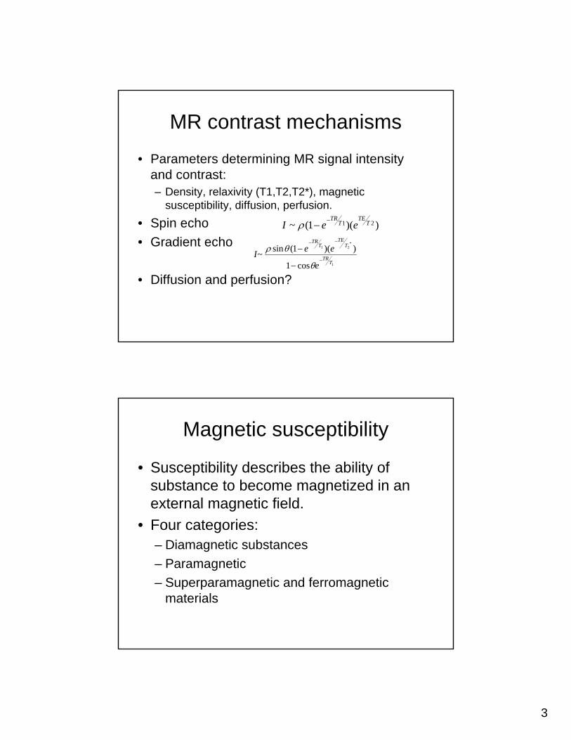
Definition and classificaiton
• Contrast agents (CA) are chemical substances introduced to the anatomical or functional region being imaged, to increase the differences between different tissues or between normal and abnormal tissue, by altering the relaxation times.
• Susceptibility describes the ability of substance to become magnetized in an external magnetic field.
• Four categories:– Diamagnetic substances – Paramagnetic – Superparamagnetic and ferromagnetic
materials
4
Magnetic susceptibility
• Diamagnetic substances (most organic compounds) -> small negative magnetic susceptibility.
• A paramagnetic ion can strongly influence the relaxation rate of nearby protons– Paramagnetic agents positive T1 relaxation,
little effect on T2 relaxation.
Magnetic susceptibility
• Superparamagnetic substance – Directly influence tissue contrast.– Large enough to be an domain.– External field ->align with the field -> large net
positive magnetization.– Removal of field->return to random
orientation->loss positive magnetization
5
Magnetic susceptibility
• Ferromagnetic compounds: – Large collection of interacting domains in a
crystalline matrix.– Extremely large net positive magnetization in
external field and remain this when removal of external field.
• Superparamagnetic and ferromagnetic compound function as negative agents.– Large net positive magnetic moments induce
spin dephasing in tissue.
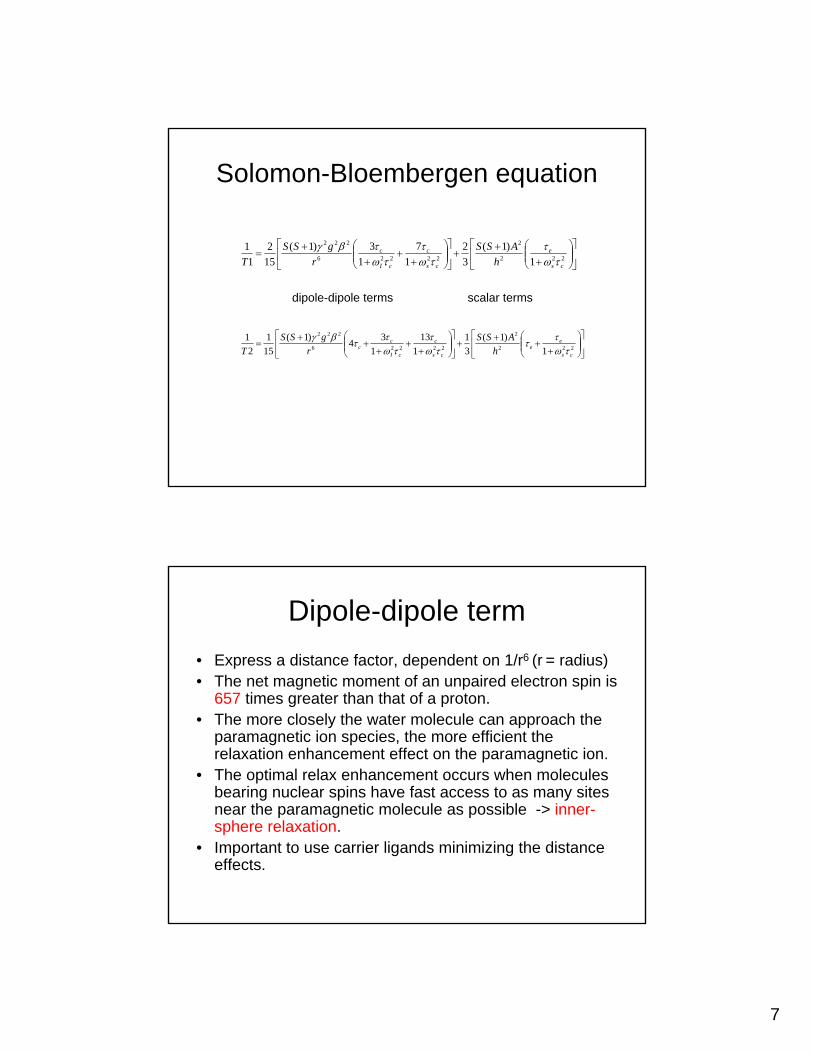
Relaxivity theory
• The contribution of a paramagnetic species to T1,T2 relaxation times arises as– Interaction between the unpaired electrons of the
paramagnetic ion and the hydrogen nuclei of water molecules.
• Interactions between paramagnetic agent and protons of water– Inner-sphere relaxation– Out-sphere relaxation
• Solomon-Bloembergen equation
6
Inner-sphere relaxation• The formation of a coordinate covalent molecular
bond between a water molecule and the paramagnetic ion.
• Lead to enhanced relaxation of the water protons on the basis of the magnetic influences and efficiency of chemical exchange.
• The more water molecules bond with paramagnetic ion, the greater its influence on relaxation enhancement.
• The shorter the residence time of water molecule with paramagnetic ion, the greater the relaxation enhancement effect (bond with other H2O)
Outer-sphere relaxation
• No direct bonding• Relative rotational and translational
diffusion of water molecules and paramagnetic ion
• The more and closer the water molecules approach (pass) the paramagnetic ion, the more efficient the relaxation enhancement.
• Dipole-dipole relaxation process
7
Solomon-Bloembergen equation
⎥⎦
⎤⎢⎣
⎡⎟⎟⎠
⎞⎜⎜⎝
⎛+
++⎥
⎦
⎤⎢⎣
⎡⎟⎟⎠
⎞⎜⎜⎝
⎛+
++
+= 222
2
22226
222
1)1(
32
17
13)1(
152
11
cs
e
cs
c
cI
c
hASS
rgSS
T τωτ
τωτ
τωτβγ
⎥⎦
⎤⎢⎣
⎡⎟⎟⎠
⎞⎜⎜⎝
⎛+
++
+⎥⎦
⎤⎢⎣
⎡⎟⎟⎠
⎞⎜⎜⎝
⎛+
++
++
= 222
2
22226
222
1)1(
31
113
134)1(
151
21
cs
ee
cs
c
cI
cc h
ASSr
gSST τω
τττω
ττω
ττβγ
dipole-dipole terms scalar terms
Dipole-dipole term• Express a distance factor, dependent on 1/r6 (r = radius)• The net magnetic moment of an unpaired electron spin is
657 times greater than that of a proton.• The more closely the water molecule can approach the
paramagnetic ion species, the more efficient the relaxation enhancement effect on the paramagnetic ion.
• The optimal relax enhancement occurs when molecules bearing nuclear spins have fast access to as many sites near the paramagnetic molecule as possible -> inner-sphere relaxation.
• Important to use carrier ligands minimizing the distance effects.
8
Scalar term• Summarize the probability that a transient
coincidence of an unpaired electron of the paramagnetic ion and the proton nucleus of nearby water molecule.
• The probability is defined by τc
• The correlation time of the interacting spins is dominated by the fastest of the three rate terms
msrc ττττ1111
++=
Scalar term
• If higher-molecular-weight ligands surround the paramagnetic metal ions
• Metal salts (MnCl2)- > metal chelates (Gd-EDTA) -> particulate agents (SPIO)->nanoparticles (USPIO).
• Nonspecific agents ->specific organ function or disease ->functional and metabolic imaging.
Gadolinium complex• Approved by FDA for use in cranial disease diagnostics
in mid-1988.• Gadolinium chelates (like Gadolinium DTPA) provides
greater contrast between normal tissue and abnormal tissue in the brain and body.
• Gadolinium chelates was developed because of:– High relaxivity of the gadolinium ion– Relax low toxicity of the complex
• Gd-DTPA, Gd-DTPA-BMA, Gd-HP-DO3A, others like Gd-DOTA
• Gadolinium chelates is eliminated through the urinary system with in six hours of the first injection
13
Nonspecific MR contrast agents
• allow measurement of vascular permeability, blood flow, and blood volume.
• Poorly suited to characterize tumor microvessels.
• differentiation of benign from malignant tissues is problematic.
14
Targeted MR contrast agents
• Many molecular targets are overexpressed in tumors and can be targeted by attaching an affinity ligand to the MR reporter.
15
Smart MR contrast agents
• Smart MR contrast agents (i.e., agents that can be activated) undergo conformational changes upon target interaction, which significantly alter their signal properties (e.g., shortening of T1 relaxation time).
Tissue specific contrast agents
• compounds with a tissue-specific distributionto detect focal anomalies or evaluate tissue function may be desirable to improve diagnostic accuracy.
16
Liver-specific agents
• (Gd) chelates improve the diagnosis of focal liver lesions. (not really specific to the liver tissue).
• Hepatocyte-specific compounds– Specific uptake in the hepatocyte– paramagnetic chelates– superparamagnetic iron oxide (SPIO)(preclinically)
• RES-specific compounds– SPIO nanoparticles
Blood-pool agents
• MR angiography (MRA)• fast imaging technologies were further
improved by using relaxation enhancers• Since imaging is still time consuming,
compounds that remain in the intravascular space are desirable.
• Several paramagnetic and superparamagnetic agents are now in clinical development.
17
Lymph node-specific agents
• Low-molecular weight Gd chelates, as well as polymeric agents, also used as blood pool agents, can be used for this indication
• Ultrasmall superparamagnetic iron oxides– darkening of the lymphatic vessels and lymph nodes– poor transport kinetics from the injection side, which
creates a tattooing effect.
Tumor-specific agents
• nontoxic, tumor-specific agents are somewhat misleading.
• Monoclonal antibodies labeled with paramagnetic atoms or superparamagneticnanoparticles are believed to be the ultimate tumor-seeking materials.
• However, the required dose of the labeled antibody is still too high to make commercial development realistic.
18
Molecular imaging
• Molecular imaging is a growing research discipline aimed at developing and testing novel tools, reagents, and methods to image specific molecular pathways in vivo, particularly those that are key targets in disease processes.
Tomography) Scan– Quantitative Autoradiography– Radionucleotide imaging combined with a
computed tomography– (CT) or a nuclear resonance imaging (NRI) scan
• MRI: uses paramagnetic-labeled CA or other CA to produces high imaging resolution
• Optical Imaging
19
Potential of imaging techniques for MI
CA for MR molecular imaging• Ligands are needed for selective binding.• Gadolinium may be used, but
– Low relaxivities; not biocompatible; potential toxicity following cellular dechelating over time
• Superparamagnetic iron oxide (SPIO) particles is preferred. – Provide most change signal (esp, T2* weighted) – Composed of biodegradable iron– Surface coating (dextran) allows directly linkage to functional groups
and ligands– Easily detected by light and electron microscopy– Can be magnetically manipulated and change their magnetic properties
according size, with potential to reveal their structural conformation– Problem:
• Prevent direct anatomical MR evaluation of tissue• Difficult to discriminate between targeted molecules and cells and image
artifacts
20
• Reference:– Magnetic resonance Imaging (2nd edition) David D. Stark,
William G. Bradley, JR.– Magnetic resonance Imaging (3nd edition) David D. Stark,
William G. Bradley, JR.– Ronald G Blasberg and Juri Gelovani Tjuvajev, “Molecular-
genetic imaging: current and future perspectives, J Clin Invest, 2003;111:1620-1629.
– T. Persigehl, W. Heindel, C. Bremer MR and optical approaches to molecular imaging. Abdom Imaging (2005) 30:342–354
– Hanns-Joachim Weinmann et al.Tissue-specific MR contrast agents. European Journal of Radiology 46 (2003) 33-44
– Bulte JW, Kraitchman DL. Iron oxide MR contrast agents for molecular and cellular imaging. NMR Biomed. 2004 Nov;17(7):484-99.
– Erik M. Shapiro,* Kathryn Sharer, Stanko Skrtic, and Alan P. Koretsky. In Vivo Detection of Single Cells by MRI. Magnetic Resonance in Medicine 55:242–249 (2006)


![How to write “Compare & Contrast” reportsCompare-and-Contrast].pdf · “Compare & Contrast” reports In compare and contrast reports, you need to describe the similaritiesand](https://static.documents.pub/doc/80x56/5fec4fdb3558df7c493bea9f/how-to-write-aoecompare-contrasta-compare-and-contrastpdf-aoecompare.jpg)

![Review Article Application of Nanoparticles on Diagnosis and ...pathways present in tumoral cells [ ]. It is known that magnetic nanoparticles also exhibit a higher longitudinal relaxivity,](https://static.documents.pub/doc/80x56/60c23a806f784d56ea1209de/review-article-application-of-nanoparticles-on-diagnosis-and-pathways-present.jpg)


