Contributions of the RNA-Binding and Linker Domains and RNA Structure to theSpecificity and Affinity of the Nucleolin RBD12/NRE Interaction†
L. David Finger,‡ Carina Johansson,‡ Bruno Rinaldi,§ Philippe Bouvet,§ and Juli Feigon*,‡
Department of Chemistry and Biochemistry, and Molecular Biology Institute, UniVersity of California,Los Angeles, California 90095-1569, and Ecole Normale Supe´rieure de Lyon, UMR CNRS 5665, 46 allee´ d’italie,
69364 Lyon, Cedex 07, France
ReceiVed January 12, 2004; ReVised Manuscript ReceiVed March 29, 2004
ABSTRACT: Nucleolin is a multidomain phosphoprotein involved in ribosome biogenesis. In vitro selectionand binding studies with pre-rRNA fragments have shown that the first two RNA-binding domains (RBDs)in nucleolin (RBD12) recognize the consensus sequence (U/G)CCCG(A/G) in the context of a stem-loopstructure (nucleolin-recognition element) NRE). Structural studies of nucleolin RBD12 in complex withan in vitro selected NRE (sNRE) and a natural pre-rRNA NRE (b2NRE) have revealed that sequence-specific binding of the consensus NRE is achieved in a similar manner in both complexes using residuesin both RBDs as well as the linker connecting them. Using fluorescence anisotropy (FA) and nuclearmagnetic resonance (NMR), we demonstrate the importance of the linker for NRE affinity by showingthat only the individual RBDs with the linker attached retain the ability to specifically bind, albeit weakly,to sNRE and b2NRE. Binding of RBD1 and RBD2 to the NREs in trans is not detected even when oneof the RBDs has the linker attached, which suggests that the linker also contributes to the affinity bytethering the two RBDs. To determine if binding of nucleolin RBD12 to natural NREs is dependent ona specific RNA stem-loop structure, as was the case for the sNRE, we conducted FA and NMR bindingassays with nucleolin RBD12 and a single-stranded NRE. The results show that nucleolin RBD12 sequence-specifically binds a single-stranded NRE with an affinity similar to that for b2NRE, indicating that astem-loop structure is not required for the nucleolin RBD12/pre-rRNA NRE interaction.
The RNA-binding domain (RBD),1 also known as theRNA-recognition motif (RRM), is the third most commonprotein motif found in nature (1, 2). Proteins containingRBDs have been shown to participate in many essentialcellular processes in RNA metabolism including pre-mRNAsplicing, mRNA stability, RNA packaging and transport (1),and rRNA processing (3, 4). Nuclear magnetic resonance(NMR) and crystallographic studies have shown that theRBDs, which are comprised of 70-100 amino acids, havea characteristicâRââRâ fold with the twoR helices packing
against a four-stranded antiparallelâ sheet (5, 6). The centraltwo â strands have conserved octameric RNP-1 and hex-americ RNP-2 motifs, which contain aromatic, hydrophobic,and basic amino acids (7) that are used to sequence-specifically bind single-stranded RNA, stem-loop RNAstructures (6), and sometimes DNA (8). Many RNA-bindingproteins contain multiple RBDs separated by linkers ofvariable amino acid composition and length (9). In the caseof the snRNP U1A protein, which has two RBDs, a singleRBD accounts for the affinity for the RNA target (10-12).In contrast, the multiple RBD proteins poly(A)-bindingprotein (PABP) (13), sex-lethal protein (Slx) (14, 15), ASF/SF2 (16, 17), hnRNP A1 (18, 19), HuC (20), HuD (21, 22),and nucleolin (3, 23) require the simultaneous binding ofmore than one RBD for optimal binding of a target RNAsequence.
Nucleolin, which contains four tandem RBDs, is the mostabundant protein in the nucleoli of vertebrate cells and isthought to participate in several steps of ribosome biogenesis,including regulation of rDNA transcription, rRNA process-ing, ribosome assembly, and nucleocytoplasmic transport(Figure 1A) (3, 24). Nucleolin associates directly withnascent pre-rRNA but is not found in cytoplasmic ribosomes(25). Attempts to understand the multiple roles of nucleolinin ribosome biogenesis have involved characterization of thepre-rRNA sequences recognized by the RBDs, because thefunctions of nucleolin are likely reflected in its RNA-bindingproperties. In vitro selection in combination with mutagenesis
† This work was supported by NIH Grant R01 GM37254 to J.F. andan ATIP from the CNRS, the Re´gion Rhone-Alpes, and the Ministe`rede la Recherche (AC nanosciences) to P.B.
* To whom correspondence should be addressed. E-mail: [email protected]. Phone: (310) 206-6922. Fax: (310) 825-0982.
‡ University of California, Los Angeles.§ Ecole Normale Supe´rieure de Lyon.1 Abbreviations: RBD, RNA-binding domain; RRM, RNA-recogni-
tion motif; NRE, nucleolin-recognition element; RNP-1, octamericrepeat found in RBD; RNP-2, hexameric repeat found in RBD; PABP,poly(A)-binding protein; Slx, sex-lethal protein; sNRE, in vitro selectedNRE; b2NRE, natural NRE found in the mouse 5′ external transcribedspacer (nts: 562 to 578 EMBL sequence database accession codeM20154); RBD1, first RBD in Chinese hamster nucleolin (residues:V298-P381); RBD2, second RBD in Chinese hamster nucleolin(residues: T394-G469); RBD12, RBD1 and RBD2; linker, 12 aminoacid residues in Chinese hamster nucleolin tethering RBD1 and RBD2(residues: K382-R393); RBD1L, protein construct of RBD1 includingthe linker residues at C terminus of RBD1; LRBD2, protein constructof RBD2 including the linker residues at the N terminus of RBD2;NMR, nuclear magnetic resonance; FA, fluorescence anisotropy; HSQC,heteronuclear single-quantum coherence;KD, dissociation constant.
and structural analysis has identified sequences in pre-rRNAto which nucleolin can bind specifically (23, 26). One familyof RNA sequences that nucleolin binds is called the nucleo-lin-recognition element (NRE), which is defined as a RNAcontaining the consensus (U/G)CCCG(A/G) in a loop ofvariable size (7-14 nucleotides) and at least a four base-pair stem (25). Two of the 39 putative mouse NREs, B1(nts 515-532) and B2 (nts 562-578), located in the 5′-ETSregion of mouse pre-rRNA (EMBL sequence databaseaccession code M20154), have been confirmed as nucleolin-binding sites by cross-linking studies (27). Subsequent studieshave shown that the interaction of nucleolin with an in vitroselected NRE only requires the first two RBDs of nucleolin(Figure 1B) in a cis arrangement for nanomolar affinity(KD ) 1-5 nM) (28). More recently, constructs of B1 andB2 NREs, b1NRE and b2NRE, respectively, have beenshown to also bind with nanomolar affinity to nucleolinRBD12, albeit approximately 100-500-fold more weaklythan the in vitro selected NRE (sNRE) (29).
We previously determined the solution structure of nucleo-lin RBD12 in complex with a 22 nucleotide RNA stem loopcontaining the 18 nucleotide in vitro selected consensussequence (sNRE) (Figure 1C) (PDB accession code 1FJE)(30) and more recently with the 21 nucleotide b2NRE stemloop (Figure 1D) (PDB accession code 1RKJ) (31). Bothstructures show that the specificity for the NRE consensussequence is determined by intermolecular stacking andhydrogen-bond interactions involving amino acids on theâ-sheet surface of the RBDs and in the linker and loops aswell. The linker connecting the two RBDs plays an importantrole in both complexes by making specific and nonspecificcontacts to the NRE consensus sequence. Furthermore, theupper part of the sNRE stem, which is not part of the NREconsensus sequence, folds into a loop E motif (S turn) (32-35). In the RBD12/sNRE complex, nucleotides in the upperpart of the loop E motif and the nonconsensus A8, which isstacked on top of the loop E motif, are also specificallyrecognized by amino acid residues in RBD1 and the linker
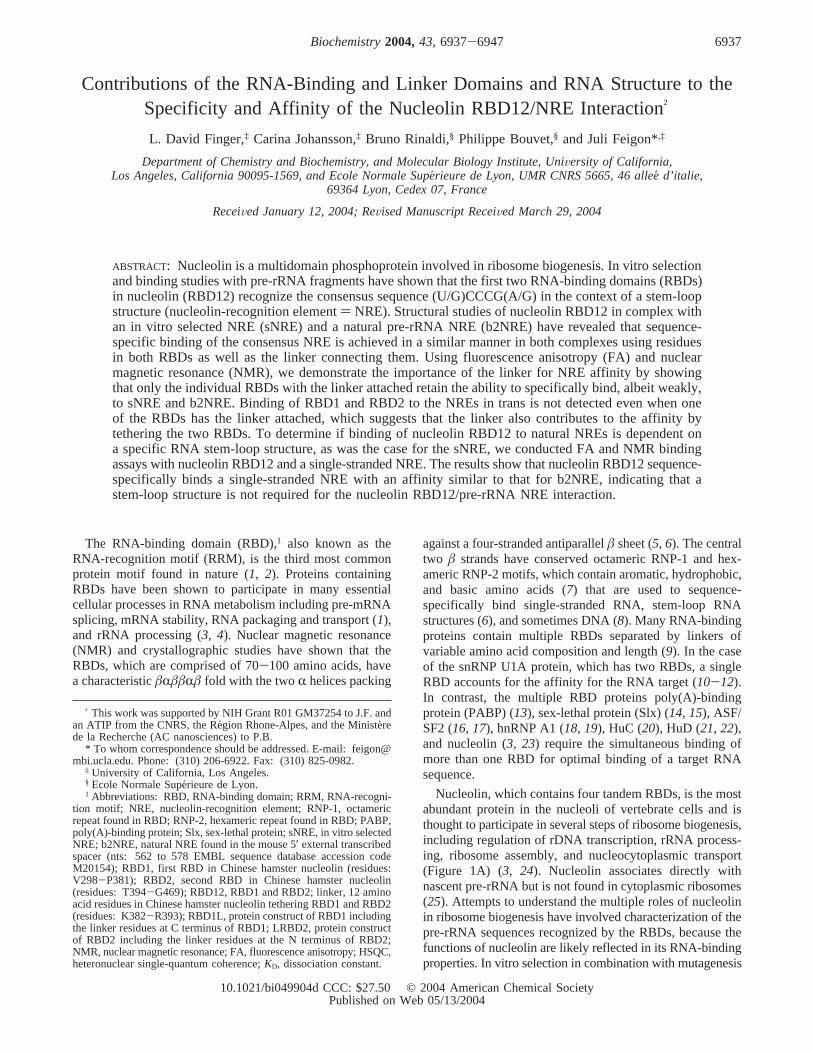
FIGURE 1: (A) Domain organization of hamster nucleolin. Some of the known functions of the nucleolin domains are indicated above. TheN terminus is known to interact with U3 snoRNP (58), RBD12 binds the NRE (28), RBD1234 binds the ECM (23), and RBD34RGG hasbeen shown to have nucleic acid annealing activity (59). (B) Amino acid sequence of hamster nucleolin RBD12 used in this paper (SWISS-PROT accession code P08199). Secondary structure elements determined for hamster nucleolin RBD12 are indicated below the sequenceand are shown in blue for RBD1, red for the linker, and green for RBD2. The hexameric RNP-2 and octameric RNP-1 motifs are highlightedin orange and purple, respectively. The linker residues are shown in a red box. V5-G175 corresponds to amino acid residues 298-468 inthe full protein. The first three amino acids at the N terminus are left after TEV cleavage of the 20 amino acid residue (His)6 purificationtag. (C) Secondary structure of the sNRE used in this paper (29). Nucleotides 3-20 are the sequence identified by in vitro selection (25).The NRE consensus is shown in orange, additional loop nucleotides shown in red, and the stem shown in black. Two G•C base pairs, whichare at the end of the stem for the purpose of transcription, are not shown. (D) Secondary structure representation of b2NRE used in thispaper. Nucleotides 4-20 correspond to the B2 sequence found in mouse pre-RNA (nts 562-578; EMBL sequence database accession codeM20154). The color scheme is the same as in C. The sequences of (C) sNRE and (D) b2NRE are numbered to correspond to the originalsNRE construct (30, 36). (E) Schematic representation of the RBD12 subdomains used in this paper, with the color scheme for the domainsbeing the same as in B. The gray box at each N terminus represents the three amino acids residues left after TEV cleavage of the affinitytag.
6938 Biochemistry, Vol. 43, No. 22, 2004 Finger et al.
(30, 36). In contrast, the intermolecular interactions in thenucleolin RBD12/b2NRE complex are restricted to the NREconsensus sequence. Kinetic and gel shift analyses haveshown that the presence of additional contacts to noncon-sensus nucleotides is responsible for the increased stabilityof the nucleolin RBD12/sNRE complex compared to thenucleolin RBD12/b2NRE complex. In addition, the gel shiftanalysis showed that the additional contacts to nonconsensusnucleotides in the sNRE are not only nucleotide-specific, butalso dependent on the RNA secondary structure (i.e.,formation of the loop E motif at the top of the stem) (31).In summary, these studies illustrate the importance of theRNA secondary structure in the nucleolin RBD12/sNREinteraction and provide a rationale for the stem-loop require-ment of the sNRE.
Prior to structure determination of the nucleolin RBD12/sNRE complex, the 12 amino acid residue linker connectingthe two RBDs was only thought to restrict the positions ofthe RBDs relative to one another and promote the associationof the two RBDs with the same RNA (37). However, thestructures of the nucleolin RBD12/sNRE and RBD12/b2NREcomplexes and gel-shift assays with RBD12 constructscontaining linker mutations demonstrated the importance ofthe linker for the nucleolin RBD12/NRE interaction (30).Most analyses of sequence-specific binding and affinity ofRNA-binding proteins containing multiple RBDs havefocused on the role of the RBDs. Here, NMR and fluores-cence anisotropy (FA) studies of the interaction of RBD1and RBD2 protein constructs with and without the linkerare used to demonstrate the importance of the linker for theaffinity, specificity, and domain cooperativity of the nucleolinRBD12/NRE interaction. The structures of other (RBD)2/RNA complexes (38-40) also show interaction of the linkerresidues to the target RNA, suggesting that the above resultsare general. FA assays and NMR were also used to determineif binding of nucleolin RBD12 to a natural pre-rRNA targetwas dependent on a specific stem-loop structure, as was thecase for the sNRE. Our results show that nucleolin RBD12can sequence-specifically bind a single-stranded NRE withaKD similar to that observed for b2NRE. We have previouslyproposed that the nucleolin/NRE interaction is important forproper folding of nascent pre-rRNA (30). The fact thatnucleolin RBD12 binds single-stranded NREs supports theproposal that nucleolin initially binds single-stranded NREsubstrates to facilitate formation of transient stem-loopstructures in pre-rRNA.
MATERIALS AND METHODS
RNA Synthesis and Purification.Unlabeled and uniformly13C,15N-labeled RNA oligonucleotides for NMR studies wereprepared by in vitro transcription as described (41), exceptfor the single-stranded substrates, which were chemicallysynthesized (Dharmacon, Boulder, CO) and purified aspreviously described (41). b2NRE and sNRE were annealedat dilute concentrations (1-10 µM) in water and adjustedto the desired salt conditions by the addition of the appropri-ate stock solution. sNRE buffer consisted of 10 mMpotassium phosphate at pH 7, 100 mM KCl, 50µMethylenediaminetetraacetic acid (EDTA), and 0.02% NaN3,and b2NRE buffer consisted of 5 mM potassium phosphateat pH 7, 50µM EDTA, and 0.02% NaN3. Single-stranded
substrates were diluted in water. All RNAs were subsequentlyconcentrated by ultrafiltration (Amicon).
Constructs.Previous attempts to cleave the affinity tagfrom the pET15b vector-expressed nucleolin RBD12 usingthrombin resulted in cleavage of the linker as well. Therefore,RBD12 and subdomains were cloned into a pProEX HTvector to allow for cleavage of the (His)6 affinity tags fromthe RBD12 subdomains using TEV protease (42) instead ofthrombin. The hamster nucleolin RBD12 subdomains weregenerated by PCR using Vent DNA polymerase and thepet15b vector containing the hamster nucleolin RBD12 gene(43). The 5′ oligonucleotide used to amplify RBD12, RBD1,and RBD1L was 5′-CATGCCATGGTGGAAGGTTCA-GAATCAACTACAC-3′; the 5′ oligo for RBD2 was 5′-CATGCCATGGCAACACTTTTAGCAAAAAATCTTTC-3′; the 5′ oligo for RBD2L was 5′-ACGGATCC-AAAAGGAAGAGATAGTAAAAAAG-3 ′; the 3′ oligo forRBD12, RBD2, and RBD2L was 5′-GCGAAGCTTCATC-CCTTCTCCCCAGTATAGTAAAG-3′; the 3′ oligo forRBD1 was 5′-ACCAAGCTTCATGGTTTTTCTAGTTTAA-TTTCATTGCC-3′; and the 3′ oligo for RBD1L was5′-ACCAAGCTTCACCTTGCAGCTCGAACTTTTTTAC-TATC-3′. After amplification, PCR fragments were sub-cloned into pProEX HT using the restriction sitesNcoI-Hind III for RBD12, RBD1, RBD1L, and RBD2 andBamH I-Hind III for RBD2L.
Protein Expression and Purification.BL21-Codon Plus-RIL (Stratagene) cells were transformed with recombinantpProEX HT (Life Technologies) plasmids containing aninsert encoding for the hamster nucleolin (His)6-RBDXsubdomains, where RBDX stands for RBD12, RBD1,RBD1L, RBD2, and LRBD2. Cells grown at 37°C in LBmedia containing 100µg/mL ampicillin and 35µg/mLchloramphenicol were induced with 1 mM isopropyl-1-thio-â-D-galactopyranoside at OD600 ) 0.6-0.8 and grown foran additional 3 h. Harvested cells were resuspended in 20mM sodium phosphate at pH 7.4, 500 mM NaCl with 0.1%Triton X-100, and 100µL of protease cocktail inhibitor(Sigma) per liter of culture, and lysed by three freeze-thawcycles and sonication. Proteins were then purified as previ-ously described (29), except that the cleavage of the (His)6
tag was accomplished with (His)6-TEV S219N protease (42).FA Assays.5′-fluorescein-labeled RNAs with sequences
corresponding to those shown in Figure 1 were obtained fromDharmacon Research, purified, deprotected, and desalted. 5′-fluoroscein-NRE substrates were diluted in TE buffer (10mM Tris-HCl at pH 8.0 and 1 mM EDTA) and quantifiedatA260 as described (44). FA was measured using the Beacon2000 Variable Temperature Fluorescence Polarization System(Panvera, Madison, WI) with fixed excitation (490 nM) andemission (535 nM). Equilibrium binding assays were per-formed as previously described (29). Polarization values wereconverted to anisotropy values using eq 1. Data are expressed
as (A - A0)/A0, whereA is the anisotropy of the fluorescein-labeled RNA at the indicated amount of protein andA0 isthe anisotropy for the free-labeled RNA or mA, where themA is 1000 times the anisotropy of the fluorescein-labeledRNA at the indicated amount of protein. Dissociation
A ) 2P3 - P
(1)
Role of RBDs, Linker, and RNA in Nucleolin-RNA Interaction Biochemistry, Vol. 43, No. 22, 20046939
constants (KD’s) were calculated by fitting data to eq 2 usingKaleidaGraph.
NMR Spectroscopy.All NMR spectra were recorded onBruker DRX 500 or 600 MHz spectrometers. Protein-RNAcomplexes were prepared by addition of aliquots of lyo-philized RNA or protein until the indicated molar ratio wasachieved. Titration of nucleolin RBD12 and RBD12 subdo-mains into 0.15 mM13C,15N-labeled sNRE or b2NRE wasmonitored at 310 K by gradient sensitivity enhanced1H-13Cheteronuclear single-quantum coherences (HSQCs) (45).Titration of SSNRE or SSNS into 0.15 mM15N-labelednucleolin RBD12 was monitored at 310 K by1H-15N HSQCswith WATERGATE (46) water suppression. Spectra wereprocessed and analyzed using Bruker XWINNMR 2.6 andFelix97 (MSI, Inc.).
RESULTS
Determination of Dissociation Constants of the IndiVidualRBDs and NREs.Many proteins that bind RNA containmultiple RBDs, and two or more tandem RBDs are oftennecessary for high-affinity binding to RNA targets (9).Although most analyses of sequence-specific recognition in(RBD)2/RNA complexes have focused on the role of theRBDs, several crystal and NMR structures of (RBD)2/RNAcomplexes show that the linker residues connecting the twoRBDs also contribute to RNA binding (30, 38-40). Forinstance, in the nucleolin RBD12/sNRE and RBD12/b2NREcomplexes, the linker residues connecting RBD1 and RBD2interact with the NREs in both sequence- and nonsequence-specific manners. In light of these observations, we decidedto further investigate the role of the linker in nucleolinRBD12/NRE binding by assaying for individual binding ofisolated RBD1 and RBD2 with and without the linkerattached. The RBD12 subdomains that were used in thispaper are shown in Figure 1E. Because the presence of the20 amino acid residue N-terminal (His)6 tag used in purifica-tion could interfere with LRBD2, RBD2, and trans binding,cleavage of the affinity tag from all constructs was necessary.TEV protease treatment resulted in RBD12 subdomains ofthe expected size (data not shown).
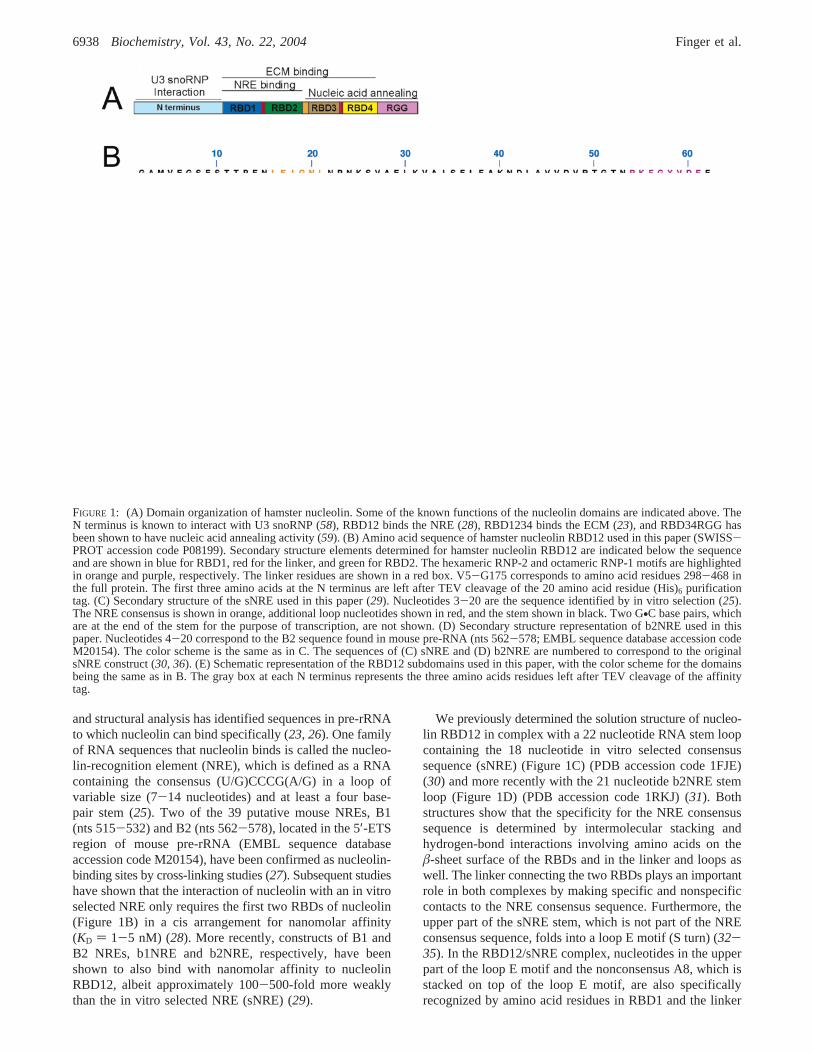
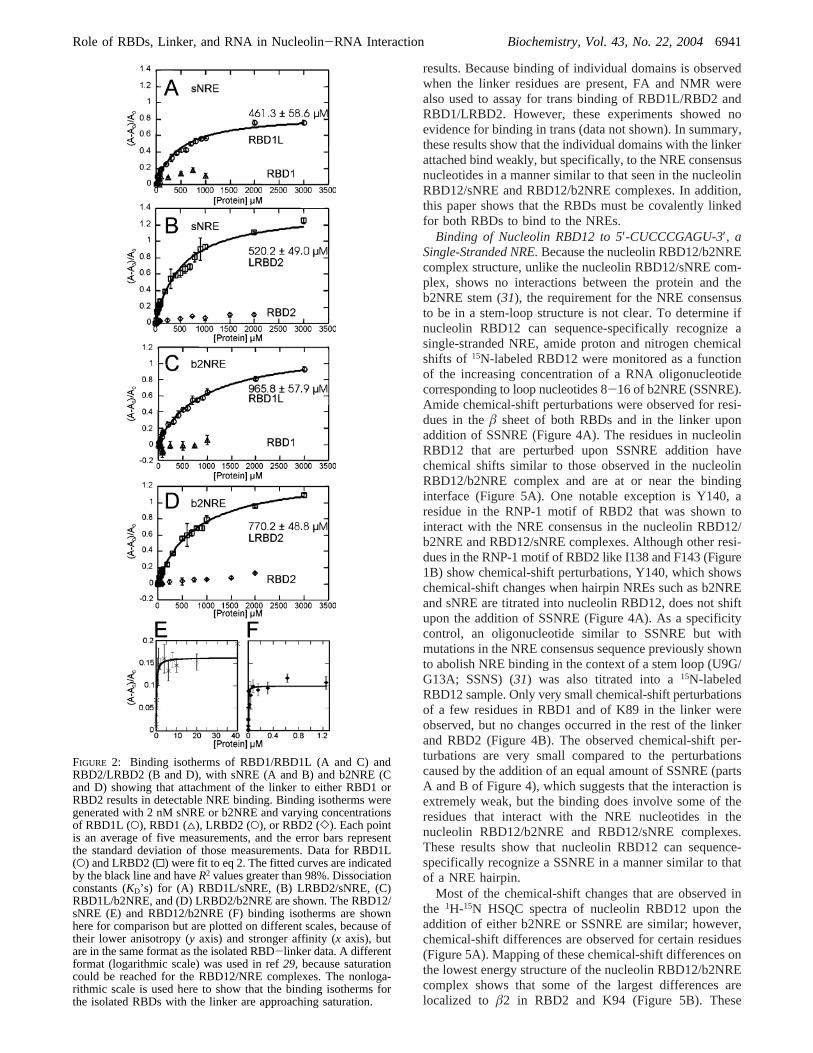
Use of the EMSA procedure previously shown to resultin band shifts for the nucleolin RBD12/sNRE and RBD12/b2NRE complexes (29) did not result in band shifts for anyof the nucleolin RBD12 subdomains with sNRE or b2NRE(data not shown). Because EMSA is a nonequilibriumbinding assay, the lack of a band shift does not necessarilyindicate the absence of binding, as low-affinity complexesmay have dissociated during the course of electrophoresis(44). Therefore, FA binding assays were employed becauseit is an equilibrium binding technique and, thus, does notrequire the separation of bound and free RNA (44). Theresults of titrations of varying concentrations of RBD1,RBD1L, RBD2, and LRBD2 into 5′-fluorescein-labeledsNRE and b2NRE are shown in Figure 2. Both RBD1L andLRBD2 bind the sNRE and b2NRE hairpins, but no binding
to the NREs is observed for the individual domains in theabsence of linker residues. Fitting the data for RBD1L andLRBD2 to eq 2 reveals that the dissociation constants (KD)for the sNRE and b2NRE interaction vary from 400 to 1000µM (parts A-D of Figure 2). Using the same FA assayprocedure, theKD’s of nucleolin RBD12 for sNRE andb2NRE have previously been reported to be 3 and 300 nM,respectively (parts E and F of Figure 2) (29). Thus, removalof one of the RBDs from nucleolin RBD12 causes anapproximate 100 000- and 1000-fold loss in affinity for sNREand b2NRE, respectively, indicating that the presence of bothRBDs is essential for nanomolar affinity.
We note that the anisotropy for the complexes of theisolated domains with linkers and hairpin NRE substrates is∼10-fold higher than that obtained with RBD12 and hairpinNRE substrates, even though the molecular weight of thecomplexes of the individual domains is smaller. This is dueto the fact that the nucleolin RBD12/NRE complexes aremore compact and spherical, while the hairpin RNA com-plexes with the individual domains and linker are asym-metric, which would give rise to a larger change inanisotropy.
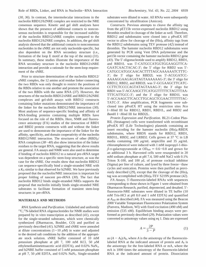
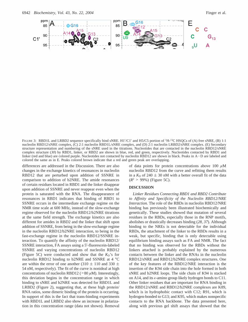
Binding of IndiVidual RBDs to NREs Studied by NMR.To test the specificity of the low-affinity interaction betweenthe individual RBDs with the linker attached and NREs,titrations of RBD1L and LRBD2 to13C,15N-labeled sNREand b2NRE were monitored by1H-13C HSQCs. Titration ofnucleolin RBD12, RBD1L, or LRBD2 into sNRE causeschemical-shift perturbations only for nucleotides in the loopand at the top of the loop E motif but leaves stem resonances(e.g., C2) mostly unaffected, except for changes in linewidths that are consistent with an increased correlation timeupon binding of the individual domains (Figure 3). Com-parison of parts A and C of Figure 3 shows that thenucleotides that are bound by RBD1 and the linker in thefull nucleolin RBD12/sNRE complex (43) are also boundby RBD1L, because as the concentration of RBD1L isincreased, the chemical shifts of C11, C12, G13, A14, andG16 change to values close to those seen in the nucleolinRBD12/sNRE complex (Figure 3B). C10 is not perturbedupon addition of RBD1L, consistent with the lack of contactsbetween C10 and RBD1 or the linker. However, U9 isaffected by RBD1L addition even though neither RBD1 northe linker contact U9 in the full nucleolin RBD12/sNREcomplex. Perturbation of U9 may be caused indirectly bythe interaction between RBD1L and nucleotides A8 and A14,which form an adenine zipperlike motif in the nucleolinRBD12/sNRE complex. When LRBD2 is titrated into sNRE,the nucleotides that are contacted by RBD2 and the linkerresidues in the full complex are perturbed (e.g., U9, C10,C11, C12, G13, and A14), whereas G16, which is neitherclose to the binding site nor contacted by RBD2 or the linker,is not affected (parts A and D of Figure 3). Similar resultswere obtained for titrations of RBD1L and LRBD2 intob2NRE. The chemical-shift changes that are observed uponthe addition of RBD1L and LRBD2 to b2NRE are alsoconsistent with the contacts that are made by the individualdomains in the nucleolin RBD12/b2NRE complex (31) (datanot shown). No chemical-shift perturbation of sNRE andb2NRE consensus loop nucleotide resonances is observedwhen the RBDs lacking the linker residues are titrated intothe RNA, an observation that is consistent with the FA
6940 Biochemistry, Vol. 43, No. 22, 2004 Finger et al.
results. Because binding of individual domains is observedwhen the linker residues are present, FA and NMR werealso used to assay for trans binding of RBD1L/RBD2 andRBD1/LRBD2. However, these experiments showed noevidence for binding in trans (data not shown). In summary,these results show that the individual domains with the linkerattached bind weakly, but specifically, to the NRE consensusnucleotides in a manner similar to that seen in the nucleolinRBD12/sNRE and RBD12/b2NRE complexes. In addition,this paper shows that the RBDs must be covalently linkedfor both RBDs to bind to the NREs.
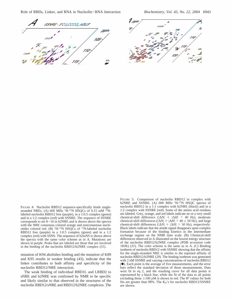
Binding of Nucleolin RBD12 to 5′-CUCCCGAGU-3′, aSingle-Stranded NRE.Because the nucleolin RBD12/b2NREcomplex structure, unlike the nucleolin RBD12/sNRE com-plex, shows no interactions between the protein and theb2NRE stem (31), the requirement for the NRE consensusto be in a stem-loop structure is not clear. To determine ifnucleolin RBD12 can sequence-specifically recognize asingle-stranded NRE, amide proton and nitrogen chemicalshifts of 15N-labeled RBD12 were monitored as a functionof the increasing concentration of a RNA oligonucleotidecorresponding to loop nucleotides 8-16 of b2NRE (SSNRE).Amide chemical-shift perturbations were observed for resi-dues in theâ sheet of both RBDs and in the linker uponaddition of SSNRE (Figure 4A). The residues in nucleolinRBD12 that are perturbed upon SSNRE addition havechemical shifts similar to those observed in the nucleolinRBD12/b2NRE complex and are at or near the bindinginterface (Figure 5A). One notable exception is Y140, aresidue in the RNP-1 motif of RBD2 that was shown tointeract with the NRE consensus in the nucleolin RBD12/b2NRE and RBD12/sNRE complexes. Although other resi-dues in the RNP-1 motif of RBD2 like I138 and F143 (Figure1B) show chemical-shift perturbations, Y140, which showschemical-shift changes when hairpin NREs such as b2NREand sNRE are titrated into nucleolin RBD12, does not shiftupon the addition of SSNRE (Figure 4A). As a specificitycontrol, an oligonucleotide similar to SSNRE but withmutations in the NRE consensus sequence previously shownto abolish NRE binding in the context of a stem loop (U9G/G13A; SSNS) (31) was also titrated into a15N-labeledRBD12 sample. Only very small chemical-shift perturbationsof a few residues in RBD1 and of K89 in the linker wereobserved, but no changes occurred in the rest of the linkerand RBD2 (Figure 4B). The observed chemical-shift per-turbations are very small compared to the perturbationscaused by the addition of an equal amount of SSNRE (partsA and B of Figure 4), which suggests that the interaction isextremely weak, but the binding does involve some of theresidues that interact with the NRE nucleotides in thenucleolin RBD12/b2NRE and RBD12/sNRE complexes.These results show that nucleolin RBD12 can sequence-specifically recognize a SSNRE in a manner similar to thatof a NRE hairpin.
Most of the chemical-shift changes that are observed inthe 1H-15N HSQC spectra of nucleolin RBD12 upon theaddition of either b2NRE or SSNRE are similar; however,chemical-shift differences are observed for certain residues(Figure 5A). Mapping of these chemical-shift differences onthe lowest energy structure of the nucleolin RBD12/b2NREcomplex shows that some of the largest differences arelocalized to â2 in RBD2 and K94 (Figure 5B). These
FIGURE 2: Binding isotherms of RBD1/RBD1L (A and C) andRBD2/LRBD2 (B and D), with sNRE (A and B) and b2NRE (Cand D) showing that attachment of the linker to either RBD1 orRBD2 results in detectable NRE binding. Binding isotherms weregenerated with 2 nM sNRE or b2NRE and varying concentrationsof RBD1L (O), RBD1 (4), LRBD2 (O), or RBD2 (]). Each pointis an average of five measurements, and the error bars representthe standard deviation of those measurements. Data for RBD1L(O) and LRBD2 (0) were fit to eq 2. The fitted curves are indicatedby the black line and haveR2 values greater than 98%. Dissociationconstants (KD’s) for (A) RBD1L/sNRE, (B) LRBD2/sNRE, (C)RBD1L/b2NRE, and (D) LRBD2/b2NRE are shown. The RBD12/sNRE (E) and RBD12/b2NRE (F) binding isotherms are shownhere for comparison but are plotted on different scales, because oftheir lower anisotropy (y axis) and stronger affinity (x axis), butare in the same format as the isolated RBD-linker data. A differentformat (logarithmic scale) was used in ref29, because saturationcould be reached for the RBD12/NRE complexes. The nonloga-rithmic scale is used here to show that the binding isotherms forthe isolated RBDs with the linker are approaching saturation.
Role of RBDs, Linker, and RNA in Nucleolin-RNA Interaction Biochemistry, Vol. 43, No. 22, 20046941
differences are addressed in the Discussion. There are alsochanges in the exchange kinetics of resonances in nucleolinRBD12 that are perturbed upon addition of SSNRE incomparison to addition of b2NRE. The amide resonancesof certain residues located in RBD1 and the linker disappearupon addition of SSNRE and never reappear even when theprotein is saturated with the RNA. The disappearance ofresonances in RBD1 indicates that binding of RBD1 toSSNRE occurs in the intermediate exchange regime on theNMR time scale at 600 MHz, instead of the slow-exchangeregime observed for the nucleolin RBD12/b2NRE titrationsat the same field strength. The exchange kinetics are alsodifferent for amides in RBD2 and the linker that shift uponaddition of SSNRE, from being in the slow-exchange regimein the nucleolin RBD12/b2NRE interaction, to being in thefast-exchange regime in the nucleolin RBD12/SSNRE in-teraction. To quantify the affinity of the nucleolin RBD12/SSNRE interaction, FA assays using a 5′-fluorescein-labeledSSNRE and varying concentrations of nucleolin RBD12(Figure 5C) were conducted and show that theKD’s fornucleolin RBD12 binding to b2NRE and SSNRE at 4°Care within the error of one another (310( 110 and 330(54 nM, respectively). The fit of the curve is nonideal at highconcentrations of nucleolin RBD12 (>80µM). Interestingly,this deviation begins in the concentration range in whichbinding to sNRE and b2NRE was detected for RBD1L andLRBD2 (Figure 2), suggesting that, at these high protein/RNA ratios, some dimeric binding of the protein is occurring.In support of this is the fact that trans-binding experimentswith RBD1L and LRBD2 also show an increase in polariza-tion in this concentration range (data not shown). Removal
of data points for protein concentrations above 100µMnucleolin RBD12 from the curve and refitting them resultsin a KD of 240( 30 nM with a better overall fit of the data(R2 > 99%) (Figure 5C).
DISCUSSION
Linker Residues Connecting RBD1 and RBD2 Contributeto Affinity and Specificity of the Nucleolin RBD12/NREInteraction.The role of the RBDs in nucleolin RBD12/NREbinding has previously been illustrated biochemically andgenetically. These studies showed that mutation of severalresidues in the RBDs, especially those in the RNP motifs,abolishes or drastically decreases binding (28, 37). Althoughbinding to the NREs is not detectable for the individualRBDs, the attachment of the linker to the RBDs results in aweak, but specific, binding that is only detectable usingequilibrium binding assays such as FA and NMR. The factthat no binding was observed for the RBDs without thelinkers attached is probably explained by the numerouscontacts between the linker and the RNAs in the nucleolinRBD12/sNRE and RBD12/b2NRE complex structures. Oneof the key features of the RBD12/NRE interaction is theinsertion of the K94 side chain into the hole formed in bothsNRE and b2NRE loops. The side chain of K94 is stackedon A14, and itsε-amino group likely hydrogen bonds to C11.Other linker residues that are important for RNA binding inthe RBD12/sNRE and RBD12/b2NRE complexes are K89,which is in hydrophobic contact with C12; R91, which ishydrogen-bonded to G13; and K95, which makes nonspecificcontacts to the RNA backbone. The data presented here,along with previous gel shift assays that showed that the
FIGURE 3: RBD1L and LRBD2 sequence specifically bind sNRE. H1′/C1′ and H5/C5 portion of1H-13C HSQCs of (A) free sNRE, (B) 1:1nucleolin RBD12/sNRE complex, (C) 2:1 nucleolin RBD1L/sNRE complex, and (D) 2:1 nucleolin LRBD2/sNRE complex. (E) Secondarystructure representation and numbering of the sNRE used in the titrations. Nucleotides that are contacted in the nucleolin RBD12/sNREcomplex structure (30) by RBD1, linker, or RBD2 are shown in blue, red, and green, respectively. Nucleotides contacted by RBD1 andlinker (red and blue) are colored purple. Nucleotides not contacted by nucleolin RBD12 are shown in black. Peaks in A-D are labeled andcolored the same as in E. Peaks colored brown indicate that a red and green peak are overlapping.
6942 Biochemistry, Vol. 43, No. 22, 2004 Finger et al.
mutation of K94 abolishes binding and the mutation of K89and K95 results in weaker binding (43), indicate that thelinker contributes to both affinity and specificity of thenucleolin RBD12/NRE interaction.
The weak binding of individual RBD1L and LRBD2 tosNRE and b2NRE was confirmed by NMR to be specificand likely similar to that observed in the structures of thenucleolin RBD12/sNRE and RBD12/b2NRE complexes. The
FIGURE 5: Comparison of nucleolin RBD12 in complex withb2NRE and SSNRE. (A) 600 MHz1H-15N HSQC spectra ofnucleolin RBD12 in a 1:1 complex with b2NRE (black) and in a1:2 complex with SSNRE (red). Some of the amino acid residuesare labeled. Grey, orange, and red labels indicate no or a very smallchemical-shift difference (|∆N| + |∆H| < 40 Hz), moderatechemical-shift differences (|∆N| + |∆H| > 40 e 50 Hz), and largechemical-shift differences (|∆N| + |∆H| > 50 Hz), respectively.Black labels indicate that the amide signal disappears upon complexformation because of the binding kinetics in the intermediateexchange regime on the NMR time scale. (B) Chemical-shiftdifferences observed in A illustrated on the lowest energy structureof the nucleolin RBD12/b2NRE complex (PDB accession code1RJK) (31). The color scheme is the same as in A. (C) Bindingisotherm of nucleolin RBD12 with SSNRE showing that the affinityfor the single-stranded NRE is similar to the reported affinity ofnucleolin RBD12/b2NRE (29). The binding isotherm was generatedwith 2 nM SSNRE and varying concentrations of nucleolin RBD12(b). Each point is the average of five measurements, and the errorbars reflect the standard deviation of those measurements. Datawere fit to eq 2, and the resulting curve for all data points isrepresented by a black line, while the fit of the data to all pointsexcluding thoseg100µM is shown in red. TheR2 values for bothfits are greater than 98%. TheKD’s for nucleolin RBD12/SSNREare shown.
FIGURE 4: Nucleolin RBD12 sequence-specifically binds single-stranded NREs. (A) 600 MHz1H-15N HSQCs of 0.15 mM15N-labeled nucleolin RBD12 free (purple), in a 1:0.5 complex (green)and in a 1:2 complex (red) with SSNRE. The sequence of SSNREcorresponds to nts 8-16 in b2NRE and is shown above the spectrawith the NRE consensus colored orange and nonconsensus nucle-otides colored red. (B)1H-15N HSQCs of15N-labeled nucleolinRBD12 free (purple) in a 1:0.5 complex (green) and in a 1:2complex (red) with SSNS. The sequence of b2ssNS is shown abovethe spectra with the same color scheme as in A. Mutations areshown in purple. Peaks that are labeled are those that are involvedin the binding of the nucleolin RBD12/b2NRE complex (31).
Role of RBDs, Linker, and RNA in Nucleolin-RNA Interaction Biochemistry, Vol. 43, No. 22, 20046943
relative affinities of the nucleolin RBD12 subdomain com-plexes studied are RBD1L/sNRE≈ LRBD2/sNRE >LRBD2/b2NRE > RBD1L/b2NRE. A comparison of thestructures of the nucleolin RBD12/sNRE and RBD12/b2NREcomplexes explains the slightly greater affinity of theindividual RBDs with the linker for sNRE compared to thosefor b2NRE. In the two complexes, the RBDs and linker makealmost identical contacts to the NRE consensus nucleotides;however, in the nucleolin RBD12/sNRE complex, RBD1 andthe linker also contact nonconsensus nucleotides in the upperportion of the loop E motif and A8, which stacks on thetrans-Hoogsteen A•A base pair in the loop E motif (36, 43).These additional contacts from the RBD1 and linker arelikely responsible for the greater affinity of RBD1L andLRBD2 for sNRE compared to that for b2NRE. RBD1L andLRBD2 bind sNRE with affinities that are within the errorof one another. This is consistent with the structure of thenucleolin RBD12/sNRE complex (43) because RBD1L andLRBD2 each contact seven nucleotides (Figure 3E). Thelower affinity of RBD1L for b2NRE compared to LRBD2is also consistent with the nucleolin RBD12/b2NRE complexstructure, in which LRBD2 contacts six nucleotides, whileRBD1L contacts four nucleotides.
Linker Is Necessary for the CooperatiVe Binding ofNucleolin RBD12 to NREs.NMR binding assays show thatthere is no trans binding even when one of the RBDs hasthe linker attached. Analysis of the nucleolin RBD12/sNREand RBD12/b2NRE complexes indicates that the two RBDsand the linker make only a few interdomain contacts, whichis consistent with the lack of trans binding. How then canthe very weak affinities of the individual RBDs with thelinker attached result in the nanomolar affinity for sNRE andb2NRE that is observed for a construct containing the RBDsin a cis arrangement? Biochemical studies of other tandemRBD proteins such as hnRNP A1 (18) and ASF/SF2 (17)have also shown that the individual RBDs maintain the abilityto bind the RNA target, albeit with a decreased affinity (14,17, 18). However, these studies did not address the role ofthe linker in binding specificity and affinity. Like thenucleolin RBD12/NRE interaction, analysis of bindingaffinities of the individual RBDs of ASF/SF2 and hnRNPA1 proteins shows that the affinity for the RNA target ofthe two RBDs in a cis arrangement is not simply the sum ofthe affinities of each individual RBD (18). The fact that thefree energy of binding of isolated RBDs cannot simply beadded to result in the affinity observed for the cis constructsof two RBDs can be attributed to the covalent attachmentof the two RBDs by the linker. Covalent attachment of twosmall molecule ligands that bind a single protein at twoseparate sites is known to increase the affinity for the proteintarget because binding of the first ligand or domain increasesthe effective concentration of the second (47, 48). Thisincrease in binding affinity from cooperative binding throughcovalent linkage or dimerization [also known as the “chelat-ing effect” (reviewed in ref47)] has been analyzed in detailfor a variety of protein-DNA and protein-RNA complexes,including helix-turn-helix domains (49), Zn2+-finger proteins(50-53), nuclear receptors (54), POU domains (55), bZipproteins (56), and RBDs (9). The cooperative bindingbehavior of covalently linked RBDs was predicted to increasethe affinity for an RNA target 10- to 1000-fold compared tothe affinity of the individual RBDs added together (9).
Assuming no linker contacts to a RNA target and that thelinker is a random coil with a interresidue distance of 3.5Å, linkers longer than 60 amino acid residues are predictedto have a negligible effect on affinity, while linkers withless than 60 residues are predicted to gradually increase theaffinity for an RNA target as the length of the linker isdecreased (9). On the basis of this analysis and the fact thatthe linker in RBD12 is comprised of 12 amino acid residues,the increase in affinity because of a cis arrangement of theRBD1, linker, and RBD2 should be approximately 2 ordersof magnitude greater than the simple addition of the bindingaffinities of the individual domains. However, the actualdifference in affinity for b2NRE and sNRE caused by a cisarrangement of RBD1, linker, and RBD2 is 1000- and100 000-fold, respectively. The 100-fold difference in theincrease of affinity between b2NRE and sNRE with a cisarrangement of the domains is simply explained by theadditional contacts from the linker and RBD1 to the loop Emotif of sNRE as discussed above. The 10- and 1000-folddifference between the predicted and actual increase inaffinity with a cis arrangement of RBD1, linker, and RBD2for b2NRE and sNRE, respectively, is partially explainedby the fact that the linkers in both complexes contact theNREs and are not in extended conformations, which weretwo of the basic assumptions of the theoretical study (9).Instead, the linkers in both NRE complexes line the majorgroove of the RNA loops and contain 310 helices. The linkerlength between RBD1 and RBD2 (K89-T101) is 17.2 Åand 20.9 Å in the RBD12/sNRE and RBD12/b2NREcomplexes, respectively, which is approximately half thepredicted length for the 12 amino acid residue linker in anextended random-coil configuration (43.5 Å). Therefore, thecontribution of the chelating effect to the affinity of thenucleolin RBD12/NRE complexes should be greater thanpredicted based on the number of linker residues alone. Inaddition, if the linker length between domains is consideredto be the last linker/RNA contact to the second RBD, thenthe chelating effect will be even greater. Calculations thatuse linker lengths measured from the last linker/RNA contactto each RBD and theKD’s shown in Figure 2 (9) can fullyaccount for the 10-fold difference between the predicted andthe actual increase in affinity because of the chelating effectfor the nucleolin RBD12/b2NRE interaction. On the otherhand, similar calculations for the sNRE complex do not fullyaccount for the 1000-fold difference between the theoreticaland actual increase in affinity because of the chelating effect.This suggests that the nucleolin RBD12/sNRE interactionis also enhanced by other factors in addition to the chelatingeffect. One possibility is that conformational changes inducedin sNRE by one RBD may positively affect the binding ofthe other covalently attached RBD (47). Another possibilityis that binding of nucleolin RBD12 to sNRE may stabilizethe adenine zipper that is only found in the lower portion ofthe RNA loop when the protein is bound (29, 30).
Importance of a Stem-Loop Structure for NucleolinRBD12/NRE Binding.Previous studies indicated that thehigher stability of the nucleolin RBD12/sNRE complexcompared to the nucleolin RBD12/b2NRE complex is dueto the additional contacts from the RBD1 and linker tonucleotides outside the NRE consensus sequence in thenucleolin RBD12/sNRE complex (31). The presence of theS-shaped backbone in the loop E motif and the adenine
6944 Biochemistry, Vol. 43, No. 22, 2004 Finger et al.
zipper-motif in sNRE provides a scaffold to which RBD1and the linker make nucleotide-specific contacts. Thesecontacts are not only dependent on the nucleotide identity,but also on the secondary structure context in which theyare presented to the protein. In contrast to the complex withsNRE, the nucleolin RBD12/b2NRE complex showed thatintermolecular contacts to the natural sequence are localizedto the NRE loop consensus sequence. Therefore, binding ofa single-stranded NRE to nucleolin RBD12 was investigated,as well. The NMR titrations show that the interaction ofnucleolin RBD12 with SSNRE is specific, and the observedchemical-shift changes suggest that the contacts betweennucleolin RBD12 and SSNRE are similar to those in thenucleolin RBD12/b2NRE complex. However, some residuesin RBD2 and the linker in the nucleolin RBD12/SSNREcomplex have chemical shifts that are different compared toboth the free RBD12 and nucleolin RBD12/b2NRE complex,suggesting that these residues have unique chemical environ-ments when nucleolin RBD12 is bound to a single-strandedNRE. Whether RBD1 residues in the nucleolin RBD12/SSNRE complex are in the same or different conformationsin comparison to the nucleolin RBD12/b2NRE complexcannot be determined with the existing data because manyof the amide resonances in RBD1 are broadened to baselineupon addition of SSNRE.
The largest chemical-shift differences between nucleolinRBD12 bound to SSNRE and b2NRE are displayed by I138and K94. I138 is located in the RNP1 motif in RBD2 andpacks closely against U9 in the nucleolin RBD12/b2NREcomplex. The large chemical-shift difference of 110 Hz forthis amide resonance may result from differences in contactswhen bound to the SSNRE and/or in the RNA conformation.Because Y140 is not perturbed upon the addition of SSNREas is the case for b2NRE and large chemical-shift differencesare observed for I138 and residues inâ2 of RBD2, it is likelythat the 5′ end of SSNRE exits theâ-sheet surface of RBD2differently than the 5′ end of the b2NRE loop, which isconstrained by the stem structure. The linker residue K94inserts its side chain into the hole of the RNA loop in theRBD12/b2NRE complex. It is therefore not surprising thatK94 displays a large chemical-shift difference in the complexwith the single-stranded NRE versus a stem-loop NRE. Theamide chemical-shift value of K94 is different from that inthe free protein, indicating that K94 still interacts with theRNA in the SSNRE complex.
Upon titration of b2NRE and sNRE into nucleolin RBD12,almost all protein amides are in the slow-exchange regimeon the NMR time scale (31), whereas the nucleolin RBD12/SSNRE titration displays fast/intermediate-exchange kineticsat the same field strength. The change from slow-exchangekinetics for the nucleolin RBD12/b2NRE complex to the fast/intermediate-exchange kinetics for the RBD12/SSNRE com-plex, even though FA assays at 4°C show that the affinitiesare within the error of one another, could be caused by threefactors. First, because chemical exchange behavior dependson the chemical-shift difference between the free and boundform, a smaller change in chemical shift could result in achange in the observed exchange regimes. However, someresonances in RBD2 have the same chemical shift in boththe nucleolin RBD12/b2NRE and RBD12/SSNRE complexes(Figure 5A) and still display a change in the exchangeregimes. Second, the on rate (kon) and off rate (koff) of the
b2NRE and SSNRE complexes could both be different butcould compensate for one another such that a difference intheKD would not be observed. Third, the contradictory resultsfrom the NMR titrations and the FA assays may be due tothe different temperatures at which the measurements wereconducted. Unfortunately, FA assays on the nucleolinRBD12/NRE binding are suboptimal at higher temperatures(29). In summary, the interaction of nucleolin RBD12 withnatural NREs such as b2NRE (300 nM) (29), which involvesno stem contacts, does not require a stem-loop structure forbinding. On the other hand, the low nanomolar binding (3nM) of nucleolin RBD12 with sNRE is dependent onsequence-specific contacts to nonconsensus nucleotides atthe bottom of the loop and in the loop E motif at the top ofthe stem.
Comparison to Other (RBD)2/RNA Complexes.Like thenucleolin RBD12/sNRE and RBD12/b2NRE complexes, thelinkers in the PABP/A11 (38), Sxl RBD12/U3-U11 (39),and HuD1,2/cfos-11 (40) complex structures also have 310
helices and interact with the target RNA. In addition, thelinkers are approximately the same length, with PABP, Sxl,and HuD having 10, 12, and 11 residues in their interdomainlinkers, respectively. Because the RBDs of these proteinsare covalently attached with relatively short linkers such asnucleolin RBD12, the chelating effect discussed above likelyapplies to these proteins as well. Furthermore, similar to thenucleolin RBD12 complexes, the linkers in the complexesdiscussed above also have lysine and/or arginine residuesthat contact the RNA targets via stacking, van der Waals,and/or specific hydrogen-bond interactions. Like the nucleo-lin RBD12/NRE complex, the secondary structure of thelinker and the RNA contacts in these (RBD)2/RNA com-plexes will result in an effective linker length shorter thanpredicted and, therefore, an even greater contribution of thechelating effect to the interaction. Thus, the linkers in several(RBD)2/RNA complexes probably contribute to RNA bindingin the same three ways as observed for the nucleolin RBD12/NRE interaction: affinity, specificity, and RBD cooperativity(chelating effect). In addition, the linkers between RBD23and RBD34 in nucleolin are also short (21 and 14 aminoacid residues, respectively) and contain positively chargedamino acids (SWISS-PROT accession code P08199). There-fore, binding of RBD3, RBD4, and linker regions in nucleolinmay contribute to the binding of other pre-rRNA sequences[e.g., the ECM (23, 26)] in a manner similar to that seen forthe nucleolin RBD12/NRE interaction.
Previous experiments using gel-shift or filter bindingassays showed that a stem-loop structure was required forhigh-affinity binding of nucleolin RBD12 to the sNRE (1-5nM) (36). Because other known (RBD)2/RNA complexessuch as PABP/(A11) (38), Sxl RBD12/(U3-U11) (39), andHuD1,2/cfos-11 (40) have substrates that are single-stranded,the nucleolin RBD12/NRE interaction was thought to beunique because it was the only (RBD)2/RNA interaction thatrequired a stem-loop structure for binding. This requirementfor a stem-loop structure is similar to that seen for theinteraction of a single RBD to the U1A/U1 hairpin II (57).However, unlike the U1A/U1 hairpin II interaction, whichabsolutely requires a stem-loop structure to sequence-specifically bind its target sequence (57), nucleolin RBD12can also bind sequence-specifically to single-stranded NREswith an affinity similar to that observed for the nucleolin
Role of RBDs, Linker, and RNA in Nucleolin-RNA Interaction Biochemistry, Vol. 43, No. 22, 20046945
RBD12/b2NRE interaction (310 nM) (29). Therefore, nu-cleolin RBD12 is unique in that it is the only tandem RBDprotein shown so far to be able to bind an RNA targetsequence both as a single strand and in the context of a stem-loop structure, an observation that is consistent with theproposed chaperone function of the nucleolin/NRE inter-action.
ACKNOWLEDGMENT
We thank Craig Blois for assistance with protein prepara-tions, Dr. Carla Theimer for helpful discussion, Haihong Wufor critical reading of the manuscript, and Evan Feinsteinfor figure and manuscript preparation. The TEV S219N clonewas a generous gift of the Doudna laboratory.
REFERENCES
1. Varani, G., and Nagai, K. (1998) RNA recognition by RNPproteins during RNA processing,Annu. ReV. Biophys. Biomol.Struct. 27, 407-445.
2. Burd, C. G., and Dreyfuss, G. (1994) Conserved structures anddiversity of functions of RNA-binding proteins,Science 265, 615-621.
3. Ginisty, H., Sicard, H., Roger, B., and Bouvet, P. (1999) Structureand function of nucleolin,J. Cell Sci. 112, 761-772.
4. Bjork, P., Bauren, G., Jin, S. B., Tong, Y. G., Burglin, T. R.,Hellman, U., and Wieslander, L. (2002) A novel conserved RNA-binding domain protein, RBD-1, is essential for ribosome bio-genesis,Mol. Biol. Cell 13, 3683-3695.
5. Perez-Canadillas, J. M., and Varani, G. (2001) Recent advancesin RNA-protein recognition,Curr. Opin. Struct. Biol. 11, 53-58.
6. Hall, K. B. (2002) RNA-protein interactions,Curr. Opin. Struct.Biol. 12, 283-288.
7. Birney, E., Kumar, S., and Krainer, A. R. (1993) Analysis of theRNA-recognition motif and RS and RGG domains: Conservationin metazoan pre-mRNA splicing factors,Nucleic Acids Res. 21,5803-5816.
8. Ding, J. Z., Hayashi, M. K., Zhang, Y., Manche, L., Krainer, A.R., and Xu, R. M. (1999) Crystal structure of the two-RRMdomain of hnRNP A1 (UP1) complexed with single-strandedtelomeric DNA,Genes DeV. 13, 1102-1115.
9. Shamoo, Y., Abdulmanan, N., and Williams, K. R. (1995) MultipleRNA binding domains (RBDs) just don’t add up,Nucleic AcidsRes. 23, 725-728.
10. Scherly, D., Boelens, W., van Venrooij, W. J., Dathan, N. A.,Hamm, J., and Mattaj, I. (1989) Identification of the RNA bindingsegment of human U1A and definition of its binding site on U1snRNA,EMBO J. 8, 4163-4170.
11. Scherly, D., Boelens, W., Dathan, N. A., van Venrooij, W. J.,and Mattaj, I. (1990) Major determinants of the specificity ofinteraction between small nuclear ribonucleoproteins U1A andU2B′′ and their cognate RNAs,Nature 345, 502-506.
12. Hall, K. B., and Stump, W. T. (1992) Interaction of N-terminalof U1A protein with an RNA stem-loop,Nucleic Acids Res. 20,4283-4290.
13. Kuhn, U., and Pieler, T. (1996)Xenopuspoly(A) binding proteinsFunctional domains in RNA binding and protein-protein interac-tion, J. Mol. Biol. 256, 20-30.
14. Samuels, M., Deshpande, G., and Schedl, P. (1998) Activities ofthe sex-lethal protein in RNA binding and protein-proteininteractions,Nucleic Acids Res. 26, 2625-2637.
15. Samuels, M. E., Bopp, D., Colvin, R. A., Roscigno, R. F.,Garciablanco, M. A., and Schedl, P. (1994) RNA binding by Sxlproteins in vitro and in vivo,Mol. Cell. Biol. 14, 4975-4990.
16. Caceres, J. F., and Krainer, A. R. (1993) Functional Analysis ofPremessenger RNA Splicing Factor Sf2/Asf Structural Domains,EMBO J. 12, 4715-4726.
17. Zuo, P., and Manley, J. L. (1993) Functional domains of the humansplicing factor Asf/Sf2,EMBO J. 12, 4727-4737.
18. Shamoo, Y., Abdulmanan, N., Patten, A. M., Crawford, J. K.,Pellegrini, M. C., and Williams, K. R. (1994) Both RNA-binding
19. Dreyfuss, G., Matunis, M. J., Pinolroma, S., and Burd, C. G. (1993)HnRNP proteins and the biogenesis of messenger RNA,Annu.ReV. Biochem. 62, 289-321.
20. Abe, R., Sakashita, E., Yamamoto, K., and Sakamoto, H. (1996)Two different RNA binding activities for the AU-rich elementand the poly(A) sequence of the mouse neuronal protein MHuC,Nucleic Acids Res. 24, 4895-4901.
21. Park, S. M., Myszka, D. G., Yu, M., Littler, S. J., and Laird-Offringa, I. A. (2000) HuD RNA recognition motifs play distinctroles in the formation of a stable complex with AU-rich RNA,Mol. Cell. Biol. 20, 4765-4772.
22. Chung, S. M., Jiang, L., Cheng, S., and Furneaux, H. (1996)Purification and properties of HuD, a neuronal RNA-bindingprotein,J. Biol. Chem. 271, 11518-11524.
23. Ginisty, H., Amalric, F., and Bouvet, P. (2001) Two differentcombinations of RNA-binding domains determine the RNAbinding specificity of nucleolin,J. Biol. Chem. 276, 14338-14343.
24. Srivastava, M., and Pollard, H. B. (1999) Molecular dissection ofnucleolin’s role in growth and cell proliferation: New insights,FASEB J. 13, 1911-1922.
25. Ghisolfi-Nieto, L., Joseph, G., Puvion-Dutilleul, F., Amaric, F.,and Bouvet, P. (1996) Nucleolin is a sequence specific RNA-binding proteinsCharacterization of targets on pre-ribosomalRNA, J. Mol. Biol. 260, 34-53.
26. Ginisty, H., Serin, G., Ghisolfi-Nieto, L., Roger, B., Libante, V.,Amalric, F., and Bouvet, P. (2000) Interaction of nucleolin withan evolutionarily conserved pre-ribosomal RNA sequence isrequired for the assembly of the primary processing complex,J.Biol. Chem. 275, 18845-18850.
27. Serin, G., Joseph, G., Faucher, C., Ghisolfi, L., Bouche, G.,Amalric, F., and Bouvet, P. (1996) Localization of nucleolinbinding sites on human and mouse pre-ribosomal RNA,Biochimie78, 530-538.
28. Serin, G., Joseph, G., Ghisolfi, L., Bauzan, M., Erard, M., Amalric,F., and Bouvet, P. (1997) Two RNA-binding domains determinethe RNA-binding specificity of nucleolin,J. Biol. Chem. 272,13109-13116.
29. Finger, L. D., Trantirek, L., Johansson, C., and Feigon, J. (2003)Solution structures of stem-loop RNAs that bind to the twoN-terminal RNA-binding domains of nucleolin,Nucleic Acids Res.31, 6461-6472.
30. Allain, F. H.-T., Bouvet, P., Dieckmann, T., and Feigon, J. (2000)Molecular basis of sequence specific recognition of RNA stem-loops by nucleolin,EMBO J. 19, 6870-6881.
31. Johansson, C., Finger, L. D., Trantirek, L., Mueller, T. D., Kim,S., Laird-Offringa, I. A., and Feigon, J. (2004) Solution structureof the complex formed by the two N-terminal domains RNA-binding domains of nucleolin and a pre-rRNA target,J. Mol. Biol.337, 799-816.
32. Wimberly, B., Varani, G., and Tinoco, I., Jr. (1993) Theconformation of loop E of eukaryotic 5S ribosomal RNA,Biochemistry 32, 1078-1087.
33. Szewczak, A. A., and Moore, P. B. (1995) The sarcin/ricin loop,a modular RNA,J. Mol. Biol. 247, 81-98.
34. Correll, C. C., Freeborn, B., Moore, P. B., and Steitz, T. A. (1997)Metals, motifs, and recognition in the crystal structure of a 5SrRNA domain,Cell 91, 705-712.
35. Correll, C. C., Munishkin, A., Chan, Y. L., Ren, Z., Wool, I. G.,and Steitz, T. A. (1998) Crystal structure of the ribosomal RNAdomain essential for binding elongation factors,Proc. Natl. Acad.Sci. U.S.A. 95, 13436-13441.
36. Bouvet, P., Allain, F. H.-T., Finger, L. D., Dieckmann, T., andFeigon, J. (2001) Recognition of preformed and flexible elementsof an RNA stem-loop by nucleolin,J. Mol. Biol. 309, 763-775.
37. Bouvet, P., Jain, C., Belasco, J. G., Amalric, F., and Erard, M.(1997) RNA recognition by the joint action of two nucleolin RNA-binding domainssGenetic analysis and structural modeling,EMBO J. 16, 5235-5246.
38. Deo, R. C., Bonanno, J. B., Sonenberg, N., and Burley, S. K.(1999) Recognition of polyadenylate RNA by the poly(A)-bindingprotein,Cell 98, 835-845.
39. Handa, N., Nureki, O., Kurimoto, K., Kim, I., Sakamoto, H.,Shimura, Y., Muto, Y., and Yokoyama, S. (1999) Structural basisfor recognition of the tra mRNA precursor by the sex-lethalprotein,Nature 398, 579-585.
6946 Biochemistry, Vol. 43, No. 22, 2004 Finger et al.
40. Wang, X. Q., and Hall, T. M. T. (2001) Structural basis forrecognition of AU-rich element RNA by the HuD protein,Nat.Struct. Biol. 8, 141-145.
41. Dieckmann, T., and Feigon, J. (1997) Assignment methodologyfor larger oligonucleotides: application to an ATP-binding RNAaptamer,J. Biomol. NMR 9, 259-272.
42. Lucast, L. J., Batey, R. T., and Doudna, J. A. (2001) Large-scalepurification of a stable form of recombinant tobacco etch virusprotease,BioTechniques 30, 544-554.
43. Allain, F. H.-T., Gilbert, D. E., Bouvet, P., and Feigon, J. (2000)Solution structure of the two N-terminal RNA-binding domainsof nucleolin and NMR study of the interaction with its RNA target,J. Mol. Biol. 303, 227-241.
44. Heyduk, T., Ma, Y., Tang, H., and Ebright, R. H. (1996) inMethods Enzymol.(Adhya, S., Abelson, J., and Simon, M., Eds.)pp 492-503, Academic Press, San Diego, CA.
45. Kay, L. E., Keifer, P., and Saarinen, T. (1992) Pure absorptiongradient enhanced heteronuclear single quantum correlationspectroscopy with improved sensitivity,J. Am. Chem. Soc. 114,10663-10665.
46. Sklenar, V., Piotto, M., Leppik, R., and Saudek, V. (1993)Gradient-tailored water suppression for1H-15N HSQC experimentsoptimized to retain full sensitivity,J. Magn. Reson., Ser. A 102,241-245.
47. Mammen, M., Choi, S. K., and Whitesides, G. M. (1998)Polyvalent interactions in biological systemssImplications fordesign and use of multivalent ligands and inhibitors,Angew.Chem., Int. Ed. 37, 2755-2794.
48. Crothers, D. M., and Metzger, H. (1972) The influence ofpolyvalency on the binding properties of antibodies,Immu-nochemistry 9, 341-357.
49. Robinson, C. R., and Sauer, R. T. (1996) Covalent attachment ofArc repressor subunits by a peptide linker enhances affinity foroperator DNA,Biochemistry 35, 109-116.
50. Liu, Q., Segal, D. J., Ghiara, J. B., and Barbas, C. F. (1997) Designof polydactyl zinc-finger proteins for unique addressing withincomplex genomes,Proc. Natl. Acad. Sci. U.S.A. 94, 5525-5530.
51. Kim, J. S., and Pabo, C. O. (1998) Getting a handhold on DNAsDesign of poly-zinc finger proteins with femtomolar dissociationconstants,Proc. Natl. Acad. Sci. U.S.A. 95, 2812-2817.
52. Moore, M., Klug, A., and Choo, Y. (2001) Improved DNA bindingspecificity from polyzinc finger peptides by using strings of two-finger units,Proc. Natl. Acad. Sci. U.S.A. 98, 1437-1441.
53. Moore, M., Choo, Y., and Klug, A. (2001) Design of polyzincfinger peptides with structured linkers,Proc. Natl. Acad. Sci.U.S.A. 98, 1432-1436.
54. Ladias, J. A. A. (1994) Convergence of multiple nuclear receptorsignaling pathways onto the long terminal repeat of humanimmunodeficiency virus-1,J. Biol. Chem. 269, 5944-5951.
55. Klemm, J. D., and Pabo, C. O. (1996) Oct-1 Pou domain DNAinteractionssCooperative binding of isolated subdomains andeffects of covalent linkage,Genes DeV. 10, 27-36.
56. Morii, T., Saimei, Y., Okagami, M., Makino, K., and Sugiura, Y.(1997) Factors governing the sequence-selective DNA binding ofgeometrically constrained peptide dimers,J. Am. Chem. Soc. 119,3649-3655.
57. Hall, K. B. (1994) Interaction of RNA hairpins with the humanU1A N-terminal RNA binding domain,Biochemistry 33, 10076-10088.
58. Ginisty, H., Amalric, F., and Bouvet, P. (1998) Nucleolin functionsin the first step of ribosomal RNA processing,EMBO J. 17, 1476-1486.
59. Hanakahi, L. A., Bu, Z. M., and Maizels, N. (2000) The C-terminaldomain of nucleolin accelerates nucleic acid annealing,Biochem-istry 39, 15493-15499.
BI049904D
Role of RBDs, Linker, and RNA in Nucleolin-RNA Interaction Biochemistry, Vol. 43, No. 22, 20046947


![B-Linker: Company Profile [April 2011]](https://static.documents.pub/doc/80x56/559569021a28ab3f2c8b45e9/b-linker-company-profile-april-2011-55965065d7f1a.jpg)


