Copyright 0 1995 by the Genetics Society of America Loss of Spatial Control of the Mitotic Spindle Apparatus in a Chlamydomonas reinhardtii Mutant Strain Lacking Basal Bodies Linda L. Ehler, Jeffrey A. Holmes' and Susan K. Dutcher Department of Molecular, Cellular, and Developmental Biology, University of Colorado, Boulder, Colorado 80309-0347 Manuscript received April 27, 1995 Accepted for publication August 4, 1995 ABSTRACT The bld2-1 mutationinthegreenalga Chlamydomonas reinhardtii is the onlyknownmutation that results in the loss of centrioles/basal bodies and the loss of coordination between spindle position and cleavage furrow position during cell division. Based on several different assays, bld2-1 cells lack basal bodies in >99% of cells.Thestereotypical cytoskeletal morphologyandprecisepositioning of the cleavage furrow observed in wild-type cells is disrupted in bld2-1 cells. The positions of the mitotic spindle and of the cleavagefurrow are not correlated with respect to each other or with a specific cellular landmark during cell division in bld2-1 cells. Actin has a variable distribution during mitosis in bld2-1 cells, but this aberrant distribution is not correlated with the spindle positioning defect. In both wild- type and bld2-1 cells, the position of the cleavage furrow is coincident with a specialized set of microtu- bules found in green algae known as the rootlet microtubules. We propose that the rootlet microtubules perform the functions of astral microtubules and that functional centrioles are necessary for the organiza- tion of the cytoskeletal superstructure critical for correct spindle and cleavage furrow placement in Chlamydomonas. T HE position of the mitotic spindle apparatus must be coordinated both temporally and spatially with the cleavage furrow to ensure correct partitioning of the cellular contents. Incorrect placement of these structures with respect to each other would result in cellular catastrophe. The elaborate mechanisms that exist for a cell to monitor the correct replication and segregation of its DNA must act in concert with cleavage furrow positioning. Furthermore, correct segregation of developmental determinantsand implementation of developmental programs may require precise cleavage furrow position within cells undergoing development. For example, in Caenarhabditis elegans, the specific orien- tation of the cleavage furrow is necessary for the correct localization of P granules to only one daughter cell in the subsequent division of the P1 cell (HYMAN and WHITE 1987). The mitotic spindles of many cells are nucleated by the centrosomes, which contain a pair of centrioles/ basal bodies andamorphous pericentriolar material (PCM) that usually nucleates a microtubule array (GOULD and BORIS 1977). Although centrosomes ap- pear to be essential for formation of the spindle and the mitotic asters, it is unclear whether the centrioles are necessary for this function. Mouse meiotic spindles do not have centrioles (CALARCO-GILLAM et al. 1983), Corresponding authm: Susan K. Dutcher, Department of Molecular, Cellular, and Developmental Biology, University of Colorado, Boul- der, CO 80309-0347. E-mail: [email protected]'Present address: Intercampus Program, Molecular Parasitology, 3333 941431204, California Street University of California, San Francisco, California, Genetics 141: 945-960 (November, 1995) Physarum and Naegleria cells do not have centrioles during mycelial growth (FULTON and DINGLE1971), and higher plant cells do not have centrioles. In addi- tion, the Drosophila haploid tissue culture cell line 1182-4, which was isolated from dead embryos of the female sterile strain mh1182, lacks centrioles when ex- amined by serial thin-section electron microscopy. The cell line undergoes growth and mitotic divisions, but it spontaneously diploidizes, has a high level of aneu- ploidy and contains large numbers of multinucleate cells (DEBEC et al. 1982; DEBEC 1984; SZOLLOSI et aZ. 1986). It is unclear whether these defects arise from the lack of centrioles or from the haploid/aneuploid nature of the cell line. In contrast, when the centrioles and most of the pericentriolar material are surgically removed from cells of the monkey cell line BSG1, an organized array of microtubules is regenerated butcen- trioles are not, and the cells do not complete cell divi- sion (MANIOTIS and SCHLIWA 1991). It is unclear whether the inability of these surgically altered cells to divide is due to a lackof centrioles or to a missing component(s) of the PCM. The evidence for the role of centrioles in cell division remains inconclusive. The exact mechanisms involved in positioning the mitotic apparatus are unknown. One current model is that the centrosomes migrate to a position previously established or maintained by a microfilament network. The interaction with the actin cytoskeleton is proposed to occur via the astral microtubules that radiate from the centrosomes (HYMAN and WHITE 1987; PALMER et al. 1992). In the P blastomeres of C. elegans, the centrosomes separate and migrate to positions directly
Transcript
Copyright 0 1995 by the Genetics Society of America
Loss of Spatial Control of the Mitotic Spindle Apparatus in a Chlamydomonas reinhardtii Mutant Strain Lacking Basal Bodies
Linda L. Ehler, Jeffrey A. Holmes' and Susan K. Dutcher
Department of Molecular, Cellular, and Developmental Biology, University of Colorado, Boulder, Colorado 80309-0347
Manuscript received April 27, 1995 Accepted for publication August 4, 1995
ABSTRACT The bld2-1 mutation in the green alga Chlamydomonas reinhardtii is the only known mutation that
results in the loss of centrioles/basal bodies and the loss of coordination between spindle position and cleavage furrow position during cell division. Based on several different assays, bld2-1 cells lack basal bodies in >99% of cells. The stereotypical cytoskeletal morphology and precise positioning of the cleavage furrow observed in wild-type cells is disrupted in bld2-1 cells. The positions of the mitotic spindle and of the cleavage furrow are not correlated with respect to each other or with a specific cellular landmark during cell division in bld2-1 cells. Actin has a variable distribution during mitosis in bld2-1 cells, but this aberrant distribution is not correlated with the spindle positioning defect. In both wild- type and bld2-1 cells, the position of the cleavage furrow is coincident with a specialized set of microtu- bules found in green algae known as the rootlet microtubules. We propose that the rootlet microtubules perform the functions of astral microtubules and that functional centrioles are necessary for the organiza- tion of the cytoskeletal superstructure critical for correct spindle and cleavage furrow placement in Chlamydomonas.
T HE position of the mitotic spindle apparatus must be coordinated both temporally and spatially with
the cleavage furrow to ensure correct partitioning of the cellular contents. Incorrect placement of these structures with respect to each other would result in cellular catastrophe. The elaborate mechanisms that exist for a cell to monitor the correct replication and segregation of its DNA must act in concert with cleavage furrow positioning. Furthermore, correct segregation of developmental determinants and implementation of developmental programs may require precise cleavage furrow position within cells undergoing development. For example, in Caenarhabditis elegans, the specific orien- tation of the cleavage furrow is necessary for the correct localization of P granules to only one daughter cell in the subsequent division of the P1 cell (HYMAN and WHITE 1987).
The mitotic spindles of many cells are nucleated by the centrosomes, which contain a pair of centrioles/ basal bodies and amorphous pericentriolar material (PCM) that usually nucleates a microtubule array (GOULD and BORIS 1977). Although centrosomes ap- pear to be essential for formation of the spindle and the mitotic asters, it is unclear whether the centrioles are necessary for this function. Mouse meiotic spindles do not have centrioles (CALARCO-GILLAM et al. 1983),
Corresponding authm: Susan K. Dutcher, Department of Molecular, Cellular, and Developmental Biology, University of Colorado, Boul- der, CO 80309-0347. E-mail: [email protected]
941431204, California Street University of California, San Francisco, California,
Genetics 141: 945-960 (November, 1995)
Physarum and Naegleria cells do not have centrioles during mycelial growth (FULTON and DINGLE 1971), and higher plant cells do not have centrioles. In addi- tion, the Drosophila haploid tissue culture cell line 1182-4, which was isolated from dead embryos of the female sterile strain mh1182, lacks centrioles when ex- amined by serial thin-section electron microscopy. The cell line undergoes growth and mitotic divisions, but it spontaneously diploidizes, has a high level of aneu- ploidy and contains large numbers of multinucleate cells (DEBEC et al. 1982; DEBEC 1984; SZOLLOSI et aZ. 1986). It is unclear whether these defects arise from the lack of centrioles or from the haploid/aneuploid nature of the cell line. In contrast, when the centrioles and most of the pericentriolar material are surgically removed from cells of the monkey cell line BSG1, an organized array of microtubules is regenerated but cen- trioles are not, and the cells do not complete cell divi- sion (MANIOTIS and SCHLIWA 1991). It is unclear whether the inability of these surgically altered cells to divide is due to a lack of centrioles or to a missing component(s) of the PCM. The evidence for the role of centrioles in cell division remains inconclusive.
The exact mechanisms involved in positioning the mitotic apparatus are unknown. One current model is that the centrosomes migrate to a position previously established or maintained by a microfilament network. The interaction with the actin cytoskeleton is proposed to occur via the astral microtubules that radiate from the centrosomes (HYMAN and WHITE 1987; PALMER et al. 1992). In the P blastomeres of C. elegans, the centrosomes separate and migrate to positions directly
946 L. L. Ehler, J. A. Holmes and S. K. Dutcher
opposite each other and subsequently both rotate 90 deg to align on the anterior-posterior axis. Rotation of the centrosomes shows sensitivity to microtubule inhibi- tors (HYMAN and WHITE 1987). Laser irradiation be- tween the centrosome and the cell cortex results in the cessation of centrosomal rotation and implicates a particular cortical site in the rotational events (HYMAN 1989). Treatment of P blastomeres with cytochalasin D, an inhibitor of microfilament assembly, results in the failure of centrosomes to rotate (HILL and STROME 1988, 1990; HYMAN 1989). Actin and actin capping pro- tein have been shown to transiently accumulate at a cortical site that also has astral microtubules in its vicin- ity during the time of centrosomal rotation; blastomeres that do not undergo centrosomal rotation do not accu- mulate capping protein at this site (WADDLE et al. 1994).
In the budding yeast, Saccharomyces cerevisicce, charac- terization of strains that have abnormal placement of the mitotic apparatus has implicated astral microtu- bules, cytoplasmic dynein, dynactin complex, and actin microfilaments in establishing or maintaining the spin- dle in the correct position before anaphase. At the re- strictive temperature, the o-tubulin mutant strain tub2- 401 loses astral microtubules preferentially, the mitotic spindles misorient; anucleate and multinucleate cells accumulate (HUFFAKER et al. 1988; SULLIVAN and HUE FAKER 1992). Deletion mutations in the cytoplasmic dynein heavy chain gene DHCI ( D Y N l ) o r f l M 1 result in minor spindle orientation defects (ESHEL et al. 1993; LI et al. 1993; MCMILLAN and TATCHELL 1994). Spindle misorientation in dhc l (dyn l ) disruption strains is ac- companied by elongated astral microtubules and an increase in cells that are binucleate. Act5 is a member of the actin-related protein family Arpl and is homolo- gous to the actin-like protein found in the vertebrate dynactin complex. Deletions of the ACT5 gene result in minor spindle placement defects (GILL et al. 1991; MUHUA et al. 1994).
Several lines of evidence suggest that the position of the cleavage furrow is determined by the position of the mitotic apparatus. The furrow always forms at right angles to the spindle, bisecting it and can be reposi- tioned by moving the mitotic apparatus before the end of anaphase (reviewed in RAPPAPORT 1986). In par mu- tant strains of C. eleguns, the mitotic apparatus fails to migrate to its normal position and the subsequent fur- row forms in a position corresponding to the abnormal location of the spindle (KFLMPHUES et al. 1986, 1988; CHENC et al. 1995). A furrow always forms between the two asters of the spindle, regardless of the original posi- tion of the mitotic apparatus. Experiments performed on invertebrate oocytes have shown that a cleavage fur- row can form between asters that belong to different mitotic spindles, as long as the asters are within a maxi- mum distance of each other (RAPPAPORT 1961) and all regions of an aster are equally effective in generating a furrow (reviewed in RAPPAPORT 1986). Cleavage furrow
formation is dependent on microtubules; loss of micro- tubules from treatment with colchicine before midana- phase prevents cleavage furrow initiation (BWS and EVANS 1940; HAMAGUCHI 1975). These results suggest that the asters and probably the astral microtubules play a key role in positioning the cleavage furrow.
We have characterized a C. reinhardtii mutant strain, bld2-I, that lacks centrioles/basal bodies. The strain was originally isolated by GOODENOUGH and ST. CLAIK (1975), has a single genetic lesion, and represents the first allele at a previously unidentified locus. Cells lack centrioles/basal bodies in >99% of the population and GOODENOUCH and ST. CWR (1975) proposed that the BLD2 gene product is required for the formation of doublet and triplet microtubules. In addition to the basal body defect, we have found that cellular organiza- tion is disrupted. The placement of the mitotic spindle is no longer correlated spatially or temporally with the cleavage furrow and is misplaced relative to the sterec- typed cytological position seen in wild-type cells. The position of the cleavage furrow is also aberrant, but it always colocalizes with specialized structures known as rootlet microtubules. The rootlet microtubules in bld2- 1 cells are disorganized and often mispositioned in rela- tion to the mitotic spindle. These observations clearly implicate the BLD2 gene product as a component in- volved in the correct positioning of the mitotic spindle. The role of the BLD2 gene product may be in the initia- tion or structure of functional centrioles, which may be necessary to maintain the elaborate cytoskeletal organi- zation critical for correct cell division in C. reinhardtii.
MATERIALS AND METHODS
Strains and culture conditions: The 137c mt+ strain of C. reinhardtii was used as the wild-type strain for this study unless otherwise noted. The bldl- l and the bld2-1 strains were origi- nally isolated by GOODENOUGH and Sr. CIAIR (1975) and obtained from the Chlamydomonas Genetics Center. The aflagellate strain 426 was isolated in a screen for cells that could not swim after heat shock treatment (42", 30 min) of a member of the F, generation from a cross of strain 137c to the polymorphic strain, CC1952 (GROSS et al. 1988). Cultures were usually grown under constant illumination on agar plates or in liquid medium at 21" as described pre\<ously (LUX and DUTCHER 1991). To obtain a higher percentage of cells i n division, two methods were used. First, wild-type cells at very low density were inoculated into a 2-liter separatory funnel with 1.5 liter of minimal medium (medium I of SAGER and GRANICK 1954) and grown at 21" under constant illumination of 25 pE/m'/sec until the culture was slightly green. The funnel was then wrapped in foil except for 1 inch at the top and cells were allowed to accumulate in the light for 2 hr. Culture medium (100 ml) was then removed from the bottom of the funnel. Twenty percent of cells in this fraction were dividing. In the second method, bld2-1 and wild-type cells were inoculated at low density into flasks containing minimal medium at 21" with constant stirring until achieving log phase growth. Cells were then resuspended in fresh minimal me- dium and fixed after 14 hr when -10% of cells were dividing. Deflagellation of wild-type cells was performed as described in WITMAY rl nl. (1972).
Spindle Position 947
Mating of Chlamydomonas strains: Mating of flagellated cells was performed as described by HARRls (1989). Aflagellate cells were induced to mate with the addition of dibutryl cyclic adenine monophosphate (CAMP) and the phosphodiesterase inhibitor 3-isobutyl-1-methylxanthine to the medium (PAS QUAI,E and GOODENOUGH 1987; DUTCHER 1995).
Mapping of the bZd2-1 allele: The bld2-1 allele is unlinked to all linkage groups except linkage group 111 (data not shown). The BIB2 locus maps to linkage group I11 in crosses to NIT2, ACl7, and TUAl: bld2-1-acl7-1: 33:0:7; bld2-I-nit2: 66:0:3; bld2-1-tual-1: 46:O:ll. Three point crosses between bld2-1, nit& and tual-l indicate that BLB2 is distal to both NIT2 and TUAl.
Reversion of the bZd2-1 allele: Ultraviolet irradiation (W) and ethyl methyl sulfonate (EMS) mutageneses were per- formed as described in LUX and DUTCHER (1991), except that cells were grown at 21" and four transfers were performed. In a separate mutagenesis, log phase cultures were exposed to a IS7Cs gamma ( 7 ) irradiation source (J. L. S. Shepherd and Associates, model 143) at 498R/min for 30 min. After irradiation, cultures were treated as for the W and EMS mutageneses.
Microscopic techniques: Observations were made and im- ages photographed on a Zeiss Axiophot microscope equipped with a X100 plan-Neofluor objective. Several different anti- bodies were used in this study. The monoclonal antibody 3A5 is specific for all forms of a-tubulin tested, was used at a 1 : l O dilution, and was a gift from M. T. FULLER (Stanford University) and J. R. MCINTOSH (University of Colorado, Boul- der). The monoclonal antibody 611B-1 is specific for ace- tylated a-tubulin (LEDIZET and PIPERNO 1986), was used at a 1:50 dilution, and was purchased from Sigma Chemical Co. The monoclonal antibody 17E10 is specific for centrin, was used at a dilution of 1:500, and was a gift from J. L. SALISBURY (Mayo Clinic). The polyclonal anti-Volvox actin antibodies were generated against the conserved amino-terminal deca- peptide of Volvocalean algal actin and have been shown by Western blot analysis to specifically recognize the 43-kD C. reinhardtii actin protein with no cross reactivity (GARLINER et al. 1994). This antibody was used at a 1:20 dilution and was a gift from D. KIRK (Washington University). Centrin staining was performed as described in SALISBURY et al. (1988). Stain- ing for tubulin alone was as described by HOLMES and DUTCHER (1989). Double labeling with actin and tubulin anti- bodies was performed using the protocol described by HARPER el al. (1992). Secondary antibodies were Texas Red-goat anti- mouse IgG (Molecular Probes), and FITC-goat an ti-rabbit IgG (Zymed). Standard control experiments were performed and no aberrant labeling or cross reactivity of any secondary anti- bodies was observed (data not shown). DAPI was used in water at 0.2 pg/pl final concentration.
Identification of mitotic stages: Assessment of mitotic stages was based on the shape, position, and appearance of the DAPI-stained DNA, as well as the conformation of the mitotic spindle. Designation of prometaphase normally in- volves distinct assessment of the attachment of kinetochores to spindle microtubules; this was not possible in these experi- ments. Therefore, the assessment of prometaphase was deter- mined by slight differences in DNA shape and spindle mor- phology from metaphase and is somewhat subjective. In mammalian cells, telophase is usually defined by the reforma- tion of the nuclear envelope. Because the nuclear envelope does not completely break down in C. reinhardtiz, no attempt was made to identify this stage. The telophase stage identified by previous authors by the absence of the mitotic spindle or the close position of the nuclei to each other (GAFFEL and EL-GAMMEL 1990; HARPER et al. 1992) was scored as cytokinesis in this study, as cleavage furrows are present in such cells.
Meiotic progeny experiment: After mating, zygotes were placed on solid agar medium for 18 hr in the light and then placed in the dark for 2 3 days. Plates were then exposed to chloroform vapors for 45 sec; nonzygotic cells are killed by this treatment. Zygotes were placed in liquid medium and allowed to germinate. The presence of flagella was monitored over time by microscopic examination of the cells.
RESULTS
Cellular organization and division in Chlamydomo- nas: The highly stereotyped organization of the Chlamy- domonas cytoskeleton results in a precise, structured organization of cell division. The interphase microtu- bule organizing center (MTOC) is located at the ante- rior of the cell and consists of the basal bodies, four specialized microtubules known as rootlet microtubules, and the nucleus-basal body connector (Figure 1 ) . Each basal body nucleates a flagellum and is associated with two rootlet microtubules, one of which contains two tubules and the other four tubules (RINGO 1967; JOHN- SON and PORTER 1968; GOODENOUGH and WEISS 1978; MOESTRUP 1978). The four rootlet microtubules are po- sitioned in a cruciate pattern; they extend toward the posterior of the cell just underneath the plasma mem- brane. The remaining cytoplasmic microtubules extend from the vicinity of the basal bodies and rootlets and form a cupshaped pattern with the basal bodies as the base. During mitosis, the duplicated pairs of centro- somes and their old rootlet microtubules migrate away from each other to the future poles of the mitotic spin- dle and the basal bodies become centrioles; the cyto- plasmic microtubules and flagella disassemble (LEDIZET and PIPERNO 1986). The direction of migration of the basal bodies and rootlet microtubules results in the posi- tioning of the mitotic spindle perpendicular to the ante- rior-posterior axis in the anterior of the cell (Figure 2) (GAFFEL and EL-GAMMEL 1990). During the migration of the basal bodies, the four-membered rootlet microtu- bules bend at approximate right angles. The distal por- tions of the rootlets orient perpendicularly to the spin- dle and the proximal portions are parallel to the spindle (Figure 2, prophase). In anaphase, new microtubule arrays begin to form perpendicularly between the nu- clei; they originate from the four-membered rootlet mi- crotubules (JOHNSON and PORTER 1968; GAFFEL and EL- GAMMEL 1990; SCHIBLER and HUANC 1991).
These microtubules spread to the interior of the cell by telophase and together with the four-membered rootlet microtubules form the phycoplast. The cleavage furrow forms along the phycoplast in a position that is correlated cytologically with the position of the distal ends of the parental four-membered rootlet microtu- bules and proceeds from the anterior of the cell to the posterior (HOLMES and DUTCHER 1989). The precise positioning of the rootlet microtubules in relation to the spindle is therefore correlated with the exact posi- tioning of the cleavage furrow that divides the cell into equal halves.
948 L. L. Ehler, J. A. Holmes and S . K. Dutcher
trans flagellum cis flagellum / \
nucleus-basal body
microtubules
interphase
Genetic characterization of the bM2 strain: The hZd2- l strain (previously designated bald-2) is aflagellate and was isolated in a screen for cells unable to mate because flagella are required for mating in Chlamydomonas (GOODENOUCH and ST. CLAIR 1975). However, this re- quirement can be bypassed by artificially raising the intracellular CAMP levels (PASQUALE and GOODENOUCH 1987). Between 0.5-30% of the hZd2-1 cells mated with wild-type cells under these conditions to form zygotes. The aflagellate phenotype of bZd2-l cells showed Men- delian segregation in 550 tetrads and is therefore likely to result from a single gene mutation. Greater than 96% of the meiotic progeny from these crosses were viable, which suggests that the mutation does not have a dominant effect on meiotic viability and that there are no unlinked modifiers of a growth or cell cycle phenotype in hZd2-l strains. The B I B 2 locus mapped 9.5 cM to the left of the centromere of linkage group 111 (see MATERIALS AND METHODS). The bZd2-l mutation also conferred a recessive meiotic phenotype; homozy- gous bZd2-I zygotes failed to germinate at all tempera- tures tested from 16 to 32". This phenotype cosegreg- ated with the aflagellate phenotype in 75 tetrads. The bld2-l mutation was recessive to the wild-type allele in stable heterozygous diploid strains for the phenotypes described below (data not shown). Dominance of actin localization and temporal control was not determined.
Penetrance of the aflagellate phenotype in bM2-2
FIGURE I.-Diagram of interphase cell mor- phology of C. rei'nhnrdtii. The microtubule or- ganizing center consists of two flagella, two ma- ture basal bodies with associated probasal bodies (not shown), and two sets of rootlet microtubules. The nucleus-basal body connector extends from the basal bodies to the nucleus. The eyespot is always associated with the cis four-membered mi- crotubule rootlet. The pyrenoid serves as a cellu- lar landmark for the stereotypical organization of the cells in these experiments. The cytoplasmic microtubules and the pericentriolar material are not included in this diagram.
strains: Several methods were used to confirm that bZd2- 1 cells lack flagella and basal bodies. By light micro- scopic examination, 6Zd2-l cells were completely afla- gellate (n = 5000). As mentioned above, mating be- tween Chlamydomonas cells normally requires the presence of flagella, and we failed to detect any mating between IO8 wild-type and loH 6Zd2-Z cells, which sug- gested that none of the 6Zd2-1 cells have flagella. GOODENOUCH and ST. CLAIR (1975) examined the bZd2- 1 strain by electron microscopy and found that most cells lack an assembled basal body. We examined two aflagellate meiotic progeny from our backcrossed strains by serial thin-section electron microscopy and no basal bodies were detected in these cells or in -1000 random thin sections (data not shown).
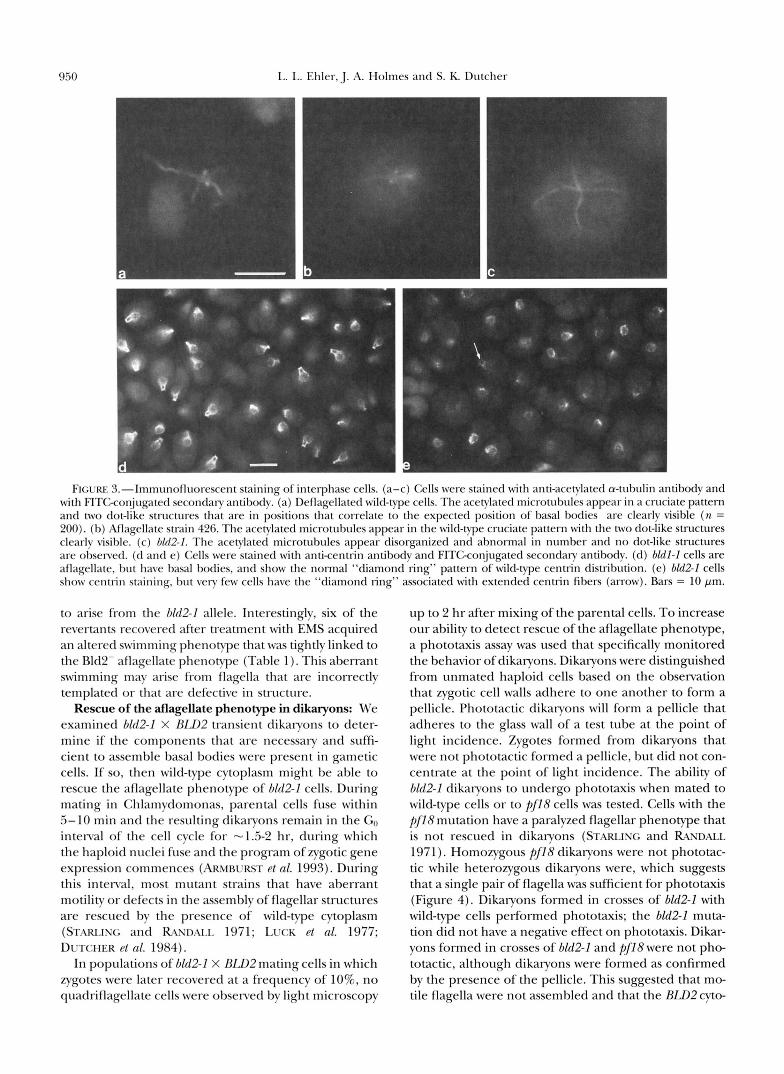
The examination of bZd2-l cells by immunofluores- cence allowed us to examine a larger number of cells for the presence of basal bodies. Using indirect immu- nofluorescence with an antibody against acetylated a- tubulin that stains basal bodies and rootlet microtu- bules (LEDMET and PIPERNO 1986), we examined de- flagellated wild-type cells, a control aflagellate strain (426), and 6Zd2-l cells. Deflagellation removes flagella at the cell wall and leaves the basal bodies intact. In 90% of deflagellated wild-type cells and in strain 426, two dot-like structures at the anterior end of the cell and the rootlet microtubules were stained with the anti- body (Figure 3, a and b). In bZd2-1 cells, acetylated mi-
Spindle Position 949
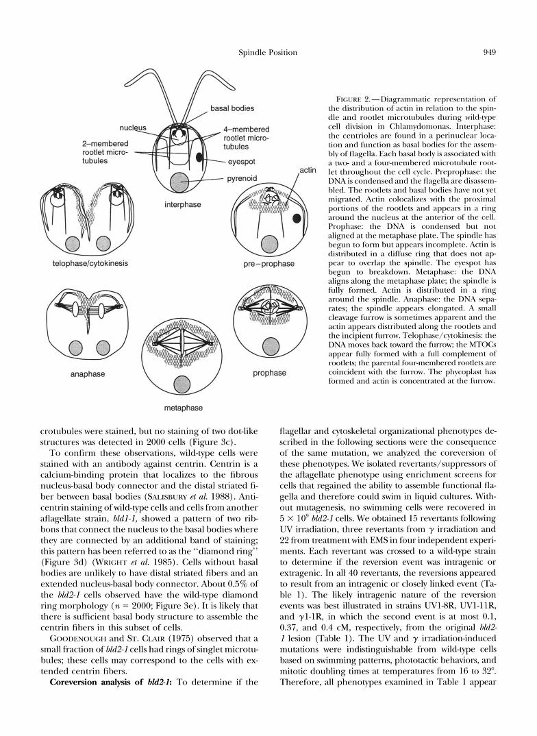
FIGURE 2.-Diagrammatic representation of the distribution of actin in relation to the spin- dle and rootlet microtubules during wild-type cell division in Chlamydomonas. Interphase: the centrioles are found in a perinuclear loca- tion and function as basal bodies for the assem- bly of flagella. Each basal body is associated with a two- and a four-membered microtubule root-
actin let throughout the cell cycle. Preprophase: the DNA is condensed and the flagella are disassem- bled. The rootlets and basal bodies have not yet migrated. Actin colocalizes with the proximal portions of the rootlets and appears in a ring around the nucleus at the anterior of the cell. Prophase: the DNA is condensed but not aligned at the metaphase plate. The spindle has begun to form but appears incomplete. Actin is distributed in a diffuse ring that does not a p
telophase/cytokinesis pre-prophase pear to overlap the spindle. The eyespot has begun to breakdown. Metaphase: the DNA aligns along the metaphase plate; the spindle is fully formed. Actin is distributed in a ring around the spindle. Anaphase: the DNA sepa- rates; the spindle appears elongated. A small cleavage furrow is sometimes apparent and the actin appears distributed along the rootlets and the incipient fM-ow. Telophase/cytokinesis: the DNA moves back toward the furrow; the MTOCs appear fully formed with a ftdl complement of rootlets; the parental four-membered rootlets are
anaphase prophase coincident with the furrow. The phycoplast has formed and actin is concentrated at the furrow.
rootlet rnicro-
metaphase
crotubules were stained, but no staining of two dot-like structures was detected in 2000 cells (Figure 3c).
To confirm these observations, wild-type cells were stained with an antibody against centrin. Centrin is a calcium-binding protein that localizes to the fibrous nucleus-basal body connector and the distal striated fi- ber between basal bodies (SALISBURY et al. 1988). Anti- centrin staining ofwild-type cells and cells from another aflagellate strain, bldl-2, showed a pattern of two rib- bons that connect the nucleus to the basal bodies where they are connected by an additional band of staining; this pattern has been referred to as the "diamond ring" (Figure 3d) (WRIGHT et al. 1985). Cells without basal bodies are unlikely to have distal striated fibers and an extended nucleus-basal body connector. About 0.5% of the bld2-I cells observed have the wild-type diamond ring morphology (n = 2000; Figure 3e). It is likely that there is sufficient basal body structure to assemble the centrin fibers in this subset of cells.
GOODENOUGH and ST. CLAIR (1975) observed that a small fraction of bld2-I cells had rings of singlet microtu- bules; these cells may correspond to the cells with ex- tended centrin fibers.
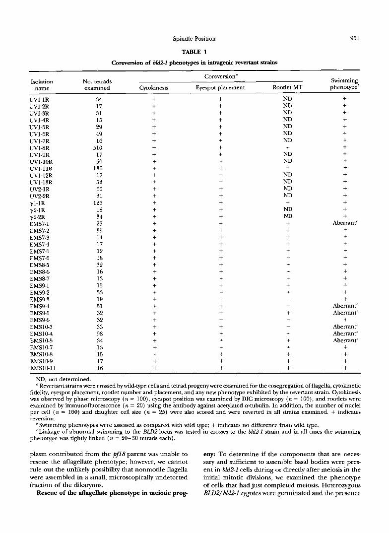
Coreversion analysis of bM2-1: To determine if the
flagellar and cytoskeletal organizational phenotypes de- scribed in the following sections were the consequence of the same mutation, we analyzed the coreversion of these phenotypes. We isolated revertant~/suppressors of the aflagellate phenotype using enrichment screens for cells that regained the ability to assemble functional fla- gella and therefore could swim in liquid cultures. With- out mutagenesis, no swimming cells were recovered in 5 x 10'' bkl2-I cells. We obtained 15 revertants following UV irradiation, three revertants from y irradiation and 22 from treatment with EMS in four independent experi- ments. Each revertant was crossed to a wild-type strain to determine if the reversion event was intragenic or extragenic. In all 40 revertants, the reversions appeared to result from an intragenic o r closely linked event (Ta- ble l). The likely intragenic nature of the reversion events was best illustrated in strains UV18R, UVl-IIR, and yl-lR, in which the second event is at most 0.1, 0.37, and 0.4 cM, respectively, from the original blit2- I lesion (Table 1). The UV and y irradiation-induced mutations were indistinguishable from wild-type cells based on swimming patterns, phototactic behaviors, and mitotic doubling times at temperatures from 16 to 32". Therefore, all phenotypes examined in Table 1 appear
950 L. L. Ehler, J. A. Holmes and S. K. Dutcher
FIGURE 3.-lmn~r1noflr1orescent staining o f interphase cells. (a-c) Cells were stained w i t h anti-acetylated a-tubulin antibody and with FITGconjugated seconckary antibody. (a) Deflagellated wild-type cells. The acetylated microtubules appear in a cruciate pattern and two dot-like structures that are in positions that correlate to the expected position of basal bodies are clearly visible (n = 200). (b) Aflagellate strain 426. The acetylated microtubules appear in the wild-type cruciate pattern with the two dot-like structures clearly visible. (c) 01*12-1. The acetylated microtubules appear disorganized and abnormal in number and no dot-like structures are observed. (d and e) Cells were stained with anticentrin antibody and FITGconjugated secondary antibody. (d) bldl-I cells are aflagellate, but have basal bodies, and show the normal “diamond ring” pattern of wild-type centrin distribution. (e) bZd2-I cells show centrin staining, but very few cells have the “diamond ring” associated with extended centrin fibers (arrow). Bars = 10 pm.
to arise from the 6112-1 allele. Interestingly, six of the revertants recovered after treatment with EMS acquired an altered swimming phenotype that was tightly linked to the Bld2- aflagellate phenotype (Table 1). This aberrant swimming mav arise from flagella that are incorrectly templated or that are defective in structure.
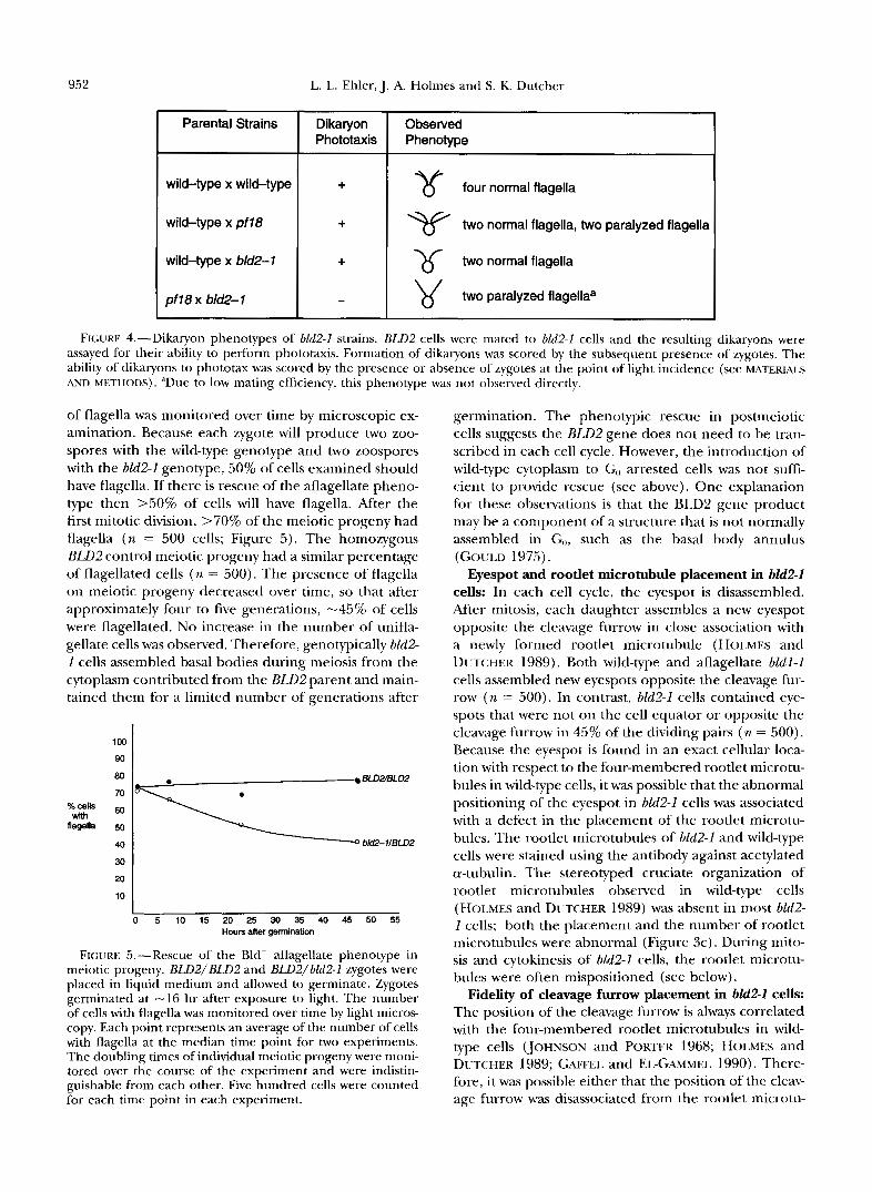
Rescue of the aflagellate phenotype in dikaryons: We examined 6112-1 X RIJ12 transient dikaryons to deter- mine if the components that are necessary and suffi- cient to assemble basal bodies were present in gametic cells. If so, then wild-type cytoplasm might be able to rescue the aflagellate phenotype of hld2-I cells. During mating in Chlamydomonas, parental cells fuse within 5-10 min and the resulting dikaryons remain in the Go interval of the cell cycle for “1.5-2 hr, during which the haploid nuclei fuse and the program of zygotic gene expression commences (ARMBURST et al. 1993). During this interval, most mutant strains that have aberrant motility or defects in the assembly of flagellar structures are rescued by the presence of wild-type cytoplasm (STARLING and RZNDALI. 1971; LUCK et al. 1977; DUTCHER ~1 nl. 1984).
In populations of 6112-1 X BZB2 mating cells in which zygotes were later recovered at a frequency of lo%, no quadriflagellate cells were observed by light microscopy
up to 2 hr after mixing of the parental cells. To increase our ability to detect rescue of the aflagellate phenotype, a phototaxis assay was used that specifically monitored the behavior of dikaryons. Dikaryons were distinguished from unmated haploid cells based on the observation that zygotic cell walls adhere to one another to form a pellicle. Phototactic dikaryons will form a pellicle that adheres to the glass wall of a test tube at the point of light incidence. Zygotes formed from dikaryons that were not phototactic formed a pellicle, but did not con- centrate at the point of light incidence. The ability of bLd2-I dikaryons to undergo phototaxis when mated to wild-type cells or to pf18 cells was tested. Cells with the pf18mutation have a paralyzed flagellar phenotype that is not rescued in dikaryons (STARLING and RCVDALL 1971). Homozygous pf18 dikaryons were not phototac- tic while heterozygous dikaryons were, which suggests that a single pair of flagella was sufficient for phototaxis (Figure 4). Dikaryons formed in crosses of bld2-I with wild-type cells performed phototaxis; the bld2-1 muta- tion did not have a negative effect on phototaxis. Dikar- yons formed in crosses of bld2-1 and pf18 were not pho- totactic, although dikaryons were formed as confirmed by the presence of the pellicle. This suggested that mo- tile flagella were not assembled and that the RLD2 cyto-
Spindle Position
TABLE 1
951
Coreversion of bM2-1 phenotypes in intragenic revertant strains
Coreversion" Isolation No. tetrads Swimming
name examined Cytokinesis Eyespot placement Rootlet MT phenotypeb
W l - 1 R W l - 2 R W1-3R UV14R W l - 5 R W l - 6 R W1-7R W l S R W l - 9 R UVl-IOR UVI-1IR W1-12R W l - 1 3 R W2-1R W2-2R yl-1R y2-1R y2-2R EMS7-1 EMS7-2 EMS7-3 EMS74 EMS7-5 EMS7-6 EMS8-5 EMS86 EMS87 EMS9-1 EMS9-2 EMS9-3 EMS94 EMS9-5 EMS9-6 EMS103 EMS 1 0 4 EMSl 0-5 EMSlO-7 EMSlO-8 EMSlO-9 EMSl 0-1 1
ND, not determined. a Revertant strains were crossed by wild-type cells and tetrad progeny were examined for the cosegregation of flagella, cytokinetic
fidelity, eyespot placement, rootlet number and placement, and any new phenotype exhibited by the revertant strain. Cytokinesis was observed by phase microscopy (n = loo), eyespot position was examined by DIC microscopy (n = loo), and rootlets were examined by immunofluorescence (n = 20) using the antibody against acetylated a-tubulin. In addition, the number of nuclei per cell (n = 100) and daughter cell size (n = 25) were also scored and were reverted in all strains examined. + indicates reversion.
Swimming phenotypes were assessed as compared with wild type; + indicates no difference from wild type. 'Linkage of abnormal swimming to the BLDP locus was tested in crosses to the bld2-1 strain and in all cases the swimming
phenotype was tightly linked (n = 20-30 tetrads each).
plasm contributed from the pf18 parent was unable to eny: To determine if the components that are neces- rescue the aflagellate phenotype; however, we cannot sary and sufficient to assemble basal bodies were pres- rule out the unlikely possibility that nonmotile flagella ent in bZd2-1 cells during or directly after meiosis in the were assembled in a small, microscopically undetected initial mitotic divisions, we examined the phenotype fraction of the dikaryons. of cells that had just completed meiosis. Heterozygous
Rescue of the atlagellate phenotype in meiotic prog- BLD2/bld2-1 zygotes were germinated and the presence
wild-type x wild-type I + l y four normal flagella
wild-type x pf18 I + two normal flagella, two paralyzed flagella
wild-type x bld2-1 1 + 1 two normal flagella
pf 18 X bld2- 1 two paralyzed flagellaa -
FIGURE 4.-Dikaryon phenotypes of bld2-1 strains. BLD2 cells were mated to bld2-1 cells and the resulting dikaryons were assayed for their ability to perform phototaxis. Formation of dikaryons was scored by the subsequent presence of zygotes. The ability of dikaryons to phototax was scored by the presence or absence of zygotes at the point of light incidence (see MATERIALS AND METHODS). “Due to low mating efficiency, this phenotype was not observed directly.
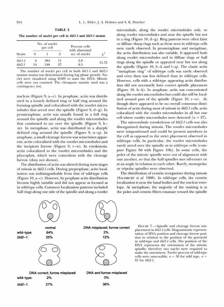
of flagella was monitored over time by microscopic ex- amination. Because each zygote will produce two zoo- spores with the wild-type genotype and two zoospores with the bZd2-1 genotype, 50% of cells examined should have flagella. If there is rescue of the aflagellate pheno- type then >50% of cells will have flagella. After the first mitotic division, >70% of the meiotic progeny had flagella (n = 500 cells; Figure 5). The homozygous B L B 2 control meiotic progeny had a similar percentage of flagellated cells ( n = 500). The presence of flagella on meiotic progeny decreased over time, so that after approximately four to five generations, -45% of cells were flagellated. No increase in the number of unifla- gellate cells was observed. Therefore, genotypically bZd2- 1 cells assembled basal bodies during meiosis from the cytoplasm contributed from the BLD2 parent and main- tained them for a limited number of generations after
loo I 90
BLD2fBLD2 0
flagella
40 bM2-1IBLD2
30 I 2o I lo I
I 0 5 10 15 20 25 30 35 40 45 50 55
Hours after germination
FIGURE 5.-Rescue of the Bld- aflagellate phenotype in meiotic progeny. BLDZ/BLD2 and BWZ/bldZ-I zygotes were placed in liquid medium and allowed to germinate. Zygotes germinated at -16 hr after exposure to light. The number of cells with flagella was monitored over time by light micros- copy. Each point represents an average of the number of cells with flagella at the median time point for two experiments. The doubling times of individual meiotic progeny were moni- tored over the course of the experiment and were indistin- guishable from each other. Five hundred cells were counted for each time point in each experiment.
germination. The phenotypic rescue in postmeiotic cells suggests the BLD2 gene does not need to be tran- scribed in each cell cycle. However, the introduction of wild-type cytoplasm to Go arrested cells was not suffi- cient to provide rescue (see above). One explanation for these observations is that the BLD2 gene product may be a component of a structure that is not normally assembled in Go, such as the basal body annulus (GWLD 1975).
Eyespot and rootlet microtubule placement in bld2-2 cells: In each cell cycle, the eyespot is disassembled. After mitosis, each daughter assembles a new eyespot opposite the cleavage furrow in close association with a newly formed rootlet microtubule (HOL~MES and DUTCHER 1989). Both wild-type and aflagellate bZdl-1 cells assembled new eyespots opposite the cleavage fur- row (n = 500). In contrast, bZd2-1 cells contained eye- spots that were not on the cell equator or opposite the cleavage furrow in 45% of the dividing pairs (n = 500). Because the eyespot is found in an exact cellular loca- tion with respect to the four-membered rootlet microtu- bules in wild-type cells, it was possible that the abnormal positioning of the eyespot in bZd2-1 cells was associated with a defect in the placement of the rootlet microtu- bules. The rootlet microtubules of bZd2-1 and wild-type cells were stained using the antibody against acetylated a-tubulin. The stereotyped cruciate organization of rootlet microtubules observed in wild-type cells (HOLMES and DUTCHER 1989) was absent in most bkd2- 1 cells; both the placement and the number of rootlet microtubules were abnormal (Figure 3c). During mito- sis and cytokinesis of bld2-1 cells, the rootlet microtu- bules were often mispositioned (see below).
Fidelity of cleavage furrow placement in bld2-I cells: The position of the cleavage furrow is always correlated with the four-membered rootlet microtubules in wild- type cells (JOHNSON and PORTER 1968; HOLMES and DUTCHER 1989; GAFFEL and EL-GAMMEL 1990). There- fore, it was possible either that the position of the cleav- age furrow was disassociated from the rootlet microtu-
Spindle Position 953
bules in bU2-I cells or that the cleavage furrow was mispositioned along with the mispositioned rootlet mi- crotubules. The location of the cleavage furrow in rela- tion to the rootlet microtubules was determined using the antibody against acetylated a-tubulin; the position of the cleavage furrow always correlated with a rootlet microtubule in dividing bU2-I cells ( n = 40). Light mi- croscopic examination of 6Zd2-1 cells showed that the cleavage furrow was placed such that unequally sized sister cells were often produced. The area of cells in wild-type and hld2-I cultures was measured and the dif- ference in area between sister cells and between unre- lated (nonsister) cells was compared (Figure 6). In wild- type populations the mean difference in area between sisters (6.4 pm') was much less than between unrelated cells (15.3 pm'). This suggests that the placement of the cleavage furrow in Chlamydomonas is nonrandom and generally results in two equal-sized cells. In hM2-I cells, the distribution of the mean differences in cell area between sister cells was similar to that of the mean difference between unrelated cells, which indicates that the areas of sister cells are not correlated to each other. Therefore, cleavage furrows are misplaced in hld2-1 cells.
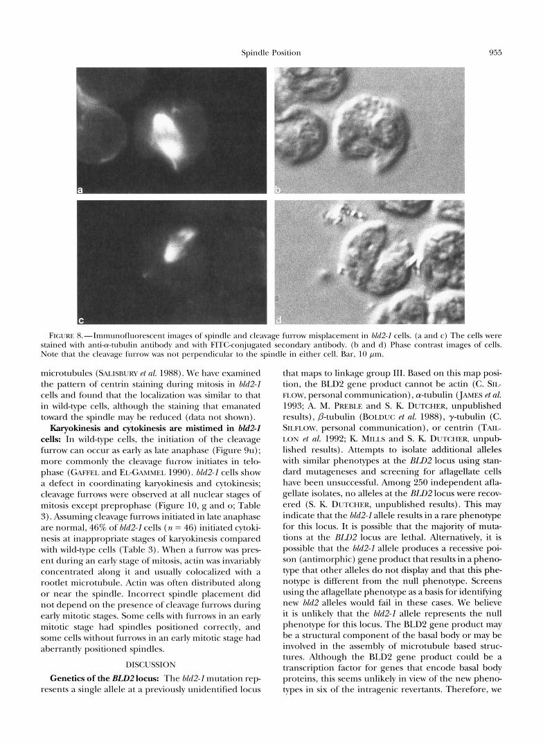
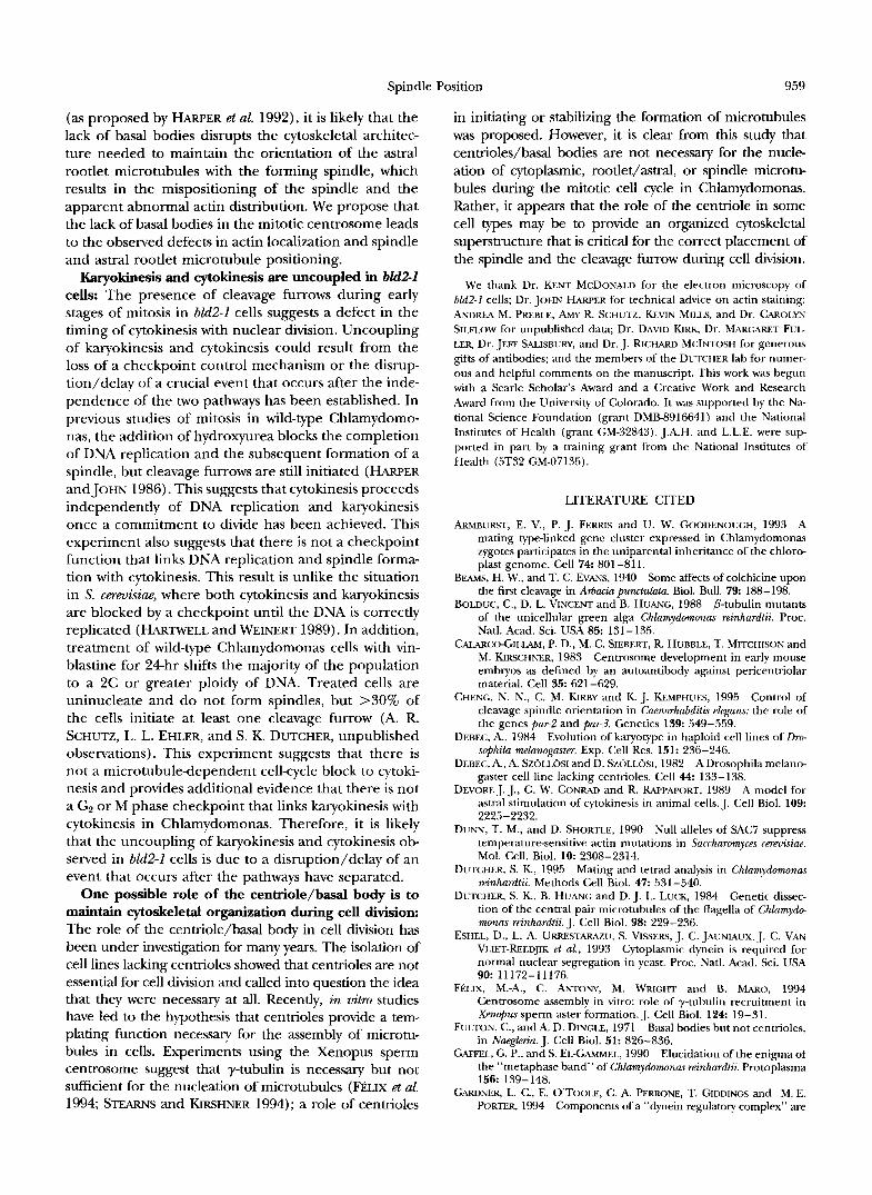
Spindle and cleavage furrow placement in bZd2-2 cells: We examined hM2-I cells to determine if the mis- placement of the cleavage furrow was accompanied by a corresponding misplacement of the spindle, as o h served in a C. Plegnns par1 mutant strain (KEMPHUES pt nl. 1986). bZd2-1 cells exhibited a longer population doubling time in log phase cultures than wild-type cells (data not shown). A significant number of nondividing bM2-I cells with 0 or 2 nuclei was observed by DAPI staining when compared with bldl-I cells (x' = 15.72, P < 0.001; Table 2), which suggests that the spindle may no longer be oriented correctly in relation to the cleavage furrow. The pyrenoid, an easily visualized or- ganelle located at the posterior end of the cell, was used as a cellular landmark. The orientation of the spindle was assessed by DAPI staining of the chromo- somes, and the orientation of the cleavage furrow by phase microscopy. Only 25% of bld2-I cells examined had the normal orientation of the spindle and the cleav- age furrow ( n = 81, Figure 7). The spindle and cleavage furrow were misplaced with respect to each other in >36% of the scored cells. In relation to the pyrenoid, the spindle was oriented incorrectly in 48% of cells and the furrow was misplaced in 63% of cells. In the most extreme cases, the furrow was positioned parallel to the newly formed nuclei, such that the nuclei would have been cleaved or anucleate cells would have been formed (Figures 8 and 10, d and g).
Distribution of actin in relation to microtubule struc- tures in mitotic cells: Actin microfilament networks are postulated to be involved in positioning the mitotic a p paratus in other organisms. The dynamics of actin local- ization during mitosis in Chlamydomonas have been described previously; however, the distribution of actin
mother cell wall
a. wild-type
3
# pairs of cells 2
1
difference in area ( !am2)
b. bld2-1
# pairs of cells 2
1
2 4 6 8 10 12 14 16 18 20 22 24 26
difference in area ( pm2)
FIGURE 6,"Fidelity of cleavage furrow placement in wild- type and bld2-I cells. The difference in the area between sister cells, which are still within the mother cell wall, was compared with that between randomly selected single cells in the popula- tion. The random population represents cells at all stages of the cell cycle as opposed to the specific stage of the sister cells and therefore results in a relatively broad distribution of cell sizes. (a) Wild-type cells. The mean difference in area for sister cells was 6.4 pm' and the variance was 6.2 pm' ( n = 95); for nonsister cell pairs the mean was 15.3 pm' and the variance was 57.8 pm' ( n = 87). (b) bld2-1 cells. The mean difference in area for sister cells was 11.93 p.m' and the vari- ance was 38.6 pm' ( n = 85); for nonsister cell pairs the mean was 13.9 pm' and the variance was 52.5 pm' ( n = 83). Area = T7d, where 7 0 = width of cell at widest point and 1 = length of cell at longest point.
in relationship to microtubule structures was not exam- ined directly (HARPER ~1 nl. 1992). The distribution of actin in relation to the position of the spindle and root- let microtubules during mitosis and cytokinesis in wild- type and in OZd2-I cells was examined by immunofluo- rescence to determine if actin localization is correlated to the defect in positioning the spindle in hld2-1 cells.
During preprophase in wild-type cells, the flagella were resorbed and cytoplasmic microtubules disassem- bled, the rootlet microtubules and basal bodies had not migrated, and the DNA was condensed. Actin colocal- ized with the proximal portions of the rootlet microtu- bules and in some cases was also in a ring around the
I sister cells
n nonsister cells
I sister cells
o nonsister cells
954 L. L,. Ehler,J. A. Holmes and S. K. Dutcher
TABLE 2
The number of nuclei per cell in bldl-1 and bld2-1 strains
No. of nuclei per cell Percent cells
with abnormal Strain 0 1 2 no. of nuclei X?
bldl-I 0 384 11 2.8 01d2-1 14 548 37 8.5 15.72
The number of nuclei per cell in both bldl-1 and bld2-I mutant strains was determined during log phase growth. Nu- clei were visualized using DAF’l to stain the DNA. Mitotic cells were not counted. The chi-square statistic used was a contingency test.
nucleus (Figure 9, a-c). In prophase, actin was distrik uted in a loosely defined ring or half ring around the forming spindle and colocalized with the rootlet micro- tubules that arced over the spindle (Figure 9, d-g). In prometaphase, actin was usually found in a full ring around the spindle and along the rootlet microtubules that continued to arc over the spindle (Figure 9, h- m). In metaphase, actin was distributed in a sharply defined ring around the spindle (Figure 9, n-q). In anaphase, a small cleavage furrow was sometimes appar- ent; actin colocalized with the rootlet microtubules and the incipient furrow (Figure 9, r-w). In cytokinesis, actin colocalized to the rootlet microtubules and the phycoplast, which were coincident with the cleavage furrow (data not shown).
The distribution of actin was altered during most stages of mitosis in bH2-1 cells. During preprophase, actin local- iiration was indistinguishable from that of wild-type cells (Figure 10, a-c). However, by prophase actin distribution became highly variable and did not appear as focused as in wild-type cells. Common localiiration patterns included half rings along one side of the spindle and along a rootlet
normal wild-type: 84% bld2- 1 25%
microtubule, along the rootlet microtubules only, or along rootlet microtubules and near the spindle but not in a ring (Figure 10, d-g). Ring patterns were often faint or diffuse; sharp rings such as those seen in wild-type cells were rarely observed. In prometaphase and metaphase, the actin distribution was also variable. It appeared both along rootlet microtubules and in diffuse rings or half rings along the spindle or appeared near but not along the spindle (Figure 10, h-k and I-q). The classic actin “metaphase ring” of wild-type cells was rarely observed and even then was less defined than in wild-type cells. However, cells with a wild-type appearing actin distribu- tion did not necessarily have correct spindle placement (Figure 10, h-k). In anaphase, actin was concentrated along the rootlet microtubules but could also still be local- ized around part of the spindle (Figure 10, r-w). Al- though there appeared to be no overall consensus distri- bution of actin during most of mitosis in bM2-1 cells, actin colocalized with the rootlet microtubules in all but one cell where rootlet microtubules were detected (n = 27).
The microtubule cytoskeleton of bZd2-I cells was also disorganized during mitosis. The rootlet microtubules were mispositioned and could be present anywhere in the cell as opposed to the strict placement observed in wild-type cells. In particular, the rootlet microtubules rarely arced over the spindle as in wild-type cells (com- pare Figure 9d with Figure 10h). In some cells, the poles of the mitotic spindle were out of alignment with one another, so that the half spindles met offcenter or at an angle in relation to each other. Rarely, monopolar or tripolar spindles were observed.
The distribution of centrin reorganizes during mitosis (SALISBURY et al. 1988). In wild-type cells, the centrin localization is near the basal bodies and the nuclear enve- lope. At metaphase, the majority of the staining is at the poles and centrin fibers emanate toward the spindle
DNA misplaced; furrow correct 2% 12%
DNA correct: furrow misplaced wild-type: 2%
bld2- 1: 27%
DNA and furrow misplaced 0%
36%
FICXIRE 7.”Spindle and cleavage furrow mis- placement in bM2-I cells. Diagrammatic represen- tation of DNA position and cleavage furrow posi- tion in relation to the position of the pyrenoid in wild-type and bId2-1 cells. The position of the DNA represents the orientation of the mitotic spindle; therefore two nuclei were required to make the assessment. Twelve percent of wild-type cells were unscorable; n = 50 for wild type, n = 81 for bId2-1.
FIGURE 8.- lm~nrlnofluo~~~sccnt imqcs ol'spintllc and clcavage furrow misplacement in M2-I cclls. ( a and c) The cells were stained with anti-cu-tubulin antibody and with FITGconjugated secondary antibody. (b and d) Phase contrast images of cells. Note that the cleavage fllrrow was not perpendicular to the spindle in either cell. Bar, 10 pm.
microtubules (SALISRURY Pt nl. 1988). We have examined the pattern of centrin staining during mitosis in bld2-I cells and found that the localization was similar to that in wild-type cells, although the staining that emanated toward the spindle may be reduced (data not shown).
Karyokinesis and cytokinesis are mistimed in bld2-I cells: In wild-type cells, the initiation of the cleavage furrow can occur as early as late anaphase (Figure 9u); more commonly the cleavage furrow initiates in telo- phase (GAFFEI. and EL-GAMMEI. 1990). bld2-1 cells show a defect in coordinating karyokinesis and cytokinesis; cleavage furrows were observed at all nuclear stages of mitosis except preprophase (Figure 10, g and 0; Table 3). Assuming cleavage furrows initiated in late anaphase are normal, 46% of 0132-1 cells ( n = 46) initiated cytoki- nesis at inappropriate stages of karyokinesis compared with wild-type cells (Table 3). "hen a furrow was pres- ent during an early stage of mitosis, actin was invariably concentrated along it and usually colocalized with a rootlet microtubule. Actin was often distributed along or near the spindle. Incorrect spindle placement did not depend on the presence of cleavage furrows during early mitotic stages. Some cells with furrows in an early mitotic stage had spindles positioned correctly, and some cells without furrows in an early mitotic stage had aberrantly positioned spindles.
DISCUSSION Genetics of the BLD2locus: The bM2-I mutation r e p
resents a single allele at a previously unidentified locus
that maps to linkage group 111. Based on this map posi- tion, the BLD2 gene product cannot be actin (C . SIL FLOW, personal communication), cy-tubulin (JAMES et nl. 1993; A. M. PRERLE and S. K. DUTCHER, unpublished results), 0-tubulin (BOLDUC et nl. 1988), y-tubulin (C . SILFLOW, personal communication), or centrin (TAIL- LON et nl. 1992; K. MILL.. and S. K. DUTCHER, unpub lished results). Attempts to isolate additional alleles with similar phenotypes at the BIB2 locus using stan- dard mutageneses and screening for aflagellate cells have been unsuccessful. Among 250 independent afla- gellate isolates, no alleles at the BZB2 locus were recov- ered (S. K. DUTCHER, unpublished results). This may indicate that the 6112-1 allele results in a rare phenotype for this locus. It is possible that the majority of muta- tions at the BZB2 locus are lethal. Alternatively, it is possible that the bM2-I allele produces a recessive poi- son (antimorphic) gene product that results in a pheno- type that other alleles do not display and that this phe- notype is different from the null phenotype. Screens using the aflagellate phenotype as a basis for identifjmg new b132 alleles would fail in these cases. We believe it is unlikely that the 6132-1 allele represent5 the null phenotype for this locus. The BLD2 gene product may be a structural component of the basal body or may be involved in the assembly of microtubule based struc- tures. Although the BLD2 gene product could be a transcription factor for genes that encode basal body proteins, this seems unlikely in view of the new pheno- types in six of the intragenic revertants. Therefore, we
PP
P
PM
M
A
Spindle Position
TUBULIN ACTIN DAP I LIGHT
9.57
FIGURE 10.-The distribution of tubdin and actin during mito- sis of bM2-1 cells. Cells were stained with antiiu-tubulin, anti- acetylated a-tubulin, and anti-ac- tin primary antibodies and with Texas Red and FITC conjugated secondary antibodies (see MATERI- AIS AND METI.IOI)S). Bar, 10 pm. (a-c) Preprophase. Actin localiza- tion was similar to that seen in wild-type at this stage. (d-g) Pro- phase. The second pole of the spindle seen in d was out of the plane of focus and was located at the anterior of the cell, just to the left of the rootlet microtubule. Ac- tin was localized along the rootlet microtubule (arrows) that was as- sociated with a prominent cleav- age furrow (g. arrowhead) that did not bisect the spindle. This cy- tokinetic event would likely have resulted in the production of an anucleate cell. (h-k) Prometa- phase. Actin was present in a half ring around the spindle and also colocalized to a rootlet microtu- bule (arrows in h and i ) . The spin- dle \vas not positioned perpendic- ular to the anterior-posterior axis of the cell as determined by the position of the pyrenoid (arrow in k). (I-q) Metaphase. ( I ) Anti-tu- bulin staining i n the focal plane of a rootlet microtubule, (m) anti- actin in the same focal plane, (p) anti-tubulin staining in the focal plane of the spindle, (4) anti-actin in the same focal plane. Actin co- localized with both rootlet micro- tubules (arrows); a cleavage fur- row has initiated in this cell. (r- v) Anaphase (anterior view). (r) Anti-tubulin staining in the focal plane of a rootlet microtubule po- sitioned along the axis of the spin- dle. The spindle was positioned so that only one pole was in the plane of focus. (s) Anti-actin in the same focal plane, (v) anti-tubulin in the focal plane of the rest of the spin- dle, and (w) anti-actin in the same focal plane. Actin appeared to l o - calize along the rootlet microtu- bule and along the side of the cell (arrows in rand s); it was also pres- ent in a ring around the top pole of the spindle.
GUCHI 1975; DE\Y)KE PL d . 1989; HARRIS and GEWALT centrosomes at the spindle poles, and are clearly not 1989; reviewed in R/WP,\POKT 1986; SA~TERWAITE and part of the spindle itself. In addition, the association POILARD 1992; STROMI.: 1993). The mitotic rootlet mi- between the rootlet microtubules and the position of crotubules of Chlamydomonas are similar to astral mi- the cleavage furrow is striking. In wild-type cells, actin crotubules and unlike the cytoplasmic microtubules colocalizes with the rootlets throughout mitosis and that disassemble in preprophase. The rootlet microtu- concentrates along them during anaphase and cytoki- bules are present throughout mitosis, originate in the nesis, and the position of the distal portions of the
958 L. L. Ehler, J. A. Holmes and S. K. Dutcher
TABLE 3
Mistiming of karyokinesis and cytokinesis in bld2-1 cells
No. of cells at mitotic stage"
Prophase Prometaphase Metaphase Anaphase Percent of total
Wild-type (wt; n = 26) and bld2-1 cells (n = 51) were observed by indirect immunofluorescence using antibodies against both acetylated and nonacetylated a-tubulin and DNA was visualized by DAPI staining. The presence or absence of cleavage furrows was determined for cells that contained mitotic spindles.
" Cells in telophase normally may have initiated furrows, so this stage was not assessed independently from cytokinesis.
rootlet microtubules invariably predicts the position of the cleavage furrow. Although the overall distribution of actin in bld2-1 cells during mitosis was variable, actin colocalized to the astral rootlet microtubules in 96% of cells, and cleavage furrows colocalized with rootlet microtubules even when the rootlet microtubules were aberrantly placed. The colocalization of actin with root- let microtubules throughout mitosis indicated that the association of rootlet microtubules with cleavage fur- rows was not coincidental. This indicates that the root- let microtubules play a role in positioning the cleavage furrow, and it is probable that the defect in cleavage furrow positioning seen in bld2-1 cells is a result of the aberrant positioning of the astral rootlet microtubules and not of a defect in actin localization.
The centrosomal defect in bld2-I results in the mispo- sitioning of the mitotic spindle via the astral microtu- bules: In bld2-1 cells, the mitotic apparatus was posi- tioned aberrantly in 248% of cells and mispositioning was observed in any stage of mitosis that had an assem- bled spindle. We suggest that this incorrect positioning resulted from centrosomes that are unable to maintain the normal cytoskeletal organization required for cor- rect spindle orientation. In wild-type cells, the rootlet microtubules are maintained in a precise orientation relative to the basal bodies (RINGO 1967; WEISS 1984; GAFFEL and EL-GAMMEL 1990) and are normally linked to the basal bodies via the striated root fibers (WEISS 1984); this linkage is not possible in bld2-1 cells. This cytoskeletal disorganization could directly result in the aberrant migration of the centrosomes, failure of the spindle to maintain a correct position, or both. It is clear from experiments in other organisms that astral microtubules have a significant role in both establishing and maintaining spindle position (HYMAN and WHITE 1987; HUFFAKER et al. 1988; HYMAN 1989; SLJLLJVAN and HUFFAKER 1992). The occasional displaced spindle halves, and monopolar and tripolar spindles observed during mitosis further indicated the deficiency of centrosomal organization in bld2-1 cells, whereas the spindle itself appeared to function normally. The spin- dle positioning and anucleate/binucleate phenotypes exhibited by bld2-1 cells are similar to the phenotypes
of a Saccharomyces cermisiae P-tubulin mutation tub2-401. The loss of astral microtubules observed at the restrictive temperature in t u b 2 4 0 1 strains is accompanied by mis- positioning of the spindle and the production of an- ucleate and multinucleate cells; however, the actin mi- crofilament network is apparently functional, as the cells continue the cell cycle (SULLIVAN and HUFFAKER 1992).
Many lines of evidence support the model that actin microfilaments have an important role in the position- ing of the mitotic apparatus via the astral microtubules (HYMAN and WHITE 1987; PALMER et al. 1992; WADDLE et al. 1994). For example, at the restrictive temperature an actl-4 mutant strain of S. cmmisiae shows complete loss of actin cables and randomization of the few corti- cal actin patches remaining (DUNN and SHORTLE 1990; PALMER et al. 1992). Actin disorganization is accompa- nied by the failure to maintain spindle position at the restrictive temperature (PALMER et al. 1992). Aside from the positioning defect, the microtubule cytoskeleton of act l -4 cells appears normal during mitosis (PALMER et al. 1992). Therefore, alteration of either astral microtu- bules or actin microfilaments results in the misposition- ing of the mitotic spindle, but the disruption of one of the cytoskeletal networks does not necessarily affect the integrity of the other.
In contrast, the disorganization of rootlet microtu- bules and the spindle positioning defect in bld2-1 cells was accompanied by abnormal localization of actin. In wild-type cells, migration of the centrosomes occurs in preprophase and prophase (GAFFEL and EL-GAMMEL. 1990); actin localizes mainly to the rootlets before centrosome migration. In bld2-1 cells, there was wild- type localization of actin before centrosomal migration and abnormal actin localization was observed only after the centrosomes attempted to migrate. This abnormal actin localization was observed as a failure either to make or maintain the actin metaphase ring. However, bld2-1 cells continued to localize actin to the rootlet microtubules during mitosis and showed no defect in the completion of cytokinesis, which indicates that the actin cytoskeleton was at least partially functional. Therefore, if actin is involved in the positioning of the spindle via the rootlet microtubules in Chlamydomonas
Spindle Position 959
(as proposed by HARPER et al. 1992), it is likely that the lack of basal bodies disrupts the cytoskeletal architec- ture needed to maintain the orientation of the astral rootlet microtubules with the forming spindle, which results in the mispositioning of the spindle and the apparent abnormal actin distribution. We propose that the lack of basal bodies in the mitotic centrosome leads to the observed defects in actin localization and spindle and astral rootlet microtubule positioning.
Karyokinesis and cytokinesis are uncoupled in bld2-2 cells: The presence of cleavage furrows during early stages of mitosis in bZd2-1 cells suggests a defect in the timing of cytokinesis with nuclear division. Uncoupling of karyokinesis and cytokinesis could result from the loss of a checkpoint control mechanism or the disrup- tion/delay of a crucial event that occurs after the inde- pendence of the two pathways has been established. In previous studies of mitosis in wild-type Chlamydomo- nas, the addition of hydroxyurea blocks the completion of DNA replication and the subsequent formation of a spindle, but cleavage furrows are still initiated (HARPER andJOHN 1986). This suggests that cytokinesis proceeds independently of DNA replication and karyokinesis once a commitment to divide has been achieved. This experiment also suggests that there is not a checkpoint function that links DNA replication and spindle forma- tion with cytokinesis. This result is unlike the situation in S. cermisiae, where both cytokinesis and karyokinesis are blocked by a checkpoint until the DNA is correctly replicated (HARTWELL and WEINERT 1989). In addition, treatment of wild-type Chlamydomonas cells with vin- blastine for 24hr shifts the majority of the population to a 2C or greater ploidy of DNA. Treated cells are uninucleate and do not form spindles, but >30% of the cells initiate at least one cleavage furrow (A. R. SCHUTZ, L. L. EHLER, and S. K. DUTCHER, unpublished observations). This experiment suggests that there is not a microtubuledependent cell-cycle block to cytoki- nesis and provides additional evidence that there is not a G2 or M phase checkpoint that links karyokinesis with cytokinesis in Chlamydomonas. Therefore, it is likely that the uncoupling of karyokinesis and cytokinesis ob- served in bld2-1 cells is due to a disruption/delay of an event that occurs after the pathways have separated.
One possible role of the centriole/basal body is to maintain cytoskeletal organization during cell division: The role of the centriole/basal body in cell division has been under investigation for many years. The isolation of cell lines lacking centrioles showed that centrioles are not essential for cell division and called into question the idea that they were necessary at all. Recently, in vitro studies have led to the hypothesis that centrioles provide a tem- plating function necessaq for the assembly of microtu- bules in cells. Experiments using the Xenopus sperm centrosome suggest that y-tubulin is necessaq but not sufficient for the nucleation of microtubules ( F ~ L I X et al. 1994; STEARNS and KIRSHNER 1994); a role of centrioles
in initiating or stabilizing the formation of microtubules was proposed. However, it is clear from this study that centrioles/basal bodies are not necessary for the nucle- ation of cytoplasmic, rootlet/astral, or spindle microtu- bules during the mitotic cell cycle in Chlamydomonas. Rather, it appears that the role of the centriole in some cell types may be to provide an organized cytoskeletal superstructure that is critical for the correct placement of the spindle and the cleavage furrow during cell division.
We thank Dr. KENT MCDONAILI for the electron microscopy of bZd2-1 cells; Dr. JOHN HARPER for technical advice on actin staining; ANDREA M. PREBLE, AMY R. SCHUTZ, KEVIN M11.1.s. and Dr. CAROLYN SII.FI.OW for unpublished data; Dr. DAVID KIRK, Dr. MARGARET FUL LER, Dr. JEFF SAI.ISBURY, and Dr. J. RICHARD MCINTOSH for generous gifts of antibodies; and the members of the DUTCHER lab for numer- ous and helpful comments on the manuscript. This work was begun with a Searle Scholar’s Award and a Creative Work and Research Award from the University of Colorado. It was supported by the Na- tional Science Foundation (grant DMB8916641) and the National Institutes of Health (grant GM-32843). J.A.H. and L.L.E. were sup- ported in part by a training grant from the National Institutes of Health (5T32 GM-07135).
LITERATURE CITED
ARMBURST, E. V., P. J. FERRIS and U. W. GOODENOUCH, 1993 A mating type-linked gene cluster expressed in Chlamydomonas zygotes participates in the uniparental inheritance of the chloro- plast genome. Cell 74: 801-811.
BEAMS, H. W., and T. C. EVANS, 1940 Some affects of colchicine upon the first cleavage in Arbacia punctulata. Biol. Bull. 7 9 188-198.
BOLDUC, C., D. L. VINCENT and B. HUANG, 1988 P-tubulin mutants of the unicellular green alga Chlamydomonas reinhardtii. Proc. Natl. Acad. Sci. USA 85: 131-135.
CAIARCO-GILIAM, P. D., M. C. SIEBERT, R. HUBBLE, T. MITCHISON and M. KIRSCHNER, 1983 Centrosome development in early mouse emblyos as defined by an autoantibody against pericentriolar material. Cell 35: 621-629.
CHENG, N. N., C. M. KIRBY and IC J. KEMPHUES, 1995 Control of cleavage spindle orientation in Cuenorhabditis ekgans: the role of the genes par-2 and par-3. Genetics 139: 549-559.
DEBEC, A,, 1984 Evolution of karyotype in haploid ccll lines of Bo- scphila melanogaster. Exp. Cell Res. 151: 236-246.
DEBEC, A., A. Szii1.1.6s1 and D. SZOILOSI, 1982 A Drosophila melano- gaster cell line lacking centrioles. Cell 4 4 133-138.
DEVORE J. J.. G. W. CONRAD and R. RAPPAPORT, 1989 A model for astral stimulation of cytokinesis in animal cells. J. Cell Biol. 109:
DUNN, T. M., and D. SHORTIE, 1990 Null alleles of SAC7 suppress temperaturesensitive actin mutations in Sarrharomycps cereuisine. Mol. Cell. Biol. 10 2308-2314.
DUTCHER, S. IC, 1995 Mating and tetrad analysis in Chlamydomonas reinhardtii. Methods Cell Biol. 47: 531 -540.
DUTCHER, S. IC, B. HUANG and D. J. L. LUCK, 1984 Genetic disser- tion of the central pair microtubules of the flagella of Chlamydo- manus reinhardtii. J. Cell Biol. 98: 229-236.
ESHEL, D., L. A. URRESTARAZU, S. VISSERS, J. C. JAUNIAUX, J. C. VAN VIIET-REEDJIK et al., 1993 Cytoplasmic dynein is required for normal nuclear segregation in yeast. Proc. Natl. Acad. Sci. USA 90: 11172-11176.
FBIJX, M.-A,, C. ANTONY, M. WRIGHT and B. MARO, 1994 Centrosome assembly in vitro: role of y-tubulin recruitment in Xenopus sperm aster formation. J. Cell Biol. 124 19-31.
FULTON, C., and A. D. DINGLE, 1971 Basal bodies but not centrioles, in Naegleria. J. Cell Biol. 51: 826-836.
GAFFEL, G. P., and S. EI,-GA“EI., 1990 Elucidation of the enigma of the “metaphase band” of Chlamydomonas ra’nhardtii. Protoplasma 156 199-148.
GARLINER, L. C., E. O’TOOLE, C. A. PERRONE, T. GIDDINGS and M. E. PORTER, 1994 Components of a “dynein regulatory complex” are
2225-2232.
960 L. L. Ehler, J. A. Holmes and S. K. Dutcher
located at the junction between the radial spokes and the dynein arms in ChZamydonrmas flagella. J. Cell Biol. 127: 1311-1326.
GIIL, S. R., T. A. SCHOER, I. SZ-, E. R. STEUER, M. P. SHEETZ et al., 1991 Dynactin, a conserved, ubiquitously expressed compo- nent of an activator of vesicle motility mediated by cytoplasmic dynein. J. Cell Biol. 115 1639-1650.
GOWENOUCH, U. W., and H. S. ST. CIAIR, 1975 bald-2: a mutation affecting the formation of doublet and triplet sets of microtu- bules in Chlamydomonas reinhardtii. J. Cell Biol. 66: 480-491.
GOODENOUGH, U. W., and R. L. WEISS, 1978 Interrelationships be- tween microtubules, a striated fiber, and the gametic mating structure of Chlamydomonas reinhardtii. J. Cell Biol. 76: 480-483.
GOULD, R. R., 1975 The basal bodies of Chlamydomonas reinhardtii: formation from probasal bodies, isolation, and partial character- ization. J. Cell Biol. 65: 65-74.
G~uI.o, R. R., and G. G. BORIS, 1977 The pericentriolar material
J. Cell Biol. 73: 601-615. in Chinese hamster ovary cells nucleates microtubule formation.
GROSS, C. H., L. P. W. RANUM and P. A. LEFEBVRE, 1988 Extensive restriction fragment length polymorphisms in a new isolate of Chlamydomonas reinhardtii. Curr. Genet. 13: 503-508.
HAMAGUC;HI, Y., 1975 Microinjection of colchicine into sea urchin eggs. Dev. Growth Differ. 17: 111-1 17.
HARPER, J. D. I., and P. C. L. JOHN, 1986 Coordination of division events in the Chlamydomonascell cycle. Protoplasma 131: 118-130.
HARPER, J. D. I . , D. W. McCumu, M. A. SANDERS, J. 1,. SALISBURY, and P. C. JOHN, 1992 Actin dynamics during the cell cycle in Chlamydomonas reinhardtii. Cell Motil. Cytoskeleton 22: 117-126.
HARRIS A. K., and S. L. GEWALT, 1989 Simulation testing of mecha- nisms for inducing the formation of the contractile ring in cytoki- nesis. J. Cell Biol. 109: 2215-2224.
HARRIS, E. H., 1989 The Chlamydomonas Sourcebwk, a Comprehensive Guide to Bioloa and Laboratq Usr. Academic Press, San Diego, CA.
HAR~WEI.~., L. H., and T. A. WEINERT, 1989 Checkpoints: controls that ensure the order of cell cycle events. Science 246 629-633.
HILL, D. P., and S. SIKOME, 1988 An analysis of the role of microfil- aments in the establishment and maintenance of asymmetry in Camorhabditis elegans zygotes. Dev. Biol. 124: 75-84.
HII.I., D. P., and S. STROME, 1990 Brief cytochalasin-induced disrup tion of microfilaments during a critical interval in 1-cell C. elegans embryos alters the partitioning of developmental instructions to the 2cell embryo. Development 108: 159-172.
HOLMES, J. A,, and S. K. DUTCHER, 1989 Cellular asymmetry in Chla- mydomonas reinhardtii. J. Cell Sci. 94: 273-285.
HUFFAKER, T. C., J. H. THOMAS and D. BOTSTEIN, 1988 Diverse ef- fects of beta-tubulin mutations on microtubule formation and function. J. Cell Biol. 106: 1997-2010.
HYMAN, A. A,, 1989 Centrosome movement in the early divisions of Camorhabditis ekgans: a cortical site determining centrosome position. J. Cell Biol. 109: 1185-1193.
HYMAN, A. A., and J. G. WHITE, 1987 Determination of cell division axes in the early embryogenesis of Caenorhabditis elegans. J. Cell Biol. 105: 2123-2135.
JAMES, S. W., C. D. SILFLOW, P. STROOM and P. A. LEFEBVRE, 1993 A mutation in the al-tubulin gene of Chlamydomonas reinhardtii confers resistance to anti-microtubule herbicides. J. Cell Sci. 106: 209-218.
JOHNSON, U. G., and K. R. PORTER, 1968 Fine structure of cell divi- sion in Chlamydomonas reinhardtii. J . Cell Biol. 38: 403-425.
KEMPHIIES, K. J., N. WOI.F, W. B. WOOD and D. HIRSH, 1986 Two loci required for cytoplasmic organization in early embryos of Cmorhabditis ekgans. Dev. Bid. 113: 449-460.
KEMPHUES. K. J., J. R. PRIESS, D. G. MORTON and N. S. CHENG, 1988 Identification of genes required for cytoplasmic localization in early C. elegans embryos. Cell 52: 311-320.
LEDMET, M., and G. PIPERNO, 1986 Cytoplasmic microtubules con- taining acetylated alpha-tubulin in Chlamydomonas reinhardtii; spa- tial arrangement and properties. J. Cell Biol. 103: 13-22.
LI, Y. Y., E. YEH, T. HAYS and K. BLOOM, 1993 Disruption of mitotic spindle orientation in a yeast dynein mutant. Proc. Natl. Acad. Sci. USA 9 0 10096-11000.
LUCK, D., G. PIPERNO, Z. RAMANIS and B. HUANC, 1977 Flagellar mutants of Chlamydomonas: studies of radial spoke-defective strains by dikaryon and revertant analysis. Proc. Natl. Acad. Sci. USA 74: 3456-3460.
LUX, F. G., 111, and S. K. DUTCHER, 1991 Genetic interactions at the FLAlO locus: suppressors and synthetic phenotypes that affect the cell cycle and flagellar function in Chlamydomonas reinhardtii. Genetics 128: 549-561.
MANIOTIS, A,, and M. SCHIJWA, 1991 Microsurgical removal of centrosomes blocks cell reproduction and centriole generation in BSC-1 cells. Cell 67: 495-504.
MCMIILAN, J. N., and K. TATCHEIL, 1994 The J N M l gene in the yeast Saccharomyces cereuisiaeis required for nuclear migration and spindle orientation during the mitotic cell cycle.]. Cell Biol. 125: 143-158.
MOESTRUP, O., 1978 On the phylogenetic validity of the flagellar apparatus in green algae and other chlorophyll a and chlorophyll b containing plants. Biosystems 10: 117-144.
MUtIITA, L., T. S. KARPOVA and J. A. COOPER, 1994 A yeast actin- related protein homologous to that in vertebrate dynactin corn-
Cell 78: 669-679. plex is important for spindle orientation and nuclear migration.
PAI.MER, R. E., D. S. SUI.I.IVAN, T. HUFFAKER and D. KosrrrAvn. 1992 Role of astral microtubules and actin in spindle orientation and migration in the budding yeast, Saccharomyces ct.rpoisia~. J. Cell Biol. 119: 583-593.
PASQIJALE, S. M., and U. W. GOODFNOIJGH, 1987 Cyclic AMP func- tions as a primary sexual signal in gametes of Ch1amydomona.r reinhardtii. J. Cell Biol. 105: 2279-2292.
RAPPAPORT, R., 1961 Experiments concerning the cleavage in sand dollar eggs. J. Exp. Zool. 148: 81 -89.
RAPPAPORT, R., 1971 Reversal of cleavage inhibition in enchintr derm eggs. J. Exp. Zool. 176: 249-255.
RAPPAPORT, R., 1973 On the rate of movement of the cleavage stimulus in sand dollar eggs. J. Exp. Zool. 183: 115-119.
RAPPAPORT, R., 1986 Establishment of the mechanism of cytokinesis in animal cells. Int. Rev. Cytol. 105: 245-281.
RINCO, D. L., 1967 Flagellar motion and fine structure of the flagel- lar apparatus in Chlamydomonas. J. Cell Biol. 33: 543-571.
SAGER, R., and S. GRANICK, 1954 Nutritional control of sexuality in Chlamydomonas reinhardtii. J. Gen. Physiol. 37: 729-742.
SALISBURY, J. L., A. T. BARON and M. A. SANDERS, 1988 The centrin- based cytoskeleton of Chlamydomonas reinhurdtii: distribution in interphase and mitotic cells. J. Cell Biol. 107: 635-641.
SATTERWAITE, L. L., and T. D. POIJAM), 1992 Cytokinesis. Curr. Opin. Cell Bid. 4: 43-52.
SCHIBLER, M. J, and B. HUANG, 1991 The col"4 and rol"15 /3-tubulin mutations in Chlamydomonas rrinhardtii confer altered sensitivities to microtubule inhibitors and herbicides by enhancing microtw bule stability. J. Cell Biol. 113: 605-614.
STARLING, D., and J. RANDAH., 1971 The flagella of temporary dikar- yons of Chlamydomonas rfinhardtii. Genet. Res. 18: 107-113.
STEAKNS, T., and M. KIRSGHNER, 1994 In vitro reconstitution of centrosome assembly and function: the central role of y-tubulin. Cell 76: 623-637.
STROME, S., 1993 Determination of cleavage planes. Cell 72: 3-6. SUI.I.WAN, D., and T. HUFFAKER, 1992 Astral microtubules are not
required for anaphase h in Sarrharomyces crrmisiar. J. Cell B i d .
S Z ~ ~ L L ~ S I , A,, H. RIS, D. SXOI.LOSI and A. DEBEC, 1986 A centriole- free Drosophila cell line: a high voltage EM study. Eur. J . Cell Biol. 40: 100-104.
TAII.ON, B. E., S. A. AIDER, J. P. SUHAN and J. W. JARVIK, 1992 Muta- tional analysis of centrin: an EF-hand protein associated with three distinct contractile fibers in the basal body apparatns o f Chlamydomonas. J. Cell Biol. 119: 1613-1624.
WADDLE, J. A,, J. A. COOPER and R. H. WATERSTON, 1994 Transient localized accumulation of actin in Cmorhabditis ekguns blasto- meres with oriented asymmetric divisions. Development 120:
WEISS, R. I,,, 1984 Ultrastructure of the flagellar roots in Cldamydo- monas gametes. J. Cell Sci. 67: 133- 143.
WITMAN, G. B., K. CARI.SON, J. BERI.INER and J. L. ROSENBAIJM, 1972 Chlamydomonas flagella. I . Isolation and electrophoretic analysis of microtubules, matrix, membranes, and mastigonemes. J. Cell Biol. 54: 507-539.
WRIGHT, R. I,., J. L. SALISBURY and J. W. JARVIK, 1985 A nucleus-basd body connector in Chlamydomonas relnhardtii that may function in basal body localization or seg7-egati0n.J. Cell Biol. 101: 1903- 1912.