Søren Nielsen, NordiQC 1 Controls and critical staining quality indicators in Immunohistochemistry Søren Nielsen Scheme Manager NordiQC Aalborg Hospital, Denmark 2 IHC – Biomarker controls What is an IHC control for diagnostic IHC ? What is recommended and best practice ? What are the pitfalls for the use of controls for IHC ? How are IHC controls used by laboratories, .NordiQC and EQA programmes ? Decalcification Preparation Pre- analytic Analytic Post- analytic Tissue Type, Dimension, Laser resection, De-differentiation Fixation Time, Type, Volume Section Thickness Storage Drying Visualization Sensitivity, Specificity Primary antibody Clone, Dilution Buffer, Time, Temp Development Sensitivity, Localization Interpretation Localization Positive/Negative - cut-off level Quantification Reporting Controlment Pre-treatment With 3 choices for 5 variables in each phase = > 4 million protocols…. Manual Stainer IHC – Biomarker controls Appropriate tissue fixation and processing Appropriate and efficient epitope retrieval Appropriate choice of antibody/clone Robust, specific & sensitive detection system Appropriate choice of control material The basal fundament for a technical optimal IHC performance: IHC – Biomarker controls 5 IHC – Biomarker controls 2011 2011 > 70 % of publications based on IHC do not describe controls used to verify data and conclusions…. 6 Reagent and tissue controls are necessary for the validation of immunohistochemical staining results. Without their use, interpretation of staining would be haphazard and the results of doubtful value. More specifically, controls determine if the staining protocols were followed correctly, whether day-to- day and worker-to-worker variations have occurred, and that reagents remain in good working order. IHC – Biomarker controls
Transcript
Søren Nielsen, NordiQC 1
Controls andcritical staining quality indicators
in Immunohistochemistry
Søren Nielsen
Scheme Manager
NordiQC
Aalborg Hospital, Denmark 2
IHC – Biomarker controls
� What is an IHC control for diagnostic IHC ?
� What is recommended and best practice ?
� What are the pitfalls for the use of controls for IHC ?
� How are IHC controls used by laboratories, .NordiQC and EQA programmes ?
> 70 % of publications based onIHC do not describe controls usedto verify data and conclusions….
6
�Reagent and tissue controls are necessary for the validation of immunohistochemical staining results.
�Without their use, interpretation of staining wouldbe haphazard and the results of doubtful value. More specifically, controls determine if the stainingprotocols were followed correctly, whether day-to-day and worker-to-worker variations have occurred, and that reagents remain in good working order.
IHC – Biomarker controls
Søren Nielsen, NordiQC 2
7
�Reagent and tissue controls are necessary for the validation of immunohistochemical staining results.
�Reagent control of the primary antibody is crucial for the producer to validate specificity and can include
� Primary ab tested on knock-out mice
� Primary ab tested on cell lines +/- antigen of interest
� Primary ab tested by western blotting
� Primary ab tested by antigen absorbtion
IHC – Biomarker controls
8
�Reagent and tissue controls are necessary for the validation of immunohistochemical staining results.
�Reagent control of the primary antibody is crucial for the producer to validate specificity and can include
� Primary ab tested on knock-out mice
� Primary ab tested on cell lines +/- antigen of interest
� Primary ab tested by western blotting
� Primary ab tested by antigen absorbtion
To secure specificity of primary ab -
Both by launch and new ab lots.
IHC – Biomarker controls
9
�Reagent and tissue controls are necessary for the validation of immunohistochemical staining results.
�Reagent control is for the laboratories of limited useand impossible to perform correctly.
� Primary ab control – negative reagent control
�Each primary ab must have its own negative
control serum, and thus all the IHC slides performed will be doubled
IHC – Biomarker controls
10
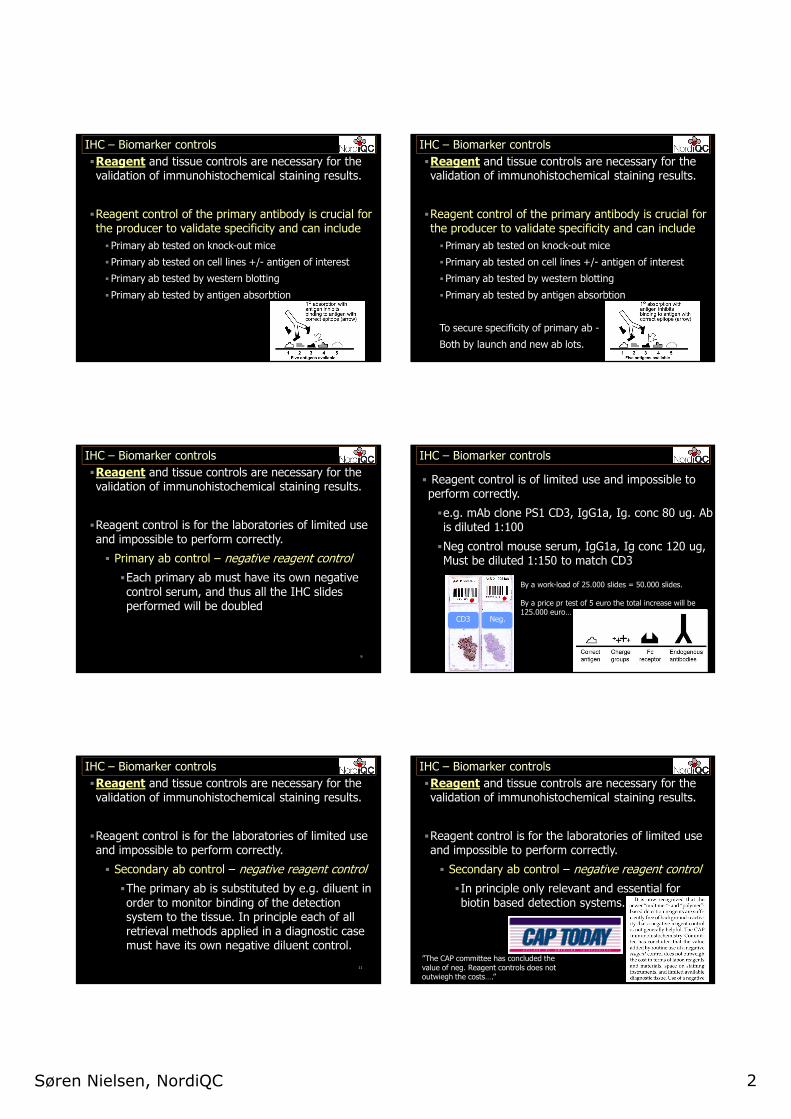
� Reagent control is of limited use and impossible to perform correctly.
�e.g. mAb clone PS1 CD3, IgG1a, Ig. conc 80 ug. Ab is diluted 1:100
�Neg control mouse serum, IgG1a, Ig conc 120 ug, Must be diluted 1:150 to match CD3
IHC – Biomarker controls
CD3 Neg.
By a work-load of 25.000 slides = 50.000 slides.
By a price pr test of 5 euro the total increase will be125.000 euro…
11
�Reagent and tissue controls are necessary for the validation of immunohistochemical staining results.
�Reagent control is for the laboratories of limited useand impossible to perform correctly.
� Secondary ab control – negative reagent control
�The primary ab is substituted by e.g. diluent in
order to monitor binding of the detectionsystem to the tissue. In principle each of all retrieval methods applied in a diagnostic case must have its own negative diluent control.
IHC – Biomarker controls
12
�Reagent and tissue controls are necessary for the validation of immunohistochemical staining results.
�Reagent control is for the laboratories of limited useand impossible to perform correctly.
� Secondary ab control – negative reagent control
�In principle only relevant and essential for
biotin based detection systems.
IHC – Biomarker controls
”The CAP committee has concluded the value of neg. Reagent controls does not outwiegh the costs….”
Søren Nielsen, NordiQC 3
13
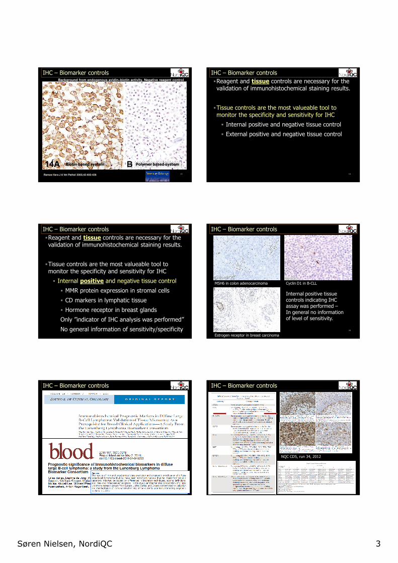
IHC – Biomarker controlsBackground from endogenous avidin–biotin activity. Negative reagent control
Ramos-Vara J A Vet Pathol 2005;42:405-426
Biotin based system Polymer based system
14
�Reagent and tissue controls are necessary for the validation of immunohistochemical staining results.
�Tissue controls are the most valueable tool to monitor the specificity and sensitivity for IHC
� Internal positive and negative tissue control
� External positive and negative tissue control
IHC – Biomarker controls
15
�Reagent and tissue controls are necessary for the validation of immunohistochemical staining results.
�Tissue controls are the most valueable tool to monitor the specificity and sensitivity for IHC
� Internal positive and negative tissue control
� MMR protein expression in stromal cells
� CD markers in lymphatic tissue
� Hormone receptor in breast glands
Only ”indicator of IHC analysis was performed”
No general information of sensitivity/specificity
IHC – Biomarker controls
16
IHC – Biomarker controls
MSH6 in colon adenocarcinoma Cyclin D1 in B-CLL
Estrogen receptor in breast carcinoma
Internal positive tissuecontrols indicating IHC assay was performed –In general no information of level of sensitivity.
IHC – Biomarker controls IHC – Biomarker controls
NQC CD5, run 34, 2012
Søren Nielsen, NordiQC 4
19
�Reagent and tissue controls are necessary for the validation of immunohistochemical staining results.
�Tissue controls are the most valueable tool to monitor the specificity and sensitivity for IHC
� Internal positive and negative tissue control
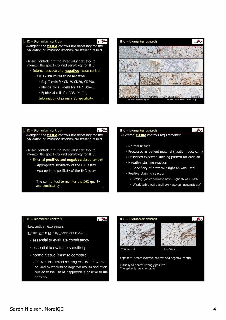
� Cells / structures to be negative
� E.g. T-cells for CD19, CD20, CD79a…
� Mantle zone B-cells for Ki67, Bcl-6…
� Epithelial cells for CD3, MUM1,…
Information of primary ab specificity
IHC – Biomarker controls IHC – Biomarker controls
MUM1 - mAb EUA32 & MUM1pMUM1 - mAb MRQ-43 SP54
Hodgkin HodgkinDLBCL DLBCL
Cerv. Ut. Ad. Carc. Carcinoid Cerv. Ut. Ad. Carc. Carcinoid
21
�Reagent and tissue controls are necessary for the validation of immunohistochemical staining results.
�Tissue controls are the most valueable tool to monitor the specificity and sensitivity for IHC
� External positive and negative tissue control
� Appropriate sensitivity of the IHC assay
� Appropriate specificity of the IHC assay
The central tool to monitor the IHC quality
and consistency
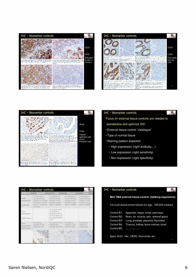
IHC – Biomarker controls
22
�External tissue controls requirements:
� Normal tissues
� Processed as patient material (fixation, decalc.,…)
� Described expected staining pattern for each ab
� Negative staining reaction
� Specificity of protocol / right ab was used…
� Positive staining reaction
� Strong (which cells and how – right ab was used)
� Weak (which cells and how - appropriate sensitivity)
IHC – Biomarker controls
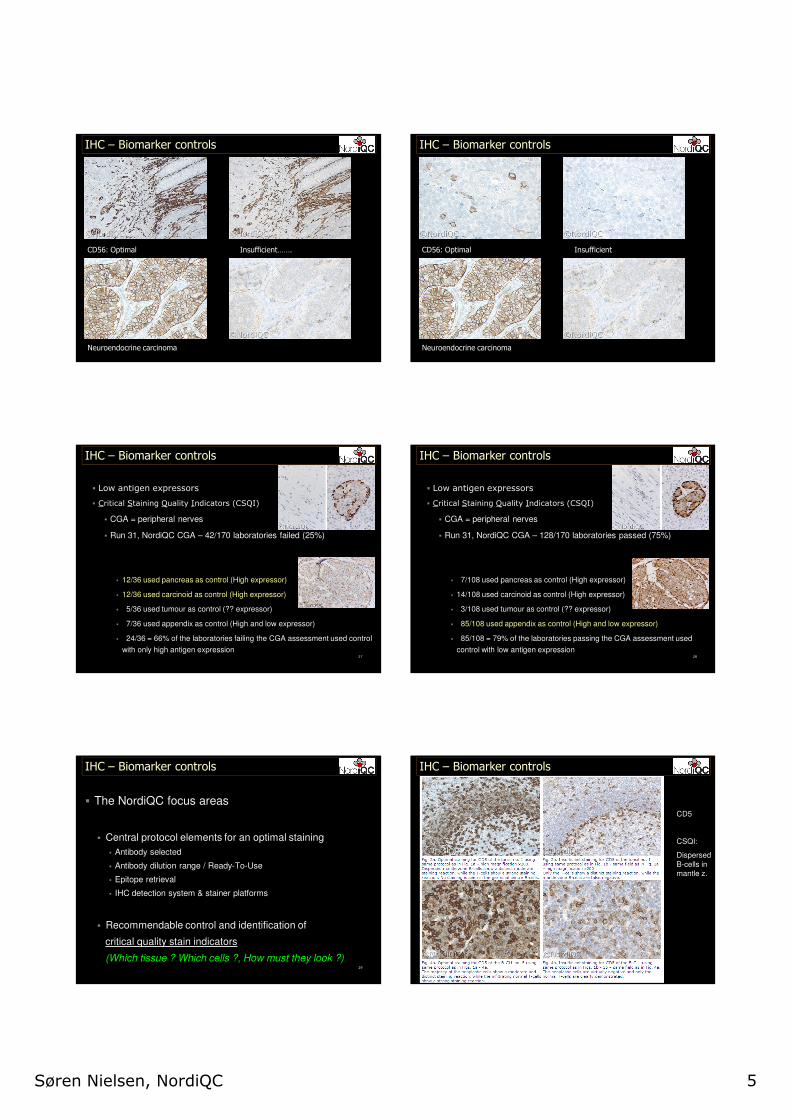
�Low antigen expressors
�Critical Stain Quality Indicators (CSQI)
� essential to evaluate consistency
� essential to evaluate sensitivity
� normal tissue (easy to compare)
� 90 % of insufficient staining results in EQA are
caused by weak/false negative results and often
related to the use of inappropriate positive tissue
controls......
IHC – Biomarker controls IHC – Biomarker controls
CD56: Optimal Insufficient…….
Appendix used as external positive and negative control
Virtually all nerves strongly positiveThe epithelial cells negative
Søren Nielsen, NordiQC 5
IHC – Biomarker controls
CD56: Optimal Insufficient…….
Neuroendocrine carcinoma
IHC – Biomarker controls
CD56: Optimal Insufficient
Neuroendocrine carcinoma
27
� Low antigen expressors
� Critical Staining Quality Indicators (CSQI)
� CGA = peripheral nerves
� Run 31, NordiQC CGA – 42/170 laboratories failed (25%)
� 12/36 used pancreas as control (High expressor)
� 12/36 used carcinoid as control (High expressor)
� 5/36 used tumour as control (?? expressor)
� 7/36 used appendix as control (High and low expressor)
� 24/36 = 66% of the laboratories failing the CGA assessment used control
with only high antigen expression
IHC – Biomarker controls
28
� Low antigen expressors
� Critical Staining Quality Indicators (CSQI)
� CGA = peripheral nerves
� Run 31, NordiQC CGA – 128/170 laboratories passed (75%)
� 7/108 used pancreas as control (High expressor)
� 14/108 used carcinoid as control (High expressor)
� 3/108 used tumour as control (?? expressor)
� 85/108 used appendix as control (High and low expressor)
� 85/108 = 79% of the laboratories passing the CGA assessment used
control with low antigen expression
IHC – Biomarker controls
29
� The NordiQC focus areas
� Central protocol elements for an optimal staining
� Antibody selected
� Antibody dilution range / Ready-To-Use
� Epitope retrieval
� IHC detection system & stainer platforms
� Recommendable control and identification of
critical quality stain indicators
(Which tissue ? Which cells ?, How must they look ?)