42
pyright © 2010 Pearson Education, Inc. Connective Tissue •Most abundant and widely distributed tissue type •Four classes • Connective tissue proper • Cartilage • Bone tissue • Blood
| Date post: | 30-Dec-2015 |
| Category: |
Documents |
| Upload: | iris-norris |
| View: | 221 times |
| Download: | 4 times |

Copyright © 2010 Pearson Education, Inc.
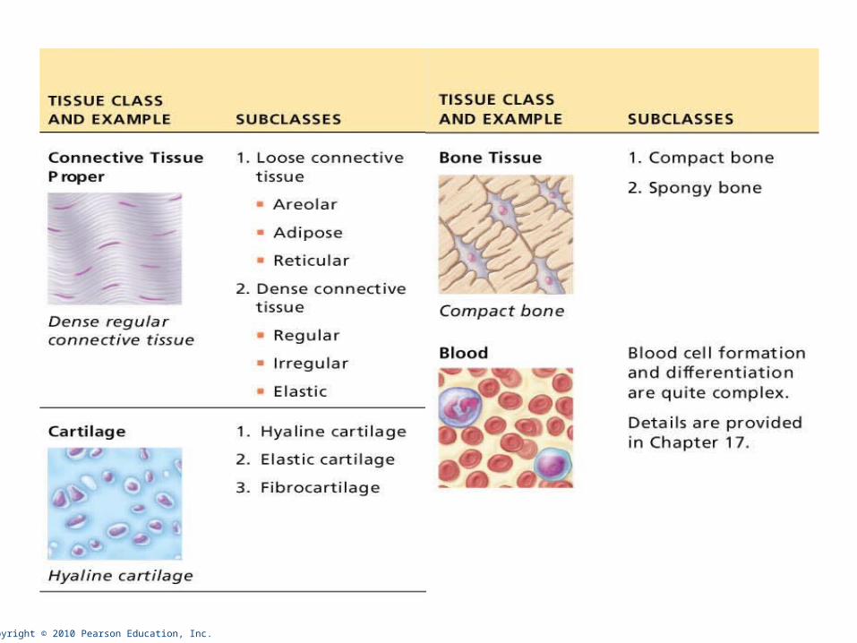
Connective Tissue
•Most abundant and widely distributed tissue type
• Four classes
• Connective tissue proper
• Cartilage
• Bone tissue
• Blood
Copyright © 2010 Pearson Education, Inc.

Copyright © 2010 Pearson Education, Inc.
Major Functions of Connective Tissue
• Binding and support
• Protection
• Insulation
• Transportation (blood)

Copyright © 2010 Pearson Education, Inc.
Characteristics of Connective Tissue
• Connective tissues have:
• Mesenchyme as their common tissue of origin
• Varying degrees of vascularity
• Cells separated by nonliving extracellular matrix (ground substance and fibers)
Copyright © 2010 Pearson Education, Inc.
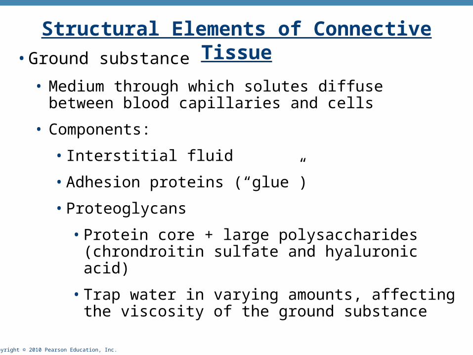
Structural Elements of Connective Tissue• Ground substance
• Medium through which solutes diffuse between blood capillaries and cells
• Components:
• Interstitial fluid
• Adhesion proteins (“glue”)
• Proteoglycans
• Protein core + large polysaccharides (chrondroitin sulfate and hyaluronic acid)
• Trap water in varying amounts, affecting the viscosity of the ground substance

Copyright © 2010 Pearson Education, Inc.
Structural Elements of Connective Tissue• Three types of fibers
• Collagen (white fibers)
• Strongest and most abundant type
• Provides high tensile strength
• Elastic
• Networks of long, thin, elastin fibers that allow for stretch
• Reticular
• Short, fine, highly branched collagenous fibers

Copyright © 2010 Pearson Education, Inc.
Structural Elements of Connective Tissue
• Cells
• Mitotically active and secretory cells = “blasts”
• Mature cells = “cytes”
• Fibroblasts in connective tissue proper
• Chondroblasts and chondrocytes in cartilage
• Osteoblasts and osteocytes in bone
• Hematopoietic stem cells in bone marrow
• Fat cells, white blood cells, mast cells, and macrophages

Copyright © 2010 Pearson Education, Inc. Figure 4.7
Macrophage
Fibroblast
Lymphocyte
Fat cell
Mast cell
Neutrophil
Capillary
Cell types Extracellularmatrix
Fibers• Collagen fiber• Elastic fiber• Reticular fiber
Ground substance
Copyright © 2010 Pearson Education, Inc.
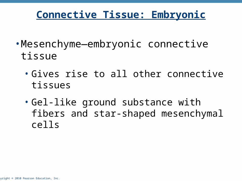
Connective Tissue: Embryonic
•Mesenchyme—embryonic connective tissue
• Gives rise to all other connective tissues
• Gel-like ground substance with fibers and star-shaped mesenchymal cells

Copyright © 2010 Pearson Education, Inc.
Overview of Connective Tissues
• For each of the following examples of connective tissue, note:
• Description
• Function
• Location
Copyright © 2010 Pearson Education, Inc.
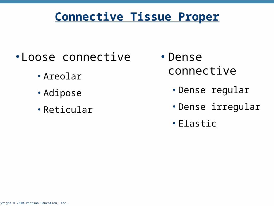
Connective Tissue Proper
• Loose connective
• Areolar
• Adipose
• Reticular
• Dense connective
• Dense regular
• Dense irregular
• Elastic

Copyright © 2010 Pearson Education, Inc.
(a) Connective tissue proper: loose connective tissue, areolar
Description: Gel-like matrix with allthree fiber types; cells: fibroblasts,macrophages, mast cells, and somewhite blood cells.
Function: Wraps and cushionsorgans; its macrophages phagocytizebacteria; plays important role ininflammation; holds and conveystissue fluid.
Location: Widely distributed underepithelia of body, e.g., forms laminapropria of mucous membranes;packages organs; surroundscapillaries.
Photomicrograph: Areolar connective tissue, asoft packaging tissue of the body (300x).
Epithelium
Laminapropria
Fibroblastnuclei
Elasticfibers
Collagenfibers
Figure 4.8a
Copyright © 2010 Pearson Education, Inc. Figure 4.8b
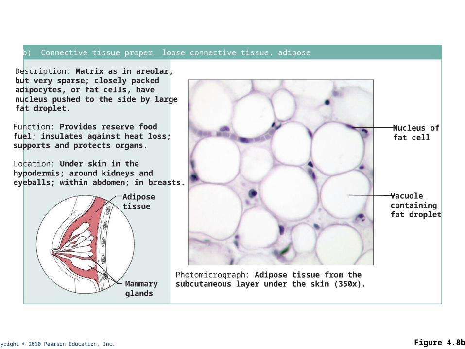
(b) Connective tissue proper: loose connective tissue, adipose
Description: Matrix as in areolar,but very sparse; closely packedadipocytes, or fat cells, havenucleus pushed to the side by largefat droplet.
Function: Provides reserve foodfuel; insulates against heat loss;supports and protects organs.
Location: Under skin in thehypodermis; around kidneys andeyeballs; within abdomen; in breasts.
Photomicrograph: Adipose tissue from thesubcutaneous layer under the skin (350x).
Nucleus offat cell
Vacuolecontainingfat droplet
Adiposetissue
Mammaryglands

Copyright © 2010 Pearson Education, Inc. Figure 4.8c
(c) Connective tissue proper: loose connective tissue, reticular
Description: Network of reticularfibers in a typical loose groundsubstance; reticular cells lie on thenetwork.
Function: Fibers form a soft internalskeleton (stroma) that supports othercell types including white blood cells,mast cells, and macrophages.
Location: Lymphoid organs (lymphnodes, bone marrow, and spleen).
Photomicrograph: Dark-staining network of reticularconnective tissue fibers forming the internal skeletonof the spleen (350x).
Spleen
White bloodcell(lymphocyte)
Reticularfibers
Copyright © 2010 Pearson Education, Inc. Figure 4.8d
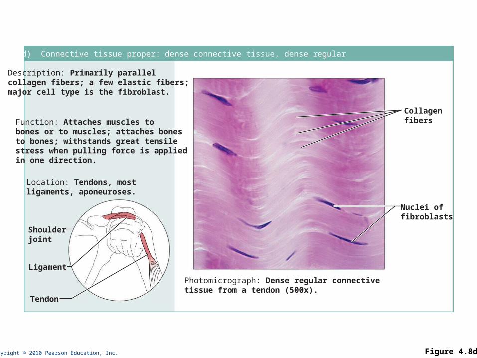
(d) Connective tissue proper: dense connective tissue, dense regular
Description: Primarily parallelcollagen fibers; a few elastic fibers;major cell type is the fibroblast.
Function: Attaches muscles tobones or to muscles; attaches bonesto bones; withstands great tensilestress when pulling force is appliedin one direction.
Location: Tendons, mostligaments, aponeuroses.
Photomicrograph: Dense regular connectivetissue from a tendon (500x).
Shoulderjoint
Ligament
Tendon
Collagenfibers
Nuclei offibroblasts

Copyright © 2010 Pearson Education, Inc. Figure 4.8e
(e) Connective tissue proper: dense connective tissue, dense irregular
Description: Primarilyirregularly arranged collagenfibers; some elastic fibers;major cell type is the fibroblast.
Function: Able to withstandtension exerted in manydirections; provides structuralstrength.
Location: Fibrous capsules oforgans and of joints; dermis ofthe skin; submucosa ofdigestive tract.
Photomicrograph: Dense irregularconnective tissue from the dermis of theskin (400x).
Collagenfibers
Nuclei offibroblasts
Fibrousjointcapsule
Copyright © 2010 Pearson Education, Inc. Figure 4.8f
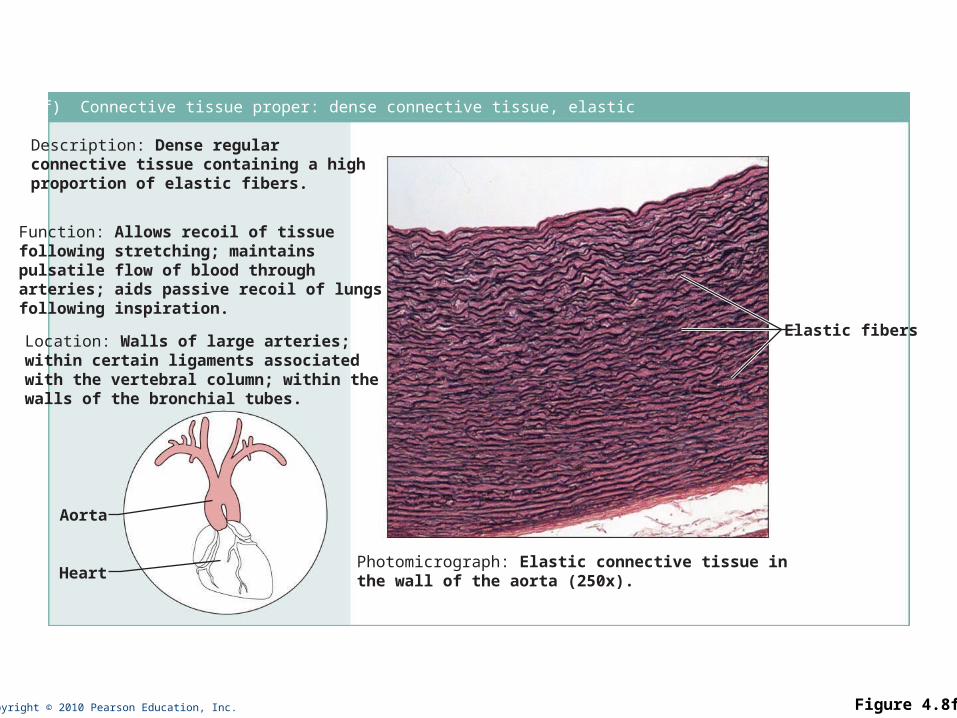
(f) Connective tissue proper: dense connective tissue, elastic
Description: Dense regularconnective tissue containing a highproportion of elastic fibers.
Function: Allows recoil of tissuefollowing stretching; maintainspulsatile flow of blood througharteries; aids passive recoil of lungsfollowing inspiration.
Location: Walls of large arteries;within certain ligaments associatedwith the vertebral column; within thewalls of the bronchial tubes.
Elastic fibers
Aorta
HeartPhotomicrograph: Elastic connective tissue inthe wall of the aorta (250x).
Copyright © 2010 Pearson Education, Inc.
Connective Tissue: Cartilage
• Three types of cartilage:
• Hyaline cartilage
• Elastic cartilage
• Fibrocartilage

Copyright © 2010 Pearson Education, Inc. Figure 4.8g
(g) Cartilage: hyaline
Description: Amorphous but firmmatrix; collagen fibers form animperceptible network; chondroblastsproduce the matrix and when mature(chondrocytes) lie in lacunae.
Function: Supports and reinforces;has resilient cushioning properties;resists compressive stress.
Location: Forms most of theembryonic skeleton; covers the endsof long bones in joint cavities; formscostal cartilages of the ribs; cartilagesof the nose, trachea, and larynx.
Photomicrograph: Hyaline cartilage from thetrachea (750x).
Costalcartilages
Chondrocytein lacuna
Matrix
Copyright © 2010 Pearson Education, Inc. Figure 4.8h
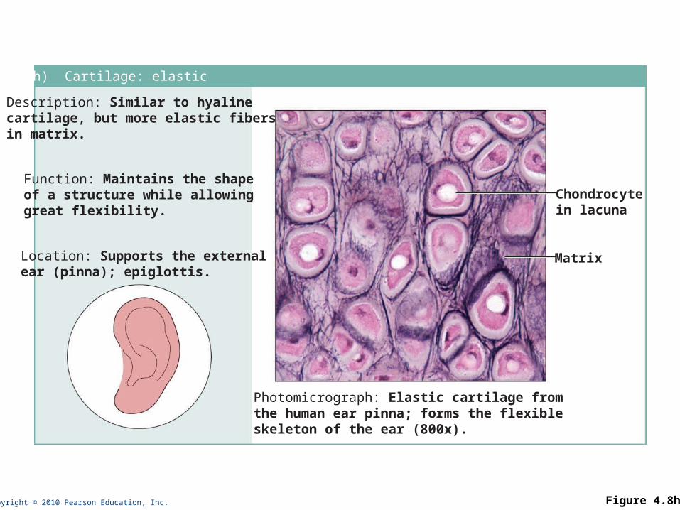
(h) Cartilage: elastic
Description: Similar to hyalinecartilage, but more elastic fibersin matrix.
Function: Maintains the shapeof a structure while allowinggreat flexibility.
Location: Supports the externalear (pinna); epiglottis.
Photomicrograph: Elastic cartilage fromthe human ear pinna; forms the flexibleskeleton of the ear (800x).
Chondrocytein lacuna
Matrix
Copyright © 2010 Pearson Education, Inc. Figure 4.8i
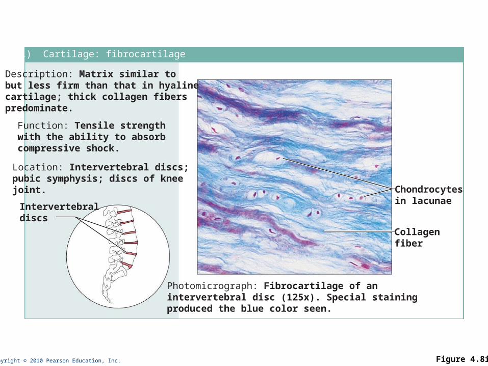
(i) Cartilage: fibrocartilage
Description: Matrix similar tobut less firm than that in hyalinecartilage; thick collagen fiberspredominate.
Function: Tensile strengthwith the ability to absorbcompressive shock.
Location: Intervertebral discs;pubic symphysis; discs of kneejoint.
Photomicrograph: Fibrocartilage of anintervertebral disc (125x). Special stainingproduced the blue color seen.
Intervertebraldiscs
Chondrocytesin lacunae
Collagenfiber

Copyright © 2010 Pearson Education, Inc. Figure 4.8j
(j) Others: bone (osseous tissue)
Description: Hard, calcifiedmatrix containing many collagenfibers; osteocytes lie in lacunae.Very well vascularized.
Function: Bone supports andprotects (by enclosing);provides levers for the musclesto act on; stores calcium andother minerals and fat; marrowinside bones is the site for bloodcell formation (hematopoiesis).Location: Bones
Photomicrograph: Cross-sectional viewof bone (125x).
Lacunae
Lamella
Centralcanal

Copyright © 2010 Pearson Education, Inc. Figure 4.8k
(k) Others: blood
Description: Red and whiteblood cells in a fluid matrix(plasma).
Function: Transport ofrespiratory gases, nutrients,wastes, and other substances.
Location: Contained withinblood vessels.
Photomicrograph: Smear of human blood (1860x); twowhite blood cells (neutrophil in upper left and lymphocytein lower right) are seen surrounded by red blood cells.
Neutrophil
Red bloodcells
Lymphocyte
Plasma
Copyright © 2010 Pearson Education, Inc.
Nervous Tissue
•Nervous system (more detail with the Nervous System)

Copyright © 2010 Pearson Education, Inc. Figure 4.9
Photomicrograph: Neurons (350x)
Function: Transmit electricalsignals from sensory receptorsand to effectors (muscles andglands) which control their activity.
Location: Brain, spinalcord, and nerves.
Description: Neurons arebranching cells; cell processesthat may be quite long extend fromthe nucleus-containing cell body;also contributing to nervous tissueare nonirritable supporting cells(not illustrated).
Dendrites
Neuron processes Cell body
Axon
Nuclei ofsupportingcells
Cell bodyof a neuron
Neuronprocesses
Nervous tissue
Copyright © 2010 Pearson Education, Inc.
Muscle Tissue
• Skeletal muscle
• Cardiac muscle
• Smooth muscle
• (more detail with the Muscular System)
Copyright © 2010 Pearson Education, Inc. Figure 4.10a
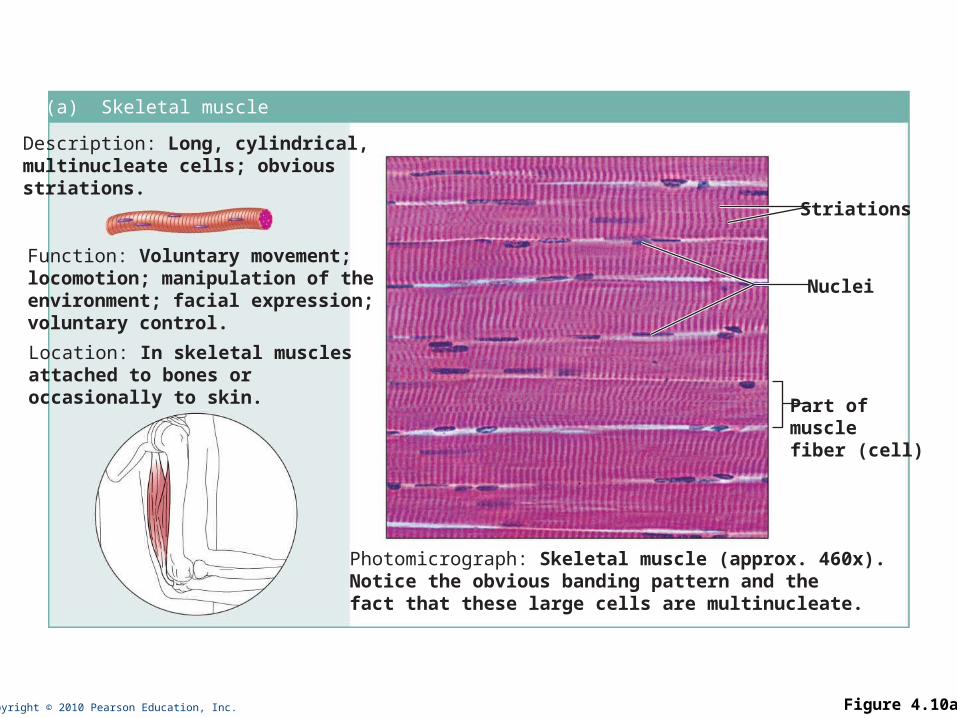
(a) Skeletal muscle
Description: Long, cylindrical,multinucleate cells; obviousstriations.
Function: Voluntary movement;locomotion; manipulation of theenvironment; facial expression;voluntary control.
Location: In skeletal musclesattached to bones oroccasionally to skin.
Photomicrograph: Skeletal muscle (approx. 460x).Notice the obvious banding pattern and thefact that these large cells are multinucleate.
Nuclei
Striations
Part ofmuscle fiber (cell)

Copyright © 2010 Pearson Education, Inc. Figure 4.10b
(b) Cardiac muscle
Description: Branching, striated, generally uninucleate cells that interdigitate atspecialized junctions (intercalated discs).
Function: As it contracts, it propels blood into the circulation; involuntary control.Location: The walls of the heart.
Photomicrograph: Cardiac muscle (500X);notice the striations, branching of cells, andthe intercalated discs.
Intercalateddiscs
Striations
Nucleus
Copyright © 2010 Pearson Education, Inc. Figure 4.10c
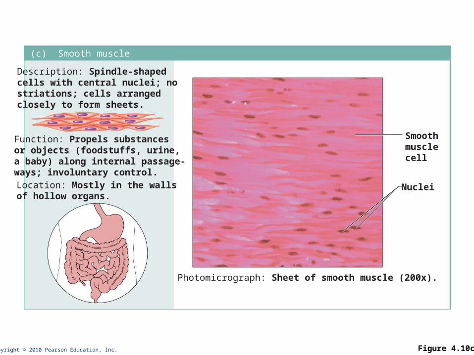
(c) Smooth muscle
Description: Spindle-shapedcells with central nuclei; nostriations; cells arranged closely to form sheets.
Function: Propels substancesor objects (foodstuffs, urine,a baby) along internal passage-ways; involuntary control.Location: Mostly in the wallsof hollow organs.
Photomicrograph: Sheet of smooth muscle (200x).
Smoothmusclecell
Nuclei
Copyright © 2010 Pearson Education, Inc.
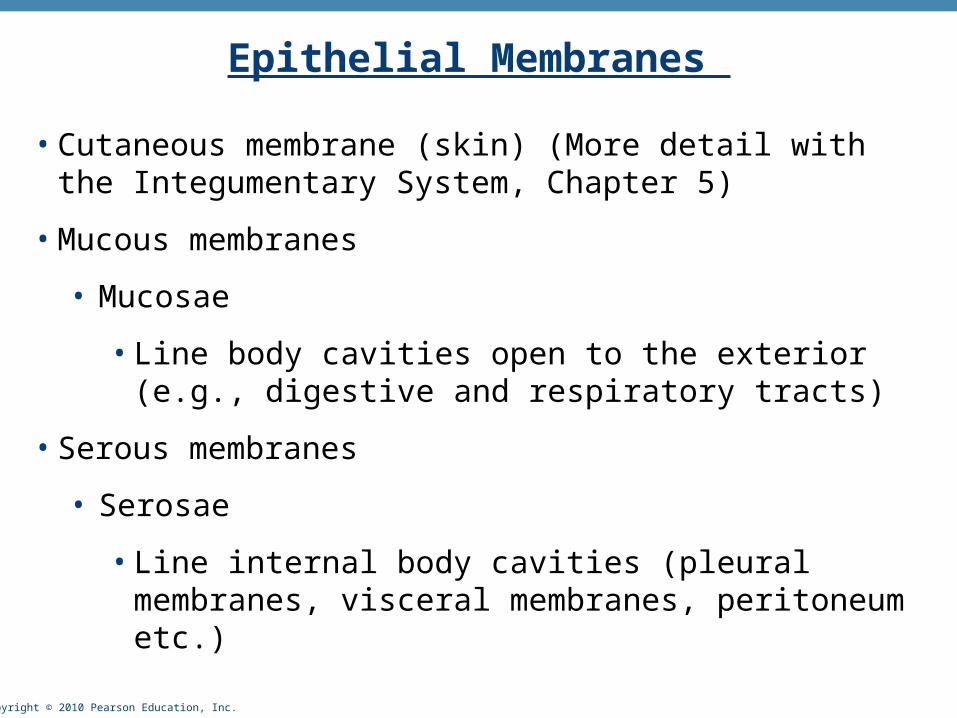
Epithelial Membranes
• Cutaneous membrane (skin) (More detail with the Integumentary System, Chapter 5)
• Mucous membranes
• Mucosae
• Line body cavities open to the exterior (e.g., digestive and respiratory tracts)
• Serous membranes
• Serosae
• Line internal body cavities (pleural membranes, visceral membranes, peritoneum etc.)
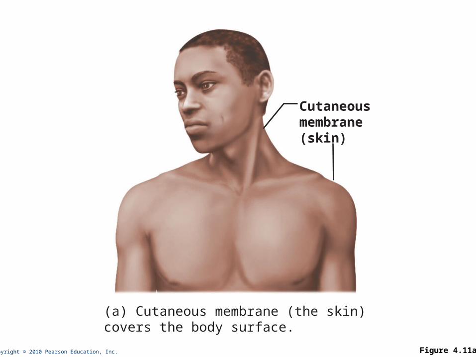
Copyright © 2010 Pearson Education, Inc. Figure 4.11a
Cutaneousmembrane(skin)
(a) Cutaneous membrane (the skin)covers the body surface.

Copyright © 2010 Pearson Education, Inc. Figure 4.11b
Mucosa ofnasal cavity
Mucosa oflung bronchi
Mucosa ofmouth
Esophaguslining
(b) Mucous membranes line body cavitiesopen to the exterior.

Copyright © 2010 Pearson Education, Inc. Figure 4.11c
Parietalpericardium
Visceralpericardium
(c) Serous membranes line body cavitiesclosed to the exterior.
Parietalperitoneum
Visceralperitoneum
ParietalpleuraVisceralpleura

Copyright © 2010 Pearson Education, Inc.
Tissue Injury
• Tissue injury = penetration of one of the bodies primary defense barriers (skin & mucosae)
• Injury triggers two types of responses:
• Inflammation: rapid, but not specific
• Immune: slower, but specific
• Tissue repair involves two major processes:
• Regeneration: replace damaged tissue with the same type of tissue
• Fibrosis: production of fibrous connective tissue called scar tissue
• Which process occurs depends on the location and severity of the tissue damage
Copyright © 2010 Pearson Education, Inc.
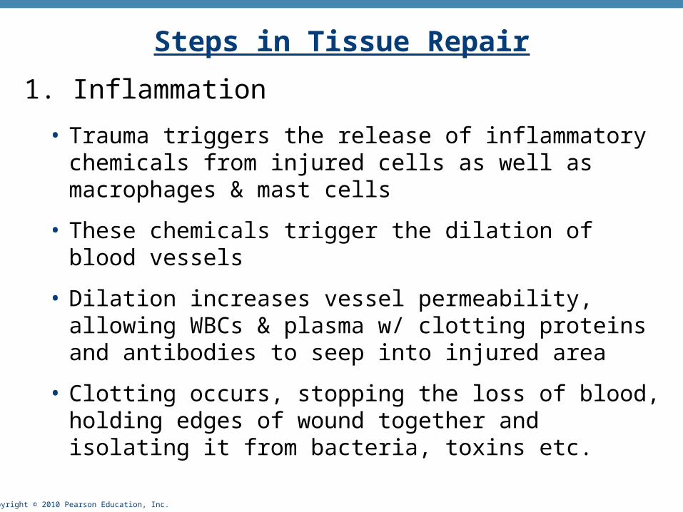
Steps in Tissue Repair
1. Inflammation
• Trauma triggers the release of inflammatory chemicals from injured cells as well as macrophages & mast cells
• These chemicals trigger the dilation of blood vessels
• Dilation increases vessel permeability, allowing WBCs & plasma w/ clotting proteins and antibodies to seep into injured area
• Clotting occurs, stopping the loss of blood, holding edges of wound together and isolating it from bacteria, toxins etc.

Copyright © 2010 Pearson Education, Inc. Figure 4.12, step 1
Scab
Blood clot inincised wound
Epidermis
Vein
Inflammatorychemicals
Inflammation sets the stage:• Severed blood vessels bleed and inflammatory chemicals arereleased.
• Local blood vessels become more permeable, allowing whiteblood cells, fluid, clotting proteins and other plasma proteinsto seep into the injured area.
• Clotting occurs; surface dries and forms a scab.
Migrating whiteblood cell
Artery
1

Copyright © 2010 Pearson Education, Inc.
Steps in Tissue Repair2. Organization and restored blood supply (beginning of actual repair)
• The blood clot is replaced with granulation tissue, which lays down a new capillary bed
• Fibroblasts within the granulation tissue produce growth factors, collagen fibers & contractile proteins to pull edges of wound together
• Epithelium begins to regenerate
• Debris is phagocytized by macrophages and undelying fibrous patch becomes scar tissue resistant to infection because it produces bacteria inhibiting fibers

Copyright © 2010 Pearson Education, Inc. Figure 4.12, step 2
Regeneratingepithelium
Area ofgranulationtissueingrowth
FibroblastMacrophage
Organization restores the blood supply:• The clot is replaced by granulation tissue, which restoresthe vascular supply.
• Fibroblasts produce collagen fibers that bridge the gap.• Macrophages phagocytize cell debris.• Surface epithelial cells multiply and migrate over thegranulation tissue.
2
Copyright © 2010 Pearson Education, Inc.
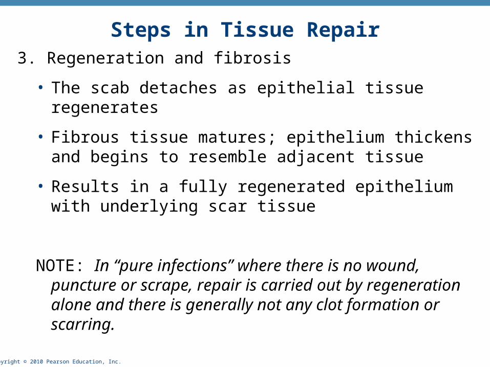
Steps in Tissue Repair3. Regeneration and fibrosis
• The scab detaches as epithelial tissue regenerates
• Fibrous tissue matures; epithelium thickens and begins to resemble adjacent tissue
• Results in a fully regenerated epithelium with underlying scar tissue
NOTE: In “pure infections” where there is no wound, puncture or scrape, repair is carried out by regeneration alone and there is generally not any clot formation or scarring.
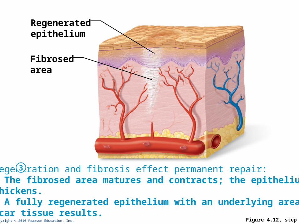
Copyright © 2010 Pearson Education, Inc. Figure 4.12, step 3
Regeneratedepithelium
Fibrosedarea
Regeneration and fibrosis effect permanent repair:• The fibrosed area matures and contracts; the epitheliumthickens.• A fully regenerated epithelium with an underlying area ofscar tissue results.
3
Copyright © 2010 Pearson Education, Inc.
Developmental Aspects of Tissues
• There are three primary germ layers: ectoderm, mesoderm, and endoderm
• Formed early in embryonic development (by the end of the second month)
• Specialize to form the four primary tissues
• Nerve tissue arises from ectoderm (mitosis basically stops after birth)
• Muscle and connective tissues arise from mesoderm (continue to divide after both)
• Epithelial tissues arise from all three germ layers (continue to divide after both)
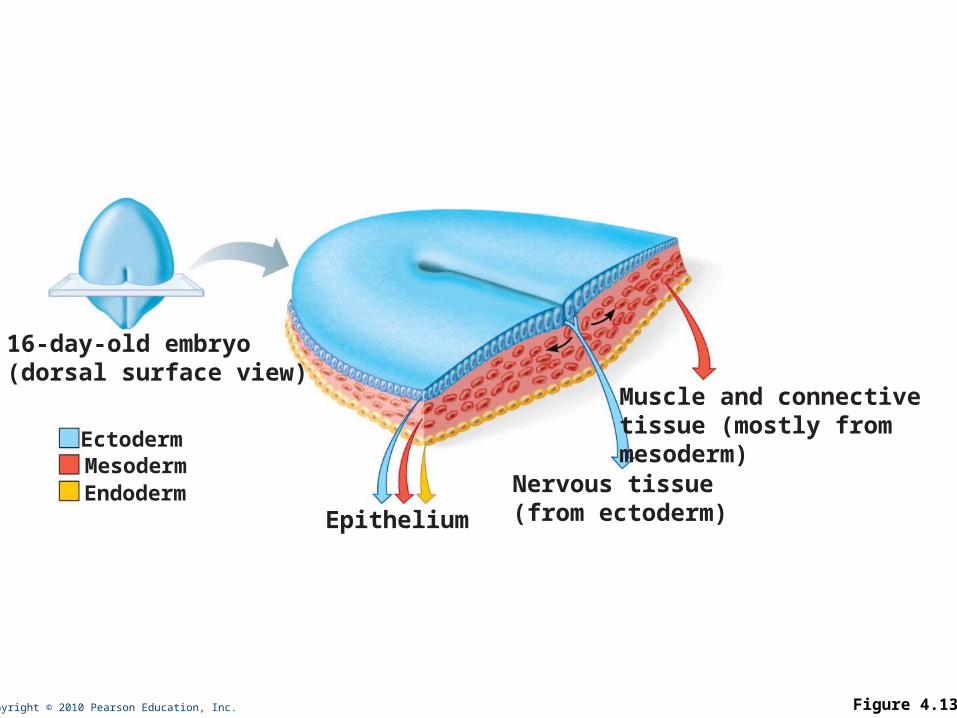
Copyright © 2010 Pearson Education, Inc. Figure 4.13
MesodermEndoderm
16-day-old embryo(dorsal surface view)
Epithelium
Nervous tissue(from ectoderm)
Muscle and connectivetissue (mostly frommesoderm)
Ectoderm


