37
pyright © 2010 Pearson Education, Inc. TISSUES Ch. 3b
| Date post: | 03-Jan-2016 |
| Category: |
Documents |
| Upload: | edwina-wilson |
| View: | 215 times |
| Download: | 1 times |

Copyright © 2010 Pearson Education, Inc.
TISSUESCh. 3b

Copyright © 2010 Pearson Education, Inc.
Tissues
• There are four types of tissue in the body
• Epithelial tissue
• Connective tissue
• Muscle tissue
• Nerve tissue
Copyright © 2010 Pearson Education, Inc.
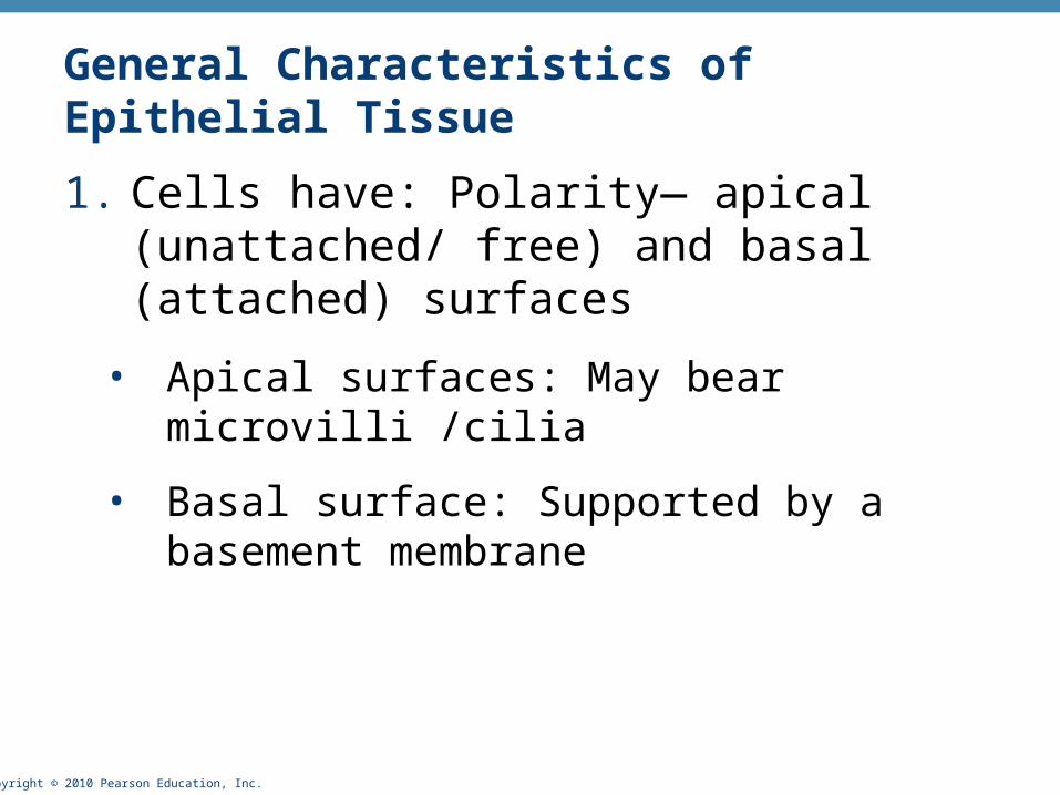
General Characteristics of Epithelial Tissue
1. Cells have: Polarity— apical (unattached/ free) and basal (attached) surfaces
• Apical surfaces: May bear microvilli /cilia
• Basal surface: Supported by a basement membrane

Copyright © 2010 Pearson Education, Inc.
Characteristics of Epithelial Tissue
2. Composed of closely packed cells
3. Avascular
4. High rate of regeneration

Copyright © 2010 Pearson Education, Inc. Figure 4.2a
Stratified
Simple
Apical surface
Basal surface
Apical surface
Basal surface

Copyright © 2010 Pearson Education, Inc.
Classification of Epithelia
• Ask two questions:
1. How many cell layers?
1 = simple epithelium
>1 = stratified epithelium

Copyright © 2010 Pearson Education, Inc.
Classification of Epithelia
2. What type of cell?
• Squamous = flat
• Cuboidal
• Columnar
• # of cell layers followed by cell shape = epithelial classification
• If E.T. is stratified, name according to the top layers of cells
Copyright © 2010 Pearson Education, Inc. Figure 4.3a
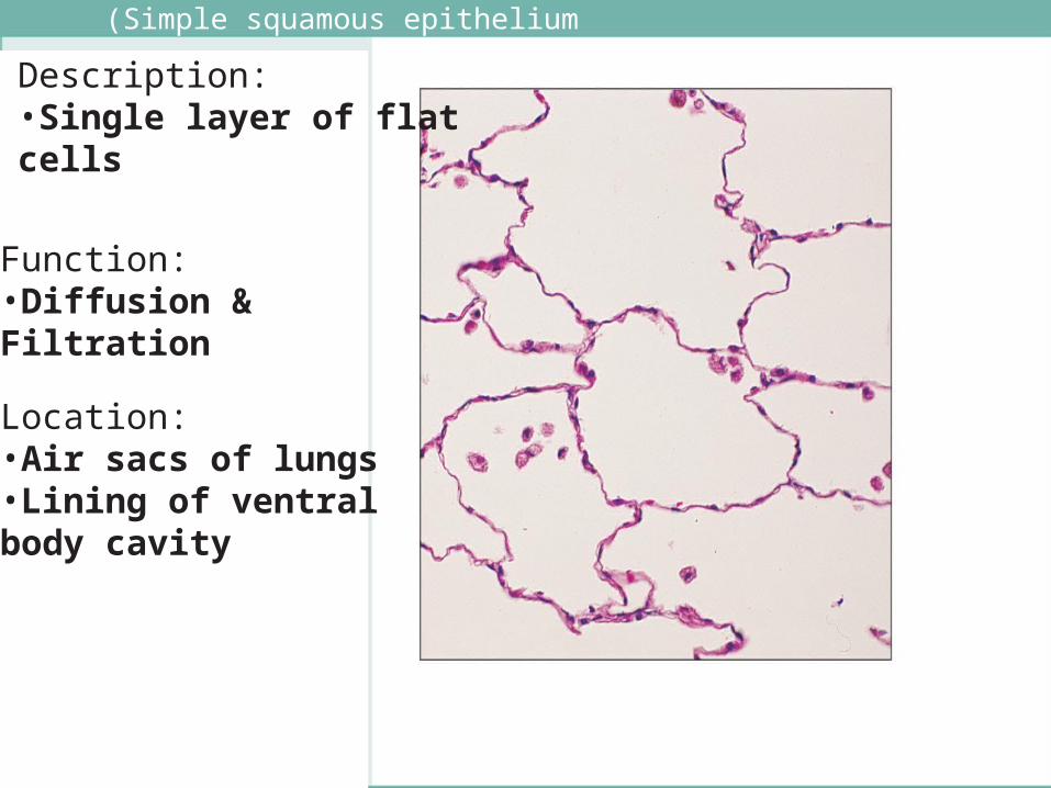
(Simple squamous epithelium
Description: •Single layer of flat cells
Function: •Diffusion & Filtration
Location: •Air sacs of lungs•Lining of ventral body cavity

Copyright © 2010 Pearson Education, Inc.
( Simple Cuboidal epithelium
Location: Glands and ducts
Function: Secretion andabsorption.
Description: Single layer of cubelike cells

Copyright © 2010 Pearson Education, Inc.
(Simple Columnar epithelium
Description: Single layer of tall cells•May have goblet cells
Function: Absorption and secretion
Location: Lines most of digestive tract

Copyright © 2010 Pearson Education, Inc.
Pseudostratified Ciliated Columnar epithelium
Trachea
Description: Single layer of cells of differing heights on same basement membrane; •May have goblet cells
Function: Secretion, propulsion of mucus byciliary action.
Location: upper respiratory tract.

Copyright © 2010 Pearson Education, Inc.
Stratified squamous epithelium
Description: Thick, surface cells squamous
•Two types:• Non-Keratinized: alive • Keratinized: surface cells dead & full of keratin
Function: Protection in areas subjected to abrasion.
Location: Nonkeratinized: Lines cavities which open to exterior•Keratinized: forms epidermis

Copyright © 2010 Pearson Education, Inc. Figure 4.3f
Transitional epithelium
Description: Basal cells cuboidal/columnar •Surface cells dome shaped or squamous (depends on degree of stretch)
Function: Stretches readily
Location: Lines hollow urinary organs

Copyright © 2010 Pearson Education, Inc.
Characteristics of Connective Tissue
• Connective Tissues are the most abundant and widely distributed tissue type
• C.T. has varying degrees of vascularity
• C.T. has cells separated by nonliving extracellular matrix (ground substance and fibers)

Copyright © 2010 Pearson Education, Inc.
Components of Extracellular Matrix• Ground substance
• Interstitial fluid, Adhesion proteins (“glue”),
• Large polysaccharides
• Three Types of Fibers can be found within the ground substance:
• Collagen
• Strongest, most abundant type; Provides tensile strength; form thick cables
• Elastic
• Long, thin, fibers; allow for stretch
• Reticular
• Short, fine, branched fibers that form an internal network (mesh) that is supportive to other cells

Copyright © 2010 Pearson Education, Inc.
Cells of Connective Tissue
• Cell Types
• “blasts” = Mitotically active and secretory cells
• “cytes” = Mature cells
• Fibroblasts in connective tissue proper
• Chondroblasts and chondrocytes in cartilage
• Osteoblasts and osteocytes in bone
• Hematopoietic stem cells in bone marrow

Copyright © 2010 Pearson Education, Inc. Figure 4.8j
Osseous tissue
Description: Hard, calcifiedmatrix; osteocytes in lacunae;
Function: Support, protection, attachment site for muscles.
Location: Bones
Lacunae
Lamella
Centralcanal

Copyright © 2010 Pearson Education, Inc.
Connective Tissue: Cartilage
• Three types of cartilage:
• Hyaline cartilage
• Elastic cartilage
• Fibrocartilage

Copyright © 2010 Pearson Education, Inc.
Cartilage: HyalineDescription: most abundant of cartilage types;less hard and more flexible than bone;Chondrocytes in lacunae.
Function: Supports,cushions, resists compressive stress.
Location: Forms most of embryonic skeleton; articular cartilage; cartilages of nose, trachea, larynx.
Costalcartilages
Chondrocytein lacuna
Matrix

Copyright © 2010 Pearson Education, Inc.
Cartilage: Elastic
Function: Also allowsgreat flexibility.
Location: Ear, epiglottis.
Chondrocytein lacuna
Matrix
Copyright © 2010 Pearson Education, Inc.
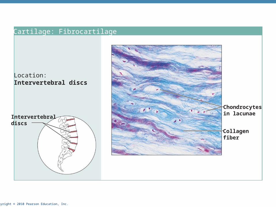
Cartilage: Fibrocartilage
Location: Intervertebral discs
Intervertebraldiscs
Chondrocytesin lacunae
Collagenfiber

Copyright © 2010 Pearson Education, Inc.
Connective tissue proper: Dense regular connective tissue
Description: Parallel collagen fibers
Function: Withstands tensilestress when pulling force is applied in one direction.
Location: Tendons, mostligaments
Shoulderjoint
Ligament
Tendon
Collagenfibers
Nuclei offibroblasts

Copyright © 2010 Pearson Education, Inc.
Connective tissue proper: Dense irregular connective tissueDescription: Irregularly arranged collagen fibers
Function: Withstandstension exerted in manydirections
Location: Fibrous capsules of organs ,joints; dermis ofskin Collagen
fibers
Nuclei offibroblasts
Fibrousjointcapsule

Copyright © 2010 Pearson Education, Inc.
Connective tissue proper: Areolar connective tissue Description: Most widely distributed variety of CT•Soft, pliable, like cobwebs
Function: Wraps and cushionsorgans
Location: Under epithelia of body, universal packaging material
Epithelium
Laminapropria
Fibroblastnuclei
Elasticfibers
Collagenfibers

Copyright © 2010 Pearson Education, Inc.
Connective tissue proper: Adipose connective tissue
Description: Closely packed adipocytes
Function: Reserve fuel insulation; supports & protects organs.
Location: Hypodermis; around kidneys and eyeballs; in abdomen; breasts.
Nucleus offat cell
Vacuolecontainingfat droplet
Adiposetissue
Mammaryglands

Copyright © 2010 Pearson Education, Inc.
Connective tissue proper: Reticular connective tissue
Description: Network of reticular fibers
Function: Fibers form a soft internal skeleton that supports other cells
Location: Lymphoid organs (lymph nodes, bone marrow, and spleen).
Spleen
White bloodcell(lymphocyte)
Reticularfibers

Copyright © 2010 Pearson Education, Inc.
Others: bloodDescription: Red and whiteblood cells in a fluid matrix(plasma).
Function: Transport ofrespiratory gases, nutrients,wastes, and other substances.
Location: Contained withinblood vessels.
Neutrophil
Red bloodcells
Lymphocyte
Plasma

Copyright © 2010 Pearson Education, Inc.
MUSCLE TISSUE

Copyright © 2010 Pearson Education, Inc.
(a) Skeletal muscleDescription: Long, cylindrical,multinucleate cells; obviousstriations.
Function: Voluntary movement
Location: Skeletal musclesattached to bones oroccasionally to skin.
Photomicrograph: (approx. 460x).
Nuclei
Striations
Part ofmuscle fiber (cell)
Copyright © 2010 Pearson Education, Inc.
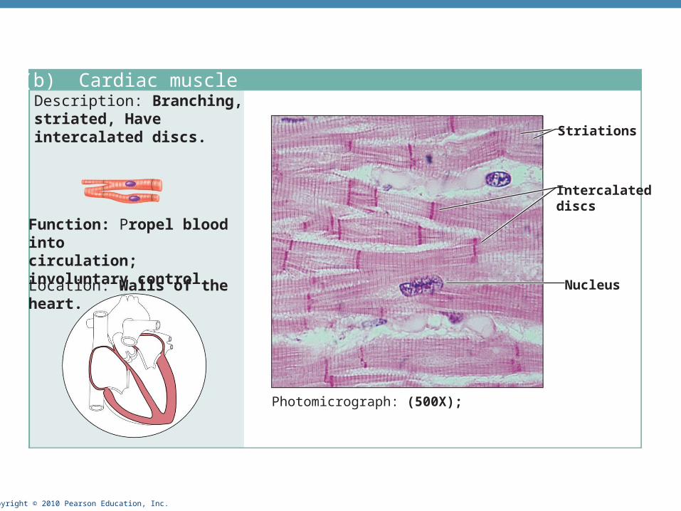
(b) Cardiac muscleDescription: Branching, striated, Have intercalated discs.
Function: Propel blood into circulation; involuntary control.
Location: Walls of the heart.
Photomicrograph: (500X);
Intercalateddiscs
Striations
Nucleus

Copyright © 2010 Pearson Education, Inc.
(c) Smooth muscleDescription: Spindle-shapedCells; no striations; cells form sheets.
Function: Propels substancesor objects; involuntary control.
Location: Mostly in wallsof hollow organs.
Photomicrograph: Sheet of smooth muscle (200x).
Smoothmusclecell
Nuclei

Copyright © 2010 Pearson Education, Inc.
NERVOUS TISSUE

Copyright © 2010 Pearson Education, Inc.
Function: Transmit electricalsignals from sensory receptorsto effectors.
Location: Brain, spinalcord, and nerves.
Description: Neurons w/ cell processes that extend from the cell body; Contains supporting cells
Dendrites
Neuron processes Cell body
Axon
Nuclei ofsupportingcells
Cell bodyof a neuron
Neuronprocesses
Nervous tissue

Copyright © 2010 Pearson Education, Inc.
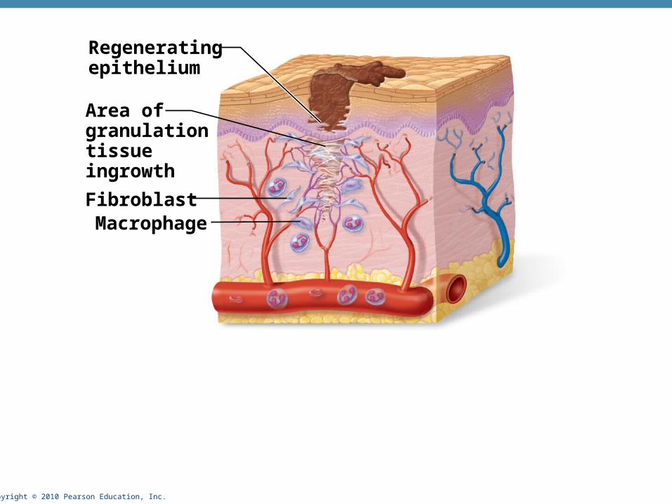
Steps in Wound Repair• Inflammation
• Increased blood flow with healing components & clotting factors to damaged area (redness, swelling, pain, heat) due to the release of inflammatory chemicals
• Clot forms and exposed portion forms scab
• Organization and restored blood supply
• Granulation tissue forms=new capillaries and phagocytes and fibroblasts
• Regeneration and fibrosis
• Surface ET regenerates, scab detaches, underlying scar tissue

Copyright © 2010 Pearson Education, Inc.
Scab
Blood clot inincised wound
Epidermis
Inflammatorychemicals
Migrating whiteblood cell
Artery
Copyright © 2010 Pearson Education, Inc.
Regeneratingepithelium
Area ofgranulationtissueingrowth
FibroblastMacrophage

Copyright © 2010 Pearson Education, Inc.
Regeneratedepithelium
Fibrosedarea


