Laboratoire de Virologie, Département des Agents Infectieux, Pôle Biologie, Centre Hospitalier Universitaire Grenoble, Grenoble, Francea; Unit of Virus-Host CellInteractions, UMI 3265, UJF-EMBL-CNRS, Grenoble, Franceb; Laboratoire CERBA, Cergy-Pontoise, Francec; Laboratoire de Virologie, Hôpital Pitié-Salpêtrière, AssistancePublique, Université Pierre et Marie Curie, Paris, Franced
For optimal antiviral therapy, the hepatitis C virus (HCV) genotype needs to be determined, as it remains a strong predictor ofsustained viral response. In this study, we assessed the number of HCV genotyping results that could not be determined usingthe commercially available line probe assay (LiPA) (Versant hepatitis C virus genotype 2.0 assay) in a large international panel ofsamples from 9,874 HCV-positive patients. In-house sequencing assays targeting the 5= untranslated region (UTR), core region,NS3 region, and NS5B region of the HCV genome and phylogenetic analyses were used to resolve these LiPA failures. Among allcases, the genotypes of 51 samples (0.52%) could not be determined with the LiPA. These undetermined results were observedmore frequently among samples from non-European regions (mainly the Arabian Peninsula). The use of sequencing assays cou-pled with phylogenetic analysis provided reliable genotype results for 86% of the LiPA failures, which exhibited higher rates ofgenotypes 4, 5, and 6 than did LiPA-resolved genotypes. As expected, the 5= UTR was not sufficiently variable for clear discrimi-nation between genotypes 1 and 6, but it also resulted in errors in classification of some genotype 3 and 4 cases using well-knownWeb-based BLAST programs. This study demonstrates the low frequency of genotyping failures with the Versant hepatitis Cvirus genotype 2.0 assay (LiPA) and also underlines the need for a complex combination of sequences and phylogenetic analysesin order to genotype these particular HCV strains correctly.
Hepatitis C virus (HCV) infection is a leading cause of chronicliver disease and affects approximately 120 million to 210
million people worldwide (1, 2). Each year, over 250,000 peopledie from HCV-related chronic liver diseases, such as end-stagecirrhosis and hepatocellular carcinoma (3, 4). Most infectionswith HCV can be cured if treatment is available, and the emer-gence of new antiviral drugs that directly target HCV will greatlyimprove treatment outcomes.
The HCV genome is characterized by extremely high sequencediversity and HCV strains are classified into genetically distinctgroups, which are known as genotypes when differences at thenucleotide level range from 31% to 33% or as subtypes whendifferences range from 20% to 25%; genetic difference below thesevalues define quasispecies (5–7). The HCV genotype (and to alesser extent, the subtype) must be determined prior to initiationof antiviral treatment because the genotype affects the choice ofagents and the duration of therapy, as well as the prognosis foreradicating the virus (8, 9). HCV typing and subtyping can beperformed using various methods, including direct sequenceanalysis, reverse hybridization, and genotype-specific reversetranscription (RT)-PCR. Several regions of the HCV genome canbe analyzed to classify strains accurately into specific genotypes.The 5= untranslated region (UTR) is the region of choice for de-tecting and quantifying HCV RNA, due to its high level of conser-vation. For this reason, it often has been used by virological labo-ratories for routine genotyping of HCV, although it now has beenclearly demonstrated that it is difficult to distinguish genotype 6from genotype 1 and to distinguish subtypes within genotypes 1,2, and 3 in this region (10). However, nucleotide sequencing cou-pled with phylogenetic analysis of genomic regions that are morevarying, such as the core/E1 and NS5B regions, has been recom-mended for HCV genotyping in consensus proposals (7). The re-
verse-hybridization Versant HCV genotype 1.0 assay (line probeassay [LiPA]) (Bayer HealthCare, Eragny, France), which is basedon a 5= UTR segment, has been upgraded and improved in version2.0 by the addition of core sequence information (11). With thisupdated version, amplification failures were described for 1.5 to2.1% of cases and rates could be lowered after retesting but, ac-cording to those reports, 4.6% and 22.8% of results could not beresolved at the genotype and subtype levels, respectively (11, 12).The present study aims (i) to evaluate the number of LiPA (ver-sion 2.0) failures in a large panel of samples from Europe and fromother parts of the world and (ii) to investigate whether the geno-types of these “difficult-to-type” samples corresponded to partic-ular HCV strains that could be typed by using a classic sequencingapproach.
MATERIALS AND METHODSClinical samples. A total of 9,874 HCV genotype analyses of samples fromEurope and other parts of the world were performed by the CERBA lab-oratory between January 2011 and May 2012. Viral loads were mea-sured with the COBAS AmpliPrep/COBAS TaqMan HCV quantitativetest, version 1.0 (Roche Diagnostics, Meylan, France), and levelsneeded to be higher than 500 IU/ml for the genotype analysis to be
Received 1 March 2013 Returned for modification 28 March 2013Accepted 17 April 2013
performed. All blood samples were centrifuged shortly after collection,and plasma was stored at �80°C before being stored and shipped ondry ice.
Versant HCV genotype 2.0 assay (LiPA). A Versant HCV genotype2.0 assay (INNO-LiPA HCV 2.0) was performed for all samples accordingto the manufacturer’s instructions. RNA was isolated from 200 �l ofplasma by using an m2000sp instrument (Abbott Molecular, Rungis,France). Extracted RNA was resuspended in 70 �l of buffer. RT-PCR wasperformed in a single tube, producing two distinct biotinylated DNA frag-ments of 240 and 270 bp, representing the 5= UTR and the core region,respectively. After denaturation, the biotinylated DNA PCR productswere hybridized to membrane-bound oligonucleotide probes specific forthe 5= UTR and the core region of HCV genotypes 1 to 6. Each stripcontained three control lines (conjugate, amplification from the 5= UTR,and amplification from the core region) and 22 DNA probe lines contain-ing sequences specific for HCV genotypes 1 to 6. After automated hybrid-ization steps, all strips were dried and scanned; results were interpretedusing Bayer LiPA-Scan HCV software (Innogenetics, Ghent, Belgium),according to the currently valid interpretation chart. When the profileobtained did not match any of the reference patterns, the genotype wasconsidered indeterminate by the software. Frozen samples were sent to theGrenoble hospital virology laboratory for sequencing.
Sequencing methods. Total nucleic acids were extracted from 1 ml ofplasma using the generic protocol of the EasyMag system (bioMérieux,Marcy l’étoile, France) and eluted in a volume of 50 �l. RT-PCR amplifi-cations of the 5= UTR, the core region, and two different regions of NS5Bwere performed with a one-step protocol using the Qiagen OneStep RT-PCR kit (Qiagen, Courtaboeuf, France), following the manufacturer’s in-structions. The NS3 protease region was amplified as described by Besse etal., using a nested PCR when necessary (13). All of the primers used arelisted in Table 1. Bidirectional sequencing of the amplicons was then per-formed with a CEQ 2000XL system (Beckman Coulter, Roissy, France).
Phylogenetic analysis. Phylogenetic analysis was performed afteralignment of the 5= UTR, core region, NS3, and two amplified regions ofNS5B and then sequences were compared with reference sequences ofgenotypes 1 to 7 using the neighbor-joining method (17) in MEGA5 soft-ware (18). The reliability of the phylogenetic clustering was evaluatedusing bootstrap analysis with 1,000 replicates. Reference sequences iden-
tified by genotype or subtype, country of identification, strain name, andPubMed number were extracted from the European HCV database (http://euhcvdb.ibcp.fr/euHCVdb/).
BLAST analysis. BLAST searches were performed in the GenBankHCV genotyping database (http://www.ncbi.nlm.nih.gov/projects/genotyping/formpage.cgi), in the Los Alamos HCV sequence database(19) (http://hcv.lanl.gov/content/sequence/BASIC_BLAST/basic_blast.html), and in the hepatitis C viral database of the Viral BioinformaticsResource Center (VBRC) (http://www.hcvdb.org/blast.asp) for all ampli-fied regions. The Max Planck Institute Informatik Geno2Pheno database(http://www.geno2pheno.org/) also was used for the NS3 and NS5B re-gions.
Criteria for final decisions on HCV genotypes. The final decision toassign an HCV genotype was made preferentially according to the NS5Bsequencing results when the results of the four BLAST analyses and thephylogenetic analysis matched. When NS5B amplification could not beobtained or when discrepancies appeared in the results, 5= UTR, core, andNS3 sequences were used, and a minimum of six consistent BLAST resultsobtained for two different regions was required to assign an HCV geno-type.
Statistical analysis. Student’s t test was used to compare HCV viralloads. Independence between qualitative parameters was assessed usingthe �2 test.
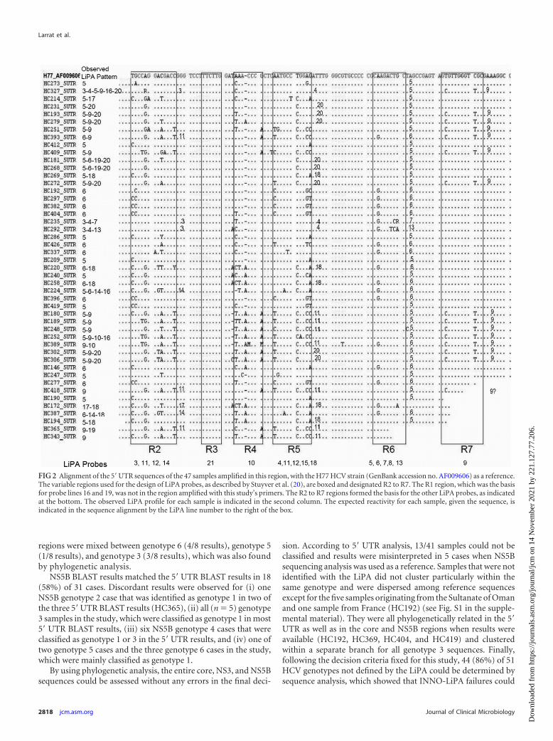
RESULTSVersant HCV genotype 2.0 assay (LiPA) failures. Between Janu-ary 2011 and May 2012, 51 (0.52%) of the 9,874 HCV genotypeanalyses performed in the CERBA laboratory using the LiPA couldnot be resolved because the results could not be interpreted.Twenty-one analyses corresponded to a single positive line in the5= UTR, 14 to a double-line profile, and the others to three or morelines (for example, see Fig. 1). Except for variable region 1, whichwas not sequenced with our primers, 5= UTR sequencing con-firmed most positive lines found with the LiPA (Fig. 2). For sevensamples, the sequencing results did not reveal a sequence thatperfectly matched the observed positive probes, possibly becausemixed infections were not detected. On the other hand, line 11 was
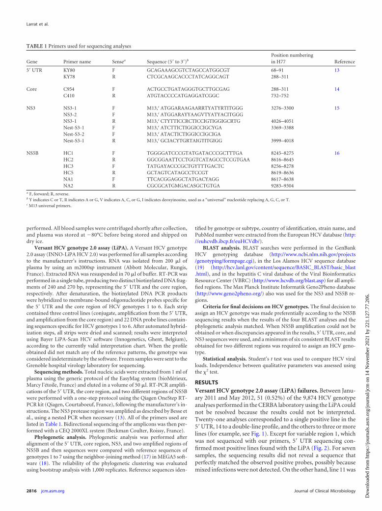
TABLE 1 Primers used for sequencing analyses
Gene Primer name Sensea Sequence (5= to 3=)b
Position numberingin H77 Reference
5= UTR KY80 F GCAGAAAGCGTCTAGCCATGGCGT 68–91 13KY78 R CTCGCAAGCACCCTATCAGGCAGT 288–311
Core C954 F ACTGCCTGATAGGGTGCTTGCGAG 288–311 14C410 R ATGTACCCCATGAGGATCGGC 732–752
NS3 NS3-1 F M13,c ATGGARAAGAARRTYATYRTITGGG 3276–3300 15NS3-2 F M13,c ATGGARAYYAAGVTYATYACITGGGNS3-1 R M13,c CTYTTICCRCTICCIGTIGGIGCRTG 4026–4051Nest-S3-1 F M13,c ATCTTICTIGGICCIGCYGA 3369–3388Nest-S3-2 F M13,c ATACTICTIGGICCIGCIGANest-S3-1 R M13,c GCIACYTGRTAIGTITGIGG 3999–4018
NS5B HC1 F TGGGGATCCCGTATGATACCCGCTTTGA 8245–8275 16HC2 R GGCGGAATTCCTGGTCATAGCCTCCGTGAA 8616–8645HC3 F TATGAYACCCGCTGYTTTGACTC 8256–8278HC5 R GCTAGTCATAGCCTCCGT 8619–8636NA1 F TTCACGGAGGCTATGACYAGG 8617–8638NA2 R CGCGCATGMGACASGCTGTGA 9283–9304
a F, forward; R, reverse.b Y indicates C or T, R indicates A or G, V indicates A, C, or G, I indicates deoxyinosine, used as a “universal” nucleotide replacing A, G, C, or T.c M13 universal primers.
never detected in this study’s samples, whereas sequencing resultsshowed that it should have been detected in eight cases. All of theseobserved profiles could not be interpreted using the manufactur-er’s interpretative chart. Forty-nine percent of these samples camefrom outside Europe, representing 0.61% of the foreign HCVgenotyping analyses conducted at the CERBA laboratory. The fail-ure rates were 0.45% and 0.27% for samples from Europe andMaghreb, respectively, whereas the rates were significantly higherfor samples from sub-Saharan Africa (1.66%) and the ArabianPeninsula (29.41%) (Table 2). Using LiPA-recommended proce-dures, no amplification failures occurred.
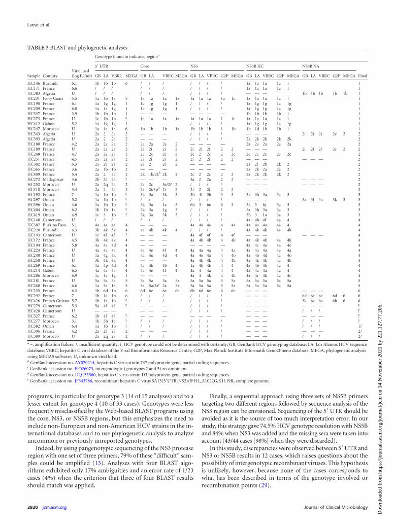
Amplification of the four different HCV genomic regions. Aspresented in Table 3, a total of 38 (74.5%) of the 51 failed LiPAsamples gave sequences that could be interpreted in the NS5Bregion, 29 (56%) with the first set of primers (HC1/HC2), 3 withthe second set of NS5B primers (HC3/HC5), and 6 with the thirdset of NS5B primers (NA1/NA2). Thirteen samples (25.5%) couldnot be correctly amplified in the NS5B region. Their mean viralload was 4.9 � 2.6 log IU/ml, compared with 5.7 � 2.7 log IU/mlfor samples amplified in NS5B (P � 0.064).
Forty-seven (98%) of 48 samples gave sequences that couldbe interpreted in the 5= UTR with the KY80/KY78 set ofprimers. For 6 of those samples, the 5= UTR was the only regionthat could be amplified and therefore the genotype could not beattributed with certainty. One sample could not be amplified
with these primers or with any others. Three samples werenot tested in the 5= UTR because not enough plasma wasavailable.
Twenty-five (61%) of 41 samples were successfully sequencedin the core region with the C954/C410 set of primers. Sixteensamples could not be amplified with these primers. Ten sampleswere not tested in the core region because not enough plasma wasavailable.
Twenty-three (79.3%) of 29 samples were successfully se-quenced in the NS3 protease region by employing the pange-notypic assay described by Besse et al. (13). Six samples couldnot be amplified with these primers. Twenty-two samples werenot tested in the NS3 region because not enough plasma wasavailable.
Sequence analysis. Concomitant amplification of the NS5Band core regions was obtained for 21 samples. At the genotypelevel, NS5B BLAST results perfectly matched the core results in 17(81%) of 21 cases and the results of phylogenetic analysis in 100%of cases (Table 3).
Eighteen samples could be amplified in the NS5B and NS3regions. Fifteen of them (83%) matched perfectly at the genotypelevel according to BLAST analysis. For two samples (HC192 andHC269), only one BLAST analysis of the four did not match in theNS3 region; in the third case (HC396), BLAST results for both
FIG 1 Four examples of LiPA strips and optical density graphs obtained with LiPA-Scan that cannot be interpreted with the manufacturer’s interpretative chart.(A) Single positive line 5 (sample HC273). (B) Single positive line 6 (sample HC192). (C) Three positive lines, 5, 9, and 20 (sample HC302). (D) Four positivelines, 5, 9, 10, and 16, and an absence of core amplification (sample HC252). The x axis corresponds to the lines of the different probes of the Inno-LiPA strip.y axis values are optical densities expressed in arbitrary units.
Sequencing of LiPA Failures
September 2013 Volume 51 Number 9 jcm.asm.org 2817
regions were mixed between genotype 6 (4/8 results), genotype 5(1/8 results), and genotype 3 (3/8 results), which was also foundby phylogenetic analysis.
NS5B BLAST results matched the 5= UTR BLAST results in 18(58%) of 31 cases. Discordant results were observed for (i) oneNS5B genotype 2 case that was identified as genotype 1 in two ofthe three 5= UTR BLAST results (HC365), (ii) all (n � 5) genotype3 samples in the study, which were classified as genotype 1 in most5= UTR BLAST results, (iii) six NS5B genotype 4 cases that wereclassified as genotype 1 or 3 in the 5= UTR results, and (iv) one oftwo genotype 5 cases and the three genotype 6 cases in the study,which were mainly classified as genotype 1.
By using phylogenetic analysis, the entire core, NS3, and NS5Bsequences could be assessed without any errors in the final deci-
sion. According to 5= UTR analysis, 13/41 samples could not beclassified and results were misinterpreted in 5 cases when NS5Bsequencing analysis was used as a reference. Samples that were notidentified with the LiPA did not cluster particularly within thesame genotype and were dispersed among reference sequencesexcept for the five samples originating from the Sultanate of Omanand one sample from France (HC192) (see Fig. S1 in the supple-mental material). They were all phylogenetically related in the 5=UTR as well as in the core and NS5B regions when results wereavailable (HC192, HC369, HC404, and HC419) and clusteredwithin a separate branch for all genotype 3 sequences. Finally,following the decision criteria fixed for this study, 44 (86%) of 51HCV genotypes not defined by the LiPA could be determined bysequence analysis, which showed that INNO-LiPA failures could
FIG 2 Alignment of the 5= UTR sequences of the 47 samples amplified in this region, with the H77 HCV strain (GenBank accession no. AF009606) as a reference.The variable regions used for the design of LiPA probes, as described by Stuyver et al. (20), are boxed and designated R2 to R7. The R1 region, which was the basisfor probe lines 16 and 19, was not in the region amplified with this study’s primers. The R2 to R7 regions formed the basis for the other LiPA probes, as indicatedat the bottom. The observed LiPA profile for each sample is indicated in the second column. The expected reactivity for each sample, given the sequence, isindicated in the sequence alignment by the LiPA line number to the right of the box.
occur with all genotypes but were found more frequently withgenotypes 4, 5, and 6 (Table 2).
DISCUSSION
In a large panel of patient samples from all over the world, theHCV genotype could not be determined using the LiPA in 0.52%of cases because the observed pattern was not included in themanufacturer’s interpretative chart; however, 86% of these LiPAfailures could be resolved using a sequencing approach with com-mon HCV sets of primers. These failures were mostly encounteredwith genotypes 4, 5, and 6 and in samples from sub-Saharan Africaand especially from the Arabian Peninsula. The number of sam-ples involved is too small to determine whether these factors werelinked or independent, however.
The first study using the second-generation line probe assay forthe 5= UTR already showed 11% undetermined LiPA profiles anda particularly high rate of subtyping errors (11% specificity) withsamples from West Africa (20). The same version of the INNO-LiPA HCV assay including core sequence hybridization was usedto evaluate HCV genotypes for 136 French blood donors and 326HCV-positive plasma samples in a Belgian study, which exhibited2 and 7 nonamplifiable samples, respectively, and no or 15 (4.6%)results that could not be clarified (11, 12). More recently, two casesof amplifiable samples giving uninterpretable LiPA results wereobserved in the Netherlands, and the authors underlined the closeresemblance of these two viral strains to viruses of genotypes 2 and3 originating from non-Western countries (21). The failure ofsome quantitative RT-PCR assays to detect or to amplify correctlyAfrican strains of HCV or HIV has been reported frequently (22–24), and the LiPA may suffer from the same type of bias.
These viral strains also are more difficult to amplify with theclassic NS5B primers. Even with the use of three sets of primerscovering two different regions, the percentage of samples ampli-
fied in the NS5B region in this study (74.5%) was lower than thevalues normally observed in our laboratory (90% with HC3/HC5primers during the same period of time) or described in otherstudies (82 to 97.3%) (10, 25). This could be explained by thegreater variety in the geographical origins of the viruses and theuse of primers not suitable for these peculiar strains. For thesesamples, the most conserved region, 5= UTR, exhibits a higheramplification rate (98%). Discrepancies between amplificationrates in the 5= UTR and NS5B region have been described and wereassociated mainly with lower levels of viremia and higher poly-morphism rates in the annealing regions of the primers (16, 25,26). In our study, the viral loads of the samples that could not beamplified in NS5B were not statistically smaller than those of am-plified samples, and genomic variability in the amplified regionappears to provide the most likely explanation for amplificationfailure. Errors in HCV genotype 6 classification using NS5B se-quencing were observed in Asia (27, 28). In our study, no geno-type 6 could be amplified with our two first sets of NS5B primersbut one could be resolved with core and NS3 amplifications andthe two others using another downstream region of NS5B. Asalready described, we found poor discrimination of genotype 6samples using only 5= UTR sequencing.
Phylogenetic analysis of these HCV strains exhibiting LiPAfailure showed that they were distributed among different geno-types but were difficult to subtype by NS3 and NS5B sequencephylogenetic analysis, probably due to the high variability in com-parison with the reference strains. For example, sequences ob-tained from the five samples from the Sultanate of Oman weregrouped, whatever the genomic region being considered, but wereclearly different from the genotype 3 reference strains.
Sequencing of the 5= UTR is well known to misclassify HCVsubtypes, and we now also demonstrate that it can lead to misclas-sification of genotypes using widely available Web-based BLAST
TABLE 2 Demographic and clinical features of the patients
programs, in particular for genotype 3 (14 of 15 analyses) and to alesser extent for genotype 4 (10 of 33 cases). Genotypes were lessfrequently misclassified by the Web-based BLAST programs usingthe core, NS3, or NS5B regions, but this emphasizes the need toinclude non-European and non-American HCV strains in the in-ternational databases and to use phylogenetic analysis to analyzeuncommon or previously unreported genotypes.
Indeed, by using pangenotypic sequencing of the NS3 proteaseregion with one set of three primers, 79% of these “difficult” sam-ples could be amplified (13). Analyses with four BLAST algo-rithms exhibited only 17% ambiguities and an error rate of 1/23cases (4%) when the criterion that three of four BLAST resultsshould match was applied.
Finally, a sequential approach using three sets of NS5B primerstargeting two different regions followed by sequence analysis of theNS3 region can be envisioned. Sequencing of the 5= UTR should beavoided as it is the source of too much interpretation error. In ourstudy, this strategy gave 74.5% HCV genotype resolution with NS5Band 84% when NS3 was added and the missing sera were taken intoaccount (43/44 cases [98%] when they were discarded).
In this study, discrepancies were observed between 5= UTR andNS3 or NS5B results in 12 cases, which raises questions about thepossibility of intergenotypic recombinant viruses. This hypothesisis unlikely, however, because none of the cases corresponds towhat has been described in terms of the genotype involved orrecombination points (29).
TABLE 3 BLAST and phylogenetic analyses
Sample CountryViral load(log IU/ml)
Genotype found in indicated regiona
5= UTR Core NS3 NS5B HC NS5B NA
FinalGB LA VBRC MEGA GB LA VBRC MEGA GB LA VBRC G2P MEGA GB LA VBRC G2P MEGA GB LA VBRC G2P MEGA
This study has some limitations. We assumed that the samplesfor which the INNO-LiPA provided a genotype were correctlyclassified, and results were not confirmed by sequencing analysis.In addition, problems genotyping HCV may result from mixedinfections in the patients’ plasma samples. Unfortunately, our se-quencing approach based on Sanger technology is not well suitedto addressing this point specifically.
In conclusion, with a low rate of HCV genotyping results thatcould not be resolved, reverse hybridization (the INNO-LiPA 2.0assay) remains a very convenient method in clinical practice, as itis produces a high rate of amplification and is relatively easy toperform. Special attention should be paid to samples from sub-Saharan Africa and the Arabian Peninsula and to genotypes 4, 5,and 6, which are less common in Western countries. Sequencingassays could be used to resolve uninterpretable LiPA results, butsequencing of multiple regions was needed to deal with more fre-quent amplification failure for these samples and the low discrim-inatory power of the 5= UTR sequences. For these difficult sam-ples, bioinformatic tools and particularly databases are crucial forinterpreting the sequences obtained.
ACKNOWLEDGMENT
We thank Linda Northrup for revision of the English manuscript.
REFERENCES1. Calvaruso V, Craxi A. 2012. 2011 European Association of the Study of
the Liver hepatitis C virus clinical practice guidelines. Liver Int. 32(Suppl1):2– 8.
2. Shepard CW, Finelli L, Alter MJ. 2005. Global epidemiology of hepatitisC virus infection. Lancet Infect. Dis. 5:558 –567.
3. Chevaliez S. 2011. Virological tools to diagnose and monitor hepatitis Cvirus infection. Clin. Microbiol. Infect. 17:116 –121.
4. El-Serag HB. 2012. Epidemiology of viral hepatitis and hepatocellularcarcinoma. Gastroenterology 142:1264 –1273.
5. Kieffer TL, Kwong AD, Picchio GR. 2010. Viral resistance to specificallytargeted antiviral therapies for hepatitis C (STAT-Cs). J. Antimicrob. Che-mother. 65:202–212.
6. Simmonds P. 1999. Viral heterogeneity of the hepatitis C virus. J. Hepatol.31(Suppl 1):54 – 60.
7. Simmonds P, Bukh J, Combet C, Deleage G, Enomoto N, Feinstone S,Halfon P, Inchauspe G, Kuiken C, Maertens G, Mizokami M, MurphyDG, Okamoto H, Pawlotsky JM, Penin F, Sablon E, Shin IT, Stuyver LJ,Thiel HJ, Viazov S, Weiner AJ, Widell A. 2005. Consensus proposals fora unified system of nomenclature of hepatitis C virus genotypes. Hepatol-ogy 42:962–973.
8. Doyle JS, Aspinall E, Liew D, Thompson AJ, Hellard ME. 2013. Currentand emerging antiviral treatments for hepatitis C infection. Br. J. Clin.Pharmacol. 75:931–943.
9. McCown MF, Rajyaguru S, Kular S, Cammack N, Najera I. 2009. GT-1aor GT-1b subtype-specific resistance profiles for hepatitis C virus inhibi-tors telaprevir and HCV-796. Antimicrob. Agents Chemother. 53:2129 –2132.
10. Murphy DG, Willems B, Deschenes M, Hilzenrat N, Mousseau R,Sabbah S. 2007. Use of sequence analysis of the NS5B region for routinegenotyping of hepatitis C virus with reference to C/E1 and 5= untranslatedregion sequences. J. Clin. Microbiol. 45:1102–1112.
11. Bouchardeau F, Cantaloube JF, Chevaliez S, Portal C, Razer A, LefrereJJ, Pawlotsky JM, De Micco P, Laperche S. 2007. Improvement ofhepatitis C virus (HCV) genotype determination with the new version ofthe INNO-LiPA HCV assay. J. Clin. Microbiol. 45:1140 –1145.
12. Verbeeck J, Stanley MJ, Shieh J, Celis L, Huyck E, Wollants E, Mo-rimoto J, Farrior A, Sablon E, Jankowski-Hennig M, Schaper C, John-son P, Van Ranst M, Van Brussel M. 2008. Evaluation of Versanthepatitis C virus genotype assay (LiPA) 2.0. J. Clin. Microbiol. 46:1901–1906.
13. Besse B, Coste-Burel M, Bourgeois N, Feray C, Imbert-Marcille BM,Andre-Garnier E. 2012. Genotyping and resistance profile of hepatitis C(HCV) genotypes 1– 6 by sequencing the NS3 protease region using asingle optimized sensitive method. J. Virol. Methods 185:94 –100.
14. Pham DA, Leuangwutiwong P, Jittmittraphap A, Luplertlop N, BachHK, Akkarathamrongsin S, Theamboonlers A, Poovorawan Y. 2009.High prevalence of hepatitis C virus genotype 6 in Vietnam. Asian Pac. J.Allergy Immunol. 27:153–160.
15. Sandres-Saune K, Deny P, Pasquier C, Thibaut V, Duverlie G, Izopet J.2003. Determining hepatitis C genotype by analyzing the sequence of theNS5b region. J. Virol. Methods 109:187–193.
16. Nakatani SM, Santos CA, Riediger IN, Krieger MA, Duarte CA, MdoCarmo Debur Carrilho FJ, Ono SK. 2011. Comparative performanceevaluation of hepatitis C virus genotyping based on the 5= untranslatedregion versus partial sequencing of the NS5B region of Brazilian patientswith chronic hepatitis C. Virol. J. 8:459.
17. Saitou N, Nei M. 1987. The neighbor-joining method: a new method forreconstructing phylogenetic trees. Mol. Biol. Evol. 4:406 – 425.
18. Tamura K, Peterson D, Peterson N, Stecher G, Nei M, Kumar S. 2011.MEGA5: molecular evolutionary genetics analysis using maximum likeli-hood, evolutionary distance, and maximum parsimony methods. Mol.Biol. Evol. 28:2731–2739.
19. Kuiken C, Yusim K, Boykin L, Richardson R. 2005. The Los Alamoshepatitis C sequence database. Bioinformatics 21:379 –384.
20. Stuyver L, Wyseur A, van Arnhem W, Hernandez F, Maertens G. 1996.Second-generation line probe assay for hepatitis C virus genotyping. J.Clin. Microbiol. 34:2259 –2266.
21. Molenkamp R, Harbers G, Schinkel J, Melchers WJ. 2009. Identificationof two hepatitis C virus isolates that failed genotyping by Versant LiPA 2.0assay. J. Clin. Virol. 44:250 –253.
22. Bourlet T, Signori-Schmuck A, Roche L, Icard V, Saoudin H, TrabaudMA, Tardy JC, Morand P, Pozzetto B, Ecochard R, Andre P. 2011.HIV-1 load comparison using four commercial real-time assays. J. Clin.Microbiol. 49:292–297.
23. Chevaliez S, Bouvier-Alias M, Castera L, Pawlotsky JM. 2009. TheCobas AmpliPrep-Cobas TaqMan real-time polymerase chain reactionassay fails to detect hepatitis C virus RNA in highly viremic genotype 4clinical samples. Hepatology 49:1397–1398.
24. Clarke JR, Galpin S, Braganza R, Ashraf A, Russell R, Churchill DR,Weber JN, McClure MO. 2000. Comparative quantification of diverseserotypes of HIV-1 in plasma from a diverse population of patients. J.Med. Virol. 62:445– 449.
25. Laperche S, Lunel F, Izopet J, Alain S, Deny P, Duverlie G, Gaudy C,Pawlotsky JM, Plantier JC, Pozzetto B, Thibault V, Tosetti F, Lefrere JJ.2005. Comparison of hepatitis C virus NS5b and 5= noncoding gene se-quencing methods in a multicenter study. J. Clin. Microbiol. 43:733–739.
26. Nolte FS, Green AM, Fiebelkorn KR, Caliendo AM, Sturchio C, Grun-wald A, Healy M. 2003. Clinical evaluation of two methods for genotyp-ing hepatitis C virus based on analysis of the 5= noncoding region. J. Clin.Microbiol. 41:1558 –1564.
27. Chao DT, Abe K, Nguyen MH. 2011. Systematic review: epidemiology ofhepatitis C genotype 6 and its management. Aliment. Pharmacol. Ther.34:286 –296.
28. Chinchai T, Noppornpanth S, Bedi K, Theamboonlers A, PoovorawanY. 2006. 222 base pairs in NS5B region and the determination of hepatitisC virus genotype 6. Intervirology 49:224 –229.
29. Morel V, Fournier C, Francois C, Brochot E, Helle F, Duverlie G,Castelain S. 2011. Genetic recombination of the hepatitis C virus: clinicalimplications. J. Viral Hepat. 18:77– 83.
Sequencing of LiPA Failures
September 2013 Volume 51 Number 9 jcm.asm.org 2821