52

CoverI_MLO201602-Cover NO LABEL.indd CoverI 1/15/2016 8:32:44 AM

I
Which HPV test
1. cobas® HPV Test [package insert]. Indianapolis, IN: Roche Diagnostics; 2014.
COBAS and LIFE NEEDS ANSWERS are trademarks of Roche.
© 2015 Roche. PP-US-05706-1215
Roche Diagnostics 9115 Hague Road Indianapolis, IN 46256
IFC-01_MLO201602_RocheAd_MECH_eb.indd CoverII 1/11/2016 4:23:56 PM
cobas — the only one for all.More screening options for more patients. The cobas HPV Test is approved for the broadest
intended use for HPV testing in cervical cancer
screening.1 Whether you co-test, genotype,
ASC-US refl ex or primary screen, we do it all.
So, clinicians can be confi dent they’re providing
the best care for all of their patients, and you can
be confi dent you’re offering the most approved
options available.
To learn more, visit www.hpv16and18.com.
is right for which patients?
IFC-01_MLO201602_RocheAd_MECH_eb.indd 1 1/11/2016 4:24:22 PM
FEBRUARY 2016 | Vol. 48, No.2The Peer Reviewed Management Source for Lab Professionals since 1969
FEATURES
SPECIAL FEATURES
16 A20 modulation: a potential biological threat that can be mitigated by immunohistochemistryBy Maj. Michael A. Washington, PhD,M(ASCP)
18 Biomarkers and personalized cancer medicineBy Nancy I. Alers, MS, MT(ASCP)CM
EDUCATION
20 Management of diabetes: the future is now By Ross Molinaro, PhD, MLS(ASCP)CM, DABCC, FACB, and Carole Dauscher
24 Paving the way for prediabetes diagnostics: biomarkers that refl ect impaired glucose toleranceBy Doug Toal, PhD
MANAGEMENT MATTERS
26 Train the trainer: taking control of your lab’s software educationBy Craig Madison
THE PRIMER
28 rRNA sequencing for bacterial identifi cationBy John Brunstein, PhD
LAB MANAGEMENT
30 The laboratory’s contribution to advanced medical analyticsBy Kim Futrell, BS, MT(ASCP)
CLINICAL ISSUES
36 Improving the molecular diagnosis and treatment of epilepsy with complex genetic testing By Aaron Elliott, PhD, and Amanda Bergner, MS
384 First validated clinical test selects best embryos for IVF and viable pregnanciesBy Elpida Fragouli, PhD
40 Soaring demand for genetic testing highlights need for streamlined data interpretationBy Michael Hadjisavas, PhD, and Ramon Felciano, PhD
CONTINUING EDUCATION
8 Five generations of HIV testsUnderstanding the CDC’s updated HIV test protocolBy Robert Kapler
14 CE TestTests can be taken online or by mail. See page 14 for testing and payment details.
8
DEPARTMENTS
4 From the editor
6 The observatory
42 Washington report
Industry leader weighs in on glucose monitor regulation controversyBy John F. McHale, Vice President, QA/RA and Technical Support, Nova Biomedical Corporation
PRODUCT FOCUS
44 Specimen Collection/Phlebotomy
MARKETPLACE
46 Product spotlights
47 Advertiser index
EXECUTIVE SNAPSHOT
48 Werner RodorffChief Executive Offi cer, CGM USSenior Vice President North AmericaIT solutions to meet the needs of clinical labs
Cover art: fi ve generations of HIV tests
FEBRUARY 2016 M L O - O N L I N E .C O M 2
02-03_MLO201602_TOC_MECH_gv.indd 2 1/12/2016 5:14:29 PM

016.A1.0101.A © 2016 Eppendorf AG. The TransferMan 4m is classifi ed in the United States as an assisted reproduction micromanipulator and microinjector medical device under 21 CFR 884.6150.
Smooth OperatorNew Eppendorf micromanipulator TransferMan® 4m
> Unprecedented movement control
with its unique DualSpeed™ joystick
> Smart functions to simplify your
workfl ow and protect your sample
> Excellent service and support
The new Eppendorf TransferMan 4m
has been designed to help you achieve
optimal micromanipulation results.
Its exceptional precision and
smoothness of movements make it
the ideal platform for use in ICSI,
PGD, and related techniques.
02-03_MLO201602_TOC_GAL_gv.indd 3 1/12/2016 8:55:11 AM

MLO - MEDICAL LABORATORY OBSERVER(ISSN: 0580-7247). Published monthly, with an additional issue in August, by NP Communications, LLC., 2477 Stickney Point Rd, Suite 221B, Sarasota, FL 34231 (941) 388-7050. Subscription rates: $127.60/year in the U.S.; $154.88 Canada/Mexico; Intl. subscriptions are $221.43/year. All issues of MLO are available on microfilm from University Microfi lms International, Box 78, 300 N. Zeeb Rd., Ann Arbor, MI 48106. Current single copies (if available) $15.40 each (U.S); and $19.80 each (Intl.). Back issues (if available) $17.60 each (U.S.); $22.00 each (Intl.). Payment must be made in U.S. funds on a U.S. bank/branch within the continental U.S. and accompany request. Subscrip-tion inquiries: [email protected]. MLO is indexed in the Cumulative Index for Nursing and Allied Health Literature and Lexis-Nexis. MLO Cover/CE, Clinical Issues, and Lab Management features are peer reviewed. Title® registered U.S. Patent Offi ce. Copyright© 2015 by NP Communications, LLC. All rights reserved. No part of this publication may be reproduced or transmitted in any form or by any means, electronic or mechanical, including photocopy, recording, or any information storage-and-retrieval system, without written permission from the publisher. Offi ce of publication: Periodicals Postage Paid at Sarasota, FL 34276 and at additional mailing offi ces. Postmaster: Send address changes to MLO MEDICAL LABORATORY OBSERVER, 2477 Stickney Point Rd, Suite 221B, Sarasota, FL 34231.Printed in U.S.A.
2477 Stickney Point Rd., Suite 221B Sarasota, FL 34231Phone: (941) 388-7050 Fax: (941) 388-7490
www.mlo-online.com
NP Communications, LLC.
MEDICAL LABORATORY OBSERVER Vol.48, No.2
Publisher/Executive Editor/PresidentKristine [email protected]
EditorAlan [email protected]
Associate EditorLisa [email protected]
Graphic ArtistEmily [email protected]
Graphic ArtistGuy [email protected]
Ad Contracts ManagerLaura [email protected]
Ad Traffi c ManagerKathleen [email protected]
LABline/eProduct InsiderMary [email protected]
ReprintsDeborah [email protected]
ADVERTISING
East Coast/Midwest Sales (except IL)Classifi ed/Recruitment AdvertisingCarol Vovcsko(941) [email protected]
South/West Coast/Illinois SalesLora Harrell(941) [email protected]
MLO EDITORIAL ADVISORY BOARD
John Brunstein, PhD, Biochemistry (Molecular Virology)President & CSOPathoID, Inc., British Columbia, Canada
John A. Gerlach, PhD, D(ABHI)Laboratory DirectorMichigan State University, East Lansing, MI
Barbara Strain, MADirector, Supply Chain AnalyticsUniversity of Virginia Health System, Charlottesville, VA
Jeffrey D. Klausner, MD, MPHAssociate Clinical Professor of Medicine Divisions of AIDS and Infectious Diseases University of California, San Francisco, CA
Susan McQuiston, JD, MT(ASCP)Instructor, Biomedical Laboratory Diagnostics ProgramMichigan State University, East Lansing, MI
Donna Beasley, DLM(ASCP)ManagerHuron Healthcare, Chicago, IL
Anthony Kurec, MS, H(ASCP)DLMClinical Associate ProfessorSUNY Upstate Medical University, Syracuse, NY
Suzanne Butch, MLS(ASCP)CM, SBBCM, DLMCM
Administrative Manager, Blood Bank and Transfusion Service, University of Michigan Health System Department of Pathology, Ann Arbor, MI
Paul R. Eden, Jr., MT(ASCP), PhDMajor, United States Air ForceToxicology Program Manager, 711 HPW/RHDJWright-Patterson AFB, OH
FEBRUARY 2016 M L O - O N L I N E .C O M 4
I received a press release from New York-based GBI Research, a good source for information on trends in the laboratory business, which focused on the somewhat controversial topic of biobank-ing. According to GBI, there are two reasons the topic is a touchy one: concerns about cost among potential investors, and ethical concerns among potential donors.
Biobanks, which are organizations that collect human bio-specimens and related data, are valuable resources for clinical research. They can be used to leverage samples and genetic data to accelerate the development of companion diagnostics, help to reduce development times and costs for new therapeutic drugs, and support cross-disciplinary scientifi c discovery. The main aim
of clinical trial-related biobanking is to identify and provide disease or trial-associ-ated biomarkers. The potential impact of biobanking will only increase as Big Data techniques are applied.
Still, the high cost of the enterprise is an obstacle; potential investors are being asked to make long-term commitments in a new business whose model is still a hazy one. According to Rodrigo Gutierrez Gamboa, Managing Analyst for GBI Research, “Most biobanks are reportedly employing relatively vague cost models, suggesting a lack of fi nancial strategy. Failure to accurately capture costs may lead to the early termination of projects, and may prove to be the downfall of various biobanks.”
The other challenge, public skepticism, may be even more diffi cult to overcome. Gutierrez Gamboa explains: “The public’s attitude towards biobanking is mixed, with some people having concerns over disclosing personal and medical informa-tion. Public support also depends to a large extent on what the samples are used for, as the treatment of disease is generally valued highly, while other interventions [e.g., the development of cosmetics] are seen as less acceptable.” To overcome these con-cerns, he says, biobanks must reassure the public that information is safely stored and encourage them to donate samples.
GBI Research concludes that despite the useful applications of biobanking, the cre-ation and endurance of biobanks depends on people’s willingness to donate and have their samples stored. In this way, community engagement is a central component of biobanking management. The participation and support of the public is important for the success of any biobank.
The full GBI Research report is called “Biobanking: Developing Smart, Sustainable and Ethically Compliant Biorepositories for the Future.” In this issue’s “Special Feature” article, “A20 modulation: a potential biological threat that can be mitigated by immunohistochemistry” (pp.16-17), Maj. Michael A. Washington, PhD, M(ASCP), Chief of Microbiology Research in the Department of Clinical Investigation at Tripler Army Medical Center in Honolulu, HI, makes a chilling observation: he suggests that we are now in “an environment in which it is possible to engineer a new biological threat agent in a matter of days, while the char-acterization of the threat and the development of countermeasures can take months to years.” The nightmare scenario of international terrorism unleashing a bio-threat that will be diffi cult to counter promptly is no longer just a staple of “techno-thriller” paperbacks; it is real. Maj. Washington goes on to describe efforts by researchers to combat that threat, particularly through the techniques of immunohistochemistry.
The author concludes that “the clinical laboratory staff is on the frontlines of bio-defense and will undoubtedly play an important role in the detection and response to future biological threats, whether natural or manmade. In order to be prepared for novel threats, it is essential that laboratory staff have a thorough understanding of what is possible and are provided with the tools to respond to unusual and novel situations.”
No lab director would prefer to use the specter of a cataclysmic bioterror event as a way to induce hospital management to loosen the budgetary purse strings. But legiti-mate threats are legitimate threats, and decision-makers should be aware of them and of the role labs can play in averting or responding to them. Can institutions afford not to invest appropriately in their labs?
Two columns for the price of one
FROM THE EDITOR By A lan Lenhof f, Edi tor
04-05_MLO201602-Editorial_FINAL.indd 4 1/12/2016 1:36:38 PM

1. Nolte FS. Clin Infect Dis. 2006; 43:1463-1467.
The Fastest Way to Better Results.
Pathogens
BacteriaEscherichia coli K1Haemophilus influenzaeListeria monocytogenesNeisseria meningitidisStreptococcus agalactiaeStreptococcus pneumoniae
FungiCryptococcus neoformans/gattii
VirusesCytomegalovirus (CMV)EnterovirusHerpes simplex virus 1 (HSV-1)Herpes simplex virus 2 (HSV-2)Human herpesvirus 6 (HHV-6)Human parechovirusVaricella zoster virus (VZV)
1 test. 14 pathogens. All in about an hour.
A fast diagnosis of meningitis is essential, but difficult
due to overlapping symptoms. The new FDA-cleared
Meningitis/Encephalitis Panel helps with this medical
emergency by detecting bacterial, viral or fungal infectious
agents in about one hour. This can influence better patient
management, leading to reduced healthcare costs and
improved outcomes1.
Learn more about the FilmArray Meningitis/Encephalitis
(ME) Panel and BioFire’s leading syndromic panels
at FilmArray.com
04-05_MLO201602-Editorial_DUM_AL.indd 5 1/12/2016 8:55:56 AM

FEBRUARY 2016 M L O - O N L I N E .C O M 6
NE WS T RENDS A N A LYSIS
EbolaStudy shows high frequency of sponta-neous mutation in Ebola virus. In late
December, nearly two years after the
epidemic began, the World Health Or-
ganization declared the African country
of Guinea to be free of Ebola virus in-
fections. But the race to fi nd a cure and
therapies to combat the disease is forg-
ing ahead as offi cials warn that inatten-
tion could lead to another epidemic.
Texas Biomedical Research Institute
scientists had been working on thera-
pies, diagnostics, and vaccines for years
before the 2014 epidemic, and a recent
study by Dr. Anthony Griffi ths published
in the Journal of Virology shows a prom-
ising mechanism for attacking the virus.
Essentially, Ebola virus has the po-
tential to evolve rapidly, but the genetic
changes result in viruses that are weak-
ened or not viable. Due to the unprec-
edented numbers of individuals infected
in the latest outbreak, researchers have
learned that Ebola virus does evolve in
humans. Therefore, a better understand-
ing of the capacity of the virus to evolve
could lead to better diagnostics and
potential therapies.
To determine whether Ebola virus was
sensitive to increasing mutation rate,
Griffi ths’ group tested a drug called riba-
virin. Preliminary experiments with mice
suggested ribavirin could be a potential
therapy and did cause the desired effect
of increasing the mutation frequency
enough to make the virus non-viable.
Further testing in monkeys showed riba-
virin reduced production of infectious
Ebola virus, but results were not strong
enough to recommend ribavirin as a
treatment protocol.
CancerCancer cells poised for growth when opportunity knocks. Researchers have
identifi ed a mechanism that allows
cancer cells to respond and grow rapidly
when levels of sugar in the blood rise.
CorrectionDue to a printing error, several lines were left out of the Continuing Education article
in the January 2016 print issue of MLO. The last paragraph on page 10 should read
as follows: Clearly, there is a pressing medical need for highly accurate detection of
cervical cancer and high grade abnormal lesions, especially in developing countries
where the use of standardized Pap tests is limited. This test must involve a low-cost,
quick, disposable, cervical cancer screening system that is suffi ciently inexpensive
to be employed as a primary screen globally. Limited laboratory infrastructure and
instrumentation should be required to quantitatively screen the cervical samples
and provide analysis quickly without the need for expensive, trained personnel.20,21
This may help to explain why people
who develop conditions in which they
have chronically high sugar levels
in their blood, such as obesity, also
have an increased risk of developing
certain types of cancer. The fi ndings
were published in the journal eLife by
Susumu Hirabayashi, who leads the
Metabolism and Cell Growth group at
the MRC Clinical Sciences Centre based
at Imperial College London, and Ross
Cagan of the Icahn School of Medicine at
Mount Sinai, in New York.
People with obesity often have per-
sistently high levels of glucose and in-
sulin in the blood. Over time this fades
to background noise and the body tunes
out, or becomes “insulin resistant.”
With the gate closed, glucose can’t be
absorbed effi ciently so it builds up in
the blood, and this accumulation can
ultimately lead to type 2 diabetes.
But not all cells tune out. In fact, Hira-
bayashi and colleagues have previously
shown that tumor cells in the fruit fl y
Drosophila melanogaster actively tune in.
Hirabayashi found that in fl ies fed a
high-sugar diet, the “normal” cells be-
came insulin-resistant, but the tumor cells
didn’t. The tumor cells actually became
more sensitive to insulin because they
turned on a metabolic switch that trig-
gered them to produce extra receptors
for insulin. With insulin binding to many
more receptors than usual, more glucose
channels opened up and the tumor cells
became a “sink” for the glucose that had
nowhere else to go in the insulin-resistant
body of the fl y.
Pregnancy/prenatal Infertility treatments do not appear to contribute to developmental delays in children. Children conceived via infertil-
ity treatments are no more likely to have
a developmental delay than children
conceived without such treatments, ac-
cording to a study by researchers at the
National Institutes of Health, the New
York State Department of Health, and
other institutions. The fi ndings, pub-
lished online in JAMA Pediatrics, may
help to allay longstanding concerns that
conception after infertility treatment
could affect the embryo at a sensitive
stage and result in lifelong disability.
Study authors found no differences
in developmental assessment scores of
more than 1,800 children born to women
who became pregnant after receiving in-
fertility treatment and those of more than
4,000 children born to women who did
not undergo such treatment.
When the researchers considered only
children conceived through ART (assisted
reproductive technology), they found that
they were at increased risk for failing any
one of fi ve domains, with the greatest
likelihood of failing the personal/social
and problem-solving domains.
However, twins were more likely to fail
a domain than were singletons (single-
born). So, when the researchers compen-
sated for the greater percentage of twins in
the ART group than in the non-treatment
group (34 percent vs. 19 percent), they
found no signifi cant difference between
the ART group and the non-treatment
group in failing any of the domains.
Similarly, the researchers found no sig-
nifi cant differences in the percentage of
singleton children in the two groups who
were referred for evaluation by develop-
mental specialists (21.2 percent vs. 20.7
percent). Of the children diagnosed with
a disability at three-to-four years old, no
signifi cant difference was found between
the treatment and non-treatment groups:
13 percent, compared to 18 percent.
Innovations in gestational diabetes test-ing may better assess risk. Susan Ham-
mond, who serves as Global Reagents
Manager for Randox Laboratories, UK,
writes in with news of a development
on testing for gestational diabetes:
“More and more women in the Unit-
ed States are waiting until they’re older
to start having children. The number of
births to women between the ages of
45 and 49 rose 14 percent in 2013 over
2012, according to the Centers for Dis-
ease Control and Prevention’s National
Vital Statistics Report. With this comes
a responsibility for clinicians and labo-
ratories to better assess those at risk of
gestational diabetes and to aid better
control of the condition for those who
already have it. Quick and precise detec-
tion of risk of gestational diabetes and
associated complications by clinical
labs will provide women with the au-
tonomy to take control of their maternal
health.
06-07_MLO201602-Observatory_FINAL.indd 6 1/12/2016 2:51:41 PM

7FEBRUARY 2016 M L O - O N L I N E .C O M
NE WS T RENDS A N A LYSIS THE OBSERVATORY
“Innovations in maternal health test-
ing have meant that analysis such as
adiponectin and enzymatic fructos-
amine are now available in automated
biochemistry formats and with more
accurate methodologies; allowing labo-
ratories to assess gestational diabetes
risk and evaluate control of the condition
with ease, speed, and accuracy. Such
analytes have historically been non-rou-
tine and not easily accessible for clinical
laboratories, but now, with little adjust-
ment within the laboratory, these can be
added to the test menu, allowing for de-
tailed patient testing profi les.
“Current innovations in the area of ges-
tational diabetes testing will ultimately
secure the health, both during and post-
pregnancy, of both mother and baby.”
Autoimmune diseaseResearchers fi nd link between pro-cessed foods and autoimmune diseases.The convenience of processed foods
may come with an even bigger price tag
than previously known, says an interna-
tional team of researchers. In fi ndings
published in Autoimmunity Reviews,
researchers from Israel and Germany
present evidence that processed foods
weaken the intestine’s resistance to
bacteria, toxins, and other hostile nu-
tritional and non-nutritional elements,
which in turn increases the likelihood of
developing autoimmune diseases.
The research team examined the ef-
fects of processed food on the intestines,
and on the development of autoimmune
diseases—conditions in which the body
attacks and damages its own tissues.
More than 100 such diseases have been
identifi ed, including type 1 diabetes, celiac
disease, lupus, multiple sclerosis, autoim-
mune hepatitis, and Crohn’s disease.
The researchers focused on the in-
crease in the use of industrial food addi-
tives aimed at improving qualities such
as taste, smell, texture, and shelf life,
and found “a signifi cant circumstantial
connection between the increased use
of processed foods and the increase in
the incidence of autoimmune diseases.”
Many autoimmune diseases stem
from damage to the functioning of the
tight-junctions that protect the intesti-
nal mucosa. When functioning normally,
tight-junctions serve as a barrier against
bacteria, toxins, allergens, and carcino-
gens, protecting the immune system
from them. Damage to the tight-junctions
(also known as “leaky gut”) leads to the
development of autoimmune diseases.
The researchers found that at least
seven common food additives weaken
the tight-junctions: glucose (sugars), so-
dium (salt), fat solvents (emulsifi ers),
organic acids, gluten, microbial transglu-
taminase (a special enzyme that serves
as food protein “glue”) and nanometric
particles.
Infectious diseaseAntibiotics pave way for C. diffi cile infections by killing benefi cial bile acid-altering bacteria. New research fi nds that
bile acids which are altered by bacteria
normally living in the large intestine in-
hibit the growth of Clostridium diffi cile.
The work sheds light on the ways in
which some commonly used antibiotics
can promote C. diff infections by killing
off the bile acid-altering microbes.
C. diff exists in the environment as
a dormant spore. To colonize the gut,
C. diff spores need to germinate and
become growing bacteria that produce
toxins and damage the large intestine.
Researchers know that the use of certain
antibiotics lead to a higher risk of C. diff
infections, particularly among hospital
patients. Casey Theriot, PhD, of North
Carolina State University, wanted to
know exactly how C. diff spores were in-
teracting with the microbiota, or natural
bacterial environment, within the gut.
“We know that within a healthy gut en-
vironment, the growth of C. diff is inhib-
ited,” Theriot says. “We wanted to learn
more about the mechanisms behind that
inhibitory effect.”
Bile acids are made from cholesterol
and aid in the digestion and absorption
of fats. They also control lipoprotein, glu-
cose, drug, and energy metabolism. Pri-
mary bile acids are made in the liver and
travel through the intestinal tract. In the
large intestine, bacteria convert these to
secondary bile acids. Theriot found many
bile acids have an inhibitory effect on
C. diff growth.
Researchers looked at the intestinal
contents of mice before and after treat-
ment with many different antibiotics.
They identifi ed 26 different primary and
secondary bile acids and defi ned the
concentrations of those acids before and
after treatment. Then they added C. diff
spores to the contents in order to fi nd
out how the bacterium may germinate
and grow in an actual gut environment.
Interestingly, the primary bile acids
in the small intestine allowed spores to
germinate, or begin to grow, regardless
of the antibiotic treatment. But when
the spores reached the large intestine,
where normal gut bacteria generate
secondary bile acids, those second-
ary bile acids stopped the C. diff from
growing. When those bacteria—and the
secondary bile acids—were not present
following antibiotic treatment, the C. diff
was able to quickly grow.
HematologyA microfl uidic biochip for blood cell counts at the point of care. The blood
cell count is among the most ubiqui-
tous diagnostic tests utilized in primary
healthcare. The “gold standard” that is
routinely used in hospitals and testing
laboratories is a hematology analyzer,
which is large and expensive equipment
and requires trained technicians and
physical sample transportation. It slows
turnaround time, limits throughput in
hospitals, and limits accessibility in
resource-limited settings.
Now, researchers from the University
of Illinois at Urbana-Champaign, led by
Rashid Bashir, PhD, have demonstrated a
biosensor capable of counting blood cells
electrically using only a drop of blood.
Bashir’s team has developed a biosen-
sor to count red blood cell, platelet, and
white blood cell counts, and its three-part
differential at the point of care, while us-
ing only 11 microL of blood.
The microfl uidic device can electri-
cally count the different types of blood
cells based on their size and membrane
properties. To count leukocyte and its dif-
ferentials, red blood cells are selectively
lysed and the remaining white blood
cells are individually counted. Specifi c
cells, like neutrophils, are counted using
multi-frequency analysis, which probes
the membrane properties of the cells. For
red blood cells and platelets, one microL
of whole blood is diluted with peripheral
blood smear on-chip and the cells are
counted electrically. The total time for
measurement is under 20 minutes.
“Our biosensor exhibits the potential
to improve patient care in a spectrum of
settings, including resource-limited set-
tings where laboratory tests are often in-
accessible due to cost, poor prevalence
of laboratory facilities, and the diffi culty
of follow-up upon receiving results that
take days to process,” says Bashir.
“There exists a huge potential to
translate our biosensor commercially for
blood cell counts applications,” says lead
study author Umer Hassan, PhD. “The
translation of our technology will result in
minimal to no experience being required
for operation of the device. In addition,
patients can perform the test at home and
share the results with their primary care
physicians via electronic means.”
06-07_MLO201602-Observatory_FINAL.indd 7 1/12/2016 2:51:26 PM

FEBRUARY 2016 M L O - O N L I N E .C O M 8
HIV
To earn CEUs, see test on page 14 or online at
www.mlo-online.com under the CE Tests tab.
LEARNING OBJECTIVESUpon completion of this article, the reader will be able to:1. Describe the history of HIV testing and the purpose for developing an HIV algorithm.2. Describe the limitations of the fi rst testing methods that were produced for the detection of HIV.3. Discuss the need for an updated algorithm.4. Identify the new objectives in the current algorithm and describe the test methods involved.
Understanding the CDC’supdated HIV test protocolBy Robert Kapler
After the human immunodefi ciency virus type 1 (HIV-1) was identifi ed as the cause of acquired immuno-
defi ciency syndrome (AIDS) in the early 1980s, publicly and privately funded scien-tists worked quickly to develop tests that could detect the antibody to the retrovirus. Although imperfect, these tests have been used for the clinical diagnosis of HIV infec-tion in both symptomatic and asymptom-atic patients and for blood-donor screen-ing for three decades.1
In 1985, the Food and Drug Admin-istration (FDA) approved for marketing the fi rst antibody-based human immu-nodefi ciency virus (HIV) screening test, an HIV-1 enzyme immunoassay (EIA). In the same year, the Centers for Disease Control and Prevention (CDC) released an interim HIV test algorithm and guidelines, and four years later published its fi rst for-mal guidelines. The algorithm is a recom-mended step-by-step testing process that is designed to overcome the limitations of any one test. It is also a way of combining the best attributes of several tests to get more accurate results than any single test can deliver. Under the algorithm, which was updated slightly in 1992, if an initial antibody assay was repeatedly reactive it was to be followed by a supplemental test. For 23 years, the protocol required labs to perform either a confi rmatory Western blot (WB) or immunofl uorescent antibody (IFA) assay on repeatedly reactive specimens.2 Market dynamics made WB the favorite.
But in June 2014, the CDC changed its algorithm to one that no longer recom-mended the use of the WB to confi rm the
presence of HIV-1 antibodies (though the test is still used for a few other applica-tions). Technological advancements had led to the approval of an HIV-1/HIV-2 antibody differentiation test that was more sensitive than the WB, especially early in an infection. Moreover, it was a rapid test, which shortened the turnaround time for confi rmation of HIV infection. Thus, technological advancements in testing championed by the private sector made the CDC’s original blood-specifi c HIV algorithm obsolete.3,4
Generations of HIV testsTo detect HIV, a lab either must detect markers of the human immune cellular re-sponse to the infection, usually antibodies, or the genetic material of the virus itself, us-ing a nucleic acid amplifi cation test (NAT), or a derivation such as a polymerase chain reaction test (PCR). From 1985 to 1999, the year the fi rst molecular tests obtained FDA approval, antibody tests and some antigen tests basically comprised the HIV test arsenal.
EIAs became the most widely used antibody tests in the United States due to their high sensitivity and standard methodology, making them suitable for high-volume testing. EIAs are designed to detect antibodies (immune cells) or antigens (proteins on the virus that stimulate an immune response) that indicate the presence of HIV infectivity by producing a color change caused by a reaction to an enzyme. The FDA has licensed approximately 10 EIAs.
Since 1985, when commercial immu-noassays for HIV-1 detection fi rst became
available, there have been fi ve new gen-erations of tests for screening and diagno-sis (Figure 1, page 12). (The CDC has pub-lished a guide that describes the quali-ties and differences among the fi rst four generations of tests.5)
Each new generation of HIV assays further reduced the detection window period—the time between
potential exposure and an accurate test result—and, therefore, the time to the di-agnosis and treatment of early infections.
• First-generation EIAs used an antigen consisting of viral lysates to detect immunoglobulin G (IgG) antibodies. The window period of infectivity detection was 56 days.
• Second-generation tests relied on recom-binant HIV proteins or synthetic peptides to detect HIV-1/2 IgG antibodies. The win-dow period was reduced to 42 days.
• Third-generation tests are basically com-bination or “combi” tests that can detect HIV-1 Group M (for “major,” the common U.S. AIDS-causing strain) and O (for “out-lier,” the rare African strain), as well as HIV-2. They also use recombinant/syn-thetic peptides to detect IgG antibodies, as well as immunoglobulin M (IgM) antibod-ies produced by B cells. The window period was reduced to 22 days.
• Fourth-generation assays, introduced in 2000, could detect HIV-1, Group M, and HIV-2 IgG and IgM antibodies, as well as the HIV-1 p24 antigen. This advancement enabled labs to detect infection in 15 to 17 days.
• The fi rst fi fth-generation assay is a multi-plexed screening test that detects and dif-ferentiates all three HIV analyte markers: HIV-1 antibodies, HIV-2 antibodies, and the HIV-1 p24 antigen.
While the Western blot was for two de-cades the confi rmatory test of choice in the majority of U.S. laboratories, that same time period saw HIV test manufacturers pouring millions of dollars into research to improve fi rst-line tests. They found new methods to make antibody-based EIAs more sensitive and specifi c and began to develop different technologies to detect both antibodies and antigens, to differen-tiate infection by type, and to shorten the time to results.
WB: popular but problematicThe fi rst WB kit for HIV-1 was licensed in the U.S. in 1991, and as a result of its ap-parent reliability and adequate specifi c-ity, WB assays proliferated and outpaced IFAs in popularity. The test, made from the inactivated virus itself, was named, in a sort of play on words, after the Southern blot, a technique used for DNA detection developed by Edwin Southern. In the WB
Continuing Education
continued on page 10
08-13_MLO201602-Continuing Education_DUM_AL.indd 8 1/12/2016 5:07:19 PM

High-Quality, Low-Cost ReagentsK-ASSAY ®. . .
[email protected] | 800-KAMIYA-5KAMIYA BIOMEDICAL COMPANY
Call today and mention this ad for a free sample !
Over 30 different assays available
Lipid Assessment Apo AIApo AII *Apo BApo CII *Apo CIII *Apo E *Lp(a)
Serum Proteinsα-1 Acid Glycoproteinα-1 Anti-Trypsinα-1 MicroglobulinHaptoglobinIgAIgGIgM
NutritionFerritinPrealbuminTransferrin
CoagulationD-DimerFibrinogenFactor XIIIPlasma FDP *Serum/Urine FDP *
Inflammation / CardiacAnti-Streptolysin OComplement C3Complement C4CRPRF
AllergyTotal IgE
DiabetesCystatin CFructosamineHemoglobin A1cInsulinMicroalbumin
Immunoassay Reagents for chemistry analyzers™
www.k-assay.com/MLO.php
* Research Use Only
08-13_MLO201602-Continuing Education_DUM_gv.indd 9 1/12/2016 8:57:30 AM

FEBRUARY 2016 M L O - O N L I N E .C O M 10
HIV
technique, a mixture of proteins is sub-jected to gel electrophoresis, which uses electricity to separate DNA, RNA, or pro-teins based on molecular weight/type as they migrate through a gel matrix. These results are then transferred to a membrane producing a band for each protein. The membrane is then incubated with antibod-ies specifi c to the protein of interest.6
By 1999, eight years after the fi rst WB test was approved, there were fi ve major WB products on the market. But despite its popularity and critical position in the protocol, the test had it vulnerabilities. For starters, it was not easy to manufacture and involved signifi cant training and a labor-intensive process to perform. It also took two to four days to produce results—visible protein bands that did not always lend themselves to easy interpretation, es-pecially if there was any cross-contamina-tion, background noise, or an impure gel matrix. Finally, the WB was vulnerable to missing early cases.
One large study of 3.6 million tests comparing the WB with IFA found that the use of the IFA had 13 times fewer in-determinate samples than the WB. And when pregnant women’s HIV tests were repeatedly reactive on an EIA, they were more likely to be negative or indeterminate on the WB.7 What’s more, the WB some-times confused the relatively rare HIV-2 type for the common HIV-1 strain. The CDC reported in 2011 that 60 percent of a study group of 163 HIV-2 cases were HIV-1 reactive on a WB.
As new generations of fi rst-line assays came on the market, laboratory supervi-sors, including those at public health labo-ratories (PHLs) in states with known hot spots for HIV incidence, began to notice a disturbing trend. More specimens were producing repeatedly reactive results on initial screening but negative or indetermi-nate results on the WB. A signifi cant num-ber of those specimens came from people known to be at high risk.
PHLs experiencing unconfi rmed HIV diagnostic results with the old algorithm often became aware of clinical manifesta-tions in the person being tested that caused them to suspect that the patient could be in an acute, or early, stage of HIV infection. If a laboratory issued a negative or indeter-minate report, it would be up to the public health provider to advise the person that he should return to provide another blood sample in two or three weeks to capture seroconversion. (The variability of indi-vidual immune responses also played a part, considering that the body starts pro-ducing antibodies between two and 12 weeks of infection). But many people who might have been infected with HIV did not
return after the initial visit, or the hospital lost touch with them entirely.8
Revisiting the algorithm: the momentum gathers The problem of the WB missing early acute-stage HIV infections, as well as miss-ing HIV-2 cases—and hospitals losing touch with patients—would eventually lead the CDC in 2008 to launch a six-year project to change its recommended HIV test algorithm. Suggestions that the CDC consider changing the protocol actually came three years earlier, in 2005, when the CDC and the Association of Public Health Laboratories (APHL) co-hosted the fi rst HIV Diagnostics Conference. Some 200 researchers, laboratorians, and industry representatives attended the meeting, which was billed as a forum for sharing “the latest information on testing technolo-gies and alternative methods to increase the uptake of testing and diagnosis of per-sons with HIV infection,” according to a summary of the 2005 conference.9
Several presentations dealt with the vulnerabilities of the WB compared with other assays, and others suggested differ-ent tests or alternative algorithms. S. Mi-chele Owen, PhD, head of the Lab Branch of the Division of HIV/AIDS Prevention of the CDC, reported the results of a study of 713 specimens that were tested by an initial EIA, retested on an alternate EIA if the original test was non-reactive, and then tested again using WB if the second test was reactive. Results showed that 675 specimens were initially reactive and deemed HIV-1positive. Of the 38 non-reactive specimens, 26 were reactive on the alternate EIA, but of that number, 17 were reactive on the WB, zero were non-reactive, and nine, or nearly one-third, of the repeatedly reactive specimens were indeterminate.
An offi cial with the American Red Cross (ARC) reported that of 12.4 million blood donations from 1989 through 1999, 11,080 were EIA repeatedly reactive, and of these, 7.1 percent were WB positive, 46.6 percent were WB indeterminate, and 46.3 percent were WB negative. She explained that the rate of indeterminate and negative WB re-sults could be partly attributed to an FDA requirement that any background discol-oration or band must be reported in addi-tion to clearly viral bands.
Several presentations explored other algorithm options that would reduce or eliminate the use of WB. These included using NAT or more advanced-generation EIA for supplemental confi rmatory test-ing; or using combinations of rapid tests; or using a particular rapid test in combi-nation with HIV-1 and HIV-2 assays and
continued from page 8
continued on page 12
INTENDED USE: The BioPlex®
2200 HIV Ag-Ab assay is a
multiplex flow immunoassay
intended for the simultaneous
qualitative detection and
differentiation of the individual
analytes HIV-1 p24 antigen, HIV-
1 (groups M and O) antibodies,
and HIV-2 antibodies in human
serum or plasma (fresh or frozen
K2 EDTA, K3 EDTA, lithium
heparin, sodium heparin; fresh
citrate). This assay is intended
as an aid in the diagnosis of
infection with HIV-1 and/or HIV-
2, including acute (primary) HIV-1
infection. The assay may also be
used as an aid in the diagnosis of
infection with HIV-1 and/or HIV-2
in pediatric subjects as young as
two years of age, and pregnant
women.
The BioPlex® 2200 HIV Ag-Ab
assay is also intended for use
in testing plasma specimens
to screen organ donors when
specimens are obtained while the
donor’s heart is still beating.
The BioPlex® 2200 HIV Ag-Ab
assay is not intended for use
in screening blood or plasma
donors, as the effectiveness of
this test for use in the screening
of these donors has not been
established. However, in urgent
situations where traditional
licensed blood donor screening
tests are unavailable or their use
is impractical, this assay can be
used as a blood donor screening
assay.
WARNING: FDA has approved
this test for use with serum and
plasma specimens only. Use of
this test kit with specimens other
than those specifically approved
for use with this test kit may result
in inaccurate test results. This test
is not intended for use in children
younger than 2 years of age.
CAUTION: United States federal
law restricts this device to sale by
or on the order of a physician, or
to a clinical laboratory.
08-13_MLO201602-Continuing Education_DUM_AL.indd 10 1/12/2016 5:07:42 PM

Bio-Rad Laboratories B I O P L E X 2 2 0 0 S Y S T E M
For more information, contact your local Bio-Rad office 1-800-224-6723 www.bio-rad.com/diagnostics
• HIV Ag-Ab overall result, with
• HIV-1 p24 Ag
• HIV-1 Ab (Groups M & O)
• HIV-2 Ab
Thanks to its radar-like sensitivity to the HIV-1 p24 antigen and
the ability to differentiate between HIV-1 and HIV-2 antibodies,
the BioPlex® 2200 HIV Ag-Ab assay detects early acute infections
and guides in the choice of supplemental testing through the
CDC diagnostic testing algorithm.
Early detection enables early treatment, saving and improving
patients’ lives.
Early detection saves livesThe 5th generation BioPlex® 2200 HIV Ag-Ab assay
sniffs out acute infection early in the window period
Bio-Rad, your authority in HIV testing
BioPlex® 2200 HIV Ag-Ab assay uses its multiplex technology to simultaneously detect and differentiate:
08-13_MLO201602-Continuing Education_DUM_gv.indd 11 1/12/2016 8:57:54 AM

FEBRUARY 2016 M L O - O N L I N E .C O M 12
HIV
higher concentration of the virus in their bodies and thus were more likely to infect others.10 There was also mounting evi-dence that the sooner an individual started therapy, the less damage was wrought on his or her immune system, and thus, the greater likelihood of an increased life span.
The objective to more accurately diag-nose HIV-2 was inspired by the fact that if a patient is misdiagnosed as being infect-ed with HIV-1, he or she might be treated with ineffective drug therapy. Some of the reverse transcriptase inhibitors used to treat HIV-1 are effective in fi ghting HIV-2. However, other classes of drugs, such as the protease inhibitors, are not.
And getting results back on the same day instead of a week later is important because a signifi cant number of people who come in for screening leave their specimen but fail to return to obtain re-sults. At the 2010 conference, Dr. Branson said that at the time about 20 percent of those tested in the U.S. never obtained WB confi rmatory results.
Revised recommendations After years of development, the CDC in June 2014 published a new HIV test al-gorithm and recommendations. (Figure 2) The new protocol is intended to help laboratories detect chronic, or estab-lished, infections, as well as acute, or new, infections up to a month sooner than the previous testing protocol.11 Thus, public health offi cials and clinicians can concen-trate on people who are in the early stage of HIV infection.12
the WB. One presentation concluded that NAT testing could not completely replace the WB, given that some HIV-infected people have non-detectable viral loads. A CDC study found that up to 3.3 percent of serology-positive test subjects were NAT negative.
Overhauling the algorithm: the critical massThe process of overhauling the algorithm began in earnest in 2008-2009, with the publication of an APHL status report that was based on the fi ndings of a group that analyzed various algorithms that labs were using and identifi ed the pros and cons of different schemes.
At the 2010 HIV Diagnostics Confer-ence in Atlanta, Bernard Branson, MD, then the Associate Director for Labora-tory Diagnostics in the CDC’s Division of HIV/AIDS Prevention, announced that the organization was seriously consider-ing a “multispot test” that produced reac-tive spots on a cube that enabled a lab to distinguish HIV-1 from HIV-2.
At the Diagnostic Conference of 2012, a group of public and private scientists presented draft recommendations and a new algorithm for diagnostic HIV test-ing. The recommendations were designed to diagnose people earlier in infection; to better and more accurately distinguish HIV-1 from HIV-2; and to get results back to people sooner. One study showed that people with early infections, particularly those who are antigen-positive but not yet antibody-positive, were found to have a
The revised algorithm enables the detection of the p24 antigen as the viral load ramps up. By exposing infectivity ear-lier, the revised algorithm helps clinicians get HIV patients into treatment faster and supports public health efforts to restrict the spread of the disease by making HIV pa-tients’ sexual partners aware that they may be at risk and should be tested. The pro-tocol also differentiates HIV-1 from HIV-2 and eliminates most indeterminate results because of the greater sensitivity of today’s supplemental test technology.
The fi rst level of testing is with an HIV-1/2 “combo” immunoassay, which can be either a fourth- or fi fth-generation test. If the serum specimen is positive in the fi rst-line test—meaning the qualitative detection of HIV-1 p24 antigen or HIV-1/2 antibodies—it is subject to the HIV-1/2 antibody differentiating assay.
A positive result of the differentiat-ing assay for either HIV-1 or HIV-2 will be interpreted and diagnosed. A negative or indeterminate test will re-quire a NAT test, which will confi rm the accurate detection of an early in-fection or indicate a false positive by the fourth-generation test. A positive test will trigger a diagnosis of acute HIV-1 infection. A negative result will indicate the individual is HIV-negative.
The FDA has approved three non-differ-entiating, fourth-generation “combo” as-says: (1) the Bio-Rad HIV-1/2 Ag/Ab EIA; (2) the Abbott Architect HIV Ag/Ab che-miluminescent assay; and (3) the ADVIA Centaur Ag/Ab CIA.
Figure 1. Five generations of HIV tests
continued from page 10
08-13_MLO201602-Continuing Education_DUM_AL.indd 12 1/12/2016 5:08:08 PM

13FEBRUARY 2016 M L O - O N L I N E .C O M
HIV CE
REFERENCES1. Starr D. Blood: An Epic History of Medicine and Com-merce. 2nd edition, New York, NY: Perennial/HarperCol-lins Publishers. p. 300.2. Centers for Disease Control and Prevention. Inter-pretation and use of the Western blot assay for serodi-agnosis of human immunodefi ciency virus type 1 infec-tions. MMWR. July 21, 1989 / 38(S-7);1-7. www.cdc.gov/mmwr/preview/mmwrhtml/00001431.htm (for the 1989 algorithm and related recommendations). Accessed December 17, 2015. 3. Some of the information used in this article was de-rived from a previous article by the author published in this journal. Kapler R. HIV test algorithm matches proto-col with latest technology. MLO. 2015;47(4):38-40. 4. The original algorithm is still used for testing other bodily fl uids.5. Centers for Disease Control and Prevention. Ad-vantages and disadvantages of different types of FDA-approved HIV immunoassays used for screening by generation and platform. www.cdc.gov/hiv/pdf/testing_Advantages&Disadvantages.pdf. Accessed December 17, 2015.6. Mahmood T, Yang P-C. Western blot: technique, theo-ry and trouble shooting. N Am J Med Sci. 2012;4(9): 429–434. www.ncbi.nlm.nih.gov/pmc/articles/PMC3456489. Accessed December 17, 2015.7. Summary of the 2010 HIV Diagnostics Conference. http://www.hivtestingconference.org/hivtesting2010/PDF/2010HIVDiagnosticsConfSummary.pdf. Accessed De-cember 17, 2015.8. Kapler R. HIV test algorithm matches protocol with
Robert Kapler spent
nearly a decade as a
science writer, editor,
and government
relations specialist
at America’s Blood
Centers. He now
works as a free-lance journalist,
copy writer, and marketing consultant
for Bio-Rad Laboratories.
Figure 2. New CDC recommendations for HIV testing in laboratories
latest technology. MLO 2015;47(4):38-40.9. Summaries of the 2005, 2007, 2010 and 2012 HIV Di-agnostic Conferences, which contain extensive data on the test algorithm development, can be found at www.hivtestingconference.org.10. Cohen MS, Chen YQ, McCauley M, et al. Preven-tion of HIV-1 infection with early antiretroviral therapy. NEJM. 2011;365:493-505.11. The three stages of HIV infection are acute (or early) HIV infection, chronic (or established) HIV infection, and acquired immunodefi ciency syndrome (AIDS).12. Bransom BM, Own SM, Wesolowski LG, et al. Laboratory testing for diagnosis of HIV infection: up-dated recommendations. www.cdc.gov/hiv/pdf/hiv-testingalgorithmrecommendation-fi nal.pdf. Accessed December 17, 2015.13. Much of this information was provided by Berry Ben-nett, MPH, head of the Retrovirology Unit of the Florida Bureau of Public Health Laboratories, during his lecture on 9/18/15 at the Bio-Rad CE Event, East Elmhurst, NY.
The FDA has approved one combo differentiating fi fth-generation assay, the BioPlex 2200 HIV Ag-Ab. This assay de-tects and differentiates antibodies to HIV-1 and HIV-2, as well as the HIV-1 p24 anti-gen. Bio-Rad’s Multispot HIV-1/2 assay is scheduled to be withdrawn from the market as of December 2016.
The FDA has approved a number of “rapid tests,” including (a) the Bio-Rad Geenius HIV-1/2 Supplemental Assay; (b) the Alere Determine HIV-1/2 Ag/AB Combo assay; (c) the INSTI HIV-1/2; (d) the DPP HIV-1/2; and the (e) OraQuick Advance HIV-1/2.
Labs may continue to use a third-gen-eration “combi” HIV-1/2 antibody-only assay, but they run the risk of missing cases in which the patient is HIV antibody-negative but HIV-1 antigen positive.13
It is clear that the pace of technological advancements in HIV testing is continu-ing to accelerate. To keep abreast of the changes, the CDC is expected to recom-mend more improvements to the HIV test algorithm in coming years.
08-13_MLO201602-Continuing Education_DUM_AL.indd 13 1/12/2016 5:08:25 PM

FEBRUARY 2016 M L O - O N L I N E .C O M 14
CON TINUING EDUCATION T ES T - UNDERSTANDING THE CDC’S UPDATED HIV TEST
PROTOCOL
MLO and Northern Illinois University (NIU), DeKalb, IL, are co-sponsors
in offering continuing education units (CEUs) for this issue’s article
UNDERSTANDING THE CDC’S UPDATED HIV TEST PROTOCOL. CEUs or
contact hours are granted by the College of Health and Human Sciences
at Northern Illinois University, which has been approved as a provider of
continuing education programs in the clinical laboratory sciences by the
ASCLS P.A.C.E.® program. Approval as a provider of continuing education
programs has been granted by the state of Florida (Provider No. JP0000496).
Continuing education credits awarded for successful completion of this
test are acceptable for the ASCP Board of Registry Continuing Competence
Recognition Program. Readers who pass the test successfully (scoring 70%
or higher) will receive a certifi cate for 1 contact hour of P.A.C.E.® credit.
Participants should allow three to fi ve weeks for receipt of certifi cate. The
fee for this continuing education test is $20. This test was prepared by Amanda Voelker, MPH, MT(ASCP), MLS, Clinical Education Coordinator, School of Allied Health and Communicative Disorders, Northern Illinois University, DeKalb, IL.
1. What was the fi rst type of HIV screening test approved by the FDA in 1985? a. HIV-1/HIV-2 enzyme
immunoassay (EIA) b. Western blot (WB) c. NAT testing d. HIV-1 EIA
2. What was the purpose for the CDC to develop an HIV algorithm? a. so that limitations of the HIV test
can be overcome b. to produce more work for the
lab technicians c. both a and b d. neither a nor b
3. In the fi rst algorithm produced, what test(s) was/were considered the most popular and most recommended confi rmatory test(s) to HIV-1 EIA? a. NAT testing b. WB test c. immunofl uorescent antibody assay
(IFA) d. both b and c
4. After more than two decades of the fi rst published HIV algorithm, what was the biggest change in the updated algorithm? a. the elimination of recommending the
WB test as a confi rmatory test b. the addition of more testing
methods, along with the WB test c. the elimination of an algorithm d. none of the above
5. EIAs are the most popular and widely used test methodology for detection of HIV antibodies because they are highly sensitive and have a standardized methodology. a. True b. False
6. How many generations of HIV immunoassays have been developed since 1985? a. 10 b. 1 c. 7 d. 5
7. What one factor was improved upon with the development of each new generation of HIV screening tests? a. decreased turnaround time of
results to the physician b. reduction of the detection
window period c. decreased amount of blood
specimen required d. none of the above
8. The improvement of fi rst-line screening tests involved the methodology to be more sensitive and more specifi c; as well as discovering technologies that could detect antibodies, along with antigens; differentiating the infection by type 1 or 2; and reducing the turnaround time of retrieving results. a. True b. False
9. What test methodology is used in the Western blot (WB) test? a. electrophoresis b. nephelometry c. agglutination d. EIA
10. The Western blot (WB) test was the most widely used confi rmatory test because it was easy to manufacture, easy to use, and provided a fast turnaround time. a. True b. False
11. What problem was identifi ed with the Western blot (WB) test, as newer generations of screening tests were being used? a. reactive screening tests, with
reactive on the WB b. nonreactive screening tests, with
reactive on the WB c. reactive screening tests, with
negative or indeterminate on the WB d. none of the above
12. What main limitations led the CDC to revisit the current HIV algorithm and make changes to the protocol in 2008? a. screening tests leading to many false
positive results b. screening tests leading to many false
negative results c. WB test results missing early
infection and HIV-2 infection d. Limitations were not identifi ed and
the protocol was not changed.
13. Recommendations for a new algorithm included the diagnosis of people early in infection, distinguishing HIV-1 from HIV-2 more accurately, and faster turnaround time of results. a. True b. False
14. What is the importance of distinguishing HIV-1 from HIV-2? a. less confi rmatory testing performed b. for follow-up appointment
for results c. for proper drug treatment protocol d. all of the above
15. What is the importance of a timely turnaround time of HIV results? a. less confi rmatory testing performed b. for follow-up appointments for results c. for proper drug treatment protocol d. none of the above
16. What is the importance of obtaining results early in the infection period of HIV? a. to reduce the infectivity of the virus
to others b. for proper drug treatment protocol c. for follow-up appointments for results d. for less confi rmatory testing
17. In what year was the most recent HIV testing algorithm published? a. 2010 b. 2011 c. 2013 d. 2014
18. What enhancements have been made to the new HIV algorithm? a. It detects chronic/established and
acute/new infections. b. It differentiates HIV-1 from HIV-2. c. It detects infection up to a month
sooner than the previous protocol. d. all of the above
19. What combination of tests is used in the new algorithm? a. HIV 1/2 quantitative immunoassay,
NAT test, WB test b. HIV 1/2 qualitative immunoassay,
WB test, HIV 1/2 differentiating assay c. HIV 1/2 qualitative immunoassay, HIV
1/2 differentiating assay, NAT test d. none of the above
TEST QUESTIONS
Send your check with this form to: University Outreach ServicesNorthern Illinois University, DeKalb, IL 60115-2860 Phone: 815-753-1382
NAME
MAILING ADDRESS
CITY STATE ZIP
INSTITUTION/FACILITY
WORK PHONE E-MAIL ADDRESS
HOME WORK
A B C D
1. O O O O2. O O O O3. O O O O4. O O O O5. O O O O6. O O O O7. O O O O8. O O O O9. O O O O10. O O O O11. O O O O12. O O O O13. O O O O14. O O O O15. O O O O16. O O O O17. O O O O18. O O O O19. O O O O
Shade circles like this: O Not like this: OCircles must be fi lled in, or test will not be graded.
P = Poor; E = Excellent
1. To what extent did the article focus on or clarify the objectives?
P E 2. To what extent was the article
well-organized and readable?
P E
3. How will you use
the CE units? state license recertifi cation employment other
Tests can be taken online or by mail.Easy registration and payment options
are available through NIUby following the links found at
www.mlo-online.com/ce.
FEE FOR THIS CE TEST IS $20.
CE Licensure Information for FL and CA:
FL: Your FL license number:____________________
(required for CE credit)
CA: Accrediting Agency: 0001
(for use in submitting your CE credits to CA)
FEE FOR THIS CE TEST IS $20
Make check payable to: Northern Illinois UniversityFEE NOT REFUNDABLE OR TRANSFERABLE
TEST ANSWER FORM UNDERSTANDING THE CDC’S UPDATED HIV TEST PROTOCOLFebruary 2016 (This form may be photocopied. It is no longer valid for CEUs after August 31, 2017.)
PRINT CLEARLY
14-15_MLO201602-CE_Test_FINAL.indd 14 1/12/2016 1:35:44 PM

Screening with HPV-Alone invites more risk into women’s lives than you may think.
One out of 5 cases of cervical cancer were missed with HPV-Alone screening in a recent landmark, retrospective study—the largest ever conducted to evaluate the effectiveness of cervical cancer screening strategies in women ages 30-65.1* And screening with Pap+HPV Together™ (co-testing) identified more than 70% of those missed cancers.1 So is HPV-Alone screening really worth the risk?
Recommend screening with Pap+HPV Together to your customers. Because every woman deserves the best possible protection from cervical cancer.
See more data at PapPlusHPV.com.
* A positive HPV screening result may lead to further evaluation with cytology and/or colposcopy.
Reference: 1. Blatt AJ, Kennedy R, Luff RD, Austin RM, Rabin DS. Comparison of cervical cancer screening results among 256,648 women in multiple clinical practices. Cancer Cytopathol. 2015 April (Study included ThinPrep®, SurePath, Hybrid Capture 2 assay).
ADS-01326-001 Rev. 001 © 2015 Hologic, Inc. All rights reserved. Hologic, Science of Sure, Pap+HPV Together, ThinPrep and associated logos are trademarks and/or registered trademarks of Hologic, Inc. and/or its subsidiaries in the United States and/or other countries. All other trademarks, registered trademarks, and product names are the property of their respective owners. This information is intended for medical professionals in the U.S. and is not intended as a product solicitation or promotion where such activities are prohibited. Because Hologic materials are distributed through websites, eBroadcasts and tradeshows, it is not always possible to control where such materials appear. For specific information on what products are available for sale in a particular country, please contact your local Hologic representative or write to [email protected].
14-15_MLO201602-CE_Test_DUM_AL.indd 15 1/12/2016 8:59:45 AM

FEBRUARY 2016 M L O - O N L I N E .C O M 16
SPECIAL FEATURE BIOM A RK ERS
A20 modulation: a potential biological threat that can be mitigated by immunohistochemistryBy Maj. Michael A. Washington, PhD, M(ASCP)
During the past two decades we have witnessed tremen-dous strides in the advancement of the biological scienc-es. In particular, molecular cloning has become a com-
mon activity, and the ability to develop genetically modifi ed organisms has migrated from the confi nes of secure biological containment suites onto the open benches of undergraduate teaching labs.
Nowhere has this progress been more dramatic than in the fi elds of immunology and microbiology. Within a short period of time, our understanding of the immune system has been transformed from that of an intractable “black box” to an in-tricate series of networks in which a specifi c and defi ned set of signal transducing proteins control the initial activation and modulation of an expanding set of effector molecules, leading to outputs that are both predictable and tractable.1
Such progress has yielded an understanding of the mecha-nisms of disease at the molecular level and the ability to diag-nose and treat both infectious and non-infectious ailments by rational means. However, detailed knowledge of the mecha-nisms of immunity can also be utilized to design specifi c and effective biological weapons, or to augment the capabilities of traditional biological threat agents.
Intentional biological attacks on the immune systemThe vertebrate immune system has been under continuous se-lective pressure for approximately 500 million years.1 The archi-tecture of this system can be divided into two distinct but inter-dependent branches: the innate and adaptive immune systems.
• The innate immune system uses germline-encoded receptors to detect pathogens via the recognition of a group of conserved constituents on the surface of invading microorganisms.
• The adaptive immune system is a complementary defense mechanism that utilizes gene rearrangement to form antigen receptors of nearly unlimited diversity, enabling the precise dis-crimination of self from non-self, the recognition of the majority of pathogens encountered throughout the life of the organism, and the constitution of an immunological memory.1
The age and complexity of the immune system, coupled with the high degree of diversity among the organisms with which it must contend, leads to the expectation that numerous genetic redundancies have accumulated over evolutionary time. How-ever, research suggests that the necessary and suffi cient func-tions of the immune system can be performed with a relatively small number of genes.2 The products of these genes are often targeted by pathogens, as a means of evading the host immune response, and it has been demonstrated that the modulation of these genes directly results in compromised immunity.2 These genes therefore represent critical vulnerabilities of the immune
The views expressed in this publication/presentation are those of the author(s)
and do not refl ect the offi cial policy or position of the Department of the Army,
Department of Defense, or the U.S. Government.
system, vulnerabilities that could potentially be exploited by adversaries through engineered biological weapons.
Currently, there are very few methods available to detect ge-netically engineered organisms or to rapidly respond to such a threat. It can be argued that the ability to manipulate biological systems has increased exponentially while the ability to detect these manipulations and develop effective countermeasures has increased linearly. This discordance has led to an environment in which it is possible to engineer a new biological threat agent in a matter of days, while the characterization of the threat and the development of countermeasures can take months to years.
A20 as a critical vulnerability of the immune systemTumor necrosis factor alpha induced protein 3 or tnfaip3 is an example of a critical vulnerability of the immune system. Tnfaip3 encodes a 790-amino acid protein known as A20.3 A20 was initially identifi ed in the early 1990s by a group working at the University of Michigan. This group was interested in the effects of a cellular mediator known as tumor necrosis factor alpha (TNF-alpha) on endothelial tissue.3 Their approach was to expose human endothelial cells to TNF-alpha, followed by molecular cloning of any gene products induced by this treat-ment. Several new proteins were identifi ed, including A20. A20 expression was detected in nearly all tissues and all cell types, with high levels present in lymphoid tissue.
Further investigation revealed that the molecular activity of A20 involves the “editing” of ubiquitin chains.3 Ubiquitin is a small peptide which is covalently attached to substrate proteins as a monomer or multimer in one of several spatial conforma-tions. Depending on the particular form of ubiquitin substitu-tion, substrate proteins are targeted for either degradation or functional modifi cation.3 Through these ubiquitin editing activ-ities, A20 has the potential to modulate the activity of a broad range of intracellular signal transducers.
Recently, a group working at the University of California in San Francisco showed that A20 plays a crucial role in the immune sys-tem. Their studies employed a “knock-out” mouse model in which the gene encoding A20 was inactivated in the mouse genome.3 The resulting mouse strain was evaluated for defects resulting from this deletion. The results of these studies showed that A20 knock-out mice exhibit systemic multi-organ infl ammation, in response to commensal fl ora, leading to early death. Interestingly, if these mice were given large doses of antibiotics, systemic infl amma-tion was reduced and the mice survived.3 This result suggested that A20 holds immune responses in check, as the absence of A20 leads to an exaggerated and ultimately fatal immune response to non-pathogenic commensal bacteria.
Later studies revealed that A20 plays a key role in limiting the extent of both innate and adaptive immune responses. It has been shown that A20 is directly involved in regulating both the morbidity and mortality resulting from acute viral infec-tion and intracellular parasitemia.4 In 2012, a Belgian group reported that the deletion of A20 in myeloid cells protects mice from lung damage during mild infl uenza A virus infection.
16-17_MLO201602-Special Feature-Washington_FINAL.indd 16 1/12/2016 12:18:24 PM

17FEBRUARY 2016 M L O - O N L I N E .C O M
BIOM A RK ERS SPECIAL FEATURE
Myeloid-specifi c A20 knock-out mice are also protected from the mortality resulting from a lethal dose of infl uenza A vi-rus.4 This study demonstrates that A20 is indeed a central gatekeeper of the immune response, since the deletion of this gene alone is able to alter the outcome of a viral infection.
The central role of A20 has been exploited by various groups of pathogens to evade the host immune response. For example, the measles P protein has been shown to up-regulate the expression of A20 as a means of dampening the innate immune response, and a group working in India report-ed that the protozoan parasite Leishmania donovani has evolved a mechanism for modulating A20 activity as a means of facilitating intracellular survival.5
A20 is also a target for biological toxins. There is data to sug-gest that A20 is involved in maintenance of epithelial barriers and in the destruction of allergens and toxins via an intracellu-lar degradation pathway involving vesicles called endosomes and lysosomes.6 It has been shown that cholera toxin forms complexes with A20 in the cytoplasm. Cholera toxin sequestra-tion inactivates A20, preventing interaction with substrate mol-ecules.6 The end result is that cholera toxin-mediated inhibition of A20 leads to the breakdown of epithelial barriers, with the concomitant release of allergens and toxins into the cytosol.
Modulation of A20 as a biological threatThe fact that A20 plays a critical and non-redundant role in the immune response to bacterial, viral, and protozoan pathogens makes this molecule an attractive target for biological weapons development. There are several means by which the activity of A20 could be modulated by a biological weapons designer. The most likely method would be to genetically modify a tra-ditional biological agent or an emerging pathogen to interfere with the activity of A20 in such a way as to enhance pathogen virulence. For example, Bacillus anthracis could be engineered to deliver an A20-activating enzyme, together with chromosome-encoded protective antigen and edema factors. Since A20 down-regulates both the innate and adaptive immune systems, the activation of A20 in the context of an anthrax infection could enable the pathogen to escape the immune response, thereby increasing virulence. In addition, this strategy might allow a smaller number of spores to initiate infection, increasing the effi ciency of either line-source or point-source dissemination.
Another possible scenario is that anthrax and cholera toxin could be delivered simultaneously as a binary weapon.6 Com-plex formation between cholera toxin and A20 would lead to a breakdown in epithelial barriers, causing increased spread of anthrax and anthrax-associated toxins resulting in increased virulence. Similarly, a viral pathogen such as infl uenza could be engineered to express an A20-degrading enzyme. This modifi -cation may have the effect of exacerbating the immune response to the virus in the upper respiratory tract, leading to increased pathology. The technology to modify Bacillus anthracis and the infl uenza virus not only exists, but is widespread. A20 is be-ing actively researched on at least three continents. Thus, there is potential for the diffusion of technical knowledge regarding A20 and its properties to potential adversaries.
A20 as a biomarker in detecting malevolent biological engineeringAlthough A20 analysis is not currently performed in the clinical setting, it is an emerging biomarker with the potential to aug-ment the analysis of novel (engineered) host-pathogen interac-tion. Since the clinical laboratory is essential to the detection of unusual pathogens in the patient population, this may be the
ideal environment for the detection of overt A20 modulation. While there are several methods which can be used to moni-tor A20 activity, immunohistochemistry is the most attractive. This is due to the fact that immunohistochemistry allows direct visualization of the protein and requires no prior knowledge of how A20 modulation may have been achieved. Normally A20 tends to localize into discrete punctate structures in the cyto-plasm.7 These structures have been visualized with the aid of fl uorescently-tagged antibodies. Such antibodies can be used to determine whether A20 is present in clinical samples, whether it is under-expressed or over-expressed, and possibly to deter-mine whether it is properly localized. In addition, antibodies can be developed to target individual A20 domains. This will make it possible to determine whether an organism is express-ing a specifi c domain, if a specifi c domain from endogenous A20 has been re-targeted, or whether A20 has been cleaved by an anti-A20 enzyme resulting in a separation of the domains.
ConclusionAs technology advances and our knowledge regarding the mo-lecular mechanisms underlying the delicate interaction between bacterial and viral pathogens and the host immune response becomes more sophisticated, the possibility of the malevolent application of this knowledge will also increase. The clinical laboratory staff is on the frontlines of biodefense and will un-doubtedly play an important role in the detection and response to future biological threats, whether natural or manmade. In order to be prepared for novel threats, it is essential that labora-tory staff have a thorough understanding of what is possible and are provided with the tools to respond to unusual and novel situations. The methods of immunohistochemistry and the application of fl uorescently tagged antibodies are uniquely suited to this task since they can be utilized in a hypothesis-free manner and to rapidly generate the data which may be used to identify the mechanism of action of an engineered agent. While many intracellular signaling molecules have been described, A20 is unique in that it appears to be a central regulator of the immune response and is thus a logical target for both natural as well as man-made biological agents. It is therefore essential that laboratory personnel be familiar with A20 and that the specifi c reagents be prepared to monitor its activity.
REFERENCES1. Flajnik MF, Kasahara M. Origin and evolution of the adaptive immune system: genetic events and selective pressures. Nature Reviews Genetics. 2010;11(1):47-59.2. Janeway CA, Travers P, Walpor MJ, Shlomchik MJ. Immunobiology: the immune system in health and disease (Vol. 2). 2001. London: Churchill Livingstone.3. Verstrepen L, Verhelst K, van Loo G, Carpentier I, Ley SC, Beyaert R. Expression, biological activities and mechanisms of action of A20 (TNFAIP3). Biochem Pharm. 2010; 80(12):2009-2020.4. Maelfait J, Roose K, Bogaert P, et al. A20 (Tnfaip3) Defi ciency in myeloid cells protects against infl uenza A virus infection. PLOS Pathog. 2012;8(3):e1002570. doi:10.1371/journal.ppat.1002570.5. Srivastave S, Kar S, Chande AG, Mukhopadhyaya R, Das PK. Leishmania donovani exploits host deubiquitinating enzyme A20, a negative regulator of TLR signaling, to subvert host immune response. J. Immunol. 2012;189: 924-934.6. Li MY, Zhu M, Zhu B, Wang ZQ: Cholera toxin suppresses expression of ubiquitin editing enzyme A20 and enhances transcytosis. Cell Physiol Biochem. 2013;31:495-504.7. Li L, Hailey DW, Soetandyo N, et al. Localization of A20 to a lysosome-associated compartment and its role in NFқB signaling. Biochimica et Biophysica Acta (BBA) Molecular Cell Research. 2008;1783.6:1140-1149.
Maj. Michael A. Washington, PhD, M(ASCP), serves as the
Chief of Microbiology Research in the Department of Clinical
Investigation at Tripler Army Medical Center in Honolulu, HI. He
is currently engaged in research designed to improve the ability
of the U.S. military to identify and respond to biological threats.
16-17_MLO201602-Special Feature-Washington_FINAL.indd 17 1/12/2016 12:18:44 PM

FEBRUARY 2016 M L O - O N L I N E .C O M 18
SPECIAL FEATURE BIOM A RK ERS
Biomarkers and personalized cancer medicineBy Nancy I. Alers, MS, MT(ASCP)CM
The American Cancer Society (ACS) estimated that 1.6 mil-lion people would be diagnosed with cancer and more than 500,000 would die from the disease in 2015.1 ACS also
projects that almost 40 percent of men and women will be diag-nosed with cancer at some point in their lifetime. With cancer having such an impact on society, it makes sense that cancer research has focused on developing more effective treatment options. Resistance to anticancer drugs, however, is a leading contributor to death in cancer patients..2 Here is a brief review of how biomarker research is addressing that problem and rev-olutionizing cancer management.
Drug resistance: one size does not fi t allThe standard of care for cancer patients has been a combination of systemic drugs, surgery, and/or radiation. Yet treatment may or may not work, based on a number of factors. For this reason, scientists have been trying to decode and understand cancer in hopes of developing more advanced diagnostic techniques and being able to offer more effective treatment options. Until rela-tively recently the knowledge base and technology needed to understand the molecular processes behind cellular resistance to treatment was simply not available.
The emergence of genome-wide fi elds of study such as pro-teomics (the study of proteins encoded in genes) and pharma-cogenomics (the study of individual genetic variations in drug response) are bridging that gap. Using new advances in technol-ogy and information learned from human genome mapping,3 re-searchers are beginning to have a better understanding of cancer cell biology, as well as some intrinsic resistance mechanisms.
In addition to increased understanding of the molecular pathogenesis of various cancer types, information on how a person’s genes affect his or her response to drugs has been in-strumental in the development of targeted treatment strategies. Targeted therapies increase the effi cacy of medication by target-ing specifi c biomarkers while reducing the toxicity associated with systemic treatments.
Predicting treatment resistanceBiomarker identifi cation is an essential element of personalized cancer medicine. Identifi cation is achieved by testing the pa-tient’s tumor tissue, blood, or other body fl uids for the presence or expression of certain molecules. Once a specifi c biomarker or genetic mutation is detected in the patient’s tumor, the informa-tion obtained is used to predict treatment effectiveness.4
Biomarker research has shed light on the genetic variations and resistance mechanisms of cancerous cells: what triggers their uncontrolled proliferation, what fuels their self-sustaining capabilities, and what makes them immune to apoptosis (pro-grammed cell death). This information has been used for the design of “smart drugs,” often used in addition to chemothera-py, which target specifi c cell growth factors—proteins involved in signaling pathways—and inhibit factors that promote an-giogenesis (formation of blood vessels to support tumors). Targeted drugs work by suppressing key activities necessary for cellular division and differentiation.5-7
For colorectal cancer patients, for example, tumor testing for KRAS mutation, a gene that stimulates cell growth as a down-stream effector of the activated EGFR (epidermal growth factor receptor) signal, has become a standard prior to using EGFR antibody therapy.4 Up to 40 percent of colorectal cancer tumors have a detected KRAS gene mutation. Patients with wild type
(i.e., normal) KRAS genes may or may not respond to such thera-pies, depending on alternative resistance mechanisms.8-12 There-fore, it is imperative to determine the patient’s KRAS mutation status prior to selecting EGFR antibody therapy as a choice.
In the case of melanoma, about 50 percent of cases have mu-tations in the BRAF gene. BRAF, a protein kinase that helps ac-tivate the RAS/MAPK pathway, is important for cell division, growth, and differentiation.13 A mutated BRAF gene in cells can potentially make normal cells turn cancerous by signaling the cells to grow and divide at abnormal rates. Treating melano-ma BRAF mutation-positive patients with a targeted therapy agent, such as Vemurafebib, helps inhibit the mutated BRAF gene function. In turn, this will increase treatment effectiveness.
Biomarkers and the labUnderstanding cancer’s pathogenicity and multiple pathways for cell growth, division, and treatment resistance at a molecu-lar level remains a complex goal for researchers. However, the potential benefi ts to patients more than justify the effort, one which will only increase as the theories and practices of per-sonalized medicine move increasingly into the clinical realm.
Laboratory testing will play a vital role in aiding physicians to select the most appropriate treatment for patients. Expect more biomarker testing to enter the clinical laboratory as more biomarkers are discovered and translated to clinical practice.
REFERENCES1. Siegel RL, Miller KD, Ahmedin . Cancer Statistics, 2015. CA Cancer J. Clin.2015;65:5–29.2. Holohan C, Van Schaeybroeck S, Longley DB, Johnston PG. Cancer drug resis-tance: an evolving paradigm. Nature Reviews Cancer. 2013;13:714–726. 3. National Cancer Institute. Biorepositories and Biospecimen Research Branch. FAQs—NCI and biorepositories. http://www.biospecimens.cancer.gov/patientcorner/faq.asp. Accessed December 31, 2015.4. Heinemann V, Douillard JY, DuCreux M, Peeters M. Targeted therapy in metastatic colorectal cancer—an example of personalized medicine in action. Cancer Treat-ment and Reviews. 2013;39(6):592-601. 5. Abramson, R. Overview of Targeted Therapies for Cancer. http://www.mycancergenome.org/content/molecular-medicine/overview-of-targeted-therapies-for- cancer. Accessed December 31, 2015.6. Cancer Support Community. Targeted cancer treatment. www.cancersupport-community.org/MainMenu/About-Cancer/Understanding-Cancer/New-Discoveries/
Targeted-Cancer-Treatment.html. Accessed December 31, 2015.7. Urruticoechea A, Alemany R, Balart J et al. Recent Advances in Cancer Therapy: an overview. Current Pharmaceutical Design. 2010; 16:3-10.8. Ness SM. Mayo Clinic. Living with cancer blog. Biomarkers help defi ne individual cancer treatment. http://www.mayoclinic.org/diseases-conditions/cancer/expert-blog/biomarkers-and-cancer-treatment/bgp-20110350. Accessed December 31, 2015. 9. Tan C, Du X. KRAS mutation testing in metastatic colorectal cancer. World J Gastroenterol. 2012;18(37):5171–5180.10. Gottesman, MM. Mechanisms of cancer drug resistance. Annual Review of Medicine. 2002;53:615-627.11. Shaib W, Mahajan R, El-Rayes B. Mar kers of resistance to anti-EGFR therapy in colorectal cancer. J Gastrointest Oncol. 2013;4(3):308–318.12. Medscape. Colorectal cancer and KRAS/BRAF. http://emedicine.medscape.com/article/1690010-overview. Accessed December 31, 2015.13. Ascierto PA, Kirkwood JM, Grob JJ et al. The role of BRAF V600 mutation in Melanoma. J of Transl Med. 2012;10:85.
Nancy I. Alers, MS, MT(ASCP)CM, works as a biorepository Quality
Assurance supervisor in
Baltimore, MD.
18-19_MLO201602-Special Feature-Alers_MECH_AL.indd 18 1/12/2016 5:34:30 PM

The Binding Site Inc. [email protected] www.thebindingsite.com
The Specialist Protein Company
Cost Effectiveness
Performance High Quality
Get a FREE VIAL!
Offer ends April 30, 2016. Mention FREEFLOW when placing your order.
Flow Cytometry ASRsThere’s Another Choice
18-19_MLO201602-Special Feature-Alers_MECH_LM.indd 19 1/12/2016 9:21:37 AM

FEBRUARY 2016 M L O - O N L I N E .C O M 20
EDUCATION DIABE T ES
Management of diabetes: the future is nowBy Ross Molinaro, PhD, MLS(ASCP)CM, DABCC, FACB, and Carole Dauscher
continued on page 22
Diabetes is a worldwide epidemic. Its prevalence continues to rise global-ly at an average rate of 8.7 percent,
and it currently affects 382 million of the world’s population. Signifi cant increases in populations diagnosed with diabetes have been reported by many nations as their lifestyle and dietary norms evolve with globalization. National healthcare budgets bear the fi nancial burden of treating diabetes and its complications, exceeding $548 billion dollars globally.1
Through the power of diagnostic test-ing to help screen, diagnose, and moni-tor, a patient’s chronic condition can be kept in balance and not allowed to esca-late to a critical state that lessens quality of life and may require hospitalization and more expensive intervention.
The impact of diabetesDiabetes is defi ned as a chronic dis-ease that occurs when the pancreas is no longer able to make insulin or when the body cannot make good use of the insulin it produces. Not being able to produce insulin or use it effectively leads to raised glucose levels in the blood (known as hyperglycemia). Over the long-term, high glucose levels are a threat to well-being, and are associated with damage to the body and failure of various organs and tissues.
Alarmingly, the growing number of people with diabetes worldwide will place even more individuals at risk for developing the comorbidities associated with diabetes. Left untreated, complica-tions may affect the proper functioning of all organ systems, including increases in the likelihood of cardio-renal syn-drome exhibited by progressive chronic
kidney disease (CKD) and cardiovascu-lar disease (CVD) that result in prema-ture mortality.2
Diabetes is the leading cause of CKD,2 which is more prevalent in diabetics than non-diabetics.3 Uncontrolled high blood glucose and high blood pressure cause damage to small blood vessels, leading to decreased kidney function.4
Kidney failure is the most costly chron-ic disease, accounting for fi ve percent of annual healthcare budgets (over $30 bil-lion in the U.S. alone). People with early CKD are generally asymptomatic; hence, early detection and treatment are cru-cial for preventing or slowing progres-sion to end stage renal disease (ESRD), complications, and premature death.5
The power of HbA1c and ACRHemoglobin A1c (HbA1c) is recognized as a reliable and convenient biomarker for the screening, diagnosis, and manage-ment of long-term diabetes. It is recom-mended as a marker for the management of diabetes in CKD patients2,6; the preva-lence of diabetes-related complications rises at higher HbA1c (> ~six percentto seven percent). Glycemic control, as refl ected by normoglycemic HbA1c con-centrations, leads to reduction in diabetic complications, including nephropathy.4
Higher levels of urine albumin (albuminuria) are determined by a measurement of urine albumin to creatinine ratio (ACR). They are possibly the earliest indication of diabetic and other glomerular kidney diseases and are associated with all-cause and CV mortality,7 adverse outcomes (ESRD, acute kidney injury, and progression of CKD), and mortality in CKD patients.8
CKD is defi ned as abnormalities in kidney structure or function of greater than three months. The Kidney Disease Improving Global Outcomes (KDIGO) guidelines recommend classifying CKD as to (1) Cause, (2) GFR category, and (3) Albuminuria category (CGA). A CKD diagnosis is made when one or both of the following are present for greater than three months: 2
• A decline in kidney function as defi ned by eGFR < 60 mL/min/1.73m2. (Normal >90 mL/min/1.73m2).
• Kidney damage (albuminuria: >30 mg urine albumin per gram of urine creati-nine (urine albumin to creatinine ratio, ACR); imaging abnormalities, genetic, or renal transplant history).
• Kidney failure (or end stage renal disease, ESRD) refers to an eGFR <15 mL/min/1.73m2 and typically requires dialysis or a transplant.2
Additionally, guidelines recommend that all CKD patients, including chil-dren, should be considered at increased risk for CVD because lower eGFR and abnormally high levels of albumin in the urine associated with CKD are also associated with cardiovascular mortality.2,7,9 The most common cause of death in the dialysis population is CVD; CVD mortality is twice as high in dialysis patients as in the general population.5 Increased risk for CVD is observed in the early stages of CKD.10, 11
Monitoring for comorbidities Diabetes is a multi-faceted chronic ill-ness requiring continuous monitoring with multi-faceted risk reduction strate-gies. Today, physicians require imme-diate access to the diagnostic tools that provide comprehensive data to diagnose and evaluate the progression of diabetes, CKD, and CVD.
Physicians require the sensitivity and specifi city of specialty assays performed in the clinical laboratory. Tests such as HbA1c, c-peptide, insulin, glycated albumin, and glucose each have their role as laboratory tests, including diagnosis and monitoring for diabetes. Creatinine, eGFR, cystatin c, and urine albumin are key assays to assess the progression of CKD within the KDIGO guidelines.
Markers to aid in the assessment of car-diovascular events and disease include BNP (B-type natriuretic peptide), NT-proBNP (N-terminal pro-brain natriuretic peptide), and cardiac troponin (cTn).2
Figure 1. ADA 2010 diabetes diagnostic criteriaReference: American Diabetes Association. Standards of Medical Care in Diabetes. Diabetes Care 2010; 33 (Suppl. 1):S11-61.
20-23_MLO201602_Education-Siemens_FINAL.indd 20 1/12/2016 3:46:14 PM
• Randox have extremely tight manufacturing tolerances to ensure uniformity across reagent batches
• Our assays are validated against gold-standard methods
• We only use the highest performing methodologies
At Randox we heavily invest in research and development to produce clinical chemistry assays that are unique to Randox. With the largest test menu of 116 assays; applications for 195 clinical chemistry analyzers; and over 450 reagent kits incorporating a range of sizes, formats and methods, we can cater for the needs of every laboratory.
GlutamateGlutamineGlutathione PeroxidaseGlutathione ReductaseGlycerolHemoglobinHaptoglobinHbA1cHDL CholesterolH-FABPHomocysteineD-3-HydroxybutyrateIgAIgEIgGIgMIronIron/ UIBCLactateLactate DehydrogenaseLDL CholesterolLipaseLipoprotein (a)LithiumMagnesiumMethadoneMethamphetamineMicroalbuminMyoglobinNEFA OpiatesPancreatic AmylasePhenobarbitalPhenytoinPhosphorusPotassiumPregnancy TestProthrombin TimeRheumatoid FactorSalicylatesLDL CholesterolSodiumSuperoxide DismutaseSyphilisTheophyllineThrombin TimeTotal Iron Binding CapacityTotal Antioxidant StatusTotal ProteinTransferrinTransthyretin (Prealbumin)TriglygceridesTxBCardio (11dhTxB2)UreaUric AcidUrinary ProteinValproic AcidZinc
AcetaminophenAcid PhosphataseAPTTAdiponectinAlbuminAldolaseAlkaline PhosphataseAlpha-I-Acid GlycoproteinAlpha-I-AntitrypsinALTAmmoniaAmylaseAntistreptolysin-OAntithrombin IIIApolipoprotein A-IApolipoprotein A-IIApolipoprotein BApolipoprotein C-IIApolipoprotein C-IIIApolipoprotein EASTBarbituratesBenzodiazepinesBeta2 MicroglobulinBile AcidsBilirubin (Total/ Direct)CalciumCannabinoidsCarbamazepineCeruloplasminChlorideCholesterolCholinesteraseCK-MBCK-NACCO2 TotalCocaine MetaboliteComplement C3Complement C4CopperCreatinineCRPCanine CRPFull Range CRPHigh Sensitivity CRPCystatin CDigoxinEcstasyEDDPEthanolFibrinogenFerritinFructosamineG-6-PDHGamma GTGentamicinGLDHGlucose
Randox Laboratories-US, Ltd. 515 Industrial Boulevard, Kearneysville, West Virginia, 25430 T: +1 304 728 2890 Toll Free 866 4 RANDOX F: +1 304 728 1890 Toll Free 866 RANDOX 1 1 E: [email protected]
randox.com/reagentsNot all products are available for diagnostic use in all countries. Contact us for more information.
randox.com
R E A G E N T S
A global market leader in the in vitro diagnostics industry for over 30 years
BE CONFIDENTIN YOUR RESULTS
THIRD PARTY BIOCHEMISTRY REAGENTS FROM RANDOX
Learn more at
20-23_MLO201602_Education-Siemens_MECH_LM.indd 21 1/12/2016 9:26:14 AM

FEBRUARY 2016 M L O - O N L I N E .C O M 22
EDUCATION DIABE T ES
POC enhances managementPhysicians’ ability to perform key assays such as HbA1c and albumin/creatinine ratio in-offi ce is important to establish an effective consultative relationship with patients and to be able to consider timely therapy modifi cations to enhance well-being and quality of life.
As healthcare becomes more connect-ed, so has the in vitro diagnostic industry. The harmonization and standardization of cost-effective end-to-end solutions across reference labs, hospitals, clinics, and physician offi ces ensure consistent, accurate, and reliable results regardless of where testing takes place.
Point-of-care (POC) informatics can maintain control and visibility from a central location to facilitate high-quality results and enhanced patient outcomes. Although there remains a resource dis-parity in many regions of the world that can affect the level of care, the availabil-ity of innovative technologies surround-ing informatics is empowering physi-cians and patients with ready delivery of customizable reports, remote 24/7 moni-toring, and control support services.
The future is here. Diagnosis, screen-ing, and monitoring the progression and treatment of diabetes has been shaped and transformed by the delivery of clini-cal and workfl ow excellence through end-to-end solutions including informat-ics, automation, and pre-post analytics. These innovations help enable patient treatment that can advance human health today.
REFERENCES
1. International Diabetes Federation, IDF Diabetes Atlas, Sixth Edition, 2013:31 .2. Levin A, Stevens PE, Co-chairs: Kidney Disease: Improving Global Outcomes Work Group. KDIGO
2012 Clinical Practice Guideline for the Evaluation and Management of Chronic Kidney Disease. Kidney International Supplements 2013;3:v-150. 3. Plantinga LC, Crews DC, Coresh J, et al. Preva-lence of chronic kidney disease in US adults with undiagnosed diabetes or prediabetes. Clin J Am Soc Nephrol. 2010;5:673-682.4. Patel A, MacMahon S, Chalmers J, et al. Inten-sive blood glucose control and vascular outcomes in patients with type 2 diabetes. N Engl J Med. 2008; 358:2560-2572. 5. United States Renal Data System, 2014 USRDS Annu-al Data Report: Epidemiology of Kidney Disease in the United States. National Institutes of Health, National Institute of Health, National Institute of Diabetes and Digestive and Kidney Diseases, Bethesda, MD, 2014. 6. Standards of Medical Care in Diabetes—2015. Diabetes Care. 2015;38:S1-92.7. van der Velde M, Matsushita K, Coresh J, et al. Lower estimated glomerular fi ltration rate and higher albumin-uria are associated with all-cause and cardiovascular mortality. A collaborative meta-analysis of high-risk population cohorts. Kidney Int. 2011;79:1341-1352.8. Astor BC, Matsushita K, Gansevoort RT, et al. Lower estimated glomerular fi ltration rate and higher albu-minuria are associated with mortality and end-stage renal disease. A collaborative meta-analysis of kidney disease population cohorts. Kidney Int. 2011;79:1331-40.9. Matsushita K, van der Velde M, Astor BC, et al. As-sociation of estimated glomerular fi ltration rate and albuminuria with all-cause and cardiovascular mor-tality in general population cohorts: a collaborative meta-analysis. Lancet. 2010;375:2073-2081. 10. Culleton BF, Larson MG, Wilson PW, Evans JC, Parfrey PS, Levy D. Cardiovascular disease and mor-tality in a community-based cohort with mild renal insuffi ciency. Kidney Int. 1999;56:2214-2219.11. Zhang L, Zuo L, Wang F, et al. Cardiovascular dis-ease in early stages of chronic kidney disease in a Chi-nese population. J Am Soc Nephrol. 2006;17:2617-2621 .
Ross Molinaro, PhD, MLS(ASCP)CM, DABCC, FACB serves as Head of Medical Affairs and
Medical Offi cer for Siemens Healthcare Laboratory Diagnostics, Inc.
Carole Dauscher serves as Senior Global
Marketing Manager for Siemens Healthcare Laboratory Diagnostics, Inc. providing
marketing and business support for clinical
chemistry and the continuum of diabetes
and renal diagnostic testing.
Figure 2. GFR Categories (mL/min/1.73m2): description and range. ACR Categories2: A1 <30 mg/g ACR—normal to mildly increased albuminuriaA2 30—300 mg/g ACR—moderately increased albuminuriaA3 >300 mg/g ACR - severely increased albuminuria
continued from page 20
Intended Use
The Bio-Rad Geenius™ HIV 1/2
Supplemental Assay is a single-
use immunochromatographic
assay for the confirmation
and differentiation of individual
antibodies to Human Immuno-
deficiency Virus Types 1 and 2
(HIV-1 and HIV-2) in fingerstick
whole blood, venous whole blood,
serum or plasma samples (EDTA,
heparin and sodium citrate). The
Geenius™ HIV 1/2 Supplemental
Assay is intended for use as an
additional, more specific test
to confirm the presence of
antibodies to HIV-1 and HIV-2 for
specimens found to be repeatedly
reactive by diagnostic screening
procedures. The assay may also
be used to confirm the presence
of antibodies to HIV-1 and/or
HIV-2 in pediatric subjects (i.e.
children as young as 2 years of
age). The results of the Geenius™
HIV 1/2 Supplemental Assay are
read and interpreted only by the
Geenius™ Reader with dedicated
software.
Restrictions
• Sale of the Geenius™ HIV 1/2
Supplemental Assay is restrict-
ed to clinical laboratories
that have an adequate
quality assurance program,
including planned systematic
activities to provide adequate
confidence that requirements
for quality will be met and
where there is assurance that
operators will receive and use
the instructional materials.
• The Geenius™ HIV 1/2 Supple-
mental Assay is approved for
use only by an agent of a clinical
laboratory.
• The Geenius™ HIV 1/2 Supple-
mental Assay is not approved
for testing of specimens
from blood, plasma, cell or
tissue donors that are
repeatedly reactive on HIV 1/2
donor screening assays.
20-23_MLO201602_Education-Siemens_FINAL.indd 22 1/12/2016 3:46:41 PM

Bio-Rad Laboratories I N F E C T I O U S D I S E A S E T E S T I N G
For more information, contact your local Bio-Rad office 1-800-224-6723 www.bio-rad.com/diagnostics
HIV testing should not be one of them
Some things are open to interpretation
The Geenius™ HIV 1/2 Supplemental
Assay, with its automated reading
and interpretation of results,
takes the guesswork out of
HIV supplemental testing.
No smudgy spots or
bands to misinterpret.
Up to six individual HIV-1 and
HIV-2 bands combined can be
objectively read and interpreted.
Full patient traceability is
achieved by the cassette
image being included
on each results report.
Geenius™ means true innovation
20-23_MLO201602_Education-Siemens_FINAL.indd 23 1/12/2016 3:48:53 PM

FEBRUARY 2016 M L O - O N L I N E .C O M 24
EDUCATION DIABE T ES
Paving the way for prediabetes diagnostics:biomarkers that refl ect impaired glucose toleranceBy Doug Toal, PhD
In 2014, the global prevalence of dia-betes was estimated to be at nine per-cent among adults age 18 and over,
with approximately 1.5 million deaths directly attributed to the disease. Fur-thermore, the World Health Organi-zation (WHO) estimates that by 2030, diabetes will be the seventh-leading cause of death worldwide.1,2 Type 2 di-abetes (T2D) has become a worldwide pandemic that continues unabated, and there remains a great public health need for biomarkers that can detect early signs of the disease (prediabetes) so that those at greatest risk can imple-ment lifestyle changes that delay or prevent the disease.
Prediabetes is currently defi ned us-ing one or more glycemic-based crite-ria, including fasting plasma glucose (FPG, 100-125 mg/dL), hemoglobin A1c (A1c, 5.7 percent to 6.4 percent) and two-hour plasma glucose (2hPG, 140-199 mg/dL).3 Unfortunately, these criteria have been shown to identify only partial overlapping groups of sub-jects and likely refl ect different patho-physiological states along the metabol-ic continuum leading to T2D. In order to complement the diagnosis of predia-betic states such as impaired glucose tolerance, it is necessary to identify and quantify disease-specifi c biomark-ers that provide more information than the currently used glycemic measures.
Impaired glucose tolerance (IGT) is a prediabetic state of hyperglycemia that occurs when blood glucose levels re-main high for an extended period after oral ingestion of glucose but not high enough to be diagnosed as T2D. It is associated with insulin resistance, obe-sity, dyslipidemia (high triglycerides and/or low HDL cholesterol), and hy-pertension.4 Persons with IGT have an increased risk of developing T2D and cardiovascular disease.5 It is estimated that 10 percent to 15 percent of adults in the United States have IGT 6.
Historically, IGT has been diagnosed via the oral glucose tolerance test (OGTT), with two-hour plasma glu-cose (2hPG) values of 140-199 mg/dL. The use of the OGTT has diminished
considerably in recent years, as it is time-consuming, expensive, and un-popular with both patients and physi-cians. Due to the greater convenience of measuring FPG and A1c, patients at risk for diabetes are more likely to have these two parameters measured in routine examinations rather than to undergo an OGTT. As a consequence, IGT subjects with normal FPG and A1c may not be identifi ed.
Since glycemic measures alone are insuffi cient in identifying IGT and the costly OGTT is inconvenient and unpopular, researchers have recently turned to metabolomics to identify IGT-specifi c biomarkers. For instance, Cobb et al7 applied global metabolomic profi ling to identify 23 candidate bio-markers from fasting plasma samples taken just prior to an OGTT from a cohort of 1,623 nondiabetic subjects. This work confi rmed the idea that mul-tiple metabolic pathways, not directly involved in glucose metabolism, are perturbed in IGT and further demon-strated that metabolites from these per-turbed pathways can be used in models predicting IGT.
Two metabolites, α-hydroxybutyric acid (α-HB) and linoleoylplycero-phosphocholine (LGPC), performed equally as well as FPG in predicting IGT. Moreover, there were a number of metabolites—including two small organic acids (α-HB and 4-methyl-2-oxopentanoic acid [4MOP]), two lipids (oleic acid and LGPC), a ketone body (ß-hydroxybutyric acid [BHBA]), an amino acid (serine), and a vitamin (pantothenic acid [vitamin B5])—that are complementary with and additive to FPG when utilized in multivariate models for the prediction of IGT. Using a multivariate logistic regression algo-rithm, a panel of the above metabolites and glucose were measured from 955 fasting plasma samples to predict IGT with a sensitivity of 78 percent and specifi city of 72 percent.
The metabolites described in that paper appear to represent a broad sam-pling of the body’s ability to dispose of a glucose load like that seen in an OGTT
Doug Toal, PhD, serves
as Vice President of CLIA
Laboratory Operations
for North Carolina-based
Metabolon, Inc., where
he leads the develop-
ment of metabolomics-
based assays for use
in the clinical laboratory. Metabolon has
launched Quantose IR and Quantose IGT
as LDTs for use in patients at risk for
development of prediabetes.
from multiple perspectives beyond that of purely glycemic parameters. Global metabolomic approaches to biomarker discovery allow translational research scientists an opportunity to improve upon the status quo, and they are lead-ing to the development of new diagnos-tics that can signifi cantly improve our ability to identify prediabetic conditions such as insulin resistance and impaired glucose tolerance. Such work will pro-vide clinicians better tools to help iden-tify at-risk patients and ultimately may help individuals to avoid the devastat-ing effects of diabetes by placing more focus on preventive measures.
REFERENCES
1. WHO. Global status report on noncommunica-ble diseases 2014. http://apps.who.int/iris/bitstream/10665/148114/1/9789241564854_eng.pdf?ua=1. Accessed January 2, 2016.2. Mathers CD, Loncar D. Projections of global mortality and burden of disease from 2002 to 2030. PLoS Med. 2006;3(11):e442.3. American Diabetes Association. Standards of medical care in diabetes—2014. Diabetes Care. 2010:33:2077-2083.4. Barr EL, Zimmet PZ, Welborn TA, et al. Risk of cardiovascular and all-cause mortality in in-dividuals with diabetes mellitus, impaired fast-ing glucose, and impaired glucose tolerance. Circulation 2007;116-151157. 5. DeFronzo RA, Abdul-Ghani M. Assessment and treatment of cardiovascular risk in prediabetes: impaired glucose tolerance and impaired fasting glucose. Am. J. Cardiol. 2011;108(3 Suppl):3B-24B. 6. Cobb J, Eckhart A, Perichon R. A novel test for IGT utilizing metabolite markers of glucose tolerance. Journal of Diabetes Science and Technology. 2015;9(1):69.
24-25_MLO201602_Education-Metabolon_FINAL.indd 24 1/12/2016 12:14:08 PM

...what, exactly,are you measuring?
200 40 60 80 100 120 140 160 180 200 220 240 260 280 300
HbA0
Other HbAHbA1c HbA2
The Next Generation HbA1cIf your HbA1c result does not look
as Clear-Cut and Precise as this one...
For fast, accurate, and automated HbA1c results contact Sebia at [email protected]
CAPILLARYS 2Flex PiercingCapillary electrophoresis HbA1c assay
1-800-835-6497
1705 Corporate DriveSuite 400
Norcross, GA 30093
24-25_MLO201602_Education-Metabolon_MECH_gv.indd 25 1/12/2016 9:32:07 AM
FEBRUARY 2016 M L O - O N L I N E .C O M 26
MANAGEMENT MATTERS
Your lab has installed new software, which you hope will improve the quality and effi ciency of its work. You want to get your staff up to speed as soon as possible so everyone
can effectively use and benefi t from the software without delay. But you fi nd that your staff’s learning curve is considerable. How can training be made painless, quick, and successful?
Training staff to use new software has inherent challenges. If the software is meant to be used by the majority of the lab’s staff, then a mid-to-large size laboratory could have dozens or even hundreds of trainees. In such cases, it may not be possible for the software vendor to train everyone. Clearly, not everyone can get off the bench at the same time, nor does everyone work the same shift. Also, many laboratory organizations consist of physically separated facilities, which can also be challenging.
These challenges can be met, however—with “train-the-trainer” sessions. In this approach, the software vendor trains a select group of “super-users,” fi rst. Those users, in turn, train the remaining staff. Some advantages of this approach include:
• Cost control: When vendors tell you the training is included in the price of the software system, this means the cost of train-ing has been added to the price of the system. If more training is needed six months or a year later, that will likely be an add-on.
• Schedule simplifi cation: Since you’re training your own lab staff with in-house trainers, there’s less potential for scheduling confl icts as opposed to using the vendor’s trainers, who must coordinate sessions with multiple lab clients.
• Solid knowledge base: While the initial group of core users provides training to others, they are themselves becoming in-creasingly profi cient with the software. They become a core team who know the product and can always help users, without having to go to IT or vendor support for front-line questions.
• Ongoing training: With the knowledge brought in-house, “refresher” training can be provided as needed. To accommodate new staff, you can schedule new user training as appropriate.
Like most good ideas, it only works, however, when it is well-executed. Here are some tips for making the most of in-house training.
Planning in advanceTrainers need to do a fair amount of planning in advance, and the vendor can help with that. The vendor can provide a list of train-ing activities, starting with the easiest activities and getting pro-gressively more sophisticated. Trainers need to determine how many sessions will be needed; there may be just one, but two or more may be needed for complicated systems. They must de-fi ne the specifi c objectives for each session—that is, the software functions that staff should be able to complete by the time each session is complete. Then, for each session, the trainers need to create step-by-step “lesson plans.” One way to do this is to start with the desired outcome, and work backwards to discover the best way to bring their students to it. Trainers can revisit their own learning: how did they absorb and integrate this material—that is, in what order and at what pace? With the material divid-ed into sessions and each session organized around objectives, the training will go more smoothly.
Grouping staff membersFor most software systems there will be different types, or sub-groups, of lab users. User types are determined based on what those particular staff members are allowed to do with the
Train the trainer: taking control of your lab’s software educationBy Craig Madison
software—such as, read-only users, editors, administrators, or system administrators. Each sub-group will have its own train-ing session(s). Rule of thumb: the more the sub-group has to learn, the smaller the number of trainees it should have.
Timing “buy-in”A common pitfall of software training sessions is trainers providing too much background information: what the software is, what it does, and why the lab has it. This strategy is meant to get the staff’s buy-in that the software is a positive change. This is, in fact, a critical part of the education of the lab’s users; however, it should take place weeks before the fi rst train-ing session. If staff are fi rst hearing that they’ll need to learn to do things differently for the fi rst time at the training session, then most of their questions and concerns will be around “why” the lab is doing it this way, instead of “how” to do it this way. Buy-in should be accomplished prior to training, or it will dominate the session, and training will suffer.
Providing hands-on interaction Users that will be directly interacting with the software need to have hands-on training, including sitting at a computer worksta-tion and actually using it. If users need to learn how to navigate through screens and retrieve or manipulate information, their training must include practice on those software functions as well. Trainers should walk users through each step on their com-puters, while providing constant support and instruction. The end goal: users can do it on their own.
Reinforcing through quizzesWhether via pop quizzes along the way, or more formal tests at the end of the training session, evaluation is an effective tool for reinforcing what your users have learned; it’s also helpful for identifying areas that may need more emphasis and review. And yes, we’re all still the middle-schoolers we once were at heart: when it is most important for staff to remember certain software functions or how to use specifi c commands or screens, successful trainers tell them, “This will be on the test.” Even the people who aren’t in the front row will take notes on these points.
Train-the-trainer is a viable approach to learning new soft-ware, and not just from a cost standpoint. It also allows easy ac-cess to in-house experts, along with the ability to set up quick training sessions on an as-needed basis. The best way to take advantage of the power of the train-the-trainer approach is to follow a template that includes these fi ve essential steps: 1. Creating a training plan with measurable objectives2. Differentiating user groups and tailoring the training3. Making sure that buy-in takes place before the training4. Structuring the training for hands-on experience5. Quizzing staff to reinforce what they’ve learned.
With this process-based approach, lab leaders can get the most out of staff software training—and that’s defi nitely the key to getting the most out of a new software system.
Craig Madison is Senior Partner for
SoftTech Health. In the last twenty
years he has founded and led fi ve
software companies in both the
medical and fi nancial fi elds.
26-27_MLO201602-Management Matters_FINAL.indd 26 1/12/2016 12:13:28 PM

Streck’s A1c-Cellular® is a time-saving ready-to-use A1c control. Reduce your prep time with no reconstitution and no thawing required. Run each vial longer with 30-day open -vial
A1c-Cellular is the only control on the market with intact red blood cells. It tests the entire HbA1c procedure, including the lysing of the red blood cell. This important step ensures the instrument, reagents and technologist’s process are accurate.
Streck's A1c-Cellular contains intact red blood cells and is stable for 30 days.
has no intact red blood cells and a short 7-day OVS.
A1c-Cellular is assayed for the most popular instruments on the market today.
Call today and mention this advertisement to request a free sample of A1c-Cellular.
866.544.0015s t r e c k . c o m
A1c-Cellular® Linearity is a ready-to-use 5-level assayed
detailed linearity report is included with product order.
ALSO AVAILABLE
SAVE TIMESTREAMLINEYOUR PROCESS
26-27_MLO201602-Management Matters_MECH_LM.indd 27 1/12/2016 9:34:52 AM

FEBRUARY 2016 M L O - O N L I N E .C O M 28
O ne branch of laboratory medicine that has remained lit-tle-changed for nearly a century is the identifi cation of bacterial species from clinical specimens. The traditional
methods are based on dilution, plating on semi-solid media (broadly supportive or selective), and incubation in the appro-priate environment—such as aerobic or anaerobic—leading to the growth of distinct individual colonies representative of the bacterial species present in the original sample. Identifi cation then proceeds on these clonal microbial colonies by a combi-nation of morphological classifi cation following microscopic examination (by properties such as shape, motility, and cell wall structure as indirectly determined through Gram stain-ing) and biochemical characterization (by properties such as ability to metabolize various carbon sources).
During the early days of microbiology, when many bacte-rial species were fi rst identifi ed, these methods were not only cutting-edge; they were the only methods available. Conse-quently, many microbes are defi ned to this day by these simple techniques, and thus these techniques remain in important use as the “gold standard” method(s) for clas-sifi cation of many bacteria. While not very fast, these methods are relatively cheap, don’t require complex labora-tory infrastructure, and even come pre-packaged in simple manual or automated biochemi-cal test panel formats readily useable even by the non-special-ist (as the author can attest from personal experience).
Despite their past primacy and assured future relevance, these methods are not, however, perfect, or ideal in all cases. Morphologic and biochemical variability can mean in some instances that bacterial species which are nearly indistin-guishable by these traditional methods can have very differ-ent clinical implications when detected in patient specimens. It has been in this context that molecular diagnostic (MDx) methods fi rst started entering clinical use more than a decade ago, and they continue to do so today.
Cell biology: a quick mini-reviewTo understand this application, a refresher on some basic biol-ogy is required. Recall that the critical “machinery” of the cell for the assembly of polypeptides (proteins) from single amino acids, as directed by messenger RNA (mRNA), which in turn is derived from the organism genome (DNA), is the ribosome. This large complex consists of an organized arrangement of a number of protein and structural (that is, non-coding and biologically active through their physical shape) RNA mol-ecules. These specifi c RNA molecules are called ribosomal RNAs (rRNAs) and are highly conserved across species. The individual rRNA types are historically classifi ed on their hy-drodynamic separation properties in Svedberg units “S,” with
prokaryotic ribosomes having 23S, 16S, and 5S rRNA compo-nents and eukaryotic ribosomes having analogous 28S, 18S, 5.8S, and 5S rRNAs.
As a cell requires many individual ribosomes to function, large numbers of these rRNA molecules are needed, and to provide this most organisms contain multiple identical cop-ies of the DNA sequences coding for them (known as rDNA) within their genomes; for example, the E. coli genome contains seven copies of its rDNA genes. While the hallmark of MDx methods is sensitivity, having a multicopy genetic target like rDNA sequences just improves lower bound sensitivity, and the polymerase chain reaction (PCR)-based amplifi cation of rDNA is particularly easy—perhaps even too easy in some cases, as we’ll see below.
In the bacterial (prokaryotic) side, sequence analysis across many species revealed that the 16S rRNA is both very high-ly conserved overall (that is, the 1542 nucleotide sequence
is identical regardless of source in almost all nucleo-tide positions) and contains particular regions which, while well conserved within a single bacterial species, have a number of small but consistent changes between species. This combination is a molecular diagnostician’s dream come true, for it pro-
vides the opportunity to design almost universally conserved PCR primers fl anking a relatively short (few hundred base pair) region which contains a few consistent variations among different bacterial species. The size of the amplicon is readily amenable to Sanger sequencing methods.
Enter molecular diagnostics As this became automated through the development of capil-lary sequencing instruments, a powerful MDx method for the identifi cation of isolated bacterial samples (colonies or broth monocultures) came into being. The method consists of ex-tracting DNA (even by very crude, rapid methods as we’re looking for a high copy number target); PCR amplifi cation with “universally conserved” primers against a part of the 16S rDNA sequence; sequencing of the amplicon; and com-parison of this sequence for an identical (or closest) match against libraries of 16S rDNA sequences from known bacte-rial organisms. While proprietary, curated libraries of 16S rDNA sequences were (and are) probably the most accu-rate libraries to compare against, publicly accessible librar-ies such as GenBank queried by open access tools (usually a variant of the old standby “Basic Local Alignment Search Tool” [BLAST]) have been demonstrated to be clinically vi-able in this approach. Lest we fear eukaryotes are left out of this approach, it should be noted that both this method and the more recent advances described below in 16S prokaryotic
Both Sanger single-product sequencing and NGS methodology have utility in the modern
molecular pathology laboratory and thus may be of use to today’s laboratorian.
rRNA sequencing for bacterial identifi cationBy John Brunstein, PhD
28-29_MLO201602-Primer_FINAL.indd 2828-29_MLO201602-Primer_FINAL.indd 28 1/12/2016 2:27:54 PM1/12/2016 2:27:54 PM

29FEBRUARY 2016 M L O - O N L I N E .C O M
John Brunstein, PhD, is a member
of the MLO Editorial Advisory
Board. He serves as President and
Chief Science Offi cer for British
Columbia-based PathoID, Inc., which provides consulting for
development and validation of
molecular assays.
context can be applied to eukaryotic 18S rDNA sequences for the identifi cation of fungal species.
As described, this approach suffers from a signifi cant failing which it shares in common with the traditional morphological/biochemical methods. That is the require-ment for isolated single colonies of bacterial species to start from. Mixed samples are not amenable to analysis prior to separation through dilution and one or more rounds of plat-ing. Bacterial species which do not grow under the condi-tions used (and even with a wide range of media and growth conditions, many bacteria remain challenging or impossi-ble to grow in the customary semi-solid plate agar format) are therefore not amenable to identifi cation either through traditional methods or this MDx technique.
A second and partially overlapping problem sometimes encountered with this method arises from the fact that PCR employs by nature DNA polymerases; these are generally de-rived by purifi cation from bacterial expression systems; DNA polymerases exhibit sequence non-specifi c DNA binding af-fi nity; rDNA sequences are often multiply repeated in bacte-rial genomes, including those bacteria used for DNA poly-merase production; and PCR is highly sensitive. Add those up, and there’s a constant risk that the method may amplify contaminating trace rDNA from the polymerase production strain. While this was a distinct nuisance in early iterations of the method, an understanding of the source of these spu-rious sequences led to the availability of specialized “low DNA content” polymerases, and to better bioinformatics in the detecting and rejection of results which most likely arise from contamination. As is always the case with lab testing, understanding and rational consideration of the test results in clinical context is required!
Resolving the problemsBoth of these problems—a need for isolated pure cultures, and a need for the capacity to discriminate results arising from bacteria associated with case-associated pathology as opposed to clinically irrelevant co-detected bacteria—are re-solved by more modern iterations of this bacterial identifi ca-tion strategy. Specifi cally, the application of next-generation sequencing (NGS) methods, with the capacity to sequence very many product amplicons simultaneously in parallel, pro-vides solutions to both while allowing for the extraction of even more useful data.
In its NGS guise, this method can work starting with a di-rect patient sample containing very small numbers of a signif-icant pathogen even in the presence of other non-signifi cant bacteria. The approach proceeds similarly as above, with bulk amplifi cation of the sample by “universally conserved” 16S primers and generation of a mixed pool of amplicons aris-ing from their respective bacterial sources. These individual amplicons are then separated and independently sequenced in parallel by the NGS process. Regardless of the exact NGS technology used, a “behind the scenes” tiling, sequence as-sembly, and bioinformatics strategy takes place to eventu-ally output both full representative sequences of each bacte-rial 16S type present in the sample, and the count or relative frequency of each sequence compared to the total number of sequences determined.
The identities of each bacterial species contributing a 16S type to the milieu are identifi ed by the same library compari-son approaches as used for the single sequence, Sanger ap-proach. The amplicon frequency values, while not linearly representative of the true frequency of the occurrence of each
underlying bacterial species type in the sample (due to is-sues such as different numbers of rDNA copies per genome of different bacterial species, or sequence-based differences in PCR amplifi cation kinetics), can be corrected back by bioinfor-matic processes to yield meaningful numbers on the relative frequencies of each bacterial species identifi ed in the sample. This in turn can help to differentiate low-level contaminant signals (such as rDNA from polymerase) from meaningful signals, and allows for the detection of known pathogenic species even when mixed with apathogenic or commensal organisms. Complex, multipathogen communities and their evolution over time in contexts such as cystic fi brosis sputum samples can be directly observed by this technique with a lev-el of detail not previously available in traditional culture and enumeration approaches.
Each of these variations on the method—Sanger single-product sequencing and NGS methodology—has utility in the modern molecular pathology laboratory. The Sanger-based method is economical both in terms of instrumentation and bioinformatics capacity, and can be helpful for the unequivo-cal differentiation of an isolated bacterial sample from (bio-chemically and morphologically) close relatives, where such differentiation is needed. The NGS methods, while more pow-erful and producing more data, require both more expensive instrumentation and bioinformatics capacity, as well as sig-nifi cantly higher associated reagent costs and times required in generation of a NGS library.
In the clinical labEither of these approaches may thus be of use to today’s clini-cal laboratory scientist. Neither, however, fully replaces tradi-tional agar plate methods for a full sample workup, primar-ily because they currently lack an effective way to evaluate absence or presence (and magnitude) of specifi c antibiotic-resistance profi les with the bacterial species detected as pres-ent. Antibiotic resistance is defi ned in a phenotypic method, such that detection even of a known antibiotic resistance de-terminant gene in the context of an isolated pure organism is not absolutely defi nitive of clinical antibiotic resistance (note, however, it can be very strongly suggestive, and would in most cases be a powerful assistant in establishing initial empiric therapy choices).
In a multiorganism NGS-based context, however, the detec-tion of antibiotic resistance markers is much less meaningful, as they generally cannot be unequivocally assigned as having come from a particular bacterial species in the mix, and thus have little capacity to inform therapy choices. Approaches to address this shortcoming through various bioinformatic tech-niques are under active development; however, until they are validated and reach mainstream use, and the cost and time to result of NGS methods both decrease, we won’t see these as a substitute for the agar plates we know so well. For now, bacterial 16S identifi cation methods remain useful adjuncts to classical microbiology rather than full replacements.
THE PRIMER
28-29_MLO201602-Primer_FINAL.indd 29 1/12/2016 2:28:08 PM

FEBRUARY 2016 M L O - O N L I N E .C O M 30
LAB MANAGEMENT L IS
The laboratory’s contribution to advanced medical analyticsBy Kim Futrell, BS, MT(ASCP)
continued on page 32
The laboratory represents only about three percent of healthcare costs, but when it is used properly, it can im-pact downstream costs far beyond the cost of performing
lab tests. As healthcare moves into the future, proactive, pre-ventive, and patient-focused care will require new innovations and a fresh perspective. Laboratorians are encouraged to see the value of the information they are processing and to become active participants in analytics that advance the value of lab information.
If you shut down labs, you shut down healthcareIn response to the continual threat of reduced lab reimburse-ments, one of the keynote speakers at the 2015 Lab Quality Confab, John Waugh, MS, MT(ASCP), of Henry Ford Health Systems, presented the question, “What if we shut down ALL laboratories?” He displayed an excerpt from “The Waugh Street Journal” (Figure 1).
His humorous parody of a Wall Street Journal article makes two excellent points that laboratorians should take heed of: 1) If labs shut down, healthcare shuts down; and, 2) Of the $3.2 tril-lion healthcare spending bill, the lab accounts for a very minis-cule portion. However, while the lab is not the problem in rising healthcare costs, it can be a huge part of the solution. Providers cannot take care of patients without the lab, so the lab will not go away as a tool in healthcare. In fact, it is just the opposite; the lab actually becomes more valuable. Because lab testing is healthcare’s highest-volume activity, providers rely heavily on laboratory data.
Labs will remain, but where they remain will be a question of cost and value. Labs remain essential no matter what happens to reimbursements. Yet what will change is which labs are suc-cessful and which facilities decide to outsource lab testing. This will come down to the level of value provided by the specifi c laboratory; and analytics can play a substantial role in demon-strating a lab’s value. Laboratory-driven analytics projects not only demonstrate lab value, but can actually drive down costs and support care improvements.
The lab is the hub of diagnosticsThe changes taking place in healthcare offer great opportuni-ty for the lab because the lab sits on the hub of clinical data that feeds diagnostic decision-making—information that will change the way we deliver and pay for care. The transi-tion to value-based reimbursements is requiring providers to use quantitative metrics to justify quality and associated reimbursements.
For example, healthcare organizations within an Accountable Care Organization (ACO) must track a multitude of measures to establish quality performance standards in order to achieve shared savings. There are interactions between ACO quality reporting and other CMS initiatives, such as the Physi-cian Quality Reporting System (PQRS) and the Meaningful Use EHR Incentive Program.1 Patient-centered Medical Homes use the Healthcare Effectiveness Data and Information Set (HEDIS) to track performance.2
Figure 1. The “Waugh Street Journal”
Figure 2. A new look at analytics
In addition to required performance measures, healthcare data is being used to glean actionable knowledge to drive deci-sions regarding patient care and fi nd ways to reduce healthcare spending. The shifts we are seeing in healthcare reimbursement and the new methodologies for tracking quality will only sub-stantiate the laboratory as an even more valuable part of the patient care plan. How can the lab, which holds the bulk of the clinical information, be more involved in gaining value from their data?
30-35_MLO201602_Lab Managment_FINAL.indd 30 1/12/2016 2:37:54 PM

Smarter QC.
Bio-Rad Laboratories Q U A L I T Y C O N T R O L UnityTM. Your Strategy for Smarter QC
It’s what’s inside U.UnityTM QC Data Management Solutions –
Your strategy to elevate your entire team’s quality performance.Streamline workflow. Automate processes.
Increase confidence in reporting patient results.
Download infographic to discover how www.qcnet.com/unity
For more information, contact your local Bio-Rad office | US: 1-800-224-6723 | www.qcnet.com/unity
30-35_MLO201602_Lab Managment_MECH_gv.indd 31 1/12/2016 9:38:08 AM

FEBRUARY 2016 M L O - O N L I N E .C O M 32
LAB MANAGEMENT L IS
continued on page 34
Internal views: a fresh perspective on data analyticsLaboratories are already known for their effi ciency and focus on reporting quality results, and are accustomed to being on a tight budget. Now it is time to branch out beyond those mastered areas and make a bigger impact. To take this to the next level, labs can use internal analytics to support the ordering provider by offering expertise in test order optimization and interpretation. This can involve one of many interventions, from offering duplicate test alerts or testing cascades at the time of Computerized Physician Order Entry (CPOE), to using variation analysis studies, test formularies, or comprehensive test utilization plans to reduce waste and promote best orders. Additionally, analytics can be used to pursue a Lean culture by tracking staffi ng to workload, inventory control, and monitoring turnaround times (TATs) (Figure 2, page 30).
continued from page 30
Figure 3. Expensive “gorillacillin” use
External impact: increasing the lab’s effectivenessLab value comes from results that drive diagnosis and treatment protocols, but results of lab testing can have a far-reach-ing infl uence because many other down-stream decisions are based on lab results. Refl ect on this concept and think careful-ly about where, within your facility, lab results can infl uence downstream deci-sions that have the potential to improve patient outcomes or reduce spending.
One of every laboratory’s goals is to make it easier for clinicians to provide the best patient care with cost-effi ciency in mind. Laboratories can provide user-friendly test menus and test-order cas-cade choices. For example, at Tri-Core Reference Laboratories in Albuquerque, NM, providers can order a pregnancy testing care package. If results are nega-tive, no further testing is performed; if they are positive, the lab takes the initia-tive to notify the provider when the 27-
week Glucose Tolerance Test (GTT) and the 36-week Strep B, and any other necessary testing, are due.3 The Tri-Core lab is involved in risk stratifi cation to iden-tify which patients need more care, and it offers order choices that make it easy for the providers to do what is best for the patients.3
Another area where the laboratory can have a positive impact and
use analytics to support this is in point-of-care testing (POCT). For example, in certain care facilities with urgent care services, adding a POCT for D-dimer to screen for deep vein thrombosis or pul-monary embolism has the potential to reduce unnecessary admissions or im-aging (which are much more costly than the POCT).
Review your lab menu with a fresh perspective and think about specifi c tests that, with a faster TAT, can have a far-reaching impact in costs and care. For example, implementation of MALDI TOF technology can increase diagnostic effi ciency via faster identifi cation of bacteria which can potentially reduce length of stay (LOS). Dr. Brad Brimhall and his colleagues at the University of Mississippi Medical Center used analytics to justify the value of this methodology. They reviewed codes for sepsis and found 710 patients with a LOS greater than two days. Their cost for an overnight hospital stay for a sepsis patient is $1,471.79. With this data, the team determined that the MALDI TOF analyzer would be paid for in only 12.6 weeks—an 80.79 percent rate of return on this project.
The lesson here is to think outside of the cost-center box. A certain methodology may appear expensive on the typical lab cost spreadsheet, but the downstream savings can be more than enough to cover the testing costs and provide a better test methodology that creates revenue for the organization.4
Getting results faster, particularly in certain scenarios, can not only improve patient satisfaction but can potentially eliminate an unnecessary hospital admission or an unnecessary CT scan, or reduce hospital LOS. The average cost of a night in the hospital is approximately $2,000,5 so if a lab can report a result faster, and this allows a patient to be discharged a day earlier, across multiple patients, that can save a signifi cant amount of money. Potential savings generated by these interventions will more than pay for the costs of the lab tests. Think through the entire patient episode of care and determine where a faster lab TAT can decrease hospital admissions, or decrease LOS. And, on the fl ip side, consider areas where preventive testing can potentially pinpoint a condition before it becomes a more costly chronic disease.
Combining lab data for greater infl uenceTake this concept of using data for actionable improvements and think about the potential of not only looking at lab data, but combining lab data with other data sets. This business intelligence Figure 4. The lab’s analytic opportunities
30-35_MLO201602_Lab Managment_FINAL.indd 32 1/12/2016 1:39:22 PM

Optimize your lab with CGM LABDAQ®
CGM LABDAQ®, the #1 installed Laboratory Information System (LIS) in the U.S., ensures regulatory compliance and is:• Adaptable, flexible and user-configurable• Recognized for fast, smooth implementations• An easy-to-use system that increases reimbursements and
interoperability
CGM LABDAQ is your strategic partner, with an innovative, comprehensive solution to help your laboratory be successful now and in the future.
Visit www.cgm.com/us.
1.800.359.0911 Ext. 3 | [email protected] © Copyright 2015 CompuGroup Medical, Inc. All rights reserved. LABDAQ is a trademark of CompuGroup Medical, Inc. CGM is a trademark of CompuGroup Medical AG.
Synchronizing Healthcare
MLO Ad 6 dd / / 6 AM
30-35_MLO201602_Lab Managment_MECH_gv.indd 33 1/12/2016 9:38:38 AM

FEBRUARY 2016 M L O - O N L I N E .C O M 34
LAB MANAGEMENT L IS
Kim Futrell, BS, MT(ASCP) currently serves as Products Marketing Manager
for Orchard Software. Kim has more than 20 years of laboratory management
experience. Prior to joining Orchard in 2012, her role was as Operations Manager of a multi-specialty physician’s offi ce in North Carolina. She is a contributing author for numerous laboratory-related magazines and white papers, as well as an advocate for laboratory leadership and value.
continued from page 32
www.psychesystems.com / 800.345.1514
NucleoLIS®, the laboratory’s true stand-alone MIMS is a fully automated solution for PCR, Immunology, FISH, Karyotyping and DNA sequencing designed to support the unique workflow, deviations, reflexing and thecomplex reporting requirements of the Molecular lab.
Don’t just keep up with innovation—stay on top of it.
NOW AVAILABLE!Psyche Systems’ Molecular Information Management
Solution (MIMS)
Streamline the diagnostically diverse complexities of Molecular
testing.
Request a demo today!800.345.1514
approach can unlock unlimited potential. Healthcare organizations are using advanced analytics that combine data sets to gain insights about their patient populations in order to improve patient outcomes and decrease spending.
Another example from the University of Mississippi involves a lucrative collaboration between the lab and the pharmacy. Dr. Brimhall partnered with colleagues in pharmacy to improve
antibiotic stewardship. Initially it was estimated that as much as $12 million to $15 million per year was being spent on unnecessary pharmaceuticals. The lab and pharmacy performed a study that analyzed the usage of the most expensive antibiotics. Looking at only two antibiotics, they uncovered 2,157 unnecessary doses which equated to a total of $705,877 (variable costs) spent on unnecessary antibiotics. Had those
patients been switched to vancomycin this would have cost $56,211, but even subtracting the cost of the vancomycin, this project still resulted in a $649,665 savings (Figure 3, page 32).4
Renew your lab strategy The lab can offer sophisticated diagnostic tools through algorithms, advanced molecular testing, guidance on test utilization, and contributions to analytics that can guide the provider in daily patient interactions and make a substantial contribution to overall organization-wide savings and patient care coordination (Figure 4, page 32). By leveraging lab data and combining that data with data from other IT systems, valuable analytic insights can be uncovered. Analytics will drive the future of patient-centered healthcare, and laboratorians are the stewards of a vast amount of the clinical data used to drive treatment plans. Make sure you are making the most of your analytic opportunities.
REFERENCES
1. Centers for Medicare and Medicaid Services. Quality measures and performance standards. CMS.gov. https://www.cms.gov/Medicare/Medicare-Fee-for-Service-Payment/sharedsavingsprogram/Quality_Measures_Standards.html. 2015. Accessed December 22, 2015.2. National Committee for Quality Assurance. HEDIS measures. NCQA.org. http://www.ncqa.org/HEDISQualityMeasurement/HEDISMeasures.aspx. Accessed December 22, 2015.3. O’Reilly, K.B. Analyze this: data shines within and without. CAP Today. 2015, November. http://www.captodayonline.com/analyze-data-shines-within-without/. Accessed December 22, 2015.4. Futrell, K. Meaningful medical analytics: Driven by laboratory data integration. June, 2015. http://www.orchardsoft.com/orchard-white-paper-meaningful-medical-analytics-driven-laboratory-data-integration/. Accessed December 22, 2015.5. The Henry J. Kaiser Family Foundation. Hospital adjusted expenses per inpatient day by ownership. 2013. http://kff.org/other/state-indicator/expenses-per-inpatient-day-by-ownership/#. Accessed December 22, 2015.
30-35_MLO201602_Lab Managment_FINAL.indd 34 1/12/2016 1:42:11 PM

(800) 856-1948
A lab system integration hub bringing
locations and diagnostic data together
Electronic integration to your clients’ EHRs
Web-based access for clients without an EHR
Training and tools to implement your own
EHR interfaces
Client Services, Patient Portal, and Mobile
modules
Seamless integration for instrument,
EHR/HIS, billing, and reference laboratory
interfaces
Rules-based decision support and
error-proofi ng technology
User-confi gurable patient report formats
Tools for data mining and business analytics
An all-inclusive CP/AP information system
Designed for clinical, molecular, and
anatomic pathology testing and reporting
Consolidated results on a single patient report
Structured data for synoptic reporting
Orchard® Web AP for remote user access
Simple software for reviewing POC testing
and integrating results with your EHR
Enables the printing of bar code labels at
the testing locations
Tools for managing your POC testing QC
Manage and track certifi cation and
competency assessment requirements
Orchard’s enterprise-class LIS built on
Microsoft® SQL© Server Enterprise
Edition architecture
Single application with a segmental
design, including Clinical, Anatomic
Pathology, and Molecular
Multi-tier structure with web-services
connectivity for maximum scalability
Advanced data mining and analytic tools
with big data archival storage
www.orchardsoft.com© 2016 Orchard Software Corporation
Call Orchard to learn more about our various lab information systems andhow they can empower your lab and help you show your lab’s value.
Our Decorated Products Empower Labs of All SizesTh e scalability and fl exibility of Orchard’s laboratory information systems facilitate a high level of confi gurability that enables Orchard to support a diverse client base. Our success and breadth of expertise is our testimony to our lab focus and our ability to deliver. Th e recent addition of Orchard® Sequoia™, our enterprise-class LIS for large labs, gives Orchard the fl exibility to expertly meet the informatics requirements of laboratories of all types and sizes.
Orchard is Growing in Many WaysOrchard installed its fi rst Laboratory Information System in Indianapolis, Indiana, in 1993. With more than 20 years of LIS experience, our commitment to and focus on the laboratory has not wavered. Since that fi rst installation, over 1,500 laboratories across the country have turned to Orchard Software—including all types and sizes of multi-site and multi-specialty clinics and physician offi ce laboratories, hospitals, regional reference labs, fertility clinics, veterinary labs, university student health services, research laboratories, pain management clinics, and public health organizations.
A Strategic Partnership: Your Diagnostic Information System VendorTo eff ectively perform their role in the future of healthcare, laboratories will need a strategic, long-term partner focused on the diagnostic laboratory and the structure of the data it provides. Orchard’s success comes from our commitment, dedication, and focus on the laboratory and partnering with our customers to develop, deliver, integrate, and support the very best laboratory information systems on the market.
Orchard’s Comprehensive Suite of Lab Information SystemsALL SHAPES & SIZES
30-35_MLO201602_Lab Managment_MECH_gv.indd 35 1/12/2016 10:40:16 AM
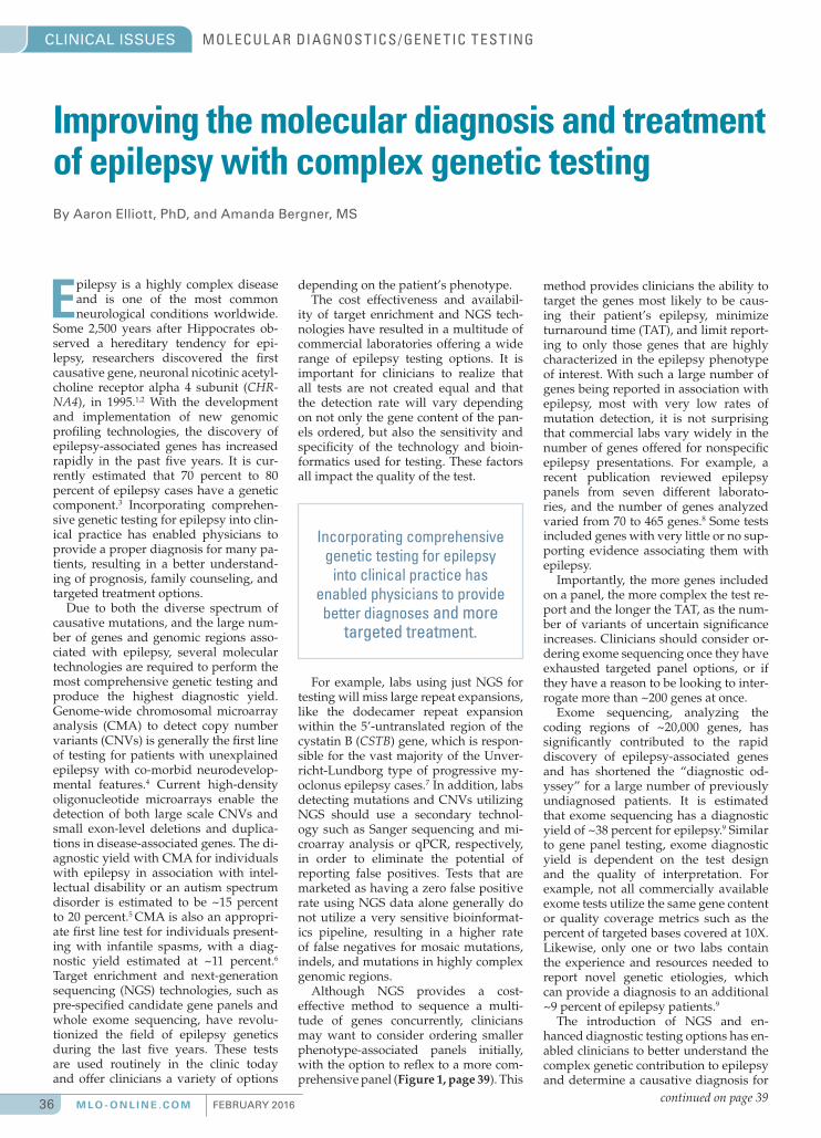
FEBRUARY 2016 M L O - O N L I N E .C O M 36
CLINICAL ISSUES MOL ECUL A R DIAGNOS TICS /GENE TIC T ES TING
Epilepsy is a highly complex disease and is one of the most common neurological conditions worldwide.
Some 2,500 years after Hippocrates ob-served a hereditary tendency for epi-lepsy, researchers discovered the fi rst causative gene, neuronal nicotinic acetyl-choline receptor alpha 4 subunit (CHR-NA4), in 1995.1,2 With the development and implementation of new genomic profi ling technologies, the discovery of epilepsy-associated genes has increased rapidly in the past fi ve years. It is cur-rently estimated that 70 percent to 80 percent of epilepsy cases have a genetic component.3 Incorporating comprehen-sive genetic testing for epilepsy into clin-ical practice has enabled physicians to provide a proper diagnosis for many pa-tients, resulting in a better understand-ing of prognosis, family counseling, and targeted treatment options.
Due to both the diverse spectrum of causative mutations, and the large num-ber of genes and genomic regions asso-ciated with epilepsy, several molecular technologies are required to perform the most comprehensive genetic testing and produce the highest diagnostic yield. Genome-wide chromosomal microarray analysis (CMA) to detect copy number variants (CNVs) is generally the fi rst line of testing for patients with unexplained epilepsy with co-morbid neurodevelop-mental features.4 Current high-density oligonucleotide microarrays enable the detection of both large scale CNVs and small exon-level deletions and duplica-tions in disease-associated genes. The di-agnostic yield with CMA for individuals with epilepsy in association with intel-lectual disability or an autism spectrum disorder is estimated to be ~15 percent to 20 percent.5 CMA is also an appropri-ate fi rst line test for individuals present-ing with infantile spasms, with a diag-nostic yield estimated at ~11 percent.6
Target enrichment and next-generation sequencing (NGS) technologies, such as pre-specifi ed candidate gene panels and whole exome sequencing, have revolu-tionized the fi eld of epilepsy genetics during the last fi ve years. These tests are used routinely in the clinic today and offer clinicians a variety of options
Improving the molecular diagnosis and treatment of epilepsy with complex genetic testingBy Aaron Elliott, PhD, and Amanda Bergner, MS
depending on the patient’s phenotype.The cost effectiveness and availabil-
ity of target enrichment and NGS tech-nologies have resulted in a multitude of commercial laboratories offering a wide range of epilepsy testing options. It is important for clinicians to realize that all tests are not created equal and that the detection rate will vary depending on not only the gene content of the pan-els ordered, but also the sensitivity and specifi city of the technology and bioin-formatics used for testing. These factors all impact the quality of the test.
For example, labs using just NGS for testing will miss large repeat expansions, like the dodecamer repeat expansion within the 5’-untranslated region of the cystatin B (CSTB) gene, which is respon-sible for the vast majority of the Unver-richt-Lundborg type of progressive my-oclonus epilepsy cases.7 In addition, labs detecting mutations and CNVs utilizing NGS should use a secondary technol-ogy such as Sanger sequencing and mi-croarray analysis or qPCR, respectively, in order to eliminate the potential of reporting false positives. Tests that are marketed as having a zero false positive rate using NGS data alone generally do not utilize a very sensitive bioinformat-ics pipeline, resulting in a higher rate of false negatives for mosaic mutations, indels, and mutations in highly complex genomic regions.
Although NGS provides a cost-effective method to sequence a multi-tude of genes concurrently, clinicians may want to consider ordering smaller phenotype-associated panels initially, with the option to refl ex to a more com-prehensive panel (Figure 1, page 39). This
method provides clinicians the ability to target the genes most likely to be caus-ing their patient’s epilepsy, minimize turnaround time (TAT), and limit report-ing to only those genes that are highly characterized in the epilepsy phenotype of interest. With such a large number of genes being reported in association with epilepsy, most with very low rates of mutation detection, it is not surprising that commercial labs vary widely in the number of genes offered for nonspecifi c epilepsy presentations. For example, a recent publication reviewed epilepsy panels from seven different laborato-ries, and the number of genes analyzed varied from 70 to 465 genes.8 Some tests included genes with very little or no sup-porting evidence associating them with epilepsy.
Importantly, the more genes included on a panel, the more complex the test re-port and the longer the TAT, as the num-ber of variants of uncertain signifi cance increases. Clinicians should consider or-dering exome sequencing once they have exhausted targeted panel options, or if they have a reason to be looking to inter-rogate more than ~200 genes at once.
Exome sequencing, analyzing the coding regions of ~20,000 genes, has signifi cantly contributed to the rapid discovery of epilepsy-associated genes and has shortened the “diagnostic od-yssey” for a large number of previously undiagnosed patients. It is estimated that exome sequencing has a diagnostic yield of ~38 percent for epilepsy.9 Similar to gene panel testing, exome diagnostic yield is dependent on the test design and the quality of interpretation. For example, not all commercially available exome tests utilize the same gene content or quality coverage metrics such as the percent of targeted bases covered at 10X. Likewise, only one or two labs contain the experience and resources needed to report novel genetic etiologies, which can provide a diagnosis to an additional ~9 percent of epilepsy patients.9
The introduction of NGS and en-hanced diagnostic testing options has en-abled clinicians to better understand the complex genetic contribution to epilepsy and determine a causative diagnosis for
continued on page 39
Incorporating comprehensive genetic testing for epilepsy
into clinical practice has enabled physicians to provide better diagnoses and more
targeted treatment.
36-39_MLO201602 Clinical Issues_FINAL.indd 36 1/12/2016 2:15:31 PM

Whole Blood Analyzer for Point-of-Care Lactate Testing
Rapid Sepsis Detection and Monitoring
Fast 13 Second Lactate Results
Small, 0.6 Microliter Whole Blood Sample
Single Use Lactate Biosensor
Easy to Use
novabiomedical.com
The Surviving Sepsis Campaign (SSC) has produced new recommendations titled International Guidelines for Management of Severe Sepsis and Septic Shock: 2012. These new updated guidelines call for lactate assays to direct therapy for septic shock. For patients with lactate greater than 4 mmol/L, SSC recommends quantitative resuscitation targeting normalization of lactate levels.
StatStrip Lactate provides a 13 second assay on a whole blood sample to allow rapid, early, goal directed therapy in septic patients. Testing is as fast and easy as bedside glucose testing. The single use StatStrip Lactate biosensor is pre-calibrated, fast and uses a very small whole blood sample (0.6 microliters) yet provides lab equivalent accuracy.
36-39_MLO201602 Clinical Issues_MECH_LM.indd 37 1/12/2016 11:11:38 AM

FEBRUARY 2016 M L O - O N L I N E .C O M 38
CLINICAL ISSUES MOL ECUL A R DIAGNOS TICS /GENE TIC T ES TING
First validated clinical test selects bestembryos for IVF and viable pregnanciesBy Elpida Fragouli, PhD
Mitochondria, organelles found in cells, play a vital role during embryo development—work-
ing as specialized “powerhouses” that supply individual cells with energy and perform other critical cellular functions. Over the past decade, understanding of mitochondrial function has advanced, yet little is known about how variations in mitochondrial DNA (mtDNA), or the genetic information found within each mitochondrion, impacts pregnancy out-comes.
With up to one-third of in vitro fertil-ization (IVF) transfers of “chromosom-ally normal” embryos failing, this writer and fellow researchers specializing in preimplantation genetic screening (PGS) and preimplantation genetic diagnosis (PGD) investigated the role of mtDNA in early fetal development. We used a new clinical diagnostic test to establish a criti-cal threshold of mtDNA quantity within embryos. Our study, published in the journal PLOS Genetics and also presented at the American Society for Reproductive Medicine (ASRM2015) annual Scientifi c Sessions, demonstrates that the level of mtDNA has a strong relationship with the ability of a human embryo to implant in the uterus following IVF.
A novel biomarkerPreimplantation development is an en-ergy-demanding process, as cells rapidly divide in early pregnancy. Because mito-chondrial functions are critical during the fi rst few days of embryo development, the study examined the possible correla-tion between mtDNA content and female age, embryo chromosome status, viabil-ity, and implantation potential among 379 embryos. It also analyzed at what stage an embryo initiates replication of its own mtDNA and carried out a detailed as-sessment of the mitochondrial genome sequence, searching for mutations, dele-tions, and polymorphisms. A combina-tion of microarray comparative genomic hybridization (aCGH), quantitative PCR, and next-generation sequencing (NGS) were used during the course of this study.
The study showed that embryos pro-duced by reproductively older women contain higher levels of mtDNA (P=0.003) than those from their younger counter-parts, implicating mitochondria in re-productive aging. Additionally, mtDNA levels were elevated in embryos with an abnormal number of chromosomes (an-
euploid embryos), independent of age (P=0.025). Aneuploidy is responsible for the majority of miscarriages and serious genetic disorders. Healthy embryos that successfully implanted in the uterus and resulted in a live birth were associated with lower levels of mtDNA than those that failed to produce a viable pregnancy (P=0.007).
Importantly, an mtDNA quantity threshold was established, above which implantation was never observed. For embryos with quantities of mtDNA below the threshold, there was a bet-ter than average chance of producing a pregnancy. Embryos that had high levels of mtDNA—above the previously estab-lished threshold—did not implant suc-cessfully, thus leading to a 100 percent negative predictive value for these failed cases. The overall pregnancy rate in the group was 38 percent when mtDNA was not considered prior to transfer.
The results of this study suggest that increased mtDNA may be related to el-evated metabolism and embryo viability; mitochondria may have a role in female reproductive aging as well as the genesis of aneuploidy. Of clinical signifi cance, mtDNA represents a novel biomarker with potential value for IVF treatment, revealing chromosomally normal em-bryos incapable of producing a viable pregnancy.
There is an urgent need for new meth-ods to improve the effi ciency and success rates of IVF. These important fi ndings show that mtDNA can help to highlight the embryos most likely to produce a pregnancy, allowing them to be given top priority for transfer to the woman’s uterus. The discovery of a new biomark-er of embryo viability, independent of standard assessments such as morphol-ogy, is a rare event and of great clinical potential.
Subsequently, the predictive value of this threshold was confi rmed in an inde-pendent blinded prospective study, indi-cating that abnormal mtDNA levels are present in 30 percent of non-implanting euploid embryos, but are not seen in embryos forming a viable pregnancy.
A new laboratory toolA fi rst validated clinical test is now avail-able that can measure the quantity of mtDNA in trophectoderm biopsies, and be applied to any biopsy specimens sent for PGS or PGD analysis. It can only be
offered for treatment cycles involving vitrifi cation of all embryos after biopsy. The turnaround time for results is two weeks or less. By helping to identify the embryos with the greatest probability of forming a successful pregnancy, this new clinical test is predicted to provide a fur-ther improvement in implantation rates, above and beyond what is currently done in the laboratory setting using PGS on its own.
Established to work on the same trophectoderm samples used for PGS, this clinical test does not require any additional work in the embryology laboratory and, as a result, embryos do not need to be subjected to any inter-ventions beyond those associated with routine chromosome screening. Since IVF can be a costly endeavor for those undergoing this medical approach to pregnancy, this new clinical test brings a simple and inexpensive approach that could help improve chances of success-ful fertility outcomes.
The success rate of IVF dramatically decreases with increasing female age. The higher levels of mtDNA observed with advancing age raise the question of whether mitochondria and their genome might play a direct role in the decline of female fertility with age. Increased levels of mtDNA may indicate compromised mitochondria that are unable to gener-ate the expected amount of energy to support embryo development.
My colleagues’ and my study dem-onstrates a clear association between mtDNA quantity and the ability of a hu-man embryo to implant in the uterus. A large (100 IVF cycles) blinded non-selection study is also being conducted in collaboration with the IVF clinic at the NYU Langone Medical Center to further assess the rate of outcome improvement if mtDNA quantifi cation is combined with PGS.
Elpida Fragouli, PhD, serves as Lab Director
at ReproG enetics UK,
in addition to holding a
research position at the
University of Oxford.
She played a key role
in the development,
validation, and clinical application of
comparative genomic hybridization (CGH),
the fi rst comprehensive chromosome
analysis method to be widely applied
to the study of human embryos.
36-39_MLO201602 Clinical Issues_FINAL.indd 38 1/12/2016 2:19:39 PM

39FEBRUARY 2016 M L O - O N L I N E .C O M
CLINICAL ISSUES MOL ECUL A R DIAGNOS TICS /GENE TIC T ES TING
patients, leading to an appropriate counseling and treatment plan. This was recently illustrated by research presented at the 2015 American Epilepsy Society Annual Meeting, which indicated that ~16 percent of epilepsy patients who underwent diagnostic sequencing harbored a mutation in a gene which had im-mediate treatment implications.10
As the number of clinicians who are comfortable utilizing complex genetic testing increases, the un-derstanding of epilepsy genetics will increase rapidly, precipitating a paradigm shift in the diagnosis, management, and treatment of the disorder.
REFERENCES
1. Riggs AJ, Riggs JE. Epilepsy’s role in the historical differentiation of religion, magic, and science. Epilepsia. 2005;46:452-453.2. Steinlein OK, Mulley JC, Propping P, et al. A missense mutation in the neuronal nicotinic acetylcholine receptor alpha 4 sub-unit is associated with autosomal dominant nocturnal frontal lobe epilepsy. Nat Genet. 1995;11(2):201-203.3. Hildebrand MS, Dahl HH, Damiano JA, et al. Recent advances in the molecular genet-ics of epilepsy. J Med Genet. 2013;50:271-279.4. Miller DT, Adam MP, Aradhya S, et al. Consensus statement: chromosomal micro-array is a fi rst-tier clinical diagnostic test for individuals with developmental disabilities or congenital anomalies. Am J Hum Genet. 2010;86(5):749-764.5. Mefford HC. CNVs in Epilepsy. Curr Genet Med Rep. 2014;2(3):162-167.6. Wirrell EC, Shelhaas RA, Joshi C, et al. How should children with West syndrome be effi ciently and accurately investigated? Results from the National Infantile Spasms Consortium. Epilepsia. 2015;56(4):617-625.
7. Lafrenière RG, Rochefort DL, Chrétien N, et al. Unstable insertion in the 5’ fl ank-ing region of the cystatin B gene is the most common mutation in progressive my-oclonus epilepsy type 1, EPM1. Nat Genet. 1997;15(3):298-302.8. Chambers C, Jansen LA, Dhami-ja R. Review of Commercially Avail-able Epilepsy Genetic Panels. J Gen-et Couns. 2015. Epub ahead of print: doi:10. 1007/ s10897-015-9906-9.9. Helbig KL, Farwell KD, Powis Z, et al. Di-agnostic exome sequencing provides a mo-lecular diagnosis for a signifi cant proportion of patients with epilepsy. Genet Med. 2015; In press.10. Demos M, Buerki SE, Guella I, et al. Tar-geted analysis of whole exome sequencing in early onset epilepsy. American Epilepsy Society. 2015; Abstract 1.311.
continued from page 36
Does this patient fit any of the following clinical categories:
chromosomal microarray
patient with unexplained epilepsy
Does this patient have epilepsy in conjunction with one or more of the following:
• developmental delay • an autism spectrum disorder • infantile spasms• intellectual disability • congenital anomaly(ies)
noyes
yes
yes no
no
Does this patient have progressive myoclonus epilepsy (PME)?
• neonatal seizures • febrile seizures
NGS panels between 10-17 genes for:• neonatal seizures• febrile seizures• infantile spasms • focal epilepsy
NGS panel of 100 genes for wide variety of epilepsies (includes dodecamer repeat expansion of CSTB)
Diagnostic Exome Sequencing
NGS panel of 3 genes for progressive myoclonus epilepsy (includes dodecamer repeat expansion of CSTB)
NGS panel of 21 genes for progressive myoclonus epilepsy (includes dodecamer repeat expansion of CSTB)
• infantile spasms• non-lesional focal epilepsy
Figure 1. Epilepsy diagnostic testing strategy
Amanda Bergner, MS, serves as Senior
Product Manager,
Neurology, for Ambry Genetics. She is a
certifi ed genetic
counselor with more
than 15 years of
clinical experience
specializing in neurogenetic disorders.
Aaron Elliott, PhD, serves as Chief
Operating Offi cer
and Interim Chief
Scientifi c Offi cer for
Ambry Genetics and
has extensive
experience in
complex molecular diagnostic test
development and implementation.
36-39_MLO201602 Clinical Issues_FINAL.indd 39 1/12/2016 2:16:30 PM
FEBRUARY 2016 M L O - O N L I N E .C O M 40
CLINICAL ISSUES MOL ECUL A R DIAGNOS TICS /GENE TIC T ES TING
Soaring demand for genetic testing highlights need for streamlined data interpretationBy Michael Hadjisavas, PhD, and Ramon Felciano, PhD
The recent surge in number of sequencing-based clinical genetic tests has put a spotlight on associated challenges in data interpretation. While advances in genomics allow
for the development of new genetic tests at a breathtaking pace and with unprecedented complexity, the interpretation of re-sults has remained a largely manual, time-consuming process that is simply not scalable. In this article, we review the cur-rent landscape of variant interpretation and the challenges it presents, as well as new developments in the fi eld that indicate signifi cant improvements may be on the horizon.
The processToday, variant interpretation is conducted by clinical geneti-cists who have tremendous skill and expertise in their fi eld. It is a testament to their dedication that current interpretations are as reliable as they are. However, this dependence on hu-man judgment, coupled with a laborious process, introduces room for error.
As lab directors know all too well, most variant analysis fol-lows the same formula: Run the genetic test, annotate results, investigate variants detected, weigh evidence, integrate and in-terpret data, and report fi nal results. It’s the middle parts of the process—variant investigation and interpretation—that pre-vent this process from becoming fully automated and scalable.
Clinical geneticists usually begin this variant interpreta-tion journey with an annotated report from the DNA results of the test, whether that’s a gene panel, exome, or even whole genome. This annotation includes a list of identifi ed high-qual-ity variants that must be pursued to determine which, if any, is causative for disease. Variant interpreters often begin with PubMed and Google, scouring the literature to fi nd mentions of these variants. Next, they have to go through each paper to fi gure out whether its information about the variant is relevant to the test at hand, tracking details about heterozygosity, dis-ease type, number of subjects, and so forth. Once any available information about the variants has been uncovered, the next stop is databases or websites that predict protein changes based on the DNA variant. This part of the process indicates whether the variant might be affecting a patient’s phenotype. With all of this information, the analysis team draws on its deep clini-cal expertise to make a judgment call about how to report each variant on the list: pathogenic or likely pathogenic, benign or likely benign, or signifi cance unknown.
Experts in this process say that this interpretation process takes about 30 minutes for each novel variant, a couple of hours for variants that have been reported in the literature, and many days for the most complicated cases. As genetic tests become increasingly complex, covering more and more of a patient’s genome, the time spent analyzing a growing list of variants for each test is expanding drastically. Challenges in effi cient and effective interpretation of genetic test data will soon gate our ability to bring these benefi ts to patients, motivating the need for robust clinical decision support solutions directed at these clinical testing labs.
Key challengesWhether a genetic test is trying to pinpoint the cause of a rare or unknown disease, fi nd evidence of hereditary disease, or suggest an appropriate treatment course, the need for rapid turnaround of results is imperative. Clinical geneticists are well aware of this, but the growing demand for genetic testing and the increasing number of variants turned up by these tests are doubly burdensome for analysis teams. As the range of testing options soars, most clinical labs no longer have a geneticist with expertise in every test indication; under these conditions, vari-ant interpretation may take even longer.
In order to ensure that results are returned to physicians quickly enough to be clinically useful, it is essential to fi nd ways to automate and streamline as much of the process as possible so that clinical geneticists deploy their expertise where it’s needed most. For instance, many large clinical labs main-tain their own variant databases, so if a variant has been seen and interpreted before, analysts can avoid the time-consuming process of researching it all over again. The development of proprietary databases by testing labs and healthcare providers that consume the data is a key emerging trend. Value is being derived mining these databases to determine variant frequen-cies and their associations with clinical profi les, outcomes, and ethnicities to enhance the value of clinical reports.
Another major challenge lies in the murky category of vari-ants of unknown signifi cance. Obviously, clinical utility is greatest when a variant falls fi rmly into either the “pathogen-ic” or “benign” category, with utility weakening as the variant moves toward the center of the spectrum. However, emerging needs are suggesting that variants be classifi ed into further subcategories such as “likely pathogenic” or “likely benign.” But in many cases, the downstream effect of variants—even those already reported in the literature—is not clear. Because many of these variants must be interpreted with minimal in-formation, it comes as no surprise that variant interpretations can differ signifi cantly from one lab to another.
In combination, these numerous secular drivers of ineffi cien-cy, together with the need for testing labs to expand their test menu, drive operational effi ciencies and turnaround time. The community needs better resources that will help defi nitively classify variants, pulling more of them out of the “unknown” category and increasing the likelihood of having consistent in-terpretation across labs. In fact, lack of commercial grade in-formation solutions is compounding these ineffi ciencies and moderating the development of the market.
Signs of improvementThere are a number of reasons to believe that the genomics and clinical communities are well on their way to addressing these challenges. For example, ClinVar1 is a publicly available database hosted by the National Center for Biotechnology Information. Users submit genetic variations and their associated phenotype, with supporting evidence, to help others in the community in-crease their confi dence in their own variant interpretations. Other
40-41_MLO201602 Clinical Issues-Qiagen_MECH_AL.indd 40 1/12/2016 5:15:23 PM

CLINICAL ISSUES
commercial efforts perform large scale curation and data integra-tion from clinical literature and other sources to power clinical test interpretation pipelines.
Meanwhile, the Clinical Sequencing Exploratory Research2 (CSER) program, funded by the National Cancer Institute and National Human Genome Research Institute, has established a consortium of laboratories conducting studies that are helping to explain variant classifi cation differences among labs. This data will be quite useful for suggesting standardized approach-es to improve lab-to-lab reproducibility of results.
Separately, the Allele Frequency Community3 (AFC) was founded by a number of organizations to share aggregated in-formation about how often variants are seen in various popula-tions, allowing analysts to factor in data for groups that may be underrepresented in existing public databases, including the Exome Aggregation Consortium4 (ExAC). The AFC operates on a share-and-share-alike model, so all members increase the value of the repository by contributing their own data about allele frequency.
Clearly, the challenge of variant analysis and interpretation affects the entire clinical community, and it will take a commu-nity-wide effort to overcome this obstacle. But we believe that clearing this hurdle is possible, and that eventually the variant interpretation process will be faster, more automated, and more defi nitive, giving clinical geneticists an even greater role in pa-tient diagnosis and care.
The trend is expected to continue and drive the use of high-complexity genetic tests toward an industrialized scale. Consistent with industrialized technology markets, the demand for software solutions will shift from open source and homebrew solutions to highly scalable commercial-grade informatics solutions that are enabled with rigorously curated knowledge bases.
REFERENCES
1. ClinVar: National Center for Biotechnology Information. http://www.ncbi.nlm.nih.gov/clinvar/. Accessed December 30, 2015.2. CSER: Clinical Sequencing Exploratory Research. National Institutes of Health. Moving the genome into the clinic. https://cser-consortium.org/. Accessed December 30, 2015.3. Allele Frequency Community. Imagine human genome interpretation…minus the false positives. http://www.allelefrequencycommunity.org/. Accessed December 2015.4. ExAC Browser (Beta) | Exome Aggregation Consortium (ExAC): http://exac.broadinstitute.org/.
Michael Hadjisavas, PhD, recently joined
QIAGEN Bioinformatics as the Vice President
of the Clinical Genomics Program. He leads
the clinical bioinformatics portfolio in strategy
to maximize molecular insight from biological
samples delivering actionable reports to
laboratory healthcare professionals.
Ramon Felciano, PhD, serves as QIAGEN’s Chief Technology Offi cer and leads strategy
for the recently launched QIAGEN Clinical
Insight (QCI) solutions, a clinical decision
support platform designed to help labs
streamline and accelerate the interpretation
of genetic test results.
40-41_MLO201602 Clinical Issues-Qiagen_MECH_AL.indd 41 1/12/2016 5:15:39 PM
FEBRUARY 2016 M L O - O N L I N E .C O M 42
WASHINGTON
REPORT
Editor’s note: In the October 2015 issue of MLO, I wrote a “Washington Report” titled “AACC urges CMS to delay enforc-ing glucose monitor regulations” (MLO. 2015;47(10):50) The essay outlined reasons why AACC and other lab stakeholders have advised the Centers of Medicare and Medic-aid Services to, in the words of former AACC president David C. Koch, “take a pragmatic, educational approach to this situation.” This month’s “Washington Report” is a response to that column, written by John F. McHale, Vice President, QA/RA and Technical Sup-port, Nova Biomedical Corporation, provider of the Nova StatStrip Glucose Hospital Meter System and Nova StatStrip Xpress Glucose Hospital Meter System.
The October 2015 MLO “Washing-ton Report” touches on a complex subject that is extremely important
to many readers. The risky, off-label use of glucose monitors with critically ill patients is under close scrutiny by the Food and Drug Administration (FDA) and Centers for Medicare and Medicaid Services (CMS), with virtually every hos-pital reevaluating its options for glucose testing.
However, the article overlooks several important facts pertaining to glucose monitor use with critically ill patients. I believe that providing these additional facts will give readers a better under-standing of the FDA/CMS enforcement actions, and what alternatives are readily available to hospitals for glucose testing with critically ill patients.
The October article’s conclusion may have been reached because several very important facts and options for hospital point-of-care (POC) glucose use were overlooked. These omissions may lead to a different conclusion. To explain fur-ther, I respond to three statements within the article.
Statement 1. “In a painful irony, the Center for Medicare and Medicaid Services’ proposed action, taken to protect patients, would actually threaten the quality of care.”
The reason for the FDA/CMS action is to protect patient care. CMS enforcement decisions came after more than 13 deaths attributed to hospital glucose monitors had been reported to the FDA.1 Two additional deaths were later reported, causing the New York State Health De-partment in January 2014 to declare the
use of hospital monitors for critically ill patients as “off label” in that state.2 Also in January 2014, the FDA issued a draft guidance document that highlighted the agency’s concerns with the use of POC blood glucose monitors for critically ill patients.3 Since that time both the FDA and CMS have taken actions to warn industry that they wanted to reduce the adverse events associated with off-label use of blood glucose monitors, which were never tested or cleared by the FDA for use with critically ill patients.
As indicated in their product labeling, glucose monitors have interferences from substances or hematocrit abnormalities that are often found in the blood of criti-cally ill patients, and this can result in the overestimation of glucose and over-dosing of insulin. According to the FDA, “Errors in BGMS (blood glucose moni-toring systems) device accuracy can lead to incorrect insulin dosing, which, when combined with other factors, can lead to increased episodes of hypoglycemia. For hospitalized patients who may be seri-ously ill, any inaccuracies in the meters would further increase the risk to these patients.”3
Statement 2. “That is, the change would suddenly make many hospitals’ use of the glu-cose monitor an ‘off-label’ use, and thus turn glucose monitoring into a high complexity test that could not be performed by nurses and oth-er bedside caregivers. Faced with this situation hospitals might have little choice but to dis-continue the point-of-care testing altogether.”
This statement implies that hospitals have no practical POC glucose alterna-tive other than discontinuing the use of off-label glucose monitors for critically ill populations. This comment overlooks the fact that the FDA has already cleared two CLIA-waived glucose monitors for POC use with critically ill patient populations: “This waived status will allow a broad variety of healthcare professionals, such as nurses and technicians, to perform the test at the point-of-care, such as at a patient’s bedside, instead of requiring that the test be performed in a hospital lab (or other lab) that meets the CLIA [Clinical Labora-tory Improvement Amendments of 1988] requirements for high complexity testing. The CLIA waiver will also allow hospital labs to safely provide blood glucose moni-toring to their critically ill patients without having to meet the signifi cant CLIA re-quirements for high complexity testing.”4
Statement 3. “In the comment letter, AACC asserts that a transitional period will allow hospitals to adjust policies, procedures, and workforce to ensure compliance with the new regulatory requirements.”
As stated earlier, the FDA issued its new draft guidance document for hospital POC glucose monitors nearly two years ago, and CMS subsequently communi-cated its intention to enforce off-label use of glucose monitors. More than half the hospitals in the U.S. have already transi-tioned to the FDA-cleared POC glucose monitor that is cleared for all patients, in-cluding critically ill. The downside of an additional transition period would per-petuate the risk of using untested glucose monitors on critically ill patients when a safe, FDA-cleared solution already exists.
AACC’s rationale that the implemen-tation of the FDA/CMS decision would cause patient harm is contrary to the facts. Off-label use of glucose monitors has been shown to cause adverse events including deaths. A delay in the enforce-ment will unnecessarily perpetuate the risky use of glucose monitors that have not been cleared for use with critically ill patients when immediate, FDA-cleared, CLIA-waived products exist.
In summary, these FDA/CMS actions do not adversely affect the quality of patient care; they improve the quality of patient care. Neither do these actions re-sult in prohibiting POC glucose testing. Hospitals have a choice of either using a POC glucose monitor tested and cleared for critically ill patients or following the regulations for off-label use in this popu-lation. Half of U.S. hospitals have already employed the FDA-cleared monitor.
REFERENCES
1. Harper CC. FDA/CDRH Public Meeting: Blood Glucose Meters. Presented at the meeting of the U.S. Food and Drug Administration/Center for Devices and Radiological Health, Gaithersburg, MD. 2010. http://diabetestechnol-ogy.org/FDA/Harper-%20PQQ%20and%20other%20inter-ferences.pdf. Accessed December 30, 2015
2. New York State Department of Health. Off-Label Use of Glucose Meters. 2014.
3. Food and Drug Administration. Blood Glucose Moni-toring Test Systems for Prescription Point-of-Care Use; Draft Guidance for Industry and Food and Drug Adminis-tration Staff. 2014. http://www.fda.gov/downloads/medi-caldevices/deviceregulationandguidance/guidanced-ocuments/ucm380325.pdF. Accessed December 30, 2014.
4. Food and Drug Administration. FDA clears glucose monitoring system for use in hospital critical care units. 2014. http://www.fda.gov/NewsEvents/Newsroom/Pres-sAnnouncements/ucm416144.htm. Accessed December 30, 2015.
Industry leader weighs in on glucose monitor regulation controversyBy John F. McHale
42-43_MLO201602_WashingtonReport_FINAL.indd 42 1/12/2016 5:37:13 PM

A New Breakthrough in Hemostasis Quality Management.
New ACL TOP Family 50 Series* Extending standardization beyond the analytical phase
The new ACL TOP Family 50 Series offers the most advanced automation and quality management for routine and specialty Hemostasis testing in mid- to high-volume labs. All models are standardized and offer automated pre-analytical sample integrity checks to identify under-filled sample tubes, abnormal sample aspiration potentially caused by clots, and assay-specific interference from hemolysis, lipemia and bilirubin. Plus, new lab accreditation tools make compliance easier. Better efficiency and quality management for you—better quality care for your patients.
The ACL TOP Family 50 Series. Quality in. Quality out.
For more information, call 1.800.955.9525 or visit www.ilus.com.©2016 Instrumentation Laboratory. All rights reserved.* Not available in all countries.
42-43_MLO201602_WashingtonReport_MECH_LM.indd 43 1/12/2016 11:46:36 AM

FEBRUARY 2016 M L O - O N L I N E .C O M 44
SPECIMEN COL L EC TION / PHL EBOTOM YPRODUCT FOCUS
Copan Diagnostics, Inc.FecalSwab is comprised of a regular
COPAN FLOQSwab and a screw cap
tube containing 2mL of Cary-Blair
medium. Because of its high pH,
Cary-Blair is the medium of choice for
transport and preservation of enteric
bacteria. FecalSwab is intended for
the collection of rectal swabs and fe-
cal specimens and to preserve the vi-
ability of enteric pathogenic bacteria during transport from the
collection site to the testing laboratory. Using the FLOQSwab,
provided in the collection kit, a small sample of feces can be
transferred into the tube by medical staff after the patient
has provided the sample or by a laboratorian after the fecal
specimen has been received in the laboratory. Alternatively,
FecalSwab can be used to take a rectal sample directly from
the patient. FecalSwab simplifi es and standardizes fecal sam-
ple collection, transport, and processing by converting solid
or semi-solid specimens into a phase suitable for automatic
specimen processors, in space-saving, instrument-ready tubes.
FecalSwab provides a compact and neat alternative to tradi-
tional large, bulky transport containers, which collect a quan-
tity far greater than needed for culture or molecular testing.
For more information, please visit: http://www.copanusa.com/
products/liquid-based-microbiology/fecalswab/
Greiner Bio-One Greiner Bio-One North America, Inc.,
announces the addition of Low Volume
Blood Collection tubes to its current
product portfolio. This addition marks a
signifi cant step into better patient care,
less bio-hazardous waste, and a reduc-
tion in blood collection volumes in the
healthcare industry. Greiner Bio-One
offers a complete line of low-volume
tubes with draw volumes of 1mL to 2.5mL to facilitate reduc-
tion in phlebotomy volumes and, subsequently, the instances
of hospital-acquired anemia which can be caused by excessive
blood draws. These blood tubes offer functional compatibility
with instrumentation by maintaining the normal 13 x 75 mm
tube size and providing optimal blood to additive ratios for
drawing reduced blood volumes.
For more information, please visit: https://shop.gbo.com/en/
usa/articles/catalogue/articles/0010_0010_0130/
Kimberly-Clark ProfessionalThe Kimtech Pure A4 Sleeve Protector
from Kimberly-Clark Professional helps
keep workers and the workplace safe
through advanced barrier protection that
shields the person and the garment from
hazardous chemicals and biologics. By
providing an additional layer of protec-
tion between gloves and lab coat sleeves,
it reduces the risk of exposure and con-
tamination. The Kimtech Pure A4 Sleeve Protector is ideal for
situations where full coverage isn’t required, but full protec-
tion is. Features include a) reinforced seams; b) advanced bar-
rier fabric—to provide enhanced protection against dangerous
liquids; c) ease of donning and doffi ng; d) a range of sizes; e)
thumb loops for full wrist coverage; and f) bulk packaging for
convenience and waste reduction. The Kimtech Pure A4 Sleeve
Protector can be diverted from landfi lls through RightCycle by
Kimberly-Clark Professional, a large-scale recycling program
for non-hazardous lab and clean room waste.
For more information, please visit: http://www.kcprofessional.
com/products/scientifi c-ppe/protective/cleanroom/49816-kim-
tech-pure-a4-cleanroom-sleeve-bulk-packaged
MarketLab The Midstream Urine Collection Kit
is designed to prevent messes and
contamination during urinalysis
collection. The patented easy-hold
handle distances the patient’s and
the technician’s hands from the cup,
lessening the chance of coming in
direct contact with urine. The sterile,
leak-proof cup holds 90mL, has a
53mm opening, and features a tight-fi tting lid to prevent spill-
age during specimen transport. Each packaged kit includes a
container, pre-attached handle, Castile soap towelettes, and
directions. Sold in packs of 50.
For more information, please visit: http://www.marketlab.com/
midstream-collection-kit/p/MidstreamCollectionKit/
Retractable Technologies, Inc. The VanishPoint blood collection set
(BCS) is a novel safety blood collection
device that features automated in-vein
retraction, which virtually eliminates
exposure to the contaminated needle.
The unique design has integrated fi nger
grips that allow for handling ease. The
automated needle retraction is activated
by depressing a retraction trigger located near the fi nger grip
area, allowing for easy one-handed activation, without changes
in hand position, while reducing the risk of inadvertent activa-
tion. In addition, the needle retraction automatically clamps the
tubing, reducing the risk of exposurel to blood. This product is
not made with DEHP or PVC.
For more information, please visit: http://www.vanishpoint.
com/Simple4.aspx?PageId=197
Visiun, Inc.Visiun’s Performance Insight provides laboratory managers
with access to detailed analytics and visual insight into their
daily specimen collection processes, including specimen de-
fect capture. Access to this data provides immediate insight
into whether or not operational standards are being achieved
by clearly identifying performance problems and root causes,
delivering actionable information to monitor and improve the
specimen collection process. Performance Insight captures
real-time data, allowing laboratory management to identify
important trends. Performance Insight provides access to real-
time analytics so that laboratory managers can identify speci-
men hemolysis and QNS rates in a matter of seconds, and
monitors the workfl ow and productivity of staffed phleboto-
mists daily on an hourly basis and alerts hospital laboratories
if specimens are not being processed and collected in a timely
manner. Its Turnaround Time module offers detailed analytics
into the percentage of service commitments achieved, while
composite reports look at the entire value stream and pin-
point where a slowdown occurred from the time the test was
ordered, to the time it was collected, to result reporting.
For more information, please visit: http://www.visiun.com/
44-45_MLO201602-Product Focus_FINAL.indd 44 1/12/2016 12:14:57 PM
Designed to reduce pain for a more comfortable blood sampling experience.
Take the Unistik ChallengeExperience the difference Unistik can make for your patients. T: 1-800-421-6936 I E: [email protected] I W: owenmumfordinc.com
dualgentle comfort normal extra preemie full term
30G1.5mm
28G1.8mm
23G1.8mm
21G2.0mm
18G1.8mm
1.0 mmDEPTH
2.5 mmLENGTH
0.85 mmDEPTH
1.75 mmLENGTH
NAME: TITLE:
FACILITY NAME:
ADDRESS:
CITY: STATE: ZIP:
PHONE: EMAIL:
To receive free samples and product literature, fax completed form to 770-977-2866 or email [email protected]
FREE SAMPLE REQUEST
Single Use Safety Lancets
Heel Incision Devices
Which samples would you like to receive?
No purchase necessary. Available while supplies last. Please allow 8 to 10 weeks for delivery of free samples. MLOFEBAD/OMI/0116/1/US.
pat entWith
ComfortZone
Technology®
comfort.comfort.
CAPILLARY SAMPLING SOLUTIONS
44-45_MLO201602-Product Focus_MECH_AL.indd 45 1/12/2016 11:48:29 AM

FEBRUARY 2016 M L O - O N L I N E .C O M 46
PRODUCT SPOTLIGHTS
When the spotlight is on quality our analyzers deliver
The RX series of chemistry
ana lyzer s guarantee
results you can trust.
Combining robust hard-
ware, intuitive software
and the extensive RX
series test menu to deliver
high quality routine and specialized testing onto
a compact easy-to-use platform. Learn more,
http://www.therxseries.com/rx-daytonaplus
Randox Laboratories Ltd.www.rsleads.com/602ml-402
Optimize your lab with CGM LABDAQ CGM LABDAQ is an innovative
LIS designed to help labs man-
age and grow their business
by offering optimized work-
fl ows in an easy-to-use, fl exi-
ble and intuitive system with quick implementation
times and tremendous scalability. With seamless inte-
gration and interoperability capabilities, labs can man-
age and grow using factual data to support business
decisions and provided improved patient care. Learn
more at www.cgm.com/us
CompuGroup Medicalwww.rsleads.com/602ml-404
D-100™ cleared by FDA for Hemoglobin A1c testing
The D-100™ System is the future of
HbA1c testing. With innovative solu-
tions to maximize workflow effi-
ciency, the D-100 System allows
high volume laboratories to quickly
and easily report HbA1c results
while also detecting hemoglobin
variants. http://www.bio-rad.com/
en-us/corporate/newsroom/bio-rad-receives-fda-
clearance-for-its-d-100-system-for-a1c-testing
Bio-Rad Laboratorieswww.rsleads.com/602ml-403
FilmArray. The fastest way to better results.
Syndromic infectious disease
testing in about an hour. With
four industry leading com-
prehensive panels (Respira-
tory, Blood Culture Identifi ca-
tion, Gastrointestinal and
Meningitis/Encephalitis), the
FilmArray eliminates the
guesswork and helps pinpoint bugs faster than ever.
Get faster results at FilmArray.com.
BioFirewww.rsleads.com/602ml-405
Temp-Chex Digital II thermometerTemp-Chex Digital II thermom-
eter features NIST-traceable
calibration and measures
temperatures from -50 °C to
200 °C. Features include an
externally mountable display
with maximum/minimum
temperatures, low/high alarm
and date/time modes. Learn
more at streck.com.
Streckwww.rsleads.com/602ml-401
Bio-Rad Mission: Control™
The FIRST objective risk
management software
available to help laborato-
ries develop a Quality
Control Plan with the best
QC rules and right QC
frequency to reduce risk of
reporting incorrect patient
results. Link: http://bit.ly/
MLO_02_2016
Bio-Rad Laboratorieswww.rsleads.com/602ml-406
Fully Automated Urine Chemistry & Sediment Analyzer
ARKRAY’s AUTION HYBRID
AU-4050 is a fully automated
urine analyzer that integrates
proven urine test strip and fl ow
cytometry technology into one
compact instrument that gener-
ates standardized and accurate
results giving operators
confidence time after time.
www.arkrayusa.com
ARKRAYwww.rsleads.com/602ml-400
Avoid False-Positive Urine Drug Screens
Use Astec® CHIROBIOTIC® V2 for the
rapid, sensitive LC/MS analysis
m e t h a m p h e t a m i n e a n d
amphetamine enantiomers in urine
without the need for derivatization.
See this application note and our
full analytical column offering at
sigma-aldrich.com/clinical
Sigma-Aldrichwww.rsleads.com/602ml-408
46-47_MLO201602 Spotlights_DUM_eb.indd 46 1/12/2016 4:05:03 PM

47FEBRUARY 2016 M L O - O N L I N E .C O M
PRODUCT SPOTLIGHTS
Stat Profi le Prime® Critical Care Blood Gas Analyzer
Nova’s Stat Profi le Prime® features
ZERØ™ maintenance cartridges
and MicroSensor technology for
exceptional value in a critical care
whole blood analyzer. This design
optimizes the life of each car-
tridge; improves uptime; and
eliminates waste, downtime, and higher costs of older
cartridgebased systems. The 10-test menu includes pH,
PCO2, PO2, Na, K, iCa, Cl, Glucose, and Lactate
Nova Biomedical
www.rsleads.com/602ml-409
Standardized Platform of Hemostasis Testing Systems.
The ACL TOP Family,
including ACL TOP 500 CTS for medi-
um- to high-volume,
routine and special-
ty testing, is a com-
plete line of Hemo-
stasis Testing Systems—designed with the same high
standards of operation and functionality for hospitals
and labs, of any size or scope.
Instrumentation Labs
www.rsleads.com/602ml-412
cobas® CT/NG TestThe PCR-based multiplex
dual probe assay is for
screening and diagnosing
chlamydia and gonorrhea
in symptomatic and
asymptomatic patients
from male urine and self-collected vaginal swabs.
It offers excellent sensitivity and high specifi city and
expands the menu for the automated cobas 4800
System, which also runs the cobas HPV Test.
Roche Diagnostics
www.rsleads.com/602ml-410
Get POC Testing Results In Your EHROrchard® Trellis™ provides the
optimal means for electronic
connectivity to more effectively
manage your POC testing. With
today’s healthcare challenges
surrounding efficiency and
accountability, Orchard Trellis serves as a cost-
effective bridge for electronically passing orders and
results between low volume, point-of-care analyzers
and your EHR.
Orchard Software
www.rsleads.com/602ml-407
Empowering YOU to lead the wayLabLeaders.com---an
online resource creat-
ed to help transform
healthcare challenges
into opportunities. It
will offer insights and
strategies from top
industry innovators to
help you redefi ne the value of your laboratory.
Pre-register now. http://lableaders.com/
Roche Diagnostics
www.rsleads.com/602ml-411
K-ASSAY® Cystatin C - Early Detectionof Chronic Kidney Disease
The K-ASSAY® Cystatin C
reagent is an accurate and
sensitive method for measuring
Cystatin C in serum and plasma,
and has many advantages over
traditional GFR methods. This
high quality, low-cost reagent
can be used on almost all
chemistry analyzers. E: [email protected]
KAMIYA Biomedical Company
www.rsleads.com/602ml-413
INDEX OF ADVERTISERS
This index is provided as a service. The publisher does not assume liability for errors or omissions.
The Binding Site ....................www.thebindingsite.com.................19
BioFire D iagnostics ................fi lmarray.com ......................................5
Bio-Rad Laboratories .............www.bio-rad.com/diagnostics ........23
Bio-Rad Laboratories .............www.bio-rad.com/diagnostics ........22
Bio-Rad Laboratories .............www.bio-rad.com/diagnostics ........ 11
Bio-Rad Laboratories .............www.bio-rad.com/diagnostics ........10
Bio-Rad Laboratories .............www.qcnet.com/unity ......................31
CompuGroup Medical ...........www.cgm.com/us.............................33
Eppendorf ........................ www.eppendorf.com/cellmanipulation ...3
Hologic, Women’s Health ......PapPlusHPV.com ..............................15
Instrumentation Laboratory ..www.ilus.com ...................................43
ADVERTISER WEB PAGE ADVERTISER WEB PAGE
Kamiya Biomedical Co. .........www.k-assay.com/MLO.php ..............9
Nova Biomedical ....................novabiomedical.com ........................37
Orchard Software ...................www.orchardsoft.com ......................35
Owen Mumford, Inc ...............www.owenmumfordinc.com...........45
Psyche Systems Corp. ...........www.psychesystems.com ...............34
Randox Laboratories .............www.randox.com/reagents .............21
Roche Diagnostics .................www.hpv16and18.com ............... IFC-1
Sebia, Inc ................................www.sebia-usa.com .........................25
Sekisui Diagnostics ................www.osomtests.com .....................IBC
Sigma-Aldrich ........................sigma-aldrich.com/clinical ..............BC
Streck ......................................www.streck.com ...............................27
46-47_MLO201602 Spotlights_DUM_eb.indd 47 1/12/2016 4:05:28 PM

FEBRUARY 2016 M L O - O N L I N E .C O M 48
By A lan Lenhof f, Edi torEXECUTIVE SNAPSHOT
WERNER RODORFF
Chief Executive Offi cer, CGM US
Senior Vice President North America
Professional: I have been in my current position since
August 2014, though I have been with
CompuGroup Medical (CGM) since 1987.
Prior to joining CGM US, I was CEO at
CompuGroup Medical in Turkey. I’ve spent
the last 23 years running different busi-
ness units and also served as CEO at sev-
eral other companies.
Education: University of Heidelberg, Master’s Degree
in Physics
Personal:I have a passion and interest in new
technology and am the “typical” early-
adopter. I also enjoy photography, hiking,
and scuba diving.
IT solutions to meet the needs of clinical labs
If you were explaining CompuGroup Medical to someone who is not fa-miliar with the organization, how would you characterize its primary areas of expertise? What are the major categories of solutions that CGM provides for its customers?CompuGroup Medical US is the U.S. divi-sion of CompuGroup Medical AG—one of the leading eHealth companies in the world. Founded in 1987 with our global headquarters in Germany, we have more than 4,500 employees, are in 43 countries, serve more than 400,000 physicians, and work across all healthcare sectors. Our mission is to synchronize the systems of the healthcare industry with the mis-sion of patient care, enabling providers to achieve the best possible outcomes for patients. In addition to our laboratory information system, CGM LABDAQ, we develop healthcare IT solutions to sup-port the clinical and practice manage-
ment activities that take place in physi-cian practices, community health centers, and hospitals; practice management soft-ware; and productivity tools and EDI/reimbursement. We’ve also recently intro-duced to the market our robust business intelligence tool, CGM ANALYTICS, as well as CGM SAM, our Software Assisted Medicine solution.
What are the core elements of eHealth for clinical labs and the larger institutions they are part of? What is the minimum that any lab should have? It’s all about connectivity and seamless integration. It’s well recog-nized that laboratory data and test results are the largest factor used when providers make healthcare decisions for patients. As patient care and outcomes become in-creasingly important and more lab testing is done outside the walls of the hospital, the need for health systems to be aligned and connected is critical.
In order to run an effi cient lab and en-sure these outcomes for the patient, it’s imperative that results from EMRs are be-ing sent and received in a streamlined and connected way. The lab requires test or-ders, administration data, and billing in-formation about the patient and interfaces to ordering locations and various result-ing locations to be effi cient. A connection to an analytics module provides an extra advantage as labs can gain insight into their effi ciency with turnaround times, QA failures, etc., to ensure their operation is running smoothly. Also, ACOs rely on this type of analytics data to determine if they’re meeting quality measures and population health goals.
What distinction would you draw between software that is neces-sary today for any lab, and options that are perhaps more appropriate for larger or specialized labs? How does CGM customize solutions for organizations with different needs? Different laboratories have very specifi c needs, based on their size and testing types. For example, physician offi ce labo-ratories typically perform fairly basic test-ing with straightforward setups. Larger laboratories like reference labs or hospital labs require multiple work stations and analyzers to be set up, necessitating in-terfaces that can share and receive orders in real time to multiple offi ces and mobile applications. For specialized labs like tox-icology and pain management laborato-ries, their unique workfl ows and specifi c customizable reports need a streamlined process. Often, larger institutions rely on
additional software like data mining or analytics tools to understand and opti-mize their lab’s performance, population management tracking, and control, to meet regulatory needs for reimbursement and much more.
CGM LABDAQ offers different pack-ages based on each lab’s specifi c size and workfl ow in order to ensure effi ciency and maximize testing reimbursements. In today’s healthcare setting, it’s critical to have an LIS that not only maximizes the lab’s operational effi ciency but offers the latest technology and is streamlined and customizable for each laboratory’s unique needs. What our customers value most about CGM LABDAQ, in addition to its ease of use and intuitive technology, is that it’s scalable and customizable to meet their individual needs, as opposed to some of the more standard dated sys-tems with a one-size-fi ts-all mentality.
We hear increasingly that aggres-sive outreach is a key difference between more and less successful clinical labs. Why is effective out-reach more important than ever? As reimbursements per test decrease, labs are making their money on higher vol-umes, unless they move into specialized testing like toxicology and pain manage-ment. However, those specialty areas re-quire different workfl ows, patients, and resources. Higher volumes help offset the lower reimbursements to maintain profi tability. In addition, as ACOs work to reduce stay times and readmission numbers, less inpatient testing is being performed at hospitals. Subsequently, they are also turning to outreach testing to provide lab testing to non-patients and generate revenue through other sources.
The new ICD-10 codes and other factors are adding challenges to re-imbursement. How do CGM solu-tions address that? CGM LABDAQ is fully ICD-10 compliant, offering dual views for users to see ICD-9 and ICD-10 results, thus allowing for dual analysis and comparison. LABDAQ also offers medical necessity checking: you can set billing rules to help with reimbursements, and can do insurance/test routing, allow-ing automated rules in house and billing interfaces so billing goes directly from the LIS to the billing software to avoid missed billing. Within the CGM applica-tion is also a custom scanning feature that allows users to scan in insurance info so you have positive ID and positive insur-ance information to eliminate insurance mistakes.
48-BC_MLO201602-Executive Snapshot_FINAL.indd 48 1/12/2016 12:15:52 PM

© 2016 Sekisui Diagnostics, LLC. All rights reserved. OSOM® is a registered U.S. trademark of Sekisui Diagnostics, LLC.
Answers for Healthcare.
Everyone needs a little extra muscle to get the job done!
Introducing three new OSOM® superstars. The OSOM® iFOB, OSOM®
Ultra Flu A&B, and OSOM® Ultra hCG Combo tests deliver enhanced
performance with the quality and service you have trusted and relied
upon for over 15 years. We understand your challenges and are
committed to bringing you new and improved diagnostic tests
that can help you improve patient care. Your expertise and our
new tests—together we can be awesome...yes, OSOM®.
For more, visit www.osomtests.com or call 800-332-1042.
48-BC_MLO201602-Executive Snapshot_MECH_AL.indd CoverIII 1/12/2016 11:50:54 AM
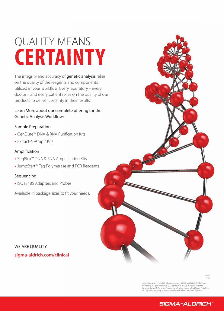
©2015 Sigma-Aldrich Co. LLC. All rights reserved. SIGMA and SIGMA-ALDRICH are trademarks of Sigma-Aldrich Co. LLC, registered in the US and other countries. GenElute, Extract-N-Amp, SeqPlex and JumpStart are trademarks of Sigma-Aldrich Co. LLC. Sigma-Aldrich Corp. is a subsidiary of Merck KGaA, Darmstadt, Germany.
CERTAIQUALITY ME
842691125
The integrity and accuracy of genetic analysis relies on the quality of the reagents and components utilized in your workflow. Every laboratory – every doctor – and every patient relies on the quality of our products to deliver certainty in their results.
Learn More about our complete offering for the Genetic Analysis Workflow:
Sample Preparation
• GenElute™ DNA & RNA Purification Kits
• Extract-N-Amp™ Kits
Amplification
• SeqPlex™ DNA & RNA Amplification Kits
• JumpStart™ Taq Polymerase and PCR Reagents
Sequencing
• ISO13485 Adapters and Probes
Available in package sizes to fit your needs.
WE ARE QUALITY.
sigma-aldrich.com/clinical
48-BC_MLO201602-Executive Snapshot_FINAL.indd CoverIV 1/12/2016 2:56:16 PM


