99
CT Venography Rich Hallett, MD Section Chief, CV Imaging, Northwest Radiology Network, Indianapolis Adjunct Clinical Assistant Professor, Stanford University, Cardiovascular Imaging Section

CT Venography
Rich Hallett, MD Section Chief, CV Imaging, Northwest Radiology Network, Indianapolis
Adjunct Clinical Assistant Professor, Stanford University, Cardiovascular Imaging Section
Introduction
• CT venography (CTV) is a technique targeted to assess venous anatomy, determine venous patency & delineate collateral circulation
• Non-invasive, simple protocols, wide anatomic coverage, short acquisition time, and ability to be combined with arterial-phase CTA

Lecture Outline
• Basic Clinical Options for Venous Imaging – Venous Imaging Modalities
• CT Scan Protocols – Indirect CTV – Direct CTV
• Selected Regional Applications – UE – Chest

Venous Imaging Modalities – The competition
• Doppler Ultrasound (US) • MR Venography • Catheter venography • Nuclear venography

Doppler US
• Well established clinical utility • No ionizing radiation • Portable • Inexpensive • Flow direction information • Operator / Patient dependent • Some areas inaccessible (pelvis, SVC) • Collateral pathways not well delineated

Doppler US
• Sens/ Spec ~ 95% for fem-pop DVT in ideal situations

Performance of Doppler vs. CTV in ICU patients – LE DVT
Sens Spec
Indiect CTV 70 96
Doppler US 70 100
Taffoni, AJR, 2004

MR Venography - Positives
• Excellent for pelvic venous system, CNS • May not require contrast • SI ratio thrombus:blood higher for MRV vs. CTV
– 3.7-8:1 vs 1.8-3.2 * • For PE: Sens 80-95%, Spec 95%, depends on
technique (Perf imaging best)+
• For DVT: Sens ~92%, Spec ~95% • 0.25 mmol/kg Gd better than 0.125 mmol/kg
+Sampson, Eur Radiol 2007; 17:175-181 * Kluge, AJR, 2006

Combo MR-PA / Indirect MRV
• MRA: TRuFISP, perfusion, MRA (0.25mmol/Kg)
• MRV: 3D FLASH w/ PV coil, voxel size of 1.2x0.8x1.1 mm – High agreement w/ CTA/CTV but requires a
change in coil and pt. position to obtain MRV after chest MRA
• Good agreement w/ Doppler in legs, moderate in pelvis
Kluge, AJR, 2006

MR Venography - Negatives
• Expensive, availability sometimes limited • Exam may be lengthy • Pt. cooperation? • Spatial resolution (vs other choices) • Limited anatomic coverage

Radionuclide Venography
• 99mTc-labeled MAA • 99mTc-labeled RBC • 99mTc-human serum albumin • 99mTc-labeled platelets
– Direct evidence of acute / active DVT – BUT: Arduous prep, false positives – pts on heparin
• 99mTc-apcitide (GIIb/IIIa receptor binding) – Can tell acute (+) vs. chronic (-) clot – Interpreter dependent?
Anatomic agents, indirect evidence

Catheter Venography
• Considered the �gold standard� • Invasive (but can treat lesions) • You only see what you can fill • Risks:
– Minor Complications: 18% – Thrombosis: 2% – Bronchospasm, Contrast reactions, etc

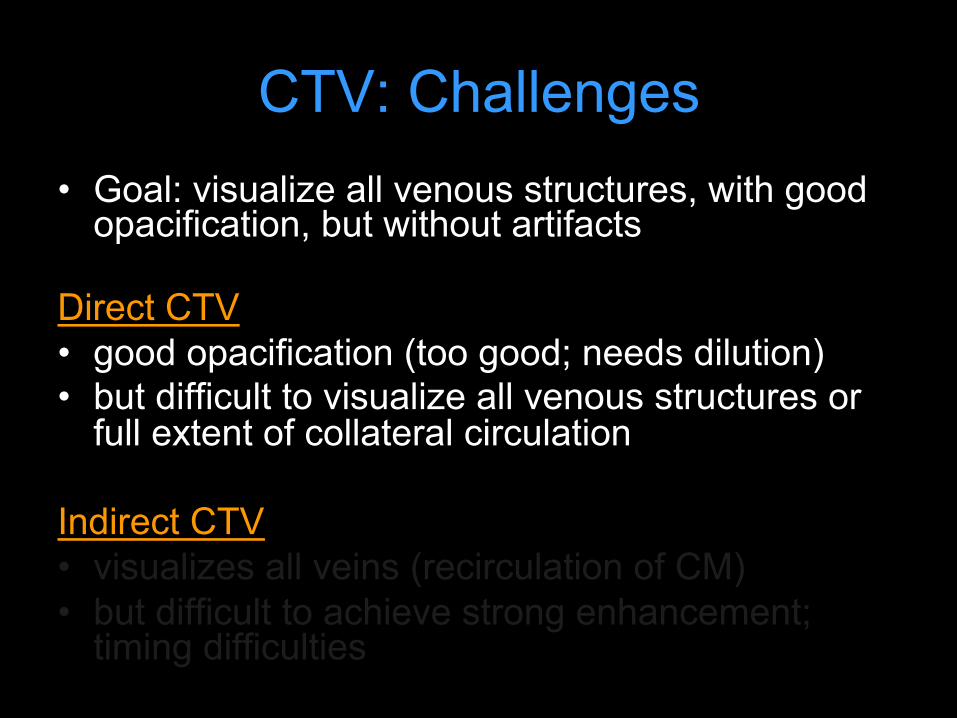
CTV: Challenges • Goal: visualize all venous structures, with good
opacification, but without artifacts
Direct CTV • good opacification (too good; needs dilution) • but difficult to show all venous structures or full
extent of collateral circulation
Indirect CTV • shows all veins; but difficult to achieve strong
enhancement; timing
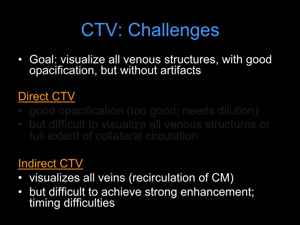
CTV: Challenges • Goal: visualize all venous structures, with good
opacification, but without artifacts
Direct CTV • good opacification (too good; needs dilution) • but difficult to visualize all venous structures or
full extent of collateral circulation
Indirect CTV • visualizes all veins (recirculation of CM) • but difficult to achieve strong enhancement;
timing difficulties

60M smoker, r/o lung cancer
Routine chest with contrast: 100cc contrast @ 2cc/sec, 40 sec diagnostic delay
CTV: Challenges • Goal: visualize all venous structures, with good
opacification, but without artifacts
Direct CTV • good opacification (too good; needs dilution) • but difficult to visualize all venous structures or
full extent of collateral circulation
Indirect CTV • visualizes all veins (recirculation of CM) • but difficult to achieve strong enhancement;
timing difficulties

L BRACHIAL, CEPHALIC VV. CLOT
LIJV STENOSIS
Indirect CTV

CTV: Imaging Techniques • Direct Venography (first pass):
– Dilute contrast (1:5 - 1:10) – Fill veins of interest (50cc or more) – Slow infusion, 1-2cc/sec – Start acquisition towards end of infusion
• Indirect Venography (recirculation) – 100-150cc contrast needed for adequate
venous opacification – Empiric imaging delay
• 60 seconds: upper extremity and pelvic veins • 3 to 3.5 min: lower extremity veins
– Smart prep off vein of interest Baldt MM, et al. Radiology 1996;200:423-428

40M prior left arm DVT. Acute pain and swelling of the left upper arm, rule out DVT.
1:5 dilution (20cc contrast + 80cc NS) @ 3cc/sec. Tourniquet around biceps region, released 15 sec before initiation of scan.
basilic
Brachial artery
basilic
cephalic
cephalic

CTV: Imaging Techniques • Direct Venography (first pass):
– Dilute contrast (1:5 or 1:6) – Fill veins of interest (50cc or more) – Slow infusion, 1-2cc/sec – Start acquisition towards end of infusion
• Indirect Venography (recirculation) – 100-150cc contrast needed for adequate
venous opacification – Empiric imaging delay
• 60 seconds: upper extremity and pelvic veins • 3 to 3.5 min: lower extremity veins
– Smart prep off vein of interest Baldt MM, et al. Radiology 1996;200:423-428

65M with metastatic lung ca and recent PEs. An IVC filter was placed but did not fully deploy. A second IVC filter was placed above the first one.
120cc contrast, diagnostic delay = 70sec

CTV: Imaging Techniques
• Direct Venography (first pass): – Dilute contrast medium (1:5 or 1:6) – Fill veins of interest (50cc or more) – Slow infusion, 1-2cc/sec – Start acquisition towards end of
infusion
Baldt MM, et al. Radiology 1996;200:423-428

CTV: Imaging Techniques • Indirect Venography (recirculation)
– ~ 150cc contrast needed for adequate venous opacification (2 mL/kg)
– Empiric imaging delay • 60 sec: thoracic • 70-80 sec: upper extremity • 11- sec: pelvis • 150 – 180 sec: lower extremity veins
– ? Smart prep off vein of interest – Want veins >80HU to be diagnostic
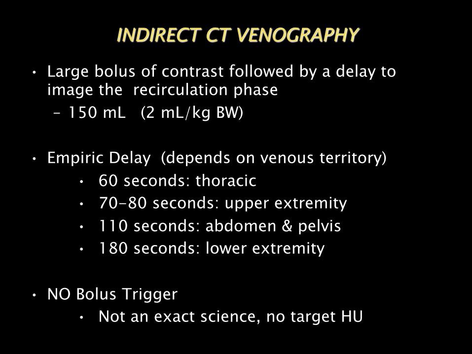
• Large bolus of contrast followed by a delay to image the recirculation phase – 150 mL (2 mL/kg BW)
• Empiric Delay (depends on venous territory) • 60 seconds: thoracic • 70-80 seconds: upper extremity • 110 seconds: abdomen & pelvis • 180 seconds: lower extremity
• NO Bolus Trigger • Not an exact science, no target HU
INDIRECT CT VENOGRAPHY

Combo Direct / Indirect CTV
• R/O LUE venous malformation; L hand and arm swelling
• 120 mL @ 5 mL/s followed by 100 mL 1:10 dilution at 2.5 mL/s via L hand IV
• Caudocranial acquisition

Combo Direct / Indirect CTV
Protocol and dataset courtesy of Scott Alexander, MD
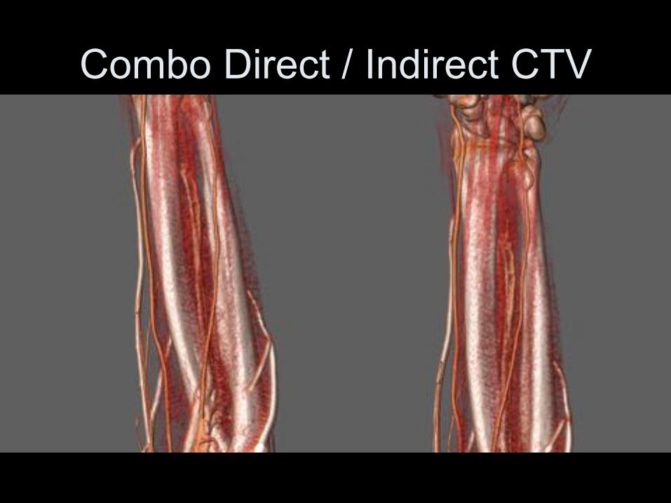
Combo Direct / Indirect CTV

Combo Direct / Indirect CTV

CTAforTOS:ComboDirect/IndirectCTA
• IpsilateralIV,armoverheadw/palmtapedup• Bolus:120mLfull-strength@4ml/s• Chase:100mLdilute(10%)[email protected]/s
• Caninjectcontralateralarmatsametime(dilute)• 65secempiricdelay,scancaudo-cranial• Armdown,immediatere-scancranio-caudal• VolumetricReview

MRAforTOS:BloodPoolMRA
• Anatomicimaging:ObliquesagandcorT1/T2• RelaxedandChallengedimaging:
§ Gadofosveset(bloodpoolagent)§ Breath-holdFSPGR,ECG-gated,highresolution(1.8mmST,448x448matrix)CORONALacquisition§ Challenged:ArmAbducted§ Relaxed:ArmDown

Arm UP Arm DOWN

Venography: Common Clinical Indications
Upper Extremity / Chest
– SVC syndrome (malignancy, post-XRT)
– Catheter-related complications (clot, stenosis)
– DVT – Thoracic Outlet syndrome – Dialysis access
Lower Extremity – DVT (+/- PE study) – May-Thurner syndrome – Pre-transplant evaluation
General - Venous stent evaluation - Vascular Malformations –
treatment planning

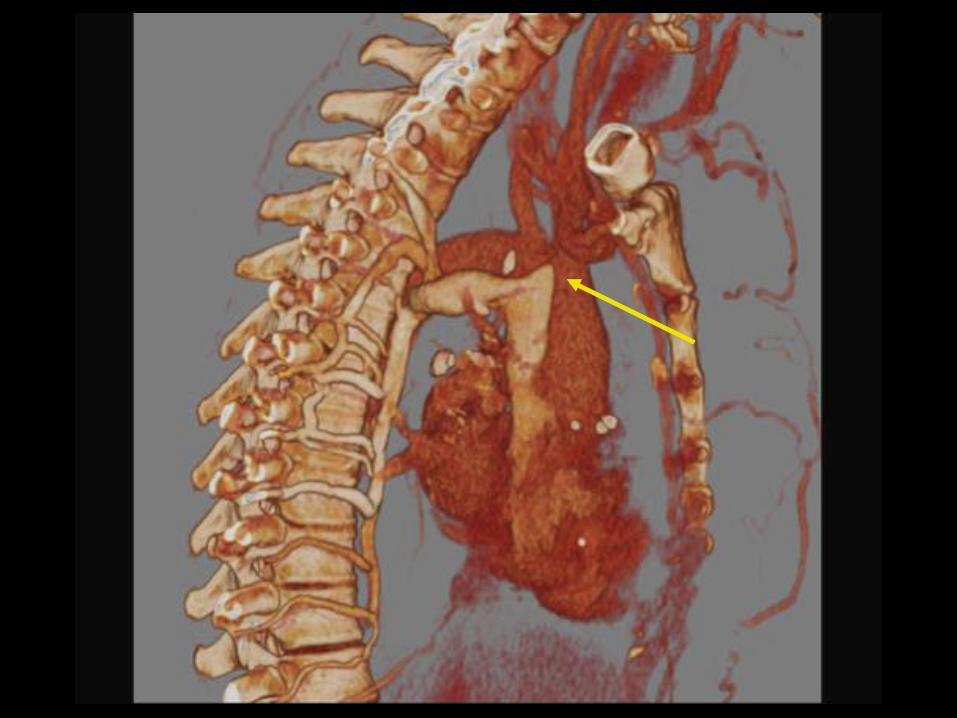
SVC Obstruction
• Stanford, et al.: Venography series with 4 main collateral pathways I. Partial SVC occlusion w/ patent Azygous v. II. Near complete obstruction SVC w/ antegrade
flow azygous à RA III. Near complete obstruction SVC w/
retrograde flow azygous IV. Complete obstruction SVC + one or more
major tributaries (e.g. azygous v.)
Stanford W, et al AJR 1987:148. 259-62.
NOT A COMPREHENSIVE SYSTEM!

SVC Occlusion
• Mass / Adenopathy • Catheter / Device (pacer / ICD leads) • Fibrosing Mediastinitis • Catheter + Mass • Catheter + pleural effusion • Thrombus • Catheter + lymph nodes
More common
Less common

SVC Syndrome from Tumor

Classification of all collateral pathways one series From: Cihangiroglu: J Comput Assist Tomogr, Volume 25(1).January/February 2001.1-8

Most common venous collaterals listed in order of frequency (n = 21). From: Cihangiroglu: J Comput Assist Tomogr, Volume 25(1).January/February 2001.1-8

A. 1 = superior vena cava 2 = inferior vena cava 3 = azygos vein 4 = hemiazygos vein 5 = accessory hemiazygos vein 6 = ascending lumbar vein 7 = lateral thoracic vein 8 = superficial epigastric vein 9 = internal mammary vein 10 = inferior epigastric vein 11 = pericardiophrenic vein 12 = right superior (highest) intercostal vein 13 = left superior (highest) intercostal vein 14 = intercostal vein 15 = inferior phrenic vein 16 = suprarenal vein
B 1 = superior vena cava 2 = brachiocephalic (innominate) vein 3 = subclavian vein 4 = internal jugular vein 5 = external jugular vein 6 = jugular venous arch 7 = superior thyroidal vein 8 = middle thyroidal vein 9 = inferior thyroidal vein 10 = facial vein 11 = anterior jugular vein 12 = vertebral venous plexus 13 = vertebral vein, and 14 = deep cervical vein From: Kim: J Comput Assist Tomogr, Volume
28(1).January/February 2004.24-33

Left Superior Intercostal Vein
Pericardiophrenic Vein

Inferior Phrenic v. (to IVC)

Lat thoracic v.
Intercostal veins
Azygous v.

Internal Mammary Veins Thoraco-acromio-Clavicular vv.
Areolar Venous Plexus

Paravertebral vv.
Capsular / Liver surface vv.
Systemic – portal collaterals

Venous collaterals organized by plexus systems – Easier, more complete to report
Cihangiroglu: J Comput Assist Tomogr, Volume 25(1).January/February 2001.1-8

The poster child for revised venous plexus nomenclature……
Chest / Upper extremity cases

ThoracicOutletSyndrome(TOS)
• Symptoma(ccompression/entrapmentofneurovascularstructuresbyboneand/orso7(ssueastheypassthroughthecervicoaxillarycanal
• 90%Neurogenic(PT,posturalTx,NSAIDs)• 10%Vascular• Venous>Arterial

Linda D D et al. Radiographics 2010;30:1373-1400
ComponentsofCervico-AxillaryCanal
• InterscaleneTriangle:#1siteofcompression
• CostoclavicularSpace:#1siteforvascularTOS
• Retro-pectoralisminorspace:#1siteformasses

CTAforTOS:ComboDirect/IndirectCTA
• IpsilateralIV,armoverheadw/palmtapedup• Bolus:120mLfull-strength@4ml/s• Chase:100mLdilute(10%)[email protected]/s
• Caninjectcontralateralarmatsametime(dilute)• 65secempiricdelay,scancaudo-cranial• Armdown,immediatere-scancranio-caudal• VolumetricReview

BilateralDirect/IndirectCTA

VenousTOS:�EffortThrombosis�
• Paget-Schroettersyndrome(PSS)• AKAaxillo-subclavianvenousthrombosis
• �Overhead�athletes• PEinupto1/3!!*• Post-thromboticsyndrome(later)
*PerlowskiAA.VascMed(2010)vol.15(6)pp.469-79

EffortThrombosis:36YOweightlifter

Post-Op1stribresection

ArterialTOS
• �Overheadathletes�• SX:Coolness,weakness,diffusearm
pain(ischemicneuritis)• Cause:Repetitivecompressioninjury
– Anatomicpredisposition(tightCCS)– Post-traumatic,bonycallus– Scalenehypertrophy

ArterialandVenousTOS:16YOVolleyballAthlete
REST
STRESS
SVC and central veins

LUNG CA with SVC syndrome
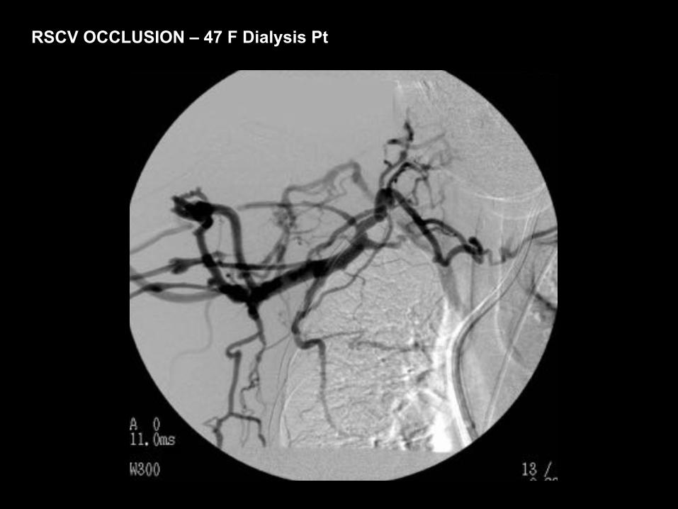
RSCV OCCLUSION – 47 F Dialysis Pt


35M hx thigh sarcoma. Facial swelling & chest wall varicosities when he bent over to tie his shoes. Documented central venous obstruction. Treatment planning: Assess vascular access, particularly axillary & subclavian veins B/L.
Simultaneous bilateral arm injection: 1: 6 dilution (30cc contrast + 170 cc NS, each arm) @ 2cc/sec. Courtesy of Anne Chin, MD

90cc contrast, 60 sec diagnostic delay. Imaging range: angle of mandible to lesser trochanters.
LT IJV

SVC Occlusion from Aneurysm

RSCV Occlusion – Previous Catheters


LT IJ injection 1:2 dilution (12cc contrast + 12cc NS @ 2cc/sec) acquired on flat-
panel detector Dyna-CT.
SVC
RIMV
L innominate Occlusion - C-Arm CT
Courtesy of Anne Chin, MD


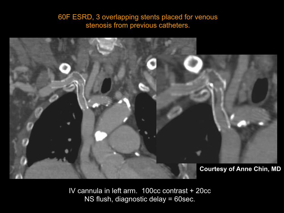
IV cannula in left arm. 100cc contrast + 20cc NS flush, diagnostic delay = 60sec.
60F ESRD, 3 overlapping stents placed for venous stenosis from previous catheters.
Courtesy of Anne Chin, MD

LIV encasement – Adenopathy

In-stent LIV / SVC thrombus

LIV Occlusion – Dialysis Patient with LUE AVF

EJ arch, lat thoracic, and pharyngeal
collaterals

100cc contrast, diagnostic delay = 60sec
62F central venous catheter for chemotherapy.

100cc contrast, diagnostic delay = 60sec
62F central venous catheter for chemotherapy.

MISC UE Cases

RUE Hemangiomatosis
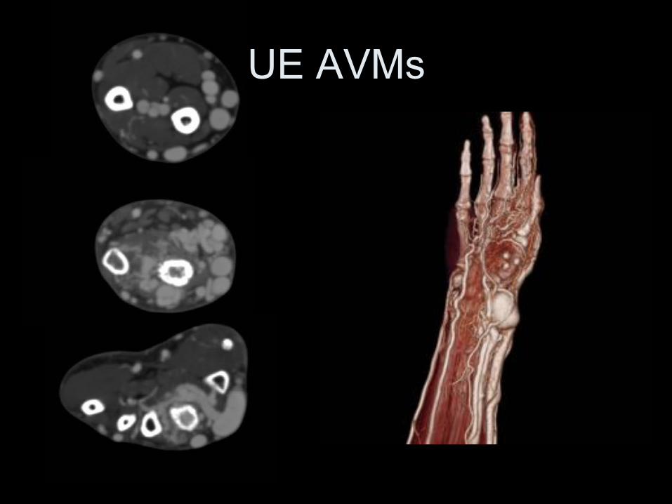
UE AVMs

Pelvis / LE Cases

MAY-THURNER : SUPERFICIAL VENOUS
VARICOSITIES


41 YO F, May - Thurner
Lack of Augmentation – �suspect upstream obstruction�
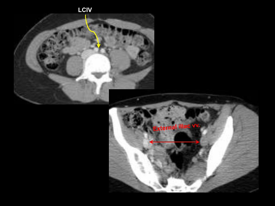
LCIV
External iliac vv.


S/P Mechnical Lysis, TPA, and PTA

Indirect Dx by arterial CTA

• 120 cc contrast • Monitoring delay = 40sec • Smart prep at infrarenal IVC
28F May-Thurner syndrome, CIV/EIV stent placement
3 years ago
Courtesy of Anne Chin, MD
F/U stenting for May Thurner

Vascular Mapping
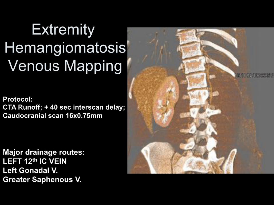
Extremity Hemangiomatosis Venous Mapping
Major drainage routes: LEFT 12th IC VEIN Left Gonadal V. Greater Saphenous V.
Protocol: CTA Runoff; + 40 sec interscan delay; Caudocranial scan 16x0.75mm

IVC Aneurysm

IVC Aneurysm • Rare • Saccular > fusiform • Cause unknown, may be related to anomalous
connections in embryologic venous systems – Acquired (trauma, AV fistulae) – May be associated with other congenital CV
anomalies • Sx: Thrombosis (7/16), pain, rupture, leg
swelling – Massive penile bleeding (1/16) – PE if thrombus

• CTV is a robust, non-invasive technique to visualize venous anatomy, and can be combined with arterial phase CTA
• Direct CTV: better opacification, less CM needed, but only the injected and downstream veins will be visualized
• Indirect CTV: all venous anatomy is delineated, empiric delay or smart-prep at ROI, opacification occasionally unpredictable
• �Combo CTV�: Perhaps the best choice for excellent and consistent venous opacification
• Provides accurate 3D visualization of venous anatomy for treatment planning
Conclusions
Thanks to:
Dominik Fleischmann, MD Frandics Chan, MD PhD

Loud PA, et al. Radiology 2001; 219:498-502. (Sens / Spec of CTV good compared to Doppler) Begemann PG, et al. J Comput Assist Tomogr 2003; 27:399-409. (Sensitivity=100%; Specificity=97% compared with ultrasonography) Baldt MM, et al. Radiology 1996; 200:423-428. (Sensitivity=100%; Specificiity=96% compared with conventional venography) Sampson, FC, et al. Eur Radiol 2007; 17:175-181. (Pooled sensitivity=91.5%; Pooled specificity=94.8% compared with conventional venography)
Key References
Kluge, A. et al. AJR 2006; 186:1686 – 1696 (Combo MRA/MRV for PE/DVT) Kim, HC, et al. J Comput Assist Tomogr 2004; 28:24-33 (Collateral Pathways) Cihangiroglu M, et al. J Comput Assis Tomogr 2001; 25: 1-8 (collaterals in SVC Obstruction) Lawler LP, et al. Radiographics 2002; 22:S45-S60 (normal and accessory chest venous pathways)

Kluge, A. et al. AJR 2006; 186:1686 – 1696 (Combo MRA/MRV for PE/DVT) Kim, HC, et al. J Comput Assist Tomogr 2004; 28:24-33 (Collateral Pathways) Cihangiroglu M, et al. J Comput Assis Tomogr 2001; 25: 1-8 (collaterals in SVC Obstruction) Lawler LP, et al. Radiographics 2002; 22:S45-S60 (normal and accessory chest venous pathways) Demos TC, et al. AJR 2004; 182:1139-1150 (Venous anomalies of chest)
Key References


