Cubital tunnel syndrome resulting from delayed intraneural hematoma of ulnar nerve
Yunqin Xu*, Yazhong Zhu, Shuiyun Feng and Zaiyue Liang
Department of Orthopedic Surgery, 98th Hospital of PLA, HuZhou City, ZheJiang Province, China 313000, China.
Accepted June 29, 2011
Cubital tunnel syndrome is the second most common peripheral compression neuropathy in adults. Close correlation between elbow trauma and cubital tunnel syndrome has been reported before. Factors causing cubital tunnel syndrome as a result of trauma may involve regional hemorrhages, edema, fibrosis, bone fracture, or displacement of bone fracture fragments into the cubital tunnel, causing narrowing of the tunnel. The pathogenesis due to delayed intraneural hematoma of ulnar nerve is extremely rare. In the present report we describe the first case of cubital tunnel syndrome due to delayed intraneural hematoma of ulnar nerve. A successful outcome may be expected if an appropriate surgical technique is chosen as early as more when worsening numbness or severe pain appears in the ring finger and little finger. Key words: Cubital tunnel syndrome, ulnar nerve, hematoma.
INTRODUCTION Cubital tunnel syndrome (CuTS) is a condition brought on by an increase in the pressure exerted upon the ulnar nerve at the elbow within the cubital tunnel. It is a well-recognized condition and is the second most common peripheral compression neuropathy
in adults after carpal
tunnel syndrome (Bozentka et al., 1998). Osborne (1957) referred to the entrapment neuropathy of the ulnar nerve at the elbow as tardy ulnar neuritis. Feindel (1958) renamed it cubital tunnel syndrome initially. Repetitive overhead activities are a primary cause of this condition (Norkus et al., 1994). The injure primarily occurs either with excessive or repetitive activity of the elbow or with acute trauma to the ulnar nerve (Barker et al., 1988; Folberg et al., 1994). Close correlation between elbow trauma and cubital tunnel syndrome has been reported in the literature (Jia et al., 2004). Factors causing cubital tunnel syndrome as a result of trauma may involve regional hemorrhages, edema, fibrosis, bone fracture, or displacement of bone fracture fragments into the cubital tunnel (Calisaneller et al., 2011), causing narrowing of the tunnel. Finally, the nerve can degenerate because of compression, pulling, or adhesion (Jia et al., 2004).
Despite the fact that several pathological entities can *Corresponding author. E-mail: [email protected]. Tel: 86-0572-7212984, Fax: 860-0591-83955075.
be potential mechanisms of the syndrome, the pathogenesis due to delayed intraneural hematoma is extremely rare. In the present report we describe the first case of cubital tunnel syndrome due to delayed intraneural hematoma of ulnar nerve. The source of the hematoma was found to be an intra- epineurium blood vessel bleed. CASE PRESENTATION A 22 year-old woman was admitted to our hospital in July 2005 reporting a 3-day history of sudden worsening numbness only in the ulnar lateral to the ring finger and little finger of right hand. The patient had suffered radiating pain from elbow to ring finger and little finger, especially at night. According to the McGowen classification, the patient had a Grade 3 ulnar neuropathy. She had no history of bleeding tendency or easy bruising. The patient had a history of falling down thrice from electromobile since two years ago; although the latest time is two months ago. A general examination revealed that a superficial scar of 3 × 4 cm in size was found in the medial to the right elbow. A subcutaneous lump about 3 × 3 cm in size can be palpated in proximal part of the ulnar nerve groove. On physical examination a remarkable tenderness was observed at the elbow, few centimeters distally to the medial epicondyle. Palpation of the lump
54 Med. Pract. Rev.
Figure 1. Pre-operative X-ray of the right elbow with no pathological findings.
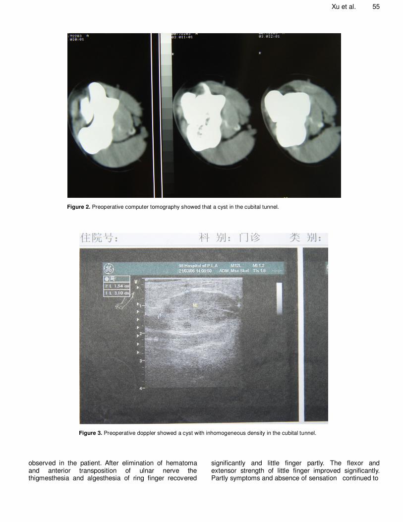
elicited radiating pain and numbness distally from the right elbow. Sensation defect in little finger and hypoesthesia in ulnar lateral to ring finger was found. Atrophy of the hypothenar or interosseous muscles was not found. The fromint syndrome of right hand is positive. Preoperative electroneuromyography (ENMG) was per-formed bilaterally using standard technique, denervation of ulnar nerve was confirmed on ENMG and the absolute motor nerve conduction velocity (NCV) of the ulnar nerve was less than 32 m/s. The X-rays evaluation of the elbow showed no pathological finding indicative for cubital tunnel syndrome (Figure 1). Preoperative computerized tomography (CT) was performed and found a cyst (3 × 3 cm in size) in the ulnar nerve groove (Figure 2). Doppler showed that a subfascia cyst (3 × 3 cm in size) without significant bleed in the ulnar nerve groove (Figure 3). Laboratory indices were as follows: white blood cell count 4300 × 10
9/L, haemoglobin 95 g/L, platelet cell count 187
× 109/L, prothrombin time 81.7% (normal 70 to 130%),
activated partial thromboplastin time 25.9 s (normal 23 to 35 s), fibrinogen 1.9 g/L (normal 2.0 to 4.0 g/L), bleeding time 1.0 min (control 1 to 3 min), D-dimer 0.25 mg/L
(normal 0 to 0.5 mg/L), CRP 7.5 mg/L (normal, <10 mg/L). Liver function tests were normal. No evidences of inherited or acquired deficiency of coagulation factors were encountered.
The patient still reported worsening numbness and severe pain after conservative treatment. And she had surgery 6 days after the first complaint of symptoms.
Surgical intervention was performed under brachial plexus anesthesia by well-trained surgeons in our institute. The patient was positioned supine, and the externally rotated arm was placed on the arm-table with the elbow flexed and the forearm in the supine position. A longitudinal axis skin incision of about 12 cm in length was made about the medial epicondyle. In this pro-cedure, a melanochroic hematoma (about 1.5 × 2.0 cm) inside the epineurium was found (Figure 4), and the inferior ulnar collateral artery accompanied the unlar nerve is intact. Epineurotomy was performed by surgeon microscope. No active bleed was found in the epineurium. The hematoma was eliminated thoroughly and the ulnar nerve was decompressed (Figure 5). The ulnar nerve was transposed anterior to the medial epicondyle subcutaneously. A pulley, erected to stabilize the ulnar nerve anteriorly, was made between the subcutaneous tissue and the fascia overlying the flexor pronator muscle mass.
The period of follow-up was 29 months. The patient was placed in sling postsurgery, and the patient was instructed to do various rehabilitative exercises, such as active range-of-motion exercises of the elbow joint, isometrics for the elbow flexor and extensor muscles, and hand, wrist and forearm strengthening as tolerated.
Neurologists and surgeons evaluated the postoperative neurological findings. A control ENMG after three month showed markedly recovery and regeneration. NCV of the ulnar nerve was more than 55 m/s. No complication was
Xu et al. 55
Figure 2. Preoperative computer tomography showed that a cyst in the cubital tunnel.
Figure 3. Preoperative doppler showed a cyst with inhomogeneous density in the cubital tunnel.
observed in the patient. After elimination of hematoma and anterior transposition of ulnar nerve the thigmesthesia and algesthesia of ring finger recovered
significantly and little finger partly. The flexor and extensor strength of little finger improved significantly. Partly symptoms and absence of sensation continued to
56 Med. Pract. Rev.
Figure 4.The entrapment of the ulnar nerve at the elbow area by melanochroic intraneural hematoma.
Figure 5. In operation the epineurium of ulnar nerve was cut open, and the hematoma was cleared completely.
have hypoaesthesia three months after operation. After rehabilitative exercises the grip strength of the right hand,
as measured with dynamometer, was fully restored to normal at six months from the operation and she returned
Xu et al. 57
Figure 6. Only slight atrophy of the hypothenar was observed in the final follow-up.
Figure 7. The fromint syndrome of right hand is negative.
to everyday textile work (Figure 6). At the time of this writing the patient is asymptomatic and no evidence of recurrence was seen at 29 months of follow-up. Only
slight atrophy of the hypothenar was observed in the final follow-up (Figure 6), and the fromint syndrome of right hand is negative (Figure 7).
58 Med. Pract. Rev. DISCUSSION
The pathogenesis of cubital tunnel syndrome due to intraneural hematoma is extremely rare. Neoplasms that have been reported as causes of cubital tunnel syndrome include: ganglions (Kato et al., 2002), haemangiomas (Nakamura et al., 1996), synovial cysts (Barss et al., 1984) and a giant cell tumour of the tendon sheaths (Mitsionis et al., 2006). The hematomaes are usually localized in brain or joint (such as the knee, the hip, the elbow and the wrist joint), but they less affect the peripheral nerve’s function. In the presented case, intraneural hematoma was responsible for the acute CuTS. Many kinds of reasons may cause the haematoma, such as the primary disease, the trauma or the inappropriate pharmacological treatment. Muscle bleed in hemophilics can result in compressive neuropathy and permanent disability if not treated properly in time (Saraf et al., 2003). Lyon and Hansen (1975) found that the brachial plexus of 4 patients was damaged due to haematoma or pseudoaneurysm-formation at the site of the arterial puncture. Puschmann and Neundörfer (1991) reported that two cases on femoral nerve lesion due to hematoma in heparin therapy. Delayed hematoma often occurred from several hours to several days after the accident (Fankhauser et al., 1983). But the delayed hematoma especially inside epineurium had never been reported. In the present study, we describe an extremely rare case of cubital tunnel syndrome due to intraneural hematoma of ulnar nerve at the elbow. To our knowledge this is the first reported case of the syndrome with such aetiology.
One of the causes of the delayed hematoma or hemorrhage is an intraneural microvasculature pseudo-aneurysm. The endangium of intraneural micro-vasculature was injured in these slight traumas (falling down from electromobile thrice). A microvasculature pseudo-aneurysm may rarely develop after slight trauma. As the disease progresses, the microvasculature pseudo-aneurysm may rupture suddenly sometime, the ulnar nerve was compressed by the hematoma in cubital tunnel. The patient may complain radiating pain from elbow to ring finger and little finger, or hypoesthesia combined with weakness in the ulnar lateral to the ring finger and little finger as an initial symptom. Long-standing compression causes damage in the myelin sheath and axonal degeneration induced by hematoma, with permanent loss of nerve function and atrophy of the denervated muscles.
Therefore, an accurate and prompt diagnosis made by sono-graphy or CT scan or MRI is the must in choosing proper strategy for the management of cubital tunnel syndrome which resulted from delayed hematoma. Surgery is used to perform chronic neuropathy associated with muscle weakness or neuropathy that does not respond to conservative measures (Shunji et al., 2005). We suggest that patients with fixed sensory loss, pain, weakness, or significant denervation on ENMG should be considered for surgery regardless of acute, sub-acute or chronic course. Preoperative
electromyography (ENMG) and nerve conduction velocity studies were performed bilaterally in all patients using standard techniques. Electrodiagnostic studies are useful to confirm the diagnosis of cubital tunnel syndrome, and to determine the severity of the disease to localize the area of compression in some cases, and to exclude other sites of compression (Simsek et al., 2011). Motor and sensory conduction velocities were determined in a standardized fashion, evaluating the ulnar nerve across the elbow from below the elbow to wrist (Puschmann et al., 1991). Many surgical treatments were performed in cubital tunnel syndrome including simple decompression (or also referred to decompression without transpoition), decompression with transpoition, medial epicon-dylectomy, cubital tunnel reconstruction (Tsujino et al., 1997), now no decision could be made with confidence concerning which mode of operation is the best treatment for CuTS. A symptomatic nerve compression duration of more than three and half days was the critical factor that determined if neurapraxia developed into severe nerve damage (Roganović et al., 2007). At this time a successful outcome may be expected if an appropriate surgical technique is chosen as early as more.
We report this case to increase high index of suspicion of cubital tunnel syndrome resulted from delayed hematoma while a patient complains of sudden worsen-ing numbness in the ulnar lateral to ring finger and little finger and/or suffers radiating pain from elbow to ring finger and little finger after trauma, perhaps this trauma is has been on several months ago. A surgical treatment should be concerned when worsening numbness or severe pain in ring finger and little finger. REFERENCES
Barker C (1988). Evaluation, treatment, and rehabilitation involving a
submuscular transposition of the ulnar nerve at the elbow. Athletic Train. JNATA, 23: 10-1112.
Barss P (1984). Ulnar compression neuropathy due to an occult post-traumatic synovial cyst. Med. J., 140: 428-429.
Calisaneller T, Ozdemir O, Caner H, Altinors N (2011). Simple decompression of the ulnar nerve at the elbow via proximal and distal mini skin incisions. Turk. Neurosurg., 21(2): 167-171.
Fankhauser H, Uske A, de Tribolet N (1983) Delayed epidural hematoma. Apropos of a series of 8 cases. Neurochirurgie. 29(4):
palsy. Can. Med. Assoc. J. 78: 351-353. Folberg CR, Weiss AP, Akelman E (1994). Cubital tunnel syndrome.
Presentation and diagnosis. Orthop. Rev., 23: 136-144. Jia ZR, Shi X, Sun XR (2004). Pathogenesis and electrodiagnosis of
cubital tunnel syndrome. Chin. Med. J. Engl. 117(9): 1313-1316. Kato H, Hirayama T, Minami A, Iwasaki N, Hirachi K (2002). Cubital
tunnel syndrome associated with medial elbow ganglia and osteoarthritis of the elbow, J. Bone Joint Surg. Am. 84A: 1413-1419.
Lyon BB, Hansen BA, Mygind T (1975). Peripheral nerve injury as a complication of axillary arteriography. Acta Neurol Scand. 51(1): 29-36.
Mitsionis G, Pakos EE, Gavriilidis I, Batistatou A (2006). Cubital tunnel syndrome due to giant cell tumour of tendon sheaths. Hand Surg, 11(1-2): 89-91.
Nakamura I, Hoshino Y (1996). Extraneural hemangioma: A case report
of acute cubital tunnel syndrome, J. Hand Surg. Am. 21: 1097-1098. Norkus SA, Meyers MC (1994). Ulnar neuropathy of the elbow. Sports
Med, 17: 189-199. Osborne G (1957). The surgical treatment of tardy ulnar neuritis. J.
Bone Joint Surg. Br., 39: 782. Puschmann E, Neundörfer B, Bauer J (1991). Femoral nerve lesion in


![Electrophysiological Features of Ulnar Tunnel Syndrome ... · Ulnar tunnel syndrome (UTS) is an uncommon ulnar entrapment neuropathy. Guyon [1] described the anatomy of the area in](https://static.documents.pub/doc/80x56/601bca5f935324075a08994b/electrophysiological-features-of-ulnar-tunnel-syndrome-ulnar-tunnel-syndrome.jpg)


