266 Chapter Cytopathology David C. Wilbur T he history of cytopathology at the Massachusetts General Hospital (MGH) is linked firmly with the beginning of the mod- ern practice of gynecologic cytopathology. Dr. George Papanicolaou, working at Cornell Medi- cal Center in New York City, is widely recognized as the father of gynecologic cytology, although the significant contributions of Dr. Aurel Babeş of Romania should also be noted. It took pio- neers in the adaptation of the Pap smear tech- nique, however, to further our understanding of its utility and role as the first widely used method of cancer screening. Much of that adoption and early refinement took place at the MGH. Papanicolaou and Babeş first presented their classic papers on the use of cytologic specimens to diagnose cervical cancer in 1928, Papanicolaou at the ird Race Betterment Conference in Bat- tle Creek, Michigan (1), and Babeş in the French literature (2). On January 5, 1928, the New York World reported that “although Dr. Papanicolaou is not willing to predict how useful the new diag- nostic method will be in the actual treatment of malignancy, it seems probable that it will prove valuable in determining cancer in the early stages of its growth when it can be more easily fought and treated.” Although the method was promis- ing, there was little interest in the medical com- munity (including pathology) for this test, and Papanicolaou returned to Cornell, where he con- tinued his own investigations related primarily to the cytologic changes in the female genital tract associated with hormones. In 1939 Dr. Joseph Hinsey, the Chairman in the Department of Anatomy at Cornell, encouraged Papanicolaou, along with Dr. Herbert F. Traut, a Cornell gyne- cologist, to engage in a clinical trial of the cyto- logic detection of cancer in the female genital tract. ese studies led to the landmark study by Papanicolaou and Traut, entitled “e Diagnos- tic Value of Vaginal Smears in Carcinoma of the Uterus,” which was presented publicly in March 1941 and published in the American Journal of Obstetrics and Gynecology in August 1941 (3). e beginnings of clinical cytopathology thus date to 1941. MGH Cytology: The Early Years e history of cytology at MGH also begins in 1941. e first events, however, took place not in the department of Pathology but in the Vincent Hospital (the Gynecologic Service at the MGH). An internist practicing at the MGH, Dr. Mau- rice Fremont-Smith (figure 19.1), who was highly interested in the relatively new concept of pre- ventive medicine, approached Dr. Joe Vincent Meigs (figure 19.2), Chief of the Vincent Service and a prominent gynecologist, with the paper by Papanicolaou and Traut. According to a letter written by Dr. Priscilla Taft (see below) in 1993, Dr. Fremont-Smith had visited Papanicolaou’s laboratory in New York. Dr. Meigs greatly val- ued diagnostic tissue pathology and was known to review specimens from his patients routinely,
Transcript
266
Chapter
CytopathologyDavid C. Wilbur
The history of cytopathology at the Massachusetts General Hospital (MGH) is
linked fi rmly with the beginning of the mod-ern practice of gynecologic cytopathology. Dr. George Papanicolaou, working at Cornell Medi-cal Center in New York City, is widely recognized as the father of gynecologic cytology, although the signifi cant contributions of Dr. Aurel Babeş of Romania should also be noted. It took pio-neers in the adaptation of the Pap smear tech-nique, however, to further our understanding of its utility and role as the fi rst widely used method of cancer screening. Much of that adoption and early refi nement took place at the MGH.
Papanicolaou and Babeş fi rst presented their classic papers on the use of cytologic specimens to diagnose cervical cancer in 1928, Papanicolaou at the Th ird Race Betterment Conference in Bat-tle Creek, Michigan (1), and Babeş in the French literature (2). On January 5, 1928, the New York World reported that “although Dr. Papanicolaou is not willing to predict how useful the new diag-nostic method will be in the actual treatment of malignancy, it seems probable that it will prove valuable in determining cancer in the early stages of its growth when it can be more easily fought and treated.” Although the method was promis-ing, there was little interest in the medical com-munity (including pathology) for this test, and Papanicolaou returned to Cornell, where he con-tinued his own investigations related primarily to the cytologic changes in the female genital tract
associated with hormones. In 1939 Dr. Joseph Hinsey, the Chairman in the Department of Anatomy at Cornell, encouraged Papanicolaou, along with Dr. Herbert F. Traut, a Cornell gyne-cologist, to engage in a clinical trial of the cyto-logic detection of cancer in the female genital tract. Th ese studies led to the landmark study by Papanicolaou and Traut, entitled “Th e Diagnos-tic Value of Vaginal Smears in Carcinoma of the Uterus,” which was presented publicly in March 1941 and published in the American Journal of Obstetrics and Gynecology in August 1941 (3). Th e beginnings of clinical cytopathology thus date to 1941.
MGH Cytology: The Early Years
Th e history of cytology at MGH also begins in 1941. Th e fi rst events, however, took place not in the department of Pathology but in the Vincent Hospital (the Gynecologic Service at the MGH). An internist practicing at the MGH, Dr. Mau-rice Fremont-Smith (fi gure 19.1), who was highly interested in the relatively new concept of pre-ventive medicine, approached Dr. Joe Vincent Meigs (fi gure 19.2), Chief of the Vincent Service and a prominent gynecologist, with the paper by Papanicolaou and Traut. According to a letter written by Dr. Priscilla Taft (see below) in 1993, Dr. Fremont-Smith had visited Papanicolaou’s laboratory in New York. Dr. Meigs greatly val-ued diagnostic tissue pathology and was known to review specimens from his patients routinely,
pathology_chap19.indd 266 8/16/11 10:22 AM
Cytopathology
267
both grossly and microscopically. He saw the potential for this new technology and assigned Ruth M. Graham (fi gure 19.1), a zoologist and medical technologist, to pursue the development and implementation of this technique in Boston.
Ruth Moore Graham was born in 1917 in Paris, Idaho, the daughter of a general medical practitioner. She received her bachelor’s degree in zoology in 1938 from the University of Michigan. Following her graduation, she studied labora-tory technology for a year at Simmons College in Boston. Her fi rst position was at the Hun-tington Memorial Hospital, where she worked as a hematology technician on projects involving chemotherapy eff ects in leukemia. In 1940 she married Dr. John B. Graham, a newly graduated Harvard Medical School (HMS) physician, who subsequently trained as a gynecologist. Th e Gra-hams initially lived in San Francisco, where Ruth
Graham worked in the coroner’s offi ce, gaining observational skills in autopsy pathology and learning histologic techniques. Th ey returned to Boston in 1941, and Ruth Graham initially became a Research Assistant of Dr. J. H. Means in the MGH department of Medicine, where her research involved investigations on respiratory enzymes (4), until the following year, when she moved to the Vincent Laboratories.
In response to Dr. Meigs’s assignment, Gra-ham spent a week in the Papanicolaou laboratory at Cornell in New York City and then returned to Boston to implement the technique. Th e Gyne-cologic Cytology Laboratory was thus established in the Vincent Laboratories during 1942, and it most probably represents the second organized cytology laboratory in the United States, after Papanicolaou’s one in New York.
Th e Vincent investigators quickly confi rmed
Figure 19.1 Members of the Vincent Laboratory staff (1947) surround (seated, left to right) Ruth Graham, George Papanicolaou, and Maurice Fremont-Smith.
(Photo courtesy of Dr. Maurice Fremont-Smith, grandson of the pictured Fremont-Smith)
Figure 19.2 Joe V. Meigs, Chief of the Vincent Service (MGH archives)
pathology_chap19.indd 267 8/16/11 10:22 AM
Keen Minds to Explore the Dark Continents of Disease
268
grossly examined, a stir was created because no gross evidence of carcinoma was present in either cervix. Upon histologic examination, however, both specimens were indeed found to have squa-mous carcinoma in situ, proving Graham correct (4). A further study performed by Graham and Dr. John J. McGraw, a research fellow in Pathol-ogy (and hence the fi rst offi cial pathologist at the MGH to practice cytopathology), was published in 1948 showing the accuracy of the cytology specimen in comparison to the corresponding biopsy for cervical squamous carcinoma (7).
In 1947 Dr. Sommers H. Sturgis was appointed Chief of the Vincent Laboratories, and the unit moved to new quarters. Also in 1947 Graham published her fi rst reports on the eff ects of radia-tion on cells in the vaginal smear (8, 9). Th ese
the original fi ndings of the Papanicolaou and Traut study in a manuscript entitled “Th e Value of the Vaginal Smear in the Diagnosis of Uterine Cancer,” which was published in Surgery Gynecol-ogy and Obstetrics in 1943 (5), and in a larger study of over 1,000 patients in 1945 (6). Th e association with the Cornell Laboratory remained strong, and Meigs and Graham reportedly insisted on including Dr. Papanicolaou in a scientifi c exhibit they were asked to prepare for the American Medical Association meeting in Chicago during 1944. Th e exhibit was entitled “Vaginal Smear in the Diagnosis of Cancer,” and for their eff orts the group (Papanicolaou, Traut, and Andrew A. Marchetti from Cornell and Meigs, Fremont-Smith, Graham, and Lois T. Janzen from the Vincent Laboratories) won a certifi cate of merit (fi gure 19.3).
One famous anecdote illustrates how cytology was cemented into routine practice at the Vincent Lab. Dr. Meigs performed two hysterectomies (one for leiomyomas, the other for endometrio-sis) on patients who also had preoperative vaginal smears that Graham read as positive for carci-noma. When the specimens were removed and
Figure 19.3 Certifi cate of Merit from the American Medical Association received by the Vincent Laboratory and Cornell Medical Center in 1944 for work verifying the cytologic method for the detection of cervical cancer
Figure 19.4 Monograph entitled Th e Cytologic Diagnosis of Cancer; the fi rst textbook of cytology,
which was published by Ruth Graham and the Vincent Laboratory staff in 1950
pathology_chap19.indd 268 8/16/11 10:22 AM
Cytopathology
269
and subsequent studies indicated that specifi c morphologic patterns of cells were prognostic and thus could guide which patients received or did not receive radiotherapy. Th is work was ini-tially accepted and reproduced by other investi-gators in Europe and Mexico, but, in the end, defi nitive studies showed that these parameters were not prognostic. Th is would become a source of confl ict in Ruth Graham’s professional career: the individual discounting her work was one of her close associates at MGH, Dr. Robert Fennell (see below), when he was later working at the University of Pittsburgh (10).
In the 1948 Vincent annual report Dr. Meigs wrote, “Mrs. Graham has really placed our Labo-ratory in a very strong position. . . . Th e Smear
Laboratory is established . . . as a routine proce-dure.” Ruth Graham and her colleagues in the Vincent Laboratories published in 1950 the fi rst comprehensive textbook of cytology, Th e Cyto-logic Diagnosis of Cancer (11), under the spon-sorship of the American Cancer Society, which was rapidly emerging as a force in the promotion of the cytologic method of screening for cervi-cal cancer (fi gure 19.4). By this time the labora-tory was not only processing vaginal smears, but was also examining exfoliative specimens from numerous other body sites. Th us, in addition to reviewing the investigations in gynecologic cytol-ogy, this monograph covered exfoliative cytology of other sites, including the respiratory, gastro-intestinal, urinary, and body cavity systems. Th e
Figure 19.5 Two pages from Th e Cytologic Diagnosis of Cancer showing photomicrographs and hand-drawn illustrations
pathology_chap19.indd 269 8/16/11 10:22 AM
Keen Minds to Explore the Dark Continents of Disease
270
monograph is beautifully illustrated with both microphotographs and drawings detailing spe-cifi c fi ne structures, and the accompanying text supplies descriptions necessary for understanding the importance and context of identifi ed features (fi gure 19.5).
The Transition to Pathology, –
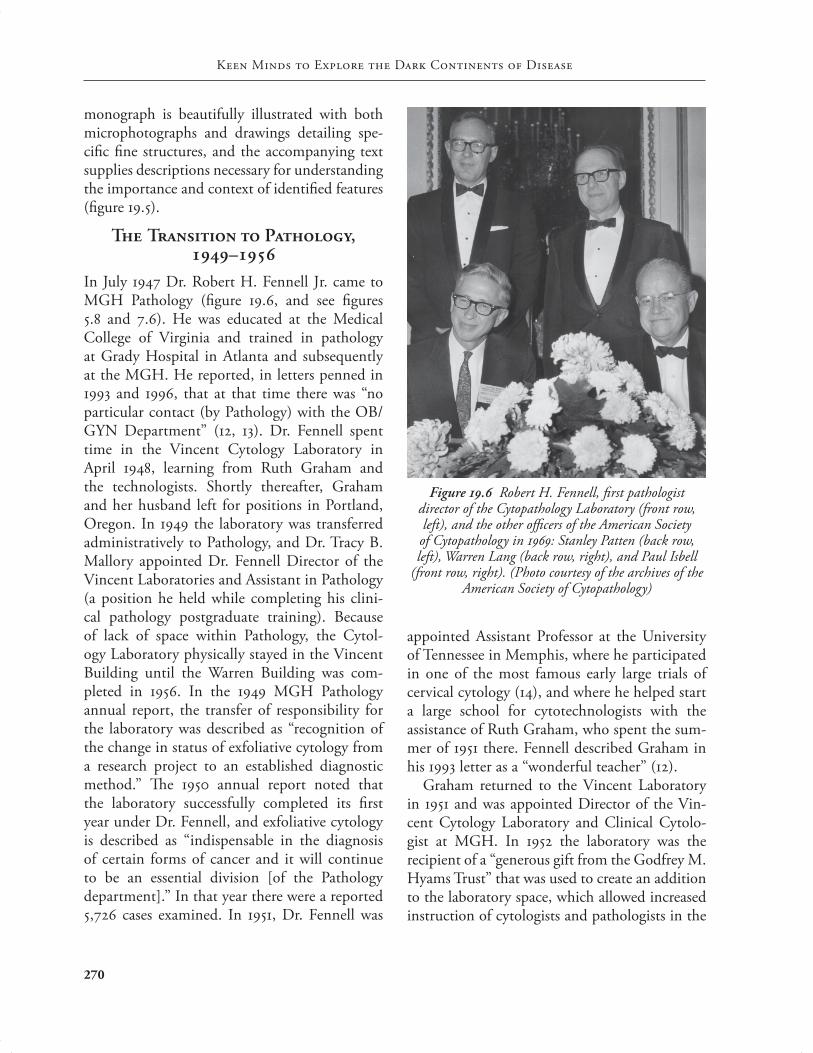
In July 1947 Dr. Robert H. Fennell Jr. came to MGH Pathology (fi gure 19.6, and see fi gures 5.8 and 7.6). He was educated at the Medical College of Virginia and trained in pathology at Grady Hospital in Atlanta and subsequently at the MGH. He reported, in letters penned in 1993 and 1996, that at that time there was “no particular contact (by Pathology) with the OB/GYN Department” (12, 13). Dr. Fennell spent time in the Vincent Cytology Laboratory in April 1948, learning from Ruth Graham and the technologists. Shortly thereafter, Graham and her husband left for positions in Portland, Oregon. In 1949 the laboratory was transferred administratively to Pathology, and Dr. Tracy B. Mallory appointed Dr. Fennell Director of the Vincent Laboratories and Assistant in Pathology (a position he held while completing his clini-cal pathology postgraduate training). Because of lack of space within Pathology, the Cytol-ogy Laboratory physically stayed in the Vincent Building until the Warren Building was com-pleted in 1956. In the 1949 MGH Pathology annual report, the transfer of responsibility for the laboratory was described as “recognition of the change in status of exfoliative cytology from a research project to an established diagnostic method.” Th e 1950 annual report noted that the laboratory successfully completed its fi rst year under Dr. Fennell, and exfoliative cytology is described as “indispensable in the diagnosis of certain forms of cancer and it will continue to be an essential division [of the Pathology department].” In that year there were a reported 5,726 cases examined. In 1951, Dr. Fennell was
appointed Assistant Professor at the University of Tennessee in Memphis, where he participated in one of the most famous early large trials of cervical cytology (14), and where he helped start a large school for cytotechnologists with the assistance of Ruth Graham, who spent the sum-mer of 1951 there. Fennell described Graham in his 1993 letter as a “wonderful teacher” (12).
Graham returned to the Vincent Laboratory in 1951 and was appointed Director of the Vin-cent Cytology Laboratory and Clinical Cytolo-gist at MGH. In 1952 the laboratory was the recipient of a “generous gift from the Godfrey M. Hyams Trust” that was used to create an addition to the laboratory space, which allowed increased instruction of cytologists and pathologists in the
Figure 19.6 Robert H. Fennell, fi rst pathologist director of the Cytopathology Laboratory (front row, left), and the other offi cers of the American Society of Cytopathology in 1969: Stanley Patten (back row, left), Warren Lang (back row, right), and Paul Isbell
(front row, right). (Photo courtesy of the archives of the American Society of Cytopathology)
pathology_chap19.indd 270 8/16/11 10:22 AM
Cytopathology
271
cytologic method. Fennell returned from Mem-phis to MGH Pathology in 1953, recruited by Dr. Benjamin Castleman because of the military service commitments of Drs. Robert Scully and Austin Vickery. During the ensuing years, Dr. Fennell worked closely with Ruth Graham, exam-ining cases of cytologic squamous carcinoma in situ for which no lesion had initially been iden-tifi ed on histologic examination. Th ese studies clearly showed that in cases where an “experi-enced cytologist diagnosed a certain lesion, mul-tiple sections would eventually show a correlation with the cytology fi ndings” (15, 16). In addition, these studies led to identifi cation and study of a subset of early invasive (microinvasive) squa-mous cell carcinomas (17). Dr. Fennell observed in his letters that although cytology now played a defi nitive role in the department, there was always skepticism of the method by the domi-nant surgical pathology–oriented group, and that the Cytology Unit held a secondary position in terms of resources and prestige (12, 13).
In 1954 the Vincent Cytology Laboratory and HMS were awarded a grant by the American Cancer Society designed as a “group attack on cervical cancer.” Th is grant was administered by John and Ruth Graham and included not only MGH but also several other hospitals in the Bos-ton area. Th e project consisted of selection of cancer patients for surgical or radiation therapy and a compilation of the results. Th at year, for her many eff orts in cytology and cervical cancer research, Ruth Graham was awarded an honor-ary doctor of sciences degree from the Women’s Medical College of Philadelphia.
In 1955 Dr. Fennell moved to the University of Pittsburgh (Magee Hospital), where he was appointed Assistant Professor of Pathology. By this time the cytology specimen volume had steadily grown to nearly 10,000 annually. Dr. Fennell went on to be an active participant in the cytol-ogy professional organizations (see below) and an active investigator in gynecologic pathology and cytology during his post-MGH career (18).
The Taft Years, –
In January 1957 cytology operations moved to the newly opened Warren Building. During that year Dr. Priscilla Dienes Taft (known as “Piri” to close friends) was appointed Director of the Cytopa-thology Laboratory and Assistant in Pathology (fi gure 19.7), after having completed an Ameri-can Cancer Society fellowship in cytology the year before, learning under Ruth Graham and her colleagues. She replaced Ruth Graham as the director when Graham left for Buff alo, New York, and a position at the Roswell Park Cancer Institute.
Dr. Taft, the daughter of the distinguished MGH microbiologist and immunologist Dr. Louis Dienes (chapter 21), was born in Budapest on May 25, 1917, but the family soon moved to the United States. She graduated from Radcliff e College in 1938 and studied medicine at Yale University, graduating in 1944. She completed
Figure 19.7 Priscilla Dienes Taft, Director of the Cytopathology Laboratory, 1957–1983 (MGH archives)
pathology_chap19.indd 271 8/16/11 10:22 AM
Keen Minds to Explore the Dark Continents of Disease
272
postgraduate training in pathology at the Mal-lory Institute of Boston City Hospital and at the University of Kansas. She was married to another long-term faculty member of MGH Pathology, Dr. Edgar Taft. During her quarter century as Director, the Cytology Laboratory specimen volume grew to over 30,000 annually, and the service became a valuable teaching unit in the Pathology department. In his request for Dr. Taft’s promotion to Assistant Professor of Pathol-ogy, Dr. Castleman described her as having done a “superb job in the organization and running of this department.” Early in her career she was involved in the research mission of the depart-ment as it related to cytology, working with Drs. Lazlo Vincze (a research fellow in Surgery, assigned to Gynecology at the Vincent Memo-rial Hospital) and Janet W. McArthur, a gyne-cologist, on the changes in squamous cells during the menstrual cycle in cytology specimens from vaginal, urinary, and buccal locations (19), and with Dr. John Raker on circulating tumor cells in the peripheral blood (20); she also wrote on opti-mal preparation and analysis methods for cyto-logic specimens (21, 22). Later in her career she was involved in research related to therapy eff ect in cells and urinary cytology in renal transplant patients (23). She also wrote an important review of the cytology of clear cell carcinoma of the gen-ital tract (24) and worked on studies detailing the cytologic eff ects of exposure to diethylstibesterol (DES) (25). Jean Buchanan (see below) described Dr. Taft as “constant—she was always there. She was knowledgeable.” Dr. Taft served as Director of the Cytology Laboratory until her retirement in 1983. In 1992 the MGH cytopathology fel-lowship was named in honor of Dr. Taft and her longtime service to the unit and to the hospital.
At the time Dr. Taft was appointed Director, the chief cytotechnologist was Lina Neri, who had studied under Ruth Graham and at Memo-rial Hospital in New York before returning to join the Vincent Laboratory team. In 1958 Neri resigned to work in the endocrinology research
laboratories at MGH, and she was replaced by Jean Pasakanas (later Buchanan), who remained the Chief Cytotechnologist until her retirement in 1989 (fi gure 19.8). In 1958 there were six cyto-technologist staff members and one secretary, processing about 10,000 exfoliative cytology specimens, of which 83 percent were gynecologic. In response to a 1972 request for a departmental report on cytology, Dr. Taft wrote that by 1970 the cytology staff had increased to 13 people, still in a space of only 415 square feet. She wrote, “Crowding exacerbates minor problems so at times it is diffi cult to obtain and keep staff ,” and “It is hoped that relief will be forthcoming.” At this point specimen volume had grown to over 23,000 annually. Under Taft, Neri, and Buchanan a number of American Cancer Society–funded trainees took a three-month course in exfoliative cytology in the laboratory. One of these trainees was Dr. Bernard Naylor, who later became the Director of Cytology at the University of Michi-gan and who had a very distinguished career in the discipline. As cytology programs developed in Boston, the number of technologists and
Figure 19.8 Jean Buchanan, Chief Supervisor of the Cytopathology Laboratory, 1959–1989 (left), with Wanda Szyfelbein, Laboratory Director, 1983–1989
(right) (Photo courtesy of Jean Buchanan)
pathology_chap19.indd 272 8/16/11 10:22 AM
Cytopathology
273
in Poland and fi nished her pathology training at Boston University Hospital and at the MGH in 1974. She was primarily interested in fi ne needle aspiration biopsy and made several important contributions to that literature, including a 1994 monograph on fi ne needle aspiration biopsy of the liver (26). She took over as Director of the Cytology Unit upon Dr. Taft’s retirement in 1983 and continued in that role until 1989. Dr. Szyfelbein was active in the department until 2003. A highlight of the Taft-Szyfelbein years was their weekly slide sessions with the residents. Th e attendance and interaction at these sessions was described as “great.” Also during this period, from 1979 until moving to St. Elizabeth’s Hospi-tal in 1982, Dr. Erika L. Whitmore was a member of the Cytology Unit. She remained a part-time member of the unit until 1990.
Expansion of Cytopathology and Recent Years
With Dr. Taft’s retirement, Dr. W. Stephen Black-Schaff er (see fi gure 19.10) joined the faculty after completing his medical education at Indiana University and pathology training at the MGH. He assisted Dr. Szyfelbein as Associate Director of Cytopathology from 1984 to 1989, and then took over as Director in 1989, a position he held until 1991. He continues on the Cytopathology staff to the present. His major clinical interests have been in respiratory and gynecologic cytol-ogy, and he has actively participated in decisions related to cytology technology and trainee educa-tion. During his tenure as Director, the labora-tory made the transition to a modern laboratory information system, implementing the CoPath system in 1990, and the fi rst cytopathology fel-lowship program was accredited, also in 1990.
Dr. Debra Bell (fi gure 19.9) followed Dr. Black-Schaff er as Director of Cytopathology. She joined Cytopathology in 1982 after completion of her pathology training at New York Univer-sity and her cytopathology fellowship at Memo-rial Sloan-Kettering Cancer Center. She held the
pathologists interested in this discipline contin-ued to grow.
In 1961 the Massachusetts Society of Cytology was founded by Jean Buchanan and other area cytotechnologists; it held its fi rst annual cytology seminar in 1962 at the Boston Lying-In Hospi-tal, with Drs. Leopold Koss, Arthur Hertig, and Paul Young as the speakers. Subsequent meetings featured luminaries in cytology and pathology, including Drs. William Johnston, Stanley Pat-ten, Myron Melamed, George Weid, Ralph Rich-art, Alan Ng, and Averill Liebow. Th e need for a formalized school for cytotechnology was recog-nized in the next several years, and the Boston School of Cytology, in association with North-eastern University, was born in 1967; Dr. Tilde Kline (of the Free Hospital for Women) was Director and Dr. Priscilla Taft was named Associ-ate Director. Although other schools of cytotech-nology had emerged in other areas of the United States before 1967, the Boston school was unique because it represented a collaborative eff ort of seven area hospitals (Boston City, MGH, Dea-coness, Free Hospital for Women, Brigham, Beth Israel, and New England Medical Center). Th e program’s fi rst class was 12 students, of whom two were assigned to MGH. In the same year MGH received a grant from the Cancer Control Branch of the NIH to support cervical cancer detection. As part of the grant, every woman 25 years or older was to receive a cervical cytology evaluation (Pap test) upon admission to MGH. An additional grant from the U.S. Public Health Service Cancer Control Branch in 1968 extended the program to outpatient clinics. Th e programs ended in 1969, and during that time over 6,000 Pap tests had been enrolled; eight cases of cervical cancer (four in situ, three stage 1, and one stage 2) were identifi ed.
By the early 1970s, the increasing specimen load necessitated the hiring of an additional pathologist dedicated to cytology, and in 1974 Dr. Wanda Szyfelbein joined the cytology staff (fi g-ure 19.8). Dr. Szyfelbein was born and educated
pathology_chap19.indd 273 8/16/11 10:23 AM
Keen Minds to Explore the Dark Continents of Disease
274
position of Director of Cytopathology from 1992 until 2001. Dr. Bell is noted for her interest and contributions in gynecologic pathology (particu-larly ovarian) and in gynecologic cytopathology. In 1992 specimen volume had further increased to greater than 35,000 annually, necessitating a much-needed increase in the unit’s space. Dur-ing Dr. Bell’s tenure as Director, the laboratory underwent a renovation and expansion following the opening of the Blake Building and the relo-cation of the Histology Laboratory (which had previously been in the contiguous space on War-ren 1) to Blake 3. Until that point, Cytopathology had occupied 992 square feet, and the renovation increased laboratory space to 1,557 square feet, refl ecting the continuing expansion of specimen volume and increasing complexity of preparation methods. In 2007 Dr. Bell left the MGH to join the pathology staff at the Mayo Clinic in Roch-ester, Minnesota.
In 2001 Dr. David C. Wilbur (fi gure 19.10) was recruited as Director of Cytopathology, a position he still holds. Dr. Wilbur received his medical education at the University of Rochester, where he also completed his pathology training under the mentorship of Drs. Stanley F. Patten Jr. and Th omas A. Bonfi glio, and rose to direct the Cytopathology Unit at that institution. Dr. Wilbur’s major interests include gynecologic cytopathology and cytology automation as well as telepathology technology. Before and during his tenure at MGH, he participated in numerous studies of new preparation and automated gyne-cologic cytology screening technology (27–30). After his arrival, Cytopathology underwent the adoption of full gynecologic cytology automa-tion, a means to a more accurate and productive practice. Dr. Wilbur is also the coeditor, with Dr. Marluce Bibbo, of the third edition of Compre-hensive Cytopathology, a major textbook in the fi eld, and, with Michael Henry, of the College of American Pathologists, Practical Guide to Gyneco-logic Cytopathology (31, 32).
During the mid-1990s, with the advent of
increasing subspecialization of anatomic pathol-ogy, an increase in the size of the Cytopathology faculty became possible. As Dr. Black-Schaff er observed, “Because of subspecialization, patholo-gists could choose a more narrow surgical pathol-ogy practice, which allowed them the opportu-nity to also focus on cytopathology as a part of their overall practice.” Th is change had the added advantage of allowing the subspecialization pro-cess to develop in Cytology, which led to a larger group of practitioners with specifi c cytologic expertise and resulted in increased academic pro-ductivity. At the time of this writing, the Cyto-pathology staff has grown from the original two or three individuals in the pre-1990 period and now comprises 10 cytopathologists (fi gure 19.10). In addition to those pathologists already men-tioned are the following: Drs. Martha B. Pitman (gastrointestinal and pancreatic pathology, Assis-tant Director of the Laboratory and Director of the Fine Needle Aspiration Service), Rosemary H. Tambouret (renal and gynecologic pathology [33], Assistant Director of the Laboratory and Cytopathology Fellowship Program Director),
Figure 19.9 Debra Bell
pathology_chap19.indd 274 8/16/11 10:23 AM
Cytopathology
275
Ronald Balassanian (fi ne needle aspiration), Elena F. Brachtel (breast pathology), Vikram Deshpande (gastrointestinal and urologic pathol-ogy (34–36), John H. Eichhorn (breast and gyne-cologic pathology), William C. Faquin (head and neck pathology), and Joseph Misdraji (gastroin-testinal and liver pathology). Dr. Faquin is also the Chief of the ENT Surgical Pathology Group and has authored two monographs on head and neck and thyroid cytopathology (37, 38). Drs. Misdraji and Balassanian have both been hon-ored with the Pathology department Award for Excellence in Resident Teaching.
Other notable staff members during the “mod-ern” era of the unit include Dr. Teri L. Cooper, who completed a cytopathology fellowship at MGH
in 1992 and joined the staff immediately thereaf-ter. She was a noted teacher, receiving the Award for Excellence in Resident Teaching in 1995, and also served as the Associate Director of the unit until her departure to Berkshire Medical Center in 1997 and also as Director of the Cytopathol-ogy Fellowship Program from 1992 to 1995. Dr. Barbara Centeno completed the Cytopathology Fellowship Program at MGH in 1993 and joined the Cytopathology staff in that year. She devel-oped interests in fi ne needle aspiration biopsy, with particular emphasis on pancreatic cytology, and served as the Cytopathology Fellowship Pro-gram Director from 1996 to 1999. Dr. Centeno is currently at the H. Lee Moffi tt Cancer Center in Tampa, Florida, where she has continued her
Figure 19.10 Th e pathologist and cytotechnologist staff of the Cytopathology Unit, 2010. Seated, left to right: Ron Balassanian, Stephen Black-Schaff er (toward front), Joseph Misdraji, Martha Pitman, David Wilbur, Rosemary
Tambouret, Elena Brachtel. Standing: Vikram Deshpande, Jill Ono (fellow), Peter Brown, Nora Popp, Diane Kuebler, Marilyn Nutter, Mary Rego, Ron Arpin, Brenda Sweeney, Rema Rao (fellow); missing from the picture
are Heather Smith, William Faquin, John Eichhorn, and Nicole Hartford.
pathology_chap19.indd 275 8/16/11 10:23 AM
Keen Minds to Explore the Dark Continents of Disease
276
career in cytopathology with notable articles in fi ne needle aspiration biopsy (39, 40).
Th e heart of any cytopathology service is the cytotechnologist staff . After the retirement of Jean Buchanan in 1989 as Chief Cytotechnolo-gist, the reins were taken up by David Beech, a graduate of the Boston School of Cytotechnol-ogy. Beech held this position until 2008, at which time Brenda Sweeney was recruited from St. Eliz-abeth’s Hospital in Boston to head the cytotech-nologist group. Her presence had an immediate eff ect on the group: the Cytopathology Quality Assurance Program was totally revamped, and Sweeney won the prize for the best paper by a cytotechnologist at the 2009 ASC meeting for her work in new immunocytochemical mark-ers for the detection of high-grade neoplasia in cervical cytology specimens (41). Th e current cytotechnologist staff consists of Ron Arpin, Peter Brown, Nicole Hartford, Diane Kuebler, Marilyn Nutter, Nora Popp, Heather Smith, and Mary Rego (fi gure 19.10).
Fine Needle Aspiration
In 1987 the Cytopathology Unit took on a new responsibility: performance of fi ne needle aspi-ration (FNA) biopsies. Dr. Ann Th or, who had trained in pathology at Vanderbilt and completed subspecialty cytopathology training at the Uni-versity of California–San Francisco, was recruited in that year. Her vision and energy began the transition from a unit composed of cytopatholo-gists who only interpreted fi ne needle aspira-tion specimens obtained by clinicians to one in which cytopathologists actually performed the procedure. Performance of the aspiration had been shown to improve the overall adequacy and accuracy of the method and provided better clinicopathological correlation, as the pathologist interpreting the specimen had an opportunity to examine the patient and take a direct history. Dr. Th or described Dr. Robert McCluskey, the Chief of Pathology at that time, as “very supportive”; he sent her to the Karolinska Hospital in Stockholm
for an additional month of FNA training during her fi rst year on the Pathology staff . Th e Karolin-ska was the “center of the world” of fi ne needle aspiration at the time. In the fi rst year of prac-tice, Dr. Th or performed about 50 aspirations, and by 1994, when she left MGH to take over as Director of Cytopathology and of the School of Cytotechnology at the University of Vermont, the number of pathologist-performed FNAs had risen to about 300 annually. In describing the early days of the service, Dr. Th or stated, “I car-ried a beeper and was essentially on most of the time.” She remembered that some opposition to the introduction of the service came from Surgi-cal Pathology staff members who did not think that an appropriate level of accuracy could be achieved with this method. Ironically, this atti-tude closely paralleled that of surgical patholo-gists in the early years of gynecologic cytology, as mentioned above. Dr. Th or remembers one member of the department calling FNAs “mal-practice.” Clinicians, on the other hand, were generally quite accepting of the procedure. Drs. Marcela Fejgl and Helen E. Cajigas were the fi rst fellows who trained in FNA under Dr. Th or. Dr. Martha Bishop Pitman, who trained as a cyto-pathology fellow in 1990–1991, was “groomed to take over the service” when Dr. Th or left. Dr. Pitman, who was educated at the Medical Uni-versity of South Carolina and did her pathology training at the MGH, also spent time during her fellowship year at the Karolinska Hospital. After taking over as Director of the Fine Needle Aspiration Service, Dr. Pitman made changes in program administration that improved the effi -ciency and delivery of services. A dedicated FNA suite opened in the Wang outpatient clinics in 2002, which allowed the pathologists to schedule patients and perform more aspirations: prepara-tion and interpretation facilities were immedi-ately adjacent. During Dr. Pitman’s tenure, the pathologists’ aspiration service grew in volume to over 1,000 procedures annually at the time of this writing. In 2009 Drs. Pitman and Ronald
pathology_chap19.indd 276 8/16/11 10:23 AM
Cytopathology
277
Balassanian trained in the use of ultrasound guid-ance for FNA performance; they planned to train additional unit members and implemented that service in 2010. Dr. Pitman has authored many papers on pancreatic fi ne needle aspiration biopsy (34, 35) and has coauthored the most recent AFIP fascicle on pancreas (42).
National Significance
MGH Cytopathology has had considerable national infl uence on, among other things, national cytology organizations. Th e American Society of Cytopathology (ASC) was founded in 1951 as the Inter-Society Cytology Council. A group of individuals interested in the cytologic method and its promulgation met at the Univer-sity Club in New York City; among them were Joe V. Meigs, Maurice Fremont-Smith, Robert Fennell, and Ruth and John Graham, all from MGH, past or present. Others in the group included such luminaries as George Papanico-laou, Frank Vellios, Arthur Hertig, and Arthur Purdy Stout. Joe Meigs and Ruth Graham were elected as the fi rst President and Assistant Secre-tary of the Council, respectively. Over time, all these individuals would hold executive or board positions, Robert Fennell being elected President in 1969. During the early years of the society, Ruth Graham worked to open the group to a cytotechnologist membership, which was fi nally accomplished in 1954 with a bylaws change that allowed cytotechnologists to become associate members. Robert Scully was elected a member of the society in 1956. He had taken a signifi cant interest in cytopathology during his year at the Pondville Hospital (chapter 10).
In later years, other members of the Cytopa-thology Unit at MGH were involved in leader-ship positions in the ASC. David Wilbur served as President of the Society in 2002–2003, Mar-tha Pitman served as Chair of the Membership (2000–2002) and Ethics (2002–2005) committees and also as Chair of the Progressive Assessment of Competency for Cytology Fellows Task Force
(2007) (a program to develop in-service tests to measure performance nationwide in fellowship programs). For her work on the Task Force, Dr. Pitman received the President’s Award of the soci-ety in 2007. Rosemary Tambouret served as Chair of the Cytopathology Fellowship Program Direc-tor’s Committee (2008–2010). Dr. Black-Schaff er was the Chair of the Task Force on Cytopathology Qualifi cations for Residency Training Programs in 2002–2005. Th is was a major eff ort on the part of the ASC aimed at overhauling the curriculum of postgraduate training in cytopathology.
Th e highest honor bestowed by the ASC is the Papanicolaou Award, and MGH has had three recipients. Drs. Joe V. Meigs and Ruth Gra-ham shared the award in 1963, and Dr. Wilbur received it in 2010.
Th e Papanicolaou Society was founded in 1992 as an all-pathologists organization dedicated to cytopathology, and again the MGH faculty has been well represented in leadership positions. Dr. Pitman served as President of that group in 2009–2010. Dr. Tambouret was the Chair of the Membership Committee in 2009–2010, Dr. Black-Schaff er was Chair of the Government Relations Task Force in 2009–2010, and Dr. Faquin was Chair of the Budget and Finance Committee in 2009–2010.
Conclusion
MGH Cytopathology is an active and robust division: in 2009 the annual volume stood at over 63,000 specimens; more than 4,000 fi ne needle aspirations were interpreted and over 1,000 of those were performed by the pathologists. Th e current success builds on an honorable history that has and continues to make its mark on the fi eld, from its seminal role in helping establish the discipline within the overall fi eld of pathol-ogy to continuing academic contributions and leadership roles by currently active staff . An excel-lent foundation for continued success in cytology practice, research, and teaching has been laid for those who follow.
pathology_chap19.indd 277 8/16/11 10:23 AM
Keen Minds to Explore the Dark Continents of Disease
278
References
1. Papanicolaou GN. New cancer diagnosis. In Pro-ceedings of the Th ird Race Betterment Conference, January 2–6, 1928. Battle Creek, Mich.: Race Bet-terment Foundation, 1928, 528–534.
2. Babeş A. Diagnostic du cancer du col utérin par les frottis. Presse Méd 29:451–454, 1928.
3. Papanicolaou GN, Traut HF. Th e diagnostic value of vaginal smears in carcinoma of the uterus. Am J Obstet Gynecol 42:193–206, 1941.
4. Naylor B. Ruth M. Graham, D.Sc. (Hon), B.S. (1917–1978). Diagn Cytopathol 12:372–374, 1995.
5. Meigs JV, Graham RM, Fremont-Smith M, Kap-nick I, Rawson RW. Th e value of the vaginal smear in the diagnosis of uterine cancer. Surg Gynecol Obstet 77:449–461, 1943.
6. Meigs JV, Graham RM, Fremont-Smith M, Jan-zen LT, Nelson CB. Th e value of the vaginal smear in the diagnosis of uterine cancer. A report of 1,015 cases. Surg Gynecol Obstet 81:337–345, 1945.
7. Graham RM, Sturgis SH, McGraw JJ. A com-parison of the accuracy in diagnosis of the vaginal smear and the biopsy in carcinoma of the cervix. Am J Obstet Gynecol 55:303–307, 1948.
8. Graham RM. Th e eff ect of radiation on vaginal cells in cervical carcinoma. I. Diagnosis of cellular changes. Surg Gynecol Obstet 84:153–165, 1947.
9. Graham RM. Th e eff ect of radiation on vaginal cells in cervical carcinoma. II. Th e prognostic sig-nifi cance. Surg Gynecol Obstet 84:165–173, 1947.
10. Fennell RH Jr., Vazquez JJ. Immunocytochemical study of the sensitization response (sr) in vaginal epithelium. Cancer 13:555–558, 1960.
11. Staff of the Vincent Memorial Laboratory. Th e Cytologic Diagnosis of Cancer. Philadelphia: W. B. Saunders, 1950.
12. Robert Fennell to Bernard Naylor, June 15, 1993. MGH Pathology archives.
13. Robert Fennell to Robert H. Young, September 4, 1996. MGH Pathology archives.
14. Dunn JE, Sprunt DH. Uterine cancer case fi nd-ing by vaginal cytology—Memphis and Shelby County, Tennessee. Publ Hlth Rep Wash 70:341–346, 1955.
15. Fennell RH, Graham RM. Serial section study of cervix in cases with positive vaginal smears and
negative biopsies. A report of 10 cases. Cancer 8:310–314, 1955.
16. Fennell RH Jr., Castleman B. Carcinoma in situ. N Engl J Med 252:1032–1037, 1955.
1 7. Fennell RH Jr. Carcinoma in situ of the cervix with early invasive changes. Cancer 8:302–309, 1955.
18. Scaramucci JC, Fennell RH Jr., Hepp JA. Car-cinoma in situ of the uterine cervix. A report of fi fty-one cases, with consideration of minimal therapy. Obstet Gynecol 12:649–655, 1958.
19. Vincze L, Taft PD, McArthur JW. A study of cor-nifi cation in vaginal, buccal, and urinary sediment smears. J Clin Endocrinol Metab 19:281–288, 1959.
20. Raker JW, Taft PD, Edmonds EE. Signifi cance of megakaryocytes in the search for tumor cells in the peripheral blood. N Engl J Med 263:993–996, 1960.
21. Taft PD, Lojananond P. An evaluation of fl uores-cence microscopy in gynecologic exfoliative cytol-ogy. Tech Bull Regist Med Technol 32:36–39, 1962.
22. Taft PD, Arizaga-Cruz JM. A comparison of cell block, Papanicolaou, and Millipore fi lter technics for the cytologic examination of serous fl uids. Tech Bull Regist Med Technol 30:189–190, 1960.
23. Taft PD, Flax MH. Urinary cytology in renal transplantation. Association of renal tubular cells and graft rejection. Transplantation 4:194–204, 1966.
24. Taft PD, Robboy SJ, Herbst AL, Scully RE. Cytol-ogy of clear-cell adenocarcinoma of the genital tract in young females. Review of 95 cases from the registry. Acta Cytol 18:279–290, 1974.
25. Robboy SJ, Friedlander LM, Welch WR, Keh PC, Taft PD, Barnes AB, Scully RE, Herbst AL. Cytol-ogy of 575 young women with prenatal exposure to diethylstilbesterol. Obstet Gynecol 48:511–515, 1976.
26. Pitman MB, Szyfelbein WS. Fine Needle Aspira-tion Biopsy of the Liver. Boston: Butterworth-Heinemann, 1994.
27. Wilbur DC, Prey MU, Miller WM, Pawlick GF, Colgan TJ. Th e AutoPap System for primary screening in cervical cytology. Comparing the results of a prospective, intended-use study with routine manual practice. Acta Cytol 42:214–220, 1998.
pathology_chap19.indd 278 8/16/11 10:23 AM
Cytopathology
279
28. Wilbur DC, Black-Schaff er WS, Luff RD, Abra-ham KP, Kemper C, Molina JT, Tench WD. Th e Becton Dickinson FocalPoint GS imaging system clinical trials demonstrate signifi cantly improved sensitivity for the detection of important cervical lesions. Am J Clin Pathol 132:767–775, 2009.
29. Eichhorn JH, Buckner L, Buckner SB, Beech DP, Harris KA, McClure DJ, Crothers BA, Wilbur DC. Internet-based gynecologic telecytology with remote automated image selection. Results of a fi rst-phase developmental trial. Am J Clin Pathol 12:686–696, 2008.
30. Parker EM, Foti JA, Wilbur DC. FocalPoint slide classifi cation algorithms show robust performance in classifi cation of high grade lesions on SurePath liquid-based cervical cytology slides. Diagn Cyto-pathol 30:107–110, 2004.
32. College of American Pathologists. Practical Guide to Gynecologic Cytopathology: Morphology, Manage-ment, and Molecular Methods. Wilbur DC, Henry MH, eds. Northfi eld, Ill.: CAP Press, 2008.
33. Tambouret RH, Misdraji J, Wilbur DC. Lon-gitudinal clinical evaluation of a novel antibody cocktail for detection of high-grade squamous intraepithelial lesions on cervical cytology speci-mens. Arch Pathol Lab Med 132:918–925, 2008.
34. Pitman MB, Desphande V. Endoscopic ultra-sound guided fi ne needle aspiration biopsy of the pancreas. A morphological and multimodal approach to the diagnosis of solid and cystic mass lesions. Cytopathology 18:331–347, 2007.
35. Pitman MB, Michaels PJ, Deshpande V, Brugge
WR, Bounds BC. Cytological and cyst fl uid anal-ysis of small (≤3 cm) branch duct intraductal pap-illary mucinous neoplasms adds value to patient management decisions. Pancreatology 8:277–284, 2008.
36. Wang WL, Farris AB, Lauwers GY, Deshpande V. Autoimmune pancreatitis-related cholecystitis. A morphologically and immunologically distinctive form of lymphoplasmacytic sclerosing cholecysti-tis. Histopathology 54:829–836, 2009.
38. Clark DP, Faquin WC. Th yroid Cytopathology. New York: Springer, 2005.
39. Centeno BA, Enkemann SA, Coppola D, Hunts-man S, Bloom G, Yeatman TJ. Classifi cation of human tumors using gene expression profi les obtained after microarray analysis of fi ne-needle aspiration biopsy samples. Cancer 105:101–109, 2005.
40. Bardales RH, Centeno B, Mallery JS, Lai R, Pochapin M, Guiter G, Stanley MW. Endoscopic ultrasound-guided fi ne-needle aspiration cytol-ogy diagnosis of solid-pseudopapillary tumor of the pancreas. A rare neoplasm of elusive origin but characteristic cytomorphologic features. Am J Clin Pathol 121:654–662, 2004.
41. Sweeney BJ, Wilbur DC, Arpin R, Tambouret R. Testing for high risk human papillomavirus, p16 and ProEx™ C on cervico-vaginal cytology. Cancer (Cancer Cytopathol) 117 suppl:386, 2009.
42. Hruban RH, Pitman MB, Kilmstra DS. Tumors of the Pancreas. AFIP Atlas of Tumor Pathology, Series 4. Washington, D.C.: American Registry of Pathology, 2007.