COMMENTARY CYTOSOLIC pH AND PANCREATIC @-CELL FUNCTION ANGELA LYNCH and LEONARD BEST* Department of Medicine, University of Manchester, Manchester Ml3 9PT, U.K. The regulation of cytosolic pH ( pHi) in mammalian cells and the importance of this process in cellular function have received a considerable amount of attention in recent years and, accordingly, have been reviewed extensively [l-5]. The purpose of this arti- cle is to examine the mechanisms by which pHi is regulated in pancreatic islet cells and the functional consequences of manipulating pHi, to assess the possible role of PI-Ii as a coupling factor in nutrient- induced insulin release, and to speculate upon the possible consequences of pHi regulation. It has become well established that pHi in mam- malian cells is closely regulated, and does not fluc- tuate under varying cellular conditions. The observed values of pHi are also considerably higher than they would be if protons were distributed across the plasma membrane according to electrochemical gradients and membrane potential. Furthermore, there is evidence that changes in pHi can be elicited by certain hormones and mitogens such that pHi may play an important role in the control of the cell cycle f3-61. Intracellular pH homeostasis is probably of critical importance for the activity of pH-dependent enzymes, ion channels and numerous cellular pro- cesses. In considering the pancreatic islet, particularly the &cell, secretory responses to nutrients such as glucose are thought to be linked closely to the high capacity of the cell to oxidise these nutrients. Thus, the possibility exists that changes in pHi as a result of the generation of acidic metabolites of these nutri- ents (such as lactic acid and COJ could play a role in the modulatign of secretory activity. In addition, it is clear that regulatory mechanisms must exist to counteract the excessive acidification which may otherwise occur. Effect.s of stimuli on islet cell pHi Conflicting reports have appeared regarding the effects of glucose on islet cell pHi, and it is likely that these inconsistencies have resulted, as least in part, from differences in experimental design, par- ticularly regarding the techniques used to assess cyto- solic pH. A number of studies have utilised a method based upon the distribution of a labelled weak acid, 5,5-dimethyl-[Z-‘“Cloxazolidine-2,Cdione ([‘4Cl- DMO), across the plasma membrane. Using such a technique, Lindstrom and Sehlin [7] reported that 20 mM glucose stimulates the equilibrium uptake of * Correspondence: Dr Leonard Best, Department of Medicine, University of Manchester, Oxford Road, Man- chester Ml3 9PT, U.K. [t4C]-DM0 in obese mouse islets corresponding to a rise in pHi of approximately 0.15 units, whilst Lebrun and colleagues [8] observed a similar increase in pHi in rat islets incubated in high concentrations of glucose. In contrast, Pace and Tarvin [9] sub- sequently reported that glucose reduces [ 14C]-DM0 equilibrium uptake in rat islets with a half-maximally effective concentration of 4 mM. A similar reduction in pHi in rat islets, assessed by [‘“C]-DMO distri- bution, was observed in response to another nutrient, acketoisocaproate [lo]. These authors also cal- culated that a-ketoisocaproate causes intracellular acidi~cation in islet cells using an indirect method based upon extracellular pH measurement and buffering capacity of islet homogenates [ll]. The use of fluorescent, pH-sensitive dyes has per- mitted the continuous monitoring of pHi, which, unlike isotope equilibrium experiments, readily enables kinetic studies to be made of responses to stimulation. In a series of studies employing flu- orescein diacetate, Deleers and colleagues [12, 131 demonstrated that glucose induces a rapid, sustained rise in pHi. A similar increase in pHi was observed in response to a+ketoisocaproate [ 131, whereas non- nutrient stimuli such as glibenclamide or high con- centrations of K* decrease intracellular pH. Subsequent studies, using the fluorescent dye 2’7’- biscarboxyethyl-5’(6’)-carboxyfluorescein (BCECF) have shown that the rise in islet cell pHi following glucose stimulation is preceded by an immediate, transient intracellular acidification f14,15]. Similar findings have been obtained subsequently, using this dye with a cultured insulinoma (HlT-TlS) ceil line [16]. In addition, the nutrients a-ketoisocaproate and glyceraldehyde, together with several non-nutri- ent stimuli including high K+ and Ba2+, were found to cause a pronounced acidification in these cells. One potentially important consideration in the measurement of cytosolic pH is the ionic composition of the medium, particularly the bicarbonate content, since at least one pHi regulatory mechanism depends upon the presence of bicarbonate (see below). Indeed, Deleers et al. 1131 found that glucose induces a rise or fall in rat islet cell pHi in the presence and absence of bicarbonate respectively. However, the glucose-induced rise in pHi in mouse islets observed by Lindstrom and Sehlin (71 persisted in the absence of bicarbonate. Similarly, omission of this anion does not appear to affect stimulus-induced changes in pHi in HIT-T15 cells (Trebilcock R, Lynch A and Best L, unpublished observations). Thus, the bicarbonate content of the medium could influence pHi responses in islet cells from certain species. 411
Transcript
COMMENTARY
CYTOSOLIC pH AND PANCREATIC @-CELL FUNCTION
ANGELA LYNCH and LEONARD BEST*
Department of Medicine, University of Manchester, Manchester Ml3 9PT, U.K.
The regulation of cytosolic pH ( pHi) in mammalian cells and the importance of this process in cellular function have received a considerable amount of attention in recent years and, accordingly, have been reviewed extensively [l-5]. The purpose of this arti- cle is to examine the mechanisms by which pHi is regulated in pancreatic islet cells and the functional consequences of manipulating pHi, to assess the possible role of PI-Ii as a coupling factor in nutrient- induced insulin release, and to speculate upon the possible consequences of pHi regulation.
It has become well established that pHi in mam- malian cells is closely regulated, and does not fluc- tuate under varying cellular conditions. The observed values of pHi are also considerably higher than they would be if protons were distributed across the plasma membrane according to electrochemical gradients and membrane potential. Furthermore, there is evidence that changes in pHi can be elicited by certain hormones and mitogens such that pHi may play an important role in the control of the cell cycle f3-61. Intracellular pH homeostasis is probably of critical importance for the activity of pH-dependent enzymes, ion channels and numerous cellular pro- cesses.
In considering the pancreatic islet, particularly the &cell, secretory responses to nutrients such as glucose are thought to be linked closely to the high capacity of the cell to oxidise these nutrients. Thus, the possibility exists that changes in pHi as a result of the generation of acidic metabolites of these nutri- ents (such as lactic acid and COJ could play a role in the modulatign of secretory activity. In addition, it is clear that regulatory mechanisms must exist to counteract the excessive acidification which may otherwise occur.
Effect.s of stimuli on islet cell pHi
Conflicting reports have appeared regarding the effects of glucose on islet cell pHi, and it is likely that these inconsistencies have resulted, as least in part, from differences in experimental design, par- ticularly regarding the techniques used to assess cyto- solic pH.
A number of studies have utilised a method based upon the distribution of a labelled weak acid, 5,5-dimethyl-[Z-‘“Cloxazolidine-2,Cdione ([‘4Cl- DMO), across the plasma membrane. Using such a technique, Lindstrom and Sehlin [7] reported that 20 mM glucose stimulates the equilibrium uptake of
* Correspondence: Dr Leonard Best, Department of Medicine, University of Manchester, Oxford Road, Man- chester Ml3 9PT, U.K.
[t4C]-DM0 in obese mouse islets corresponding to a rise in pHi of approximately 0.15 units, whilst Lebrun and colleagues [8] observed a similar increase in pHi in rat islets incubated in high concentrations of glucose. In contrast, Pace and Tarvin [9] sub- sequently reported that glucose reduces [ 14C]-DM0 equilibrium uptake in rat islets with a half-maximally effective concentration of 4 mM. A similar reduction in pHi in rat islets, assessed by [‘“C]-DMO distri- bution, was observed in response to another nutrient, acketoisocaproate [lo]. These authors also cal- culated that a-ketoisocaproate causes intracellular acidi~cation in islet cells using an indirect method based upon extracellular pH measurement and buffering capacity of islet homogenates [ll].
The use of fluorescent, pH-sensitive dyes has per- mitted the continuous monitoring of pHi, which, unlike isotope equilibrium experiments, readily enables kinetic studies to be made of responses to stimulation. In a series of studies employing flu- orescein diacetate, Deleers and colleagues [12, 131 demonstrated that glucose induces a rapid, sustained rise in pHi. A similar increase in pHi was observed in response to a+ketoisocaproate [ 131, whereas non- nutrient stimuli such as glibenclamide or high con- centrations of K* decrease intracellular pH.
Subsequent studies, using the fluorescent dye 2’7’- biscarboxyethyl-5’(6’)-carboxyfluorescein (BCECF) have shown that the rise in islet cell pHi following glucose stimulation is preceded by an immediate, transient intracellular acidification f14,15]. Similar findings have been obtained subsequently, using this dye with a cultured insulinoma (HlT-TlS) ceil line [16]. In addition, the nutrients a-ketoisocaproate and glyceraldehyde, together with several non-nutri- ent stimuli including high K+ and Ba2+, were found to cause a pronounced acidification in these cells.
One potentially important consideration in the measurement of cytosolic pH is the ionic composition of the medium, particularly the bicarbonate content, since at least one pHi regulatory mechanism depends upon the presence of bicarbonate (see below). Indeed, Deleers et al. 1131 found that glucose induces a rise or fall in rat islet cell pHi in the presence and absence of bicarbonate respectively. However, the glucose-induced rise in pHi in mouse islets observed by Lindstrom and Sehlin (71 persisted in the absence of bicarbonate. Similarly, omission of this anion does not appear to affect stimulus-induced changes in pHi in HIT-T15 cells (Trebilcock R, Lynch A and Best L, unpublished observations). Thus, the bicarbonate content of the medium could influence pHi responses in islet cells from certain species.
411
412 A. LYWH and L. Bt:s’l
There are clearly a number of mechanisms by which a stimulus could influence islet cytosolic pH. As mentioned earlier, it would be predicted that metabolism of nutrients such as glucose. with the resultant production of acidic metabolites, would cause a fall in pHi. Whilst such an acidification, albeit transient, has been detected in certain studies. the majority of reports suggest that the long-term response to glucose and possibly ~-ketoisocaproate is an intracellular alkalinisation. Thus. it appears that the P-cell possesses at least one mechanism by which the acidification which would be expected to result from nutrient oxidation can be masked and overcome. The most probable candidates for such a regulation of cytosolic pH are Na+/H+ exchange and Na+-dependent Cl -/HCO.T exchange [2-S].
Several studies have provided evidence that regu- lation of pHi in rat pancreatic islet cells [E-15] and in HIT cells [16] is highly sensitive to amiloride, an inhibitor of Na+/H+ exchange. Furthermore, when islets or HIT cells were incubated in the presence of amiloride, a prcfgyessive intracellular acidification. rather than aikallnisation. was observed 113, 16, f7j. suggesting that glucose may, in some way, activate Na”/H’ exchange. The mechanism by which glucose might activate this process is unknown, although a number of possibilities exist. It has been proposed that activity of the antiporter is enhanced by the binding of protons to an allosteric “modifier” site on its cytoplasmic face [ 18. 191. Such a mechanism could also explain the “overshoot” rise in pHi which has been observed in HIT cells following intracellular acidification [16]. An alternative possibility is that Na+/H+ exchang! can be stimulated by diacyl- glycerol via an activation of protein kinase C [ 191. It is well established that glucose, in common with several other nutrients, induces inositol lipid hydrolysis in islets [20. 211, and it therefore seems likely that diacylglycerol levels could be elevated in nutrient-stimulated islet cells.
A possible involvement of Cl-/HCO; exchange in islet cell pHi homeostasis was suggested by the observation that glucose also provoked an intra- cellular acidification in islets incubated in the absence of bicarbonate 1131. In addition, an increase in ‘“COIH- uptake has been observed in gIucose-stimu- lated islets [121, suggesting that uptake of bicar- bonate, perhaps via the anion antiporter. could at least contribute toward the neutralisation of acid equivalents derived from nutrient oxidation. Such an exchanger would presumably be the Na”-dependent system which acts as a cell alkalinising mechanism [5]. In contrast, the Na’-independent Cl-/HCO, exchange system acts as a cell-acidifying mechanism. promoting recovery from an alkaline load f5]. In recent experiments using BCECF-loaded HIT-TlS cells incubated in the absence of chloride or bicar- bonate, we have failed to observe any modification of pHi responses to nutrients, weak acids or bases. thereby questioning the existence of either type of Cl-/HCO; exchange system in these cells (Trehilcock R, Lynch A and Best L, unpublished observations).
Whether Na’/H’ or Cl-/HCO_? exchange is pre- domi~~~ntly responsible for the dissipation of protons
from the cytosol. the net effect of’ nutrient ~)~~d~ition in islet cells would be the extruion of H’ from the cell into the extracellular medium. Malaisse and colleagues 1221 have indeed demonstrated that plu-
case causes a concentration-related incrrasc in out- put of Hi from pancreatic islets. wttiist IIO citmnpc in
pHi was detected. thus providing evidence for one or more regulatory mechanisms in islet cells responsible
for pHi homeostasis. It should he emphasised that processes other than
nutrient oxidation could influence islet cell @ii. For example. the H+-coupled transport of nutrients or metabolites across the plasma membrane would be predicted to cause an intracellular acidification. Such a process could at least c~~ntribut~ towards the fall in pHi observed upon stimulating islets or HfT cells with m-ketoisocaproate [IO. 11, 161, pyruvate and lactate [16]. In particular. the pHi response of HIT cells to pyruvate and lactate strongly resembles the effects of addition of weak acids. suggesting that these mctaboiites could be H--cotransported, or otherwise enter the islet cell in the undissociated form.
The acidi~cation of islet and HIT cells following exposure to non-nutrient secretagogues such as gli- bendamide [13,23], tolbutamide [23], high K- [13, 16, 231, calcium ionophores 1231 or Ba’” [ 161 is unlikely to be the result of H’ transport into the cell. but rather a consequence of increased cytosolic [Ca’+], a response common to the above treatments. It is well established that changes in [Ca’-] can influence pHi [24.25], presumably owing to protons and calcium ions competing for common intr~~ceiiu~~~r binding sites 125,261.
Thus, the overall effect of a given agonist on islet cell pHi is likely to be determined by a number of factors including proton co-transport. oxidative metabolism, changes in cytosolic calcium con- centration and activation of Na+/H’ and possibly Cl /HCO,7 exchange. Such a multifactorial regu- lation could account for the diversity of and time- dependence of cytosolic pH responses to different stimuli.
Manipulation of pHi; conseyucmw for islel cell cd- uation
In attempting to investigate the role of cytosolic pH in the regulation of islet cell function. several studies have examined the consequences of phar- macological manipulation of pHi. Such manipu- lations can be achieved in a number of ways (Fig. I). The inhibition of Nat/H+ exchange by amiloride results in an intracellular acidification 19, 13-171 and prevents recovery from an acid load [ 161, although it should also be noted that amiloride has been shown to inhibit other Na+-dependent processes including Na+/Ca?+ exchange 1271. (Na’. K+)-ATPase activity and Na’-coupled amino acid transport [ZS].
In all except one study [29], amiloride treatment of insulin-secreting cells resulted in an enhanced basal rate of insulin release [14--17.30, 311 although. paradoxically, secretion evoked by a high con- centration of glucose (16.7 mM) was suppressed [31]. In an attempt to rationalise these conflicting findings, we have shown recently that whilst glucose did indeed potentiate insulin release in the presence of
Cytosotic pH and pancreatic p-cell function 413
e/c i/c
H+++-/--/- NH3---- NH;
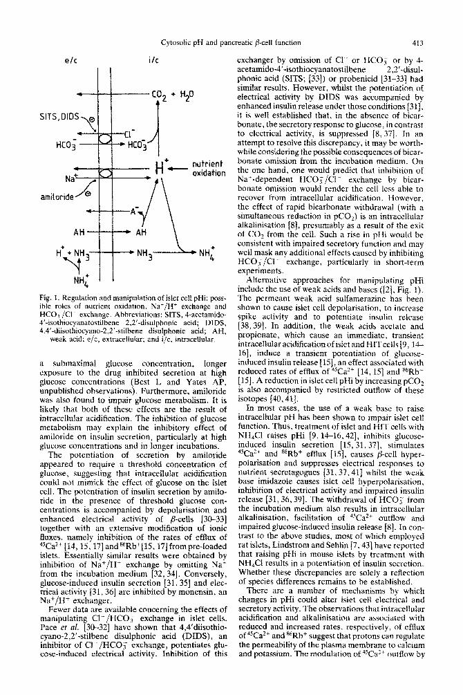
Pig. 1. Regulation and manipulation of islet ceil pHi: poss- ible roles of nutrient oxidation, Nat/H+ exchange and HCOs/Cl- exchange. Abbreviations: SITS, 4-acetamido* 4’-isothiocyanatostilbene 2,2’-disulphonic acid; DIDS, 4.4’-diisothiocyano-2,2’-stilbene disulphonic acid; AH,
weak acid: e/c, extraceltular; and i/c, i~~ra~~~l~~ar.
a submaximal glucose concentration, i0flgCX
exposure to the drug inhibited secretion at high glucose concentrations (Best L and Yates AP, unpublished observations). Furthermore, amiloride was also found to impair glucose metabolism. It is likely that both of these effects are the result of intracellular acidification. The inhibition of glucose metabolism may explain the inhibitory effect of amdoride on insulin secretion, particularly at high glucose concentrations and in longer incubations.
The potentiation of secretion by amiloride appeared to require a threshold concentration of glucose, suggesting that intracellular acidification could not mimick the effect of glucose on the islet cell. The potentiation of insulin secretion by amilo- ride in the presence of threshold glucose con- centrations is accompanied by depolarisation and enhanced electrical activity of pcells [3&33] together with an extensive modi~cation of ionic fluxes, nameiy inhibition of the rates of efflux of %azf [14,15,17] ands6Rb+[15, 17jfrom pre-loaded islets. Essentially similar results were obtained by inhibition of Na*/H+ exchange by omitting NaC from the incubation medium [32,34]. Conversely, glucose-induced insulin secretion [31,35] and elec- trical activity 131,361 are inhibited by monensin, au Na’/H+ exchanger.
Fewer data are available concerning the effects of manipulating Cl-/HCO; exchange in islet cells. Pace et al. [M-32] have shown that 4,4’diisothio- cyano-2,2’-stilbene disulphonic acid (DIDS), an inhibitor of Cl”*/HCO; exchange, potentiates glu- cose-induced electrical activity. Inhibition of this
exchanger by omission of Cl or HCOi or by 4- acetamido-4’-isothiocyanatostilbene 2,2’-disul- phonic acid (SITS; 1331) or probenicid [31-33] had similar results. However, whilst the potentiation of electrical activity by DIDS was accompanied by enhanced insulin release under those conditions [31], it is well established that, in the absence of bicar- bonate, the secretory response to glucose, in contrast to electrical activity, is suppressed [8,37]. In an attempt to resolve this discrepancy, it may be worth- while considering the possible consequences of bicar- bonate omission from the incubation medium. Qn the one hand, one would predict that inhibition of Na+-dependent HCOi/Cl- exchange by bicar- bonate omission would render the cell less able to recover frcrm intracellular acidification. However, the effect af rapid bicarbonate withdrawal (with a simultaneous reduction in pC&) is an intra~llular alkalinisation IS]> presumably as a result of the exit of CO* from the cell. Such a rise in pHi would be consistent with impaired secretory function and may well mask any additional effects caused by inhibiting HCO;/Cl- exchange, particularly in short-term experiments.
Alternative approaches for manipulating pNi include the use of weak acids and bases ([Z], Fig. 1). The permeant weak acid sulfamerazine has been shown to cause islet cell depolarisation, to increase spike activity and to potentiate insulin release [38,39]. In addition, the weak acids acetate and propionate, which cause an immediate, transient intracellular acidification of islet and HITceils 19, ?.4- 16], induce a transient potentiation of glucose- induced insulin release [lS], an effect associated with reduced rates of efflux of 4sCaZ4 [14,15] and s6Rb+ [ 151. A reduction in islet cell pIIi by increasing pCOz is also accompanied by restricted outflow of these isotopes [40,41].
In most cases, the use of a weak base to raise intracellular pH has been shown to impair islet cell function. Thus, treatment of islet and HIT ceils with NH4Cl raises pHi [9,14-l& 421, inhibits glucose- induced insulin secretion [ 15,31,37], stimulates 45Ca2+ and a6Rb+ efflux [IS], causes @cell hyper- polarisation and suppresses electrical responses to nutrient secretagogues [31,X’, 411 whilst the weak base imidazole causes islet cell hyperpoIarisat~on~ inhibition of electrical activity and impaired insulin release [3l, 36,391. The withdrawal of HCO, from the incubation medium also results in intracellular alkalinisation, facilitation of 45Ca’+ outflow and impaired glucose-induced insulin release [8]. In con- trast to the above studies, most of which employed rat islets, Lindstrom and Sehlin [7,43] have reported that raising pHi in mouse islets by treatment with NH4CI results in a potentiation of insulin secretion. Whether these discrepancies are solely a rellection of species differences remains to be established.
There are a number of mechanisms by which changes in pHi could alter islet cell electrical and secretory activity. The observations that intracehular acidification and alkalinisation are associated with reduced and increased rates, respectively, of efflux of 45Ca2+ and *6Rb+ suggest that protons can regulate the permeability of the plasma membrane to calcium and potassium. The modulation of %a’+ outflow by
413 A. LYNCI~ and L. Bt.s’t
pHi is largely dependent upon the presence of Na+ in the medium [8, 15.40], suggesting that protons inhibit the activity of the Na+/Ca?+ exchange system 1441. Such an action would be expected to result in an increased cytosoiic calcium concentration, which would be consistent with the potentiation of insulin secretion caused by lowering pHi. The reduction of “Rb-+ efflux by intracellular acidification is likely to be the result of inhibition of the calcium-activated K’ channel by protons [42,45], which would again explain the de~olarisation and potentiation of elec- trical and secretory activity accompanying intra- cellular acidification. The possibility that protons may exert additional actions upon islet cells cannot, of course, be ruled out.
Thus, there is little doubt that manipulations of pHi can influence ionic fluxes and hence electrical and secretory activity in insulin-secreting cells. How- ever. the marked dependence of these effects upon the presence of glucose (or another nutrient) must question whether pHi acts as a coupling factor per se in nutrient-stimulated insulin release. The lack of a clear correlation between observed changes in pHi and secretory activity casts further doubt on this possibility.
Conclusions and perspectives
In common with a wide variety of mammalian cell types, pancreatic islet cells have a number of mechanisms for the regulation of pHi. It is likely that an abilitv to maintain intracellular pH homeostasis is of particular importance in the case of the p-cell, where nutrient-stimulated insulin release is inti- mately associated with the high capacity of the cetl to oxidise those nutrients, a process resulting in the generation of acidic metabolites.
The operation of such regulatory mechanisms, which is apparent from measurements of pHi in nutrient-stimulated islet cells, makes it unlikely that intracellular acidification plays a major role in coup- ling nutrient oxidation to insulin release except, per- haps, for the initial phase of secretion. This is not to deny the possibility that other, non-nutrient secre- tagogues and pharmacological agents may exert their influence on islet cell activity, at least in part, by promoting intracellular acidification.
In the case of nutrients such as glucose, the pre- dominant response of islet cell pHi to nutrient stimuli appears to be a gradual alkalinisation, presumably as a result of activation of a regulatory mechanism(s). Whether this rise in pHi plays any active or per- missive roie in the stimulus-secretion coupling pro- cess remains to be established. One possibility is that a rise in pHi produces an enhanced rate of nutrient oxidation, which might be conducive to greater secretory activity. A rise in pHi could also be linked to the process of insulin biosynthesis, DNA rep- lication or islet cell growth, all of which are regulated by nutrients [46-4X].
One aspect of pHi regulation which could be of considerable importance arises from the fact that the two principal systems responsible for intracellular pH homeostasis, namely Na+/H+ and HCO,/Cl- exchange (both Na+-dependent and -independent), are electroneutral. Since the metabolism of glucose or another nutrient will result in the production of
glucose II glucose
1
:,* Na+
II T
+ Lacdp+e- r-l bicarbonate-
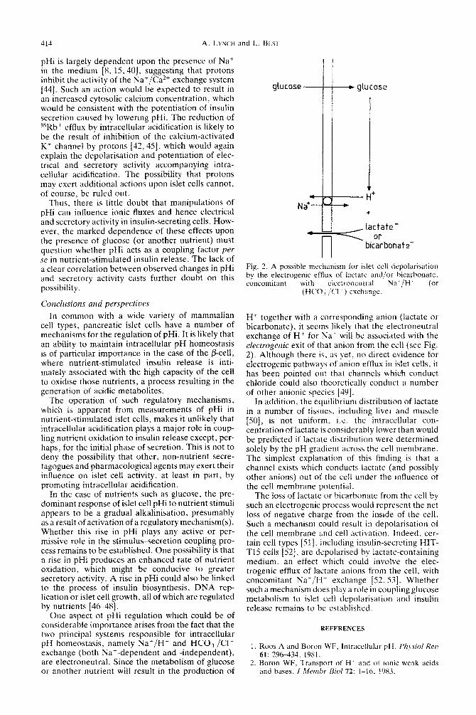
Fig. 2. A possible mechanism for islet ccl1 depolarisation by the electrogenic efllux of lactate and/or hicarbonate, concomitant with cicctroneutral P&‘/H’ (or
(HCO, $3 ) exchanpe.
E-I+ together with a corresponding anion (lactate or bicarbonate), it seems likely that the electroneutral exchange of H+ for Na+ will be associated with the electrogenic exit of that anion from the cell (see Fig. 2). Although there is. as yet, no direct evidence for electrogenic pathways of anion efflux in islet cells, it has been pointed out that channels which conduct chloride could also theoretically conduct a number of other anionic species [4Y].
In addition, the equilibrium distribution of lactate in a number of tissues, including liver and muscle [SO], is not uniform, i.e. the intracellular con- centration of lactate is considerably lower than would be predicted if lactate distribution were determined solely by the pH gradient across the cell membrane. The simplest explanation of this finding is that a channel exists which conducts lactate (and possibly other anions) out of the cell under the influence of the cell membrane potential.
The loss of lactate or bicarbonate from the cell by such an electrogenic process would represent the net loss of negative charge from the inside of the cell. Such a mechanism could result in depolarisation of the ceil membrane and cell activation. Indeed. cer- tain cell types [51], including insulin-secreting HIT- T15 cells [52], are depolarised by lactate-containing medium, an effect which could involve the elec- trogenic efflux of lactate anions from the cell, with concomitant Nat/H* exchange 12. X3]. Whether such a mechanism does play a role in coupling glucose metabolism to islet cell depolarisation and insulin release remains to be established.
REFERENCES
1. Roos A and Boron WF. Intracellular pH. Ph,vsio/ Reo 61: 29&434. 1981.
2. Boron WF, Transport of H and of ionic weak acids and hases. J .Menzhr Riol 72: i-16. 1’183.
Cytosolic pH and pancreatic /3-cell function 415
Busa WB, Mechanisms and consequences of pH- mediated cell regulation. Annu Reu Physiol 48: 389- 402, 1986. Madshus IH, Regulation of intracellular pH in euka- ryotic cells. Eiochem J 250: 1-8, 1988. Frelin C, Vigne P, Ladoux A and Lazdunski M, The regulation of the intracellular pH in cells from ver- tebrates. Eur J Biochem 174: F14. 1988. Rosengurt E, Early signals in the microgenic response. Science 234: 161-166, 1986.
I Lindstrom P and Sehlin J, Effect of glucose on the
8.
9.
10.
11.
intracellular pH of pancreatic islet cells.%iochemJ218: 887-892. 1984. Lebrun P, Malaisse WJ .md Herchuelz A, Effect of the absence of bicarbonate upon intracellular pH and calcium fluxes in pancreatic islet cells. Biochim Biophys Acta 721: 357-365, 1982. Pace CS and Tarvin JT, Amiloride-sensitive regulation of intracellular pH in /?-cells: Activation by glucose. Metabolism 35: 176181, 1986. Hutton JC, Sener A and Malaisse WJ, The metabolism of 4-methyl-2-oxopentanoate in rat pancreatic islets. Biochem J 184: 291-301. 1979. Hutton JC, Sener A, Herchuelz A, Valverde I, Bos- chero AC and Malaisse WJ, The stimulus-secretion coupling of glucose-induced insulin release. XLII. Effects of extracellular pH on insulin release: Their dependency on nutrient concentration. Harm Metab Res 12: 294-299. 1980.
12.
13.
14.
15.
16
Deleers M. Lebrun P and Malaisse WJ, Increase in HCO< influx and cellular pH in glucose-stimulated pancreatic islets. FEBS Lett 154: 97-100. 1983. Deleers M, Lebrun P and Malaisse WJ. Nutrient- induced changes in the pH of pancreatic islet cells. Horm Metab Res 17: 391-395. 1985 Best L, Bone EA, Meats JE and Tomlinson S, Is intracellular pH a coupling factor in nutrient-stimulated pancreatic islets? J Mol Endocr 1: 33-38, 1988. Best L, Yates AP. Gordon C and Tomlinson S, Modu- lation by cytosolic pH of calcium and rubidium fluxes in rat pancreatic islets. Biochem PharmacoJ 37: 4611- 4615, 1988. Lynch AM, Meats JE, Best L and Tomlinson S, Effects of nutrient and non-nutrient stimuli on cytosolic pH in cultured insulinoma (HIT-TlS) cells. Biochim Biophys Acta 1012: 166-170. 1989.
I,. Lebrun P. van Ganse E. Juvent M. Deleers M and 17
Herchuelz A, Na+-H’ exchange in the process of glu- cose-induced insulin release from the pancreatic /?-cell. Effects of amiloride on R6Rb’, ‘5Ca++ fluxes and insulin release. Biochim Biophys Acta 886: 44%456, 1986.
18. Aaronson PS, Nee J and Suhm MA, Modifier role of internal H+ in activating the Na+/H+ exchanger in renal microvillus membranes. Nature 299: 161-163, 1982.
19. Grinstein S and Rothstein A, Mechanisms of regulation of the Na’/H’ exchanger. J Membr Riot 90: 1-12, 1986.
20. Best L and Malaisse WJ, Phospholipids and islet func- tion. Diabetologia 25: 299-305, 1983.
21. Prentki M and Marschinski FM, Ca++, CAMP and phospholipid-derived messengers in coupling mech- anisms of insulin secretion. Physiol Reu 67: 1185-1248, 1987.
22. Malaisse WJ, Hutton JC, Kawazu S, Herschuelz A, Valverde I and Sener A. The stimulus-secretion coup- ling of glucose-induced insulin release. XXXV. Links between metabolic and cationic events. Diabetologia 16: 331-341,1979.
23. Deleers M. Lebrun P and Malaisse WJ, Effects of cations, ionophores and hypoglycemic sulfonylureas on the fluorescence of fluorescein-labelled pancreatic islets. Res Commun Chem Path01 Pharmacol 44: 83- 92, 1984.
24. Meech RW and Thomas RC, The effect of calcium
injection on the intracellular sodium and pH of snail neurones. J Phvsiol (Land) 265: 867-879. 1977.
25. Ives HE and Daniel TO,’ Interrelationship between growth factor-induced pH changes and intracellular Ca++. Proc Nat1 Acad Sci USA 84: 1950-1954, 1987.
26. Hellman B, The significance of calcium for glucose stimulation of insulin release. Endocrinology 97: 392- 398, 1975.
27. Siegl PKS, Cragoe EJ, Trumble MJ and Kaczorowski GJ, Inhibition of Na+/Ca++ exchange in membrane vesicle and papillary muscle preparations from guinea pig heart by analogues of amiloride. Proc Nat1 Acad Sci USA 81: 3238-3242, 1984.
28. Renner EL, Lake JR, Cragoe EJ Jr and Scharschmidt BF, Amiloride and amiloride analogues inhibit Na+/ K+-transporting ATPase and Na+-coupled alanine transport in rat hepatocytes. Biochim Biophys Acta 938: 386394, 1988.
29. Bidem TJ, Janjic D and Wollheim CB, Sodium require- ment for insulin release: Putative role in the regulation of intracellular pH. Am J Physiol 250: C207-C213, 1986.
30. Pace CS, Role of pH as a transduction device in trig- gering electrical and secretory responses in islet B cells. Fedn Proc 43: 2379-2384. 1984.
31. Pace CS, Tarvin JT and Smith JS, Stimulus-secretion coupling in /3-cells: Modulation by pH. Am J Physiol 244: E3-E18, 1983.
32. Pace CS and Tarvin JT, pH modulation of glucose- induced electrical activity’in B-cells: Involvement of Na/H and HCO,/CI antioorters. J Membr Eiol73: 3% 49,‘1983. _’ *
33. Eddlestone GT and Beigelman PM, Pancreatic p-cell electrical activity: The role of anions and the control of pH. Am J Physiol244: C188-C197, 1983.
34. Lebrun P, Plasman PO and Herchuelz A, Effect of extracellular sodium removal upon R6Rb outflow from pancreatic islet cells. Biochim Biophys Actu 1011: 6- 11, 1989.
35. Kanatsuka A, Makino H, Sakurada M, Hashimoto N, Yamaguchi T and Yoshida S, Biphasic insulin response to high glucose and a role for protons and calcium. Endocrinology 120: 77-82, 1987.
36. Tarvin JT, Sachs G and Pace CS, Glucose-induced electrical activity in the pancreatic /I-cell: Modulation by pH. Am J Physiol241: C264-C268, 1981.
37. Henquin JC and Lambert AE, Extracellular bicar- bonate ions and insulin secretion. Eiochim Biophys Acta 381: 437-442, 1975.
38. Pace CS and Goldsmith KT, Effect of substitution of a permeant weak acid for the permissive role of glucose in amino acid-induced electrical activity in /I-cells. Endocrinology 119: 2433-2438, 1986.
39. Smith JS and Pace CS. Modification of glucose-induced insulin release by alteration of pH. Diabetes 32: 61-66, 1983.
40. Carpinelli AR, Sener A, Herchuelz A and Malaisse WJ, The stimulus-secretion coupling of glucose- induced insulin release. XLI. Effect of intracellular acidification unon calcium efflux from islet cells. Metub- olism 29: 546545, 1980.
41. Caroinelli AR and Malaisse WJ. Reeulation of %Rb+ outHow from pancreatic islets. II. Ef&ct of changes in extracellular and intracellular pH. Diabete Metab 6: 193-198, 1980.
42. Rosario LM and Rojas E, Modulation of K+ con- ductance by intracellular pH in pancreatic pcells. FEES Lett 200: 203-209, 1986.
43. Lindstrom P and Sehlin J, Effect of intracellular alka- linisation on pancreatic islet calcium uptake and insulin secretion. Biochem J 239: 19%204, 1986.
44. Malaisse WJ, Herchuelz A and Sener A, The possible significance of intracellular pH in insulin release. Life Sci 26: 1367-1371, 1980.
416 A. LYNCH and L. BEST
45. Cook DL, Musatoshi I and Fujimoto WY, Lowering of pHi inhibits Ca++- activated channels in pancreatic /3- cells. Nature 311: 269-271, 1984.
46. Lin BJ and Haist RE, Insulin biosynthesis: Effects of carbohydrates and related compounds. Can J Physiol Pharmacol47: 791-801, 1969.
47. Nielsen JH, Growth and function of the pancreatic p- cell in uitro: Effects of glucose, hormones and serum factors on mouse, rat and human pancreatic islets in organ culture. Acta Endocrol (Copenh) 108 (Suppl 266): l-39, 1985.
48. Swenne I. Glucose-stimulated DNA replication of the pancreatic islets during the development of the rat foetus: Effects of nutrients. growth hormone and T3. Diabetes 34: 803-809, 1985.
49. Wright EM and Diamond JM, Anion selectivity in biological systems. Physiol Reu 57: 109-156, 1977.
50. Edlund GL and Halestrap AP, The kinetics of transport of lactate and pyruvate into rat hepatocytes. Biochem J 249: 117-126, 198X.
51. Bashford CL and Pasternak CA, Plasma membrane potential of some animal cells is generated by ion pumping, not by ion gradients. 7rend.~ Riochem Sci 11:
113-l 16. 1986. 52. Meats JE, Tuersley MD, Best L. Lynch AM and Tom-
linson S. Lactate alters plasma membrane potential, increases the concentration of cytosolic Ca’ and stimulates the secretion of insulin hy the hamster B- cell line HIT-Tl5. J Mol Endocr 3: 121-12X. 1989.
53. Best L, Yates AP, Meats JE and Tomlinson S. Effects of lactate on pancreatic Islets: Lactate efflux as a poss- ible determinant of islet cell depolarisation hy glucose. Riochem J 259: 507-51 I. 1989.