Page 1
42
DAFTAR PUSTAKA
1. WHO. Methanol Poisoning Outbreaks [Internet]. 2011. Available from:
http://www.who.int/environmental_health_emergencies/poisoning/methano
l_information.pdf
2. Barceloux DG, Krenzelok EP, Olson K, Watson W. American Academy of
Clinical Toxicology Practice Guidelines on the Treatment of Ethylene
Glycol Poisoning. Ad Hoc Committee. J Toxicol Clin Toxicol.
1999;37(5):537–60.
3. Goldfrank L. Goldfrank’s Toxicologic Emergencies. 9th ed. 2011.
4. El-Bakary A a, El-Dakrory S a, Attalla SM, Hasanein N a, Malek H a.
Ranitidine as an alcohol dehydrogenase inhibitor in acute methanol toxicity
in rats. Hum Exp Toxicol [Internet]. 2010;29(2):93–101. Available from:
http://www.ncbi.nlm.nih.gov/pubmed/20026516
5. Li FX, Lu J, Xu YJ, Tong ZQ, Nie CL HR. Formaldehyde-mediated
chronic damage may be related to sporadic neurodegeneration. 2008;
6. Yang MF, Lu J, Miao JY, Rizak J, Yang JZ, Zhai RW, et al. Alzheimer’s
Disease and Methanol Toxicity (Part 1): Chronic Methanol Feeding Led to
Memory Impairments and Tau Hyperphosphorylation in Mice. 2014;
7. Rubinstein D, Escott E, Kelly JP. Methanol intoxication with putaminal
and white matter necrosis: MR and CT findings. AJNR Am J Neuroradiol
[Internet]. 1995;16(7):1492–4. Available from:
http://www.ncbi.nlm.nih.gov/pubmed/7484638
8. Susan Standring, PhD Ds. Gray’s Anatomy 40th edition [Internet].
Churchill Livingstone. 2009. Available from:
http://www.us.elsevierhealth.com/anatomy/gray-anatomy-expert-
consult/9780443066849/
9. Bartsch T. The Clinical Neurobiology of the Hippocampus: An Integrative
View, Volume 151 [Internet]. OUP Oxford; 2012 [cited 2016 Jan 14]. 310
p. Available from:
https://books.google.com/books?id=_J0PcQtq5m8C&pgis=1
10. Wright A. Section 4: Homeostasis and Higher Brain Function, Chapter 5.
Page 2
42
Limbic System: Hippocampus. In: Neuroscience Online: an Electronic
Textbook for Neuroscience. 1997.
11. Catani M, Dell’Acqua F, Thiebaut de Schotten M. A revised limbic system
model for memory, emotion and behaviour. Neuroscience and
Biobehavioral Reviews. 2013. p. 1724–37.
12. Rolls ET. Limbic systems for emotion and for memory, but no single
limbic system. Cortex. 2015. p. 119–57.
13. Mega MS, Cummings JL, Salloway S, Malloy P. The limbic system: an
anatomic, phylogenetic, and clinical perspective. J Neuropsychiatry Clin
Neurosci. 1997;9(3):315–30.
14. Isaacson RL, Neil J Smelser, Paul B Baltes. Limbic System. Int Encycl Soc
Behav Sci [Internet]. 2001;2:8858–62. Available from:
http://www.sciencedirect.com/science/article/B7MRM-4MT09VJ-
4XS/2/9e7fa303f8769577cb0e460f32fb60c7
15. Barger N, Hanson KL, Teffer K, Schenker-Ahmed NM, Katerina
semendeferi. Evidence for evolutionary specialization in human limbic
structures. Front Hum Neurosci [Internet]. 2014;8(707):277. Available
from:
http://journal.frontiersin.org/article/10.3389/fnhum.2014.00277/abstract\np
apers3://publication/doi/10.3389/fnhum.2014.00277
16. Shorvon S. The Human Hippocampus. Functional Anatomy,
Vascularization and Serial Sections with MRI. J Anat. 2000;197:513–8.
17. Isaacson RL, Larry RS. Hippocampus. Encycl Neurosci [Internet].
2004;1119–27. Available from:
http://www.sciencedirect.com/science/article/pii/B978008045046902026X
18. Andersen P, Morris R, Amaral D, Bliss T, O’ Keefe J. The Hippocampus
Book. The Hippocampus Book. 2009. 1-852 p.
19. Papp E, Leergaard B, Calabrese E, Johnson G BJ. Waxholm space atlas of
the sprague dawley rat brain. National Institutes of Health Public Access.
2014.
20. Paulsen F WJ. Sobotta atlas of human anatomy Volume 1 Head, Neck,
Upper Limb. 14th Ed. 2006.
Page 3
43
21. Hall JE. Textbook of Medical Physiology. 13th Ed. 2015.
22. Soebowo, Sarjadi, Wijaya I, Amarwati S, Miranti I PA. Pedoman Kuliah
Mahasiswa Patologi Anatomi 1. 2014.
23. Babu KM, Rosenbaum CD, Boyer EW. Head CT in patient with metabolic
acidosis. J Med Toxicol [Internet]. 2008;4(4):275–6. Available from:
http://www.ncbi.nlm.nih.gov/pubmed/19031380
24. Troncoso J, Rubio A, Fowler DR. Essential Forensic Neuropathology.
2010.
25. Auer RN. Effect of Age and Sex on N-Methyl-D-Aspartate Antagonist-
Induced Neuronal Necrosis in Rats. Stroke [Internet]. 1996;27(4):743–6.
Available from: http://stroke.ahajournals.org/content/27/4/743.full
26. Fuchs E, Flugge G, Czeh B. Remodeling of neuronal networks by stress.
Front Biosci. 2006;11(August 2015):2746–58.
27. Upton JW, Kaiser WJ, Mocarski ES. Virus inhibition of RIP3-dependent
necrosis. Cell Host Microbe. 2010;7(4):302–13.
28. Rohleder N, Kirschbaum C. Effects of nutrition on neuro-endocrine stress
responses. Curr Opin Clin Nutr Metab Care [Internet]. 2007;10(4):504–10.
Available from: http://www.ncbi.nlm.nih.gov/pubmed/17563471
29. Armstrong D, Halliday W, Hawkings C, Takashima S. Pediatric
Neuropathology: A Text-Atlas [Internet]. Springer Science & Business
Media; 2008 [cited 2016 Jan 25]. 443 p. Available from:
https://books.google.com/books?id=VJY20uS-BagC&pgis=1
30. Methanol Toxicological Overview [Internet]. [cited 2016 Jan 19].
Available from:
https://www.gov.uk/government/uploads/system/uploads/attachment_data/f
ile/456293/Methanol_TO_PHE_260815.pdf
31. ACUTE TOXICITY SUMMARY METHANOL [Internet]. [cited 2016 Jan
19]. Available from: http://oehha.ca.gov/air/acute_rels/pdf/67561a.pdf
32. McCoy HG, Cipolle RJ, Ehlers SM, Sawchuk RJ, Zaske DE. Severe
methanol poisoning. Application of a pharmacokinetic model for ethanol
therapy and hemodialysis. Am J Med. 1979;67(5):804–7.
33. Korabathina K. Methanol Toxicity [Internet]. 2015. Available from:
Page 4
44
http://emedicine.medscape.com/article/1174890-overview
34. Dally AM. Fatal Methanol Intoxication – Two Exceptional Cases.
Toxichem Krimtech. 2015;
35. Kraut JA, Kurtz I. Toxic alcohol ingestions: Clinical features, Diagnosis,
and management. Clin J Am Soc Nephrol. 2008;3(1):208–25.
36. Acid-Base Physiology: 8.6 Metabolic Acidosis due to Drugs and Toxins
[Internet]. [cited 2016 Jan 19]. Available from:
http://www.anaesthesiamcq.com/AcidBaseBook/ab8_6a.php
37. Strum WB. Ranitidine. JAMA [Internet]. 1983;250(14):1894–6. Available
from: http://jama.jamanetwork.com/article.aspx?articleid=388242
38. van Hecken AM, Tjandramaga TB, Mullie A, Verbesselt R, de Schepper
PJ. Ranitidine: single dose pharmacokinetics and absolute bioavailability in
man. Br J Clin Pharmacol. 1982;14(2):195–200.
39. Ranitidine [Internet]. 2016. Available from:
http://www.drugs.com/pro/ranitidine.html
40. Ding J. Handbook of Metabolic Pathways of Xenobiotics. 2nd ed. John
Wiley & Sons; 2014.
41. Azeemuddin M, Naqi R. Case Series MRI findings in methanol
intoxication : a report of three cases. :1099–101.
42. Lwanga S.K., Lemeshow S. Sample size determination in health studies A
practicle manual. World Health Organization. 1991. p. 38.
43. WHO. Methanol poisoning outbreaks. 2014;
44. Nabila N. Pengaruh Pemberian Metanol Dan Etanolterhadap Tingkat
Kerusakan Sel Hepar Tikus Wistar. 2011;1–16.
45. Patnaik R, Mohanty S SH. Blockade of histamine H2 receptors attenuate
blood-brain barrier permeability, cerebral blood flow disturbances, edema
formation and cell reactions following hyperthermic brain injury in the rat.
2000;
46. Ding D, Moskowitz SI, Li R, Lee SB, Esteban M, Tomaselli K, et al.
Acidosis induces necrosis and apoptosis of cultured hippocampal neurons.
Exp Neurol [Internet]. 2000;162(1):1–12. Available from:
http://www.ncbi.nlm.nih.gov/pubmed/10716884
Page 5
45
47. Singh M, Bhatia R. Emergencies in Neurology. Byword Books Private
Limited; 2011. 384 p
Page 6
46
Lampiran
Lampiran 1. Hasil Analisis SPSS
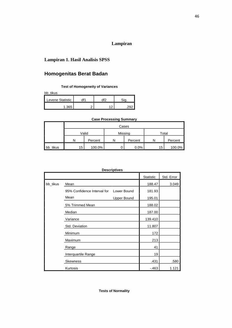
Homogenitas Berat Badan
Test of Homogeneity of Variances
bb_tikus
Levene Statistic df1 df2 Sig.
1.365 2 12 .292
Case Processing Summary
Cases
Valid Missing Total
N Percent N Percent N Percent
bb_tikus 15 100.0% 0 0.0% 15 100.0%
Descriptives
Statistic Std. Error
bb_tikus Mean 188.47 3.049
95% Confidence Interval for
Mean
Lower Bound 181.93
Upper Bound 195.01
5% Trimmed Mean 188.02
Median 187.00
Variance 139.410
Std. Deviation 11.807
Minimum 172
Maximum 213
Range 41
Interquartile Range 19
Skewness .431 .580
Kurtosis -.463 1.121
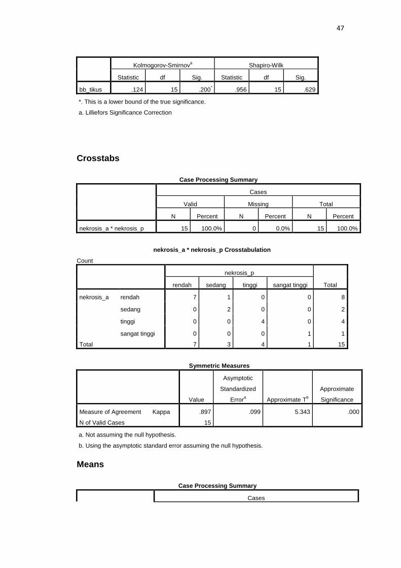
Tests of Normality
Page 7
47
Kolmogorov-Smirnova Shapiro-Wilk
Statistic df Sig. Statistic df Sig.
bb_tikus .124 15 .200* .956 15 .629
*. This is a lower bound of the true significance.
a. Lilliefors Significance Correction
Crosstabs
Case Processing Summary
Cases
Valid Missing Total
N Percent N Percent N Percent
nekrosis_a * nekrosis_p 15 100.0% 0 0.0% 15 100.0%
nekrosis_a * nekrosis_p Crosstabulation
Count
nekrosis_p
Total rendah sedang tinggi sangat tinggi
nekrosis_a rendah 7 1 0 0 8
sedang 0 2 0 0 2
tinggi 0 0 4 0 4
sangat tinggi 0 0 0 1 1
Total 7 3 4 1 15
Symmetric Measures
Value
Asymptotic
Standardized
Errora Approximate T
b
Approximate
Significance
Measure of Agreement Kappa .897 .099 5.343 .000
N of Valid Cases 15
a. Not assuming the null hypothesis.
b. Using the asymptotic standard error assuming the null hypothesis.
Means
Case Processing Summary
Cases
Page 8
48
Included Excluded Total
N Percent N Percent N Percent
nekrosis_p * perlakuan 15 100.0% 0 0.0% 15 100.0%
Report
nekrosis_p
perlakuan Mean Std. Deviation Median Minimum Maximum
kontrol negatif 1.20 .447 1.00 rendah sedang
kontrol positif 3.20 .447 3.00 tinggi sangat tinggi
perlakuan 1.40 .548 1.00 rendah sedang
Total 1.93 1.033 2.00 rendah sangat tinggi
Explore perlakuan
Case Processing Summary
perlakuan
Cases
Valid Missing Total
N Percent N Percent N Percent
nekrosis_p kontrol negatif 5 100.0% 0 0.0% 5 100.0%
kontrol positif 5 100.0% 0 0.0% 5 100.0%
perlakuan 5 100.0% 0 0.0% 5 100.0%
Descriptives
perlakuan Statistic Std. Error
nekrosis_p kontrol negatif Mean 1.20 .200
95% Confidence Interval for
Mean
Lower Bound .64
Upper Bound 1.76
5% Trimmed Mean 1.17
Median 1.00
Variance .200
Std. Deviation .447
Minimum 1
Maximum 2
Range 1
Interquartile Range 1
Skewness 2.236 .913
Page 9
49
Kurtosis 5.000 2.000
kontrol positif Mean 3.20 .200
95% Confidence Interval for
Mean
Lower Bound 2.64
Upper Bound 3.76
5% Trimmed Mean 3.17
Median 3.00
Variance .200
Std. Deviation .447
Minimum 3
Maximum 4
Range 1
Interquartile Range 1
Skewness 2.236 .913
Kurtosis 5.000 2.000
perlakuan Mean 1.40 .245
95% Confidence Interval for
Mean
Lower Bound .72
Upper Bound 2.08
5% Trimmed Mean 1.39
Median 1.00
Variance .300
Std. Deviation .548
Minimum 1
Maximum 2
Range 1
Interquartile Range 1
Skewness .609 .913
Kurtosis -3.333 2.000
Tests of Normality
perlakuan
Kolmogorov-Smirnova Shapiro-Wilk
Statistic df Sig. Statistic df Sig.
nekrosis_p kontrol negatif .473 5 .001 .552 5 .000
kontrol positif .473 5 .001 .552 5 .000
perlakuan .367 5 .026 .684 5 .006
a. Lilliefors Significance Correction
NPar Tests
Page 10
50
Kruskal-Wallis Test
Ranks
perlakuan N Mean Rank
nekrosis_p kontrol negatif 5 5.00
kontrol positif 5 13.00
perlakuan 5 6.00
Total 15
Test Statisticsa,b
nekrosis_p
Chi-Square 10.857
df 2
Asymp. Sig. .004
a. Kruskal Wallis Test
b. Grouping Variable:
perlakuan
NPar Tests Mann-Whitney Test
Ranks
perlakuan N Mean Rank Sum of Ranks
nekrosis_p kontrol negatif 5 3.00 15.00
kontrol positif 5 8.00 40.00
Total 10
Test Statisticsa
nekrosis_p
Mann-Whitney U .000
Wilcoxon W 15.000
Z -2.785
Asymp. Sig. (2-tailed) .005
Exact Sig. [2*(1-tailed Sig.)] .008b
a. Grouping Variable: perlakuan
b. Not corrected for ties.
NPar Tests
Page 11
51
Mann-Whitney Test
Ranks
perlakuan N Mean Rank Sum of Ranks
nekrosis_p kontrol negatif 5 5.00 25.00
perlakuan 5 6.00 30.00
Total 10
Test Statisticsa
nekrosis_p
Mann-Whitney U 10.000
Wilcoxon W 25.000
Z -.655
Asymp. Sig. (2-tailed) .513
Exact Sig. [2*(1-tailed Sig.)] .690b
a. Grouping Variable: perlakuan
b. Not corrected for ties.
NPar Tests Mann-Whitney Test
Ranks
perlakuan N Mean Rank Sum of Ranks
nekrosis_p kontrol positif 5 8.00 40.00
perlakuan 5 3.00 15.00
Total 10
Test Statisticsa
nekrosis_p
Mann-Whitney U .000
Wilcoxon W 15.000
Z -2.739
Asymp. Sig. (2-tailed) .006
Exact Sig. [2*(1-tailed Sig.)] .008b
a. Grouping Variable: perlakuan
b. Not corrected for ties.
Page 12
52
Lampiran 2. Cara kerja sediaan histopatologi
1) Menyiapkan wadah yang di isi dengan larutan formalin 10% buffer dengan
minimal lima kali volume jaringan
2) Testis yang telah diambil, segera dimasukkan ke dalam wadah tersebut
3) Memberi identitas pada semua wadah dengan identitas masing-masing
kelompok perlakuan
4) Dikirim ke Sentra Diagnostik Patologi Anatomi disertai dengan formulir
pengantar
5) Preparat kemudian dipotong dengan ketebalan maksimal 3-4 cm
6) Setelah dipotong diletakkan di dalam kaset jaringan, dan dimasukkan ke
wadah yang berisi formalin 10% buffer
7) Dilakukan proses pembuatan blok parafin, kemudian didinginkan di dalam
lemari es
8) Blok parafin dipotong menjadi lebih tipis menggunakan mikrotom sesuai
kebutuhan
9) Pita parafin dilebarkan dengan ditempelkan langsung pada kaca benda yang
telah dibasahi dengan air
10) Dimulai dengan proses pengecatan Hematoksilin dan Eosin (HE)
11) Perparat diberi cat Hematoksilin
12) Kemudian didiferensiasi dengan menggunakan air kran
13) Diberi cat Eosin
14) Kemudian di dehidrasi menggunakan alkohol 70%
15) Pada proses “clearing” menggunakan larutan xylol
16) Mouting adalah tahap terakhir yang kemudian dapat diamati di mikroskop
Page 13
53
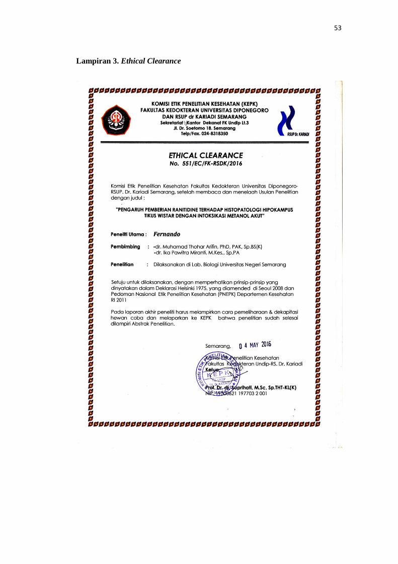
Lampiran 3. Ethical Clearance
Page 14
54
Lampiran 4. Surat Keterangan Penelitian
Page 15
55
Lampiran 5. Hasil Pengamatan Nekrosis Sel Hipokampus
Subjek Jumlah Nekrosis (persen) Kategori
Kontrol Negatif 1 6 1
Kontrol Negatif 2 17 1
Kontrol Negatif 3 11 1
Kontrol Negatif 4 26 2
Kontrol Negatif 5 9 1
Kontrol Positif 1 51 3
Kontrol Positif 2 52 3
Kontrol Positif 3 83 4
Kontrol Positif 4 59 3
Kontrol Positif 5 64 3
Perlakuan 1 32 2
Perlakuan 2 39 2
Perlakuan 3 12 1
Perlakuan 4 23 1
Perlakuan 5 21 1
Page 16
56
Lampiran 6. Dokumentasi Penelitian