49
Database Comparisons: Age effects
| Date post: | 13-Dec-2015 |
| Category: |
Documents |
| Upload: | eunice-jocelin-fletcher |
| View: | 229 times |
| Download: | 0 times |

Database Comparisons:Age effects

EEG age effects by hemisphere

Dominant (“alpha”) rhythm
• Normal EEG pre-school to Adolescence Occipital rhythm
• 3-4 Hz activity at 3-4 months of age– responds to stimulation at 5-6 mo.
• At 6 mo, typically 5-6 Hz• At 1 year, 6-7 Hz.
• Frequency range increases as child ages. • Typical 10y old averages about 10 Hz.

Dominant frequencies during infancy

Othmer clinic data

Normal Child has theta rhythm, maximal in posterior sites

Normal Adult has 8-12 Hz rhythm, maximal in posterior sites


Posterior sites

Neurometic Analysis(Comparing someone to a QEEG Database to identify statistical
abnormality, commonly > 2 Std deviations, plus or minus)
• Baseline conditions– Eyes closed– Eyes open– Motor control– Stimulus control
• Task conditions or challenge conditions– Problem solving– Performance


Eyes closed replications support macrostate concept (however…)

Correct for state transitions
Stabilize state before recording, and include stabilization in database!
Look at the data
More is better
(Some db use 30 s only)

Client comparison requires similar recording methodology

Criticisms of Neurometrics
• Color maps are deceptive1
• Too many statistical tests (inflating Type I errors)
• Some normals appear abnormal to controls
• Overly sensitive to artifact• More removed from the data,
the more errors that can creep in– E.g., unreliable discriminant
functions
• Methodology differences between client and database recordings
• Artifact management differences
• Normative database not representative2
• Differences in basic parameters (power/magn), coherence calculation
Not ready for prime time …if misused!

Brain maps can be deceptive …..
Some activation needed at left medial temporal site or right occipitoparietal juncture?

But they are convincing, concise, and accessible to laypersons

5 Neurometric databases in common use for Neurotherapy
• “My kid acting like an astronaut” story

Deciphering Neural codingWe transform from time to frequency because we believe
mental or psychophysiological phenomena are best captured by latter domain

Brainwave frequencies and tentative mental correspondences
1
0.5 – 4 Hz DELTA Sleep
4-7 Hz THETA Inward focus, distracted, daydream
8-12 Hz ALPHA Relaxed, not actively processing
12-15 Hz SMR Relaxed, external attention (low beta)
15-18 Hz BETA Active external attention
19-35 Hz HIGH High correlation with anxiety, BETA intensity, or lots of muscle tension.

Alpha = activation or arousal

Activation is inversely proportional to alpha activity incidence
• To some extent, regardless of topography

10-10 International System of Electrode Placement
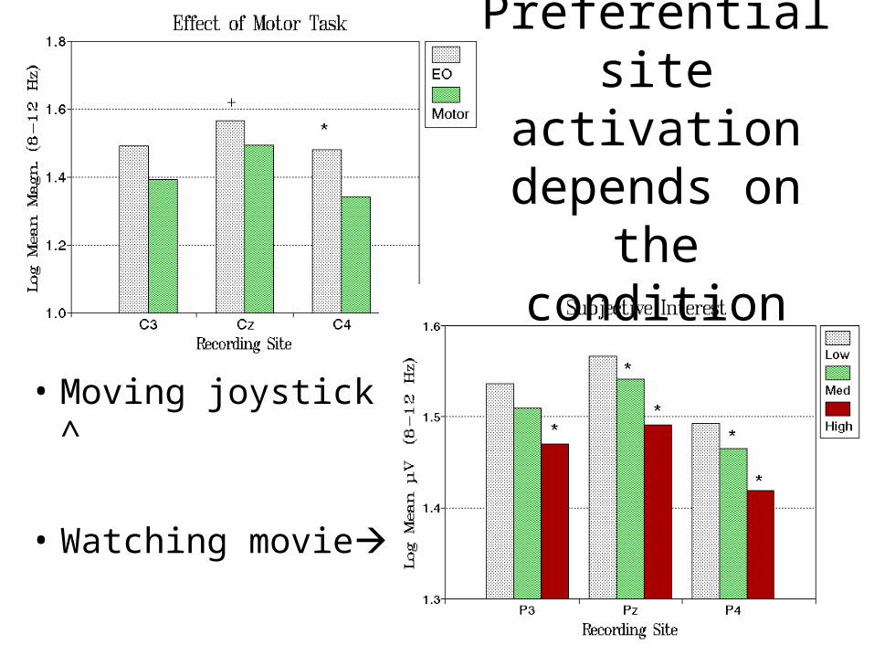
Preferential site activation
depends on the condition
• Moving joystick ^
• Watching movie

Topographic Activation Patterns

Laterality differences
• “Everyone is a left-brainers until films make them integrationalists”

Gender differences

Cerebral Organization Variation (and obstacles to neurometric assessment)
Trait and State variables
– TRAIT• Gender• Handedness• Age• Education• Experience• Neurological
present/history• Bilingual• Diagnosis!
– STATE• Task competence
– practice
• Task strategies• Time of Day• Drugs• Sleep debt

Be aware of the plasticity spectrum – people change

Inclusion/Exclusion criteria for normals
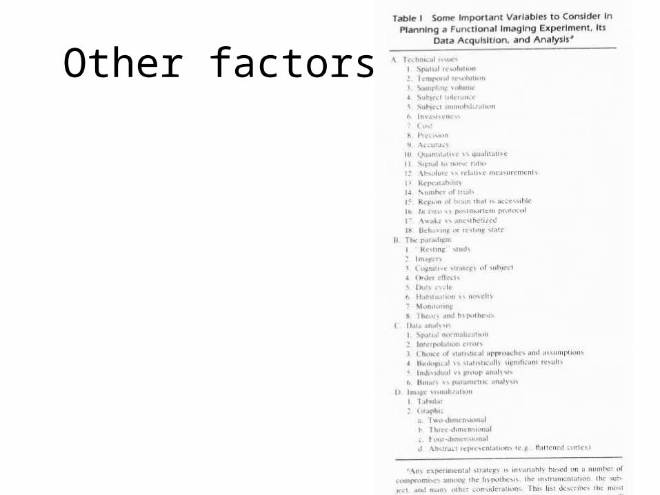
Other factors


Jared’s spindles


Measurement parameters
• Relative vs Absolute – Power/magnitude
• Connectivity or linear dependency – Asymmetry– Coherence, comodulation

Synchrony measures between two signals


Coherence


Coherence Database

Comodulation (Functional grouping in dominant frequency activity)

Consistency or stationarity of amplitudes between two signals in
frequency band


MVA or Youthful

LORETALow resolution EEG tomographical array (source imaging of maximal smoothness)


Current EEG Applications
• CLINICAL• Epilepsy (1930s)• Sleep (1940s)• Patient monitoring, anaesthesia• Head injury assessment• Neurological assessment (AEP, ERP)• Neurotherapy• Psychiatric assessment

Current EEG Applications
• SCIENTIFIC
• Attention
• Workload
• Circadian rhythms
• Cognition
• Learning & Memory
• Neuroimaging co-registration

Future EEG Assessment
• Subtype clinical conditions• Monitor attentional state• Lie Detection• Parole disposition


