SummaryBecause forensic anthropologists and pathologists can be confronted in their pro-
fessional practices with bodies or mortal remains in different states of preservationand/or decay, it is essential for this book to have a chapter that fully documents thepathway of a body from its death until disintegration.
The different ways a corpse can progress from putrefaction directly (or not) toskeletonization, passing through conservation processes such as saponification or mum-mification are presented here, always taking into account the forensic relevance of eachstage, the time and conditions needed, as well as the duration. Full, illustrated examplesof cases that have contributed to solve forensic questions are provided. Factors thatmight influence the speed of putrefaction and the interrelations—through chemicalreactions between these processes—are also debated.
It is common for forensic anthropologists and pathologists to be con-fronted in their professional practice with bodies or human remains states ofpreservation and/or decay that are not entirely to their liking, such states beingoutside their knowledge and experience. The forensic pathologist generallyfeels more at ease with a fresh body, whereas the forensic anthropologist wouldcertainly prefer to work with dry bones. Ideally, the forensic anthropologist
86 Pinheiro
should always be called whenever a body appears whose morphological char-acteristics do not permit any identification. Such cases are usually in anadvanced state of decay: with adipocere, mummified, carbonized, skeleton-ized, or with a mixture of all of these. Indeed, the same cadaver may revealvarious states of preservation at the same time. This fact is closely connectedto the different transformations that may take place in a body from the momentof death to skeletonization, upon the action of various extrinsic and intrinsicfactors, which are analyzed here.
For this reason, it is fundamental that the pathologist has prior knowl-edge of the various alterations that take place postmortem (the object of thestudy of forensics called taphonomy). These alterations particularly affect thesoft tissues, and are decisive not only for the time taken for skeletonization tooccur, but also for the state of preservation of the cadaver (1). Between afresh cadaver and a heap of loose bones, there are a series of stages of decom-position and/or preservation that may occur when the environmental condi-tions are right. Various authors have drawn attention to the need to understandthis process (2,3), whereas some of the definitions of forensic anthropologyitself, such as the one of Bass (4), presuppose the existence of cases otherthan those skeletonized: “…the science that focuses principally on the identi-fication of remains that are more or less skeletonized, in the legal context.”
This journey along the taphonomic process will certainly be useful in itsearlier stages for anthropologists that are not used to working with almost-fresh cadavers, and in the final phase (skeletonization) for pathologists, whoare normally not too fond of working with bones.
2. DECOMPOSITION
The process by means of which a cadaver becomes a skeleton, throughthe destruction of the soft tissue, is quite complex. In discussing the decom-position process, it is important to remember that, as with everything in biol-ogy, the exception is the rule, or rather, that there are no two individualsalike, nor any two decomposition processes alike. This is why this stage canbe difficult.
The decomposition of a body is a mixed process that varies from cellu-lar autolysis by endogenous chemical destruction to tissue autolysis, by eitherthe release of enzymes or external processes, resulting from the bacteria andfungus in the intestines or from outside (5). Predators, ranging from insects tomammals, participate in the process and may accelerate it. It can therefore besaid, with Di Maio (6), that decomposition involves autolysis (the destructionof cells and organs by an aseptic chemical process) and putrefaction (because
Decay Process of a Cadaver 87
of bacteria and fermentation). Thus, whereas common sense understandsdecomposition to be synonymous with putrefaction, in the forensic context, ithas a much broader meaning, covering all stages from the moment of death tothe dissolution of all body parts.
It is a process that varies greatly from body to body, environment toenvironment, according to whether the body is clothed or naked, the circum-stances of the death and the place where the body is found, the climate, and soforth. For example, it is known that putrefaction occurs much faster in bodiesthat are left in the open air than those immersed in water, whereas buriedbodies decay at a much slower rate (7–9). In these cases, factors such as thelength of time before the body was buried (thus allowing putrefaction to be-gin), the temperature at the site, the presence or absence of oxygen, the depthof the body, topography of the soil (rather than its composition), and the typeof coffin used, considerably affect the speed of decomposition (6). However,whereas many exhumations have little to offer to certain investigations, thisshould not devalue their importance. Indeed, this can never be anticipated,because there are cases in which bodies are in truly surprising states of con-servation. In an autopsy performed by the author, it was possible, some monthsafter the burial, to undertake a detailed examination, entirely unexpectedly,of a spontaneous brain hemorrhage in a body that had been buried in winter inan area of harsh climate (atmospheric temperatures between –3 and 10 C).
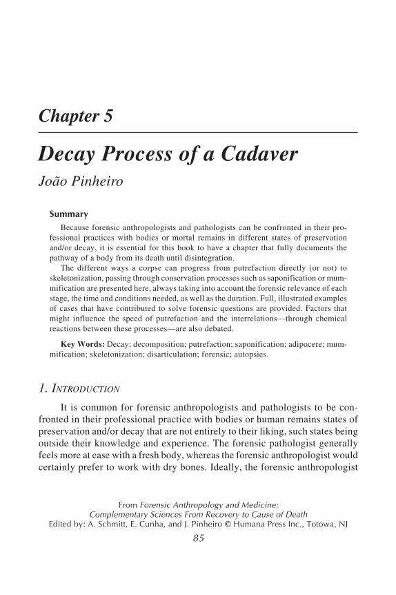
Decomposition may also vary within the same cadaver, with some partsof the body showing adipocere, other parts mummified, and still others onlyputrefied (Fig. 1). This will depend on the different “microenvironments”that develop around them, in accordance with the place where they are found.There are also various possible interconnections between these states, whichmakes it difficult to estimate the date of death.
The calculation of the postmortem interval (PMI), one of the most contro-versial and difficult problems in legal medicine, becomes more acute in cases ofdecomposition. Excluding the precious assistance provided by forensic entomol-ogy (a separate discipline that is not dealt with here), various methods have beenused to calculate this interval, while of course taking into account the subjectivenature of the individual assessment. Prieto (9) lists the evaluation of biomarkerslike lipids, nitrogen, amino acid content, neurotransmitters, decompositionalbyproducts, persistence of blood remnants in bone tissue; extent of DNA deterio-ration; changes sustained by microanatomical skeletal structure; and carbon 14.Others have tried to study the variations of factors that influence decompositionin certain cases, either prospectively (through the formation of adipocere [10,11])or retrospectively (by analyzing cases that have already been solved in order tostudy particularly extrinsic factors that affect it [9,12]).
88 Pinheiro
In all cases, and despite the relevance of some of these methods, theestablishment of PMI continues, for most pathologists, to be based on indi-vidual analysis and experience obtained in similar cases. And, whereas it islegitimate to suggest a date for past populations with some margin of varia-tion, it is always very difficult to risk a prognosis in forensic cases, becausethere are so many factors involved, and the range of variation is so broad.This is supported by a number of authors (8,10–13), who, because of themultiplicity of factors involved, find it impossible to attribute a credible timeinterval for each of the stages of decomposition.
Fig. 1. Coexistence of three states of decomposition in the same body:skeletonization of the head, adipocere in the trunk organs, and mummificationof the limbs. Note skin’s leathery appearance. The body belongs to a 93-yr-oldwoman found facedown in the countryside, whose positive identification wasachieved. Cause of death was ascertained.
Decay Process of a Cadaver 89
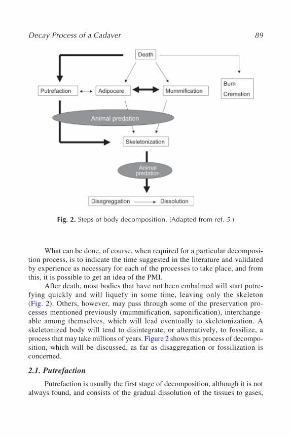
What can be done, of course, when required for a particular decomposi-tion process, is to indicate the time suggested in the literature and validatedby experience as necessary for each of the processes to take place, and fromthis, it is possible to get an idea of the PMI.
After death, most bodies that have not been embalmed will start putre-fying quickly and will liquefy in some time, leaving only the skeleton(Fig. 2). Others, however, may pass through some of the preservation pro-cesses mentioned previously (mummification, saponification), interchange-able among themselves, which will lead eventually to skeletonization. Askeletonized body will tend to disintegrate, or alternatively, to fossilize, aprocess that may take millions of years. Figure 2 shows this process of decompo-sition, which will be discussed, as far as disaggregation or fossilization isconcerned.
2.1. Putrefaction
Putrefaction is usually the first stage of decomposition, although it is notalways found, and consists of the gradual dissolution of the tissues to gases,
Fig. 2. Steps of body decomposition. (Adapted from ref. 5.)
90 Pinheiro
liquids, and salts (7). Not only is it the subject of numerous publications, it isalso covered in any book about legal medicine and forensic pathology.
Many authors distinguish phases and stages in the putrefaction process,some of which are based on the study of the decomposition of animal car-casses. For example, Shean et al. (14) distinguish 4 phases (decomposition ofthe soft tissue, exposure of bone, remains only with connective tissue, andbone only) that are broken down into 15 stages, whereas Galloway et al. (15)distinguish 5 phases and 21 stages, to cite only some. Others describe thesestages with great temporal precision, which, although revealing great erudi-tion and being very successful in lessons or lectures, are inadequate for prac-tical application given the enormous variability of the development of thisprocess. These discrepancies between suggested periods and phases of de-composition, along with the study of animals, naturally limit the value ofthese findings. In the author’s opinion, what is much more important thanknowing the stage of putrefaction, or how long it has taken to get there, is theability to recognize the elements that characterize clearly and objectively thestage of putrefaction, and the artifacts that this may induce, and to know itspotential and limits in terms of thanatological research.
Therefore, described here chronologically are the alterations undergoneby a body after the death, in a place with temperate weather. It should beemphasized that the times mentioned are merely indications and in no wayexact because some of the characteristics described may appear considerablyearlier or later than suggested.
2.1.1. FIRST WEEK
One of the earliest signs of putrefaction is the discoloration of the lowerabdominal wall in the right iliac fossa because of the proximity of the cecumto the surface. Intestinal bacteria break down the hemoglobin intosulfohemoglobin and other colored pigments (the “green abdominal stain,”as it is known in some countries), which extends from the right iliac fossa tothe whole of the abdomen and thorax (Fig. 3). These bacteria are also respon-sible for the formation of gases, provoking edema of the face and neck. Thegases released in this process (sulfuretted hydrogen, phosphoretted hydro-gen, methane, carbon dioxide, ammonia and hydrogen (7), and some mer-captans) are responsible for the unpleasant odor that is characteristic of thesebodies. Other effects produced by gases include a marked increase in thevolume of the abdomen, which is under tension, and of the scrotum andpenis, which may gain extraordinary dimensions. The face and neck alsoincrease greatly, with protrusion of the eyes and tongue, making identifica-tion difficult.
Decay Process of a Cadaver 91
The phenomenon known as “marbling” in Anglo-Saxon literature (orBrouardel’s “posthumous circulation,” as it is better known in the Latin coun-tries), which results from the colonization of the venous system by intestinalbacteria that hemolyze the blood, is very characteristically found at this time.It appears on the thighs and sidewalls of the abdomen, chest, and shoulders(see Fig. 3), first with a reddish color and, later, green.
Skin blisters containing reddish purplish serous liquid erupt in the slop-ing regions (see Figs. 3 and 9). These should be distinguished from thephlyctenae that result from burns; phlyctenae containing a serous liquid areof course characteristic of second-degree burns, but they are usually surroundedby an erythematous ring, something that is not found in putrefactive blisters.
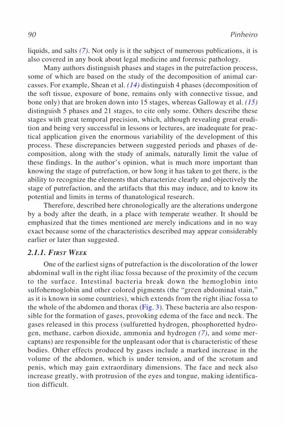
The epidermis becomes fragile and tears easily, which means that it maycome off in large areas, leaving the red dermis visible, similar to what hap-pens with first- and second-degree burns (Fig. 4). Such patches may also becaused by the bursting of the phlyctenae, when these are large and containliquid under pressure. The skin may also come off on the fingertips, which, ofcourse, hinders the taking of fingerprints.
Fig. 3. The colors of putrefaction: abdominal green, which had begun in theright inguinal region; red marbling, typically on the lateral part of the trunk,shoulders, and upper limbs; reddish, purplish of the putrefactive phlyctenae.Note the skin slippage. (See ebook for color version of this figure.)
92 Pinheiro
In hairy areas, hairs will come off at the slightest pressure. This phase ofputrefaction may also provide some curious aspects, such as the “gloves”made of skin on the hands (see Fig. 4), or the use of the hair by birds for nestbuilding (6).
2.1.2. SECOND AND THIRD WEEKS
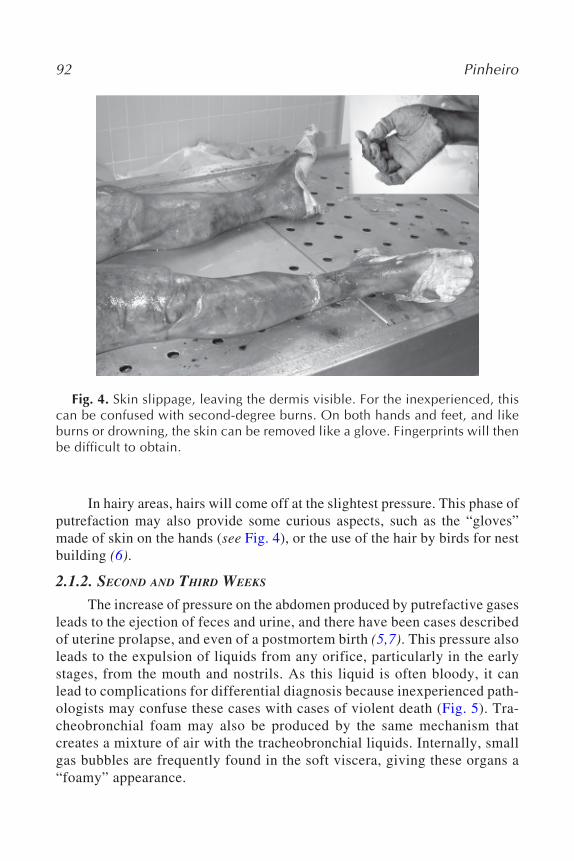
The increase of pressure on the abdomen produced by putrefactive gasesleads to the ejection of feces and urine, and there have been cases describedof uterine prolapse, and even of a postmortem birth (5,7). This pressure alsoleads to the expulsion of liquids from any orifice, particularly in the earlystages, from the mouth and nostrils. As this liquid is often bloody, it canlead to complications for differential diagnosis because inexperienced path-ologists may confuse these cases with cases of violent death (Fig. 5). Tra-cheobronchial foam may also be produced by the same mechanism thatcreates a mixture of air with the tracheobronchial liquids. Internally, smallgas bubbles are frequently found in the soft viscera, giving these organs a“foamy” appearance.
Fig. 4. Skin slippage, leaving the dermis visible. For the inexperienced, thiscan be confused with second-degree burns. On both hands and feet, and likeburns or drowning, the skin can be removed like a glove. Fingerprints will thenbe difficult to obtain.
Decay Process of a Cadaver 93
2.1.3. FOLLOWING WEEKS
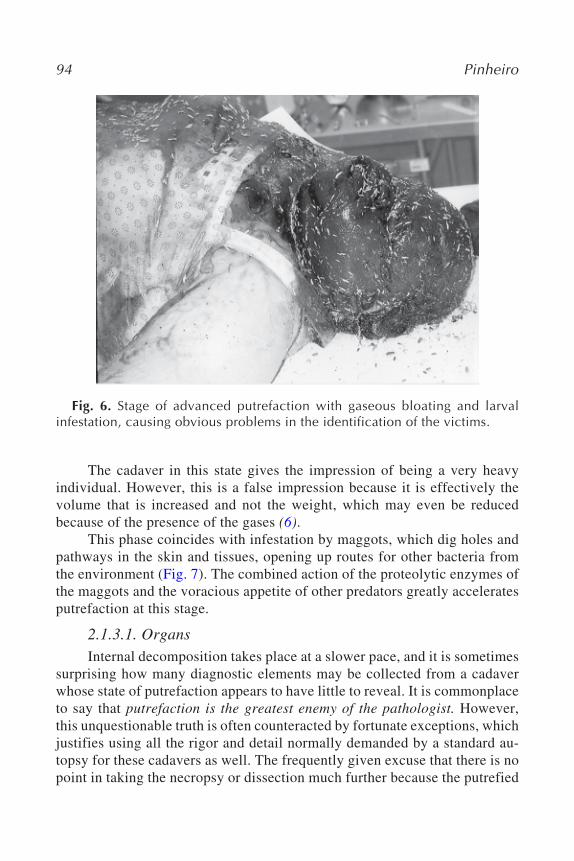
The green color gradually darkens to black, making identification evenmore difficult. The association of this with edema and the formation of gas inthe head lead to an increase in its size and the flattening of anatomical promi-nences, causing an “africanization” of features, known in some places as“blackman’s head” (Fig. 6). This phenomenon may arise, however, much ear-lier. The swelling of the face, in fact, begins immediately in the first week,depending on environmental conditions, and is accompanied by protrusion ofthe tongue between the dental arches.
Fig. 5. Purging of a bloody liquid from nostrils because of the gaseous dila-tation of the abdomen. Note the abrasions under the breasts, which could beerroneously considered antemortem, but were produced, in fact, by skin pres-sure with postmortem exsiccation.
94 Pinheiro
The cadaver in this state gives the impression of being a very heavyindividual. However, this is a false impression because it is effectively thevolume that is increased and not the weight, which may even be reducedbecause of the presence of the gases (6).
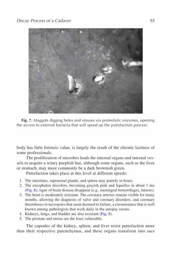
This phase coincides with infestation by maggots, which dig holes andpathways in the skin and tissues, opening up routes for other bacteria fromthe environment (Fig. 7). The combined action of the proteolytic enzymes ofthe maggots and the voracious appetite of other predators greatly acceleratesputrefaction at this stage.
2.1.3.1. OrgansInternal decomposition takes place at a slower pace, and it is sometimes
surprising how many diagnostic elements may be collected from a cadaverwhose state of putrefaction appears to have little to reveal. It is commonplaceto say that putrefaction is the greatest enemy of the pathologist. However,this unquestionable truth is often counteracted by fortunate exceptions, whichjustifies using all the rigor and detail normally demanded by a standard au-topsy for these cadavers as well. The frequently given excuse that there is nopoint in taking the necropsy or dissection much further because the putrefied
Fig. 6. Stage of advanced putrefaction with gaseous bloating and larvalinfestation, causing obvious problems in the identification of the victims.
Decay Process of a Cadaver 95
body has little forensic value, is largely the result of the chronic laziness ofsome professionals.
The proliferation of microbes leads the internal organs and internal ves-sels to acquire a winey purplish hue, although some organs, such as the liveror stomach, may more commonly be a dark brownish green.
Putrefaction takes place at this level at different speeds:
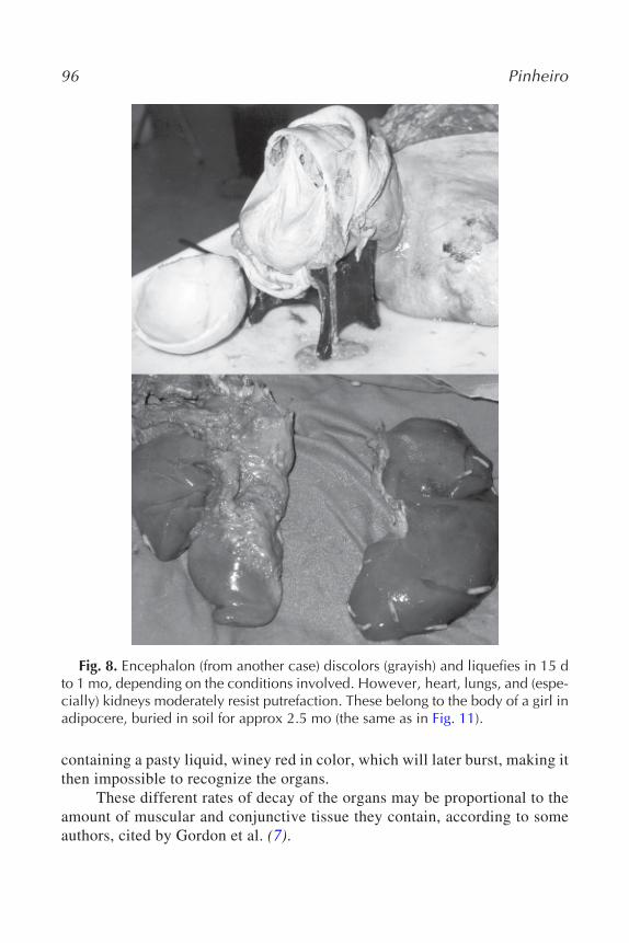
1. The intestines, suprarenal glands, and spleen may putrefy in hours.2. The encephalon discolors, becoming grayish pink and liquefies in about 1 mo
(Fig. 8); signs of brain disease disappear (e.g., meningeal hemorrhages, tumors).3. The heart is moderately resistant. The coronary arteries remain visible for many
months, allowing the diagnosis of valve and coronary disorders, and coronarythromboses in necropsies that seem doomed to failure, a circumstance that is wellknown among pathologists that work daily in the autopsy rooms.
4. Kidneys, lungs, and bladder are also resistant (Fig. 8).5. The prostate and uterus are the least vulnerable.
The capsules of the kidney, spleen, and liver resist putrefaction morethan their respective parenchymas, and these organs transform into sacs
Fig. 7. Maggots digging holes and sinuses via proteolytic enzymes, openingthe access to external bacteria that will speed up the putrefaction process.
96 Pinheiro
containing a pasty liquid, winey red in color, which will later burst, making itthen impossible to recognize the organs.
These different rates of decay of the organs may be proportional to theamount of muscular and conjunctive tissue they contain, according to someauthors, cited by Gordon et al. (7).
Fig. 8. Encephalon (from another case) discolors (grayish) and liquefies in 15 dto 1 mo, depending on the conditions involved. However, heart, lungs, and (espe-cially) kidneys moderately resist putrefaction. These belong to the body of a girl inadipocere, buried in soil for approx 2.5 mo (the same as in Fig. 11).
Decay Process of a Cadaver 97
2.1.4. LATER ALTERATIONS (MONTHS)
The viscera and soft tissues disintegrate, whereas organs such as theuterus, heart, and prostate last longer, as do tendon tissues and ligaments at-tached to the bones.
The body will then finally enter into the phase of skeletonization, depend-ing on the place where it is found and the season of the year. Some fragmentsof skin, protected by clothing or that come between the body and the supportsurface, may remain preserved, mummified, or with adipocere (see Fig. 1).
2.1.5. PUTREFACTION OF A BODY EXPOSED TO THE AIR
The rate at which a body decomposes is extremely variable. In theauthor’s experience, for the bodies of patients who have died in the hospitalin the summer and are not refrigerated (Portugal has a temperate, Mediterra-nean climate), a single afternoon is enough for the process to start. In fact, theearlier it starts, the faster it will be.
Various factors influence the speed of putrefaction: the atmospheric tem-perature and humidity level (10), the movement of air, state of hydration ofthe tissues and nutritional state of the victim, age, and respective cause ofdeath (5,7). Thus, low temperatures, which inhibit the growth of bacteria,retard the process considerably. The optimum temperature for the activationof bacteria responsible for putrefaction is 37.5 C (7). In a simultaneous doublehomicide autopsied by the author, the effect of temperature on the rate ofputrefaction in each of the corpses found at home was clearly perceptible(Fig. 9). Exposure to warm humid air, and the movement of this, also acceler-ates putrefaction (7).
In tissues that are greatly hydrated, with a higher liquid content, such asoccurs in cases of deaths through chronic congestive heart failure, putrefac-tion is faster. Victims who are dehydrated or who had suffered from vomitingand diarrhea resist much longer.
The process is faster in children than in adults and also in more obesepeople than in thinner individuals (6,7). However, newborn infants show someresistance to the start of the process.
Di Maio (7) claims that bodies wearing heavy clothes putrefy morequickly than those that are more lightly dressed, whereas other authors stressthat a clothed body decomposes less quickly than a naked one (15). It is alsonecessary to take into account the kind of fiber that is used in the clothing,whether natural or synthetic.
Of course, someone who dies of septicemia or from some acute infec-tion will already contain a proliferation of bacteria, which means that the
98 Pinheiro
process will be significantly accelerated. This acceleration will be greater inthe trunk than in the limbs, certainly for the same reason (that is, the absenceof bacteria in the muscular tissue of the limbs, as opposed to the abundance inthe organs of the trunk, especially the abdomen).
The presence of traumatic lesions caused by a blunt instrument or fire-arm may also affect the speed of decomposition (15), in that they open upholes through which air and insects may enter. Flies, however, tend to preferthe natural openings.
Fig. 9. Different stages of putrefaction observed in two bodies, both shot athome and recovered 3 d after the crime, on a moderate winter day. The man onthe top had a heater working near him; the girl lay on the floor of her bedroom,without any source of heating.
Decay Process of a Cadaver 99
2.1.6. DECOMPOSITION OF AN IMMERSED BODY
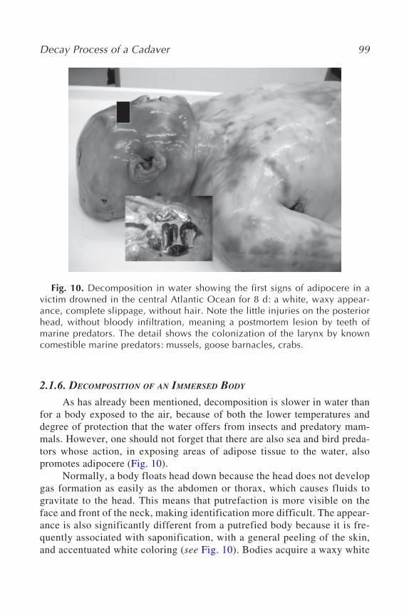
As has already been mentioned, decomposition is slower in water thanfor a body exposed to the air, because of both the lower temperatures anddegree of protection that the water offers from insects and predatory mam-mals. However, one should not forget that there are also sea and bird preda-tors whose action, in exposing areas of adipose tissue to the water, alsopromotes adipocere (Fig. 10).
Normally, a body floats head down because the head does not developgas formation as easily as the abdomen or thorax, which causes fluids togravitate to the head. This means that putrefaction is more visible on theface and front of the neck, making identification more difficult. The appear-ance is also significantly different from a putrefied body because it is fre-quently associated with saponification, with a general peeling of the skin,and accentuated white coloring (see Fig. 10). Bodies acquire a waxy white
Fig. 10. Decomposition in water showing the first signs of adipocere in avictim drowned in the central Atlantic Ocean for 8 d: a white, waxy appear-ance, complete slippage, without hair. Note the little injuries on the posteriorhead, without bloody infiltration, meaning a postmortem lesion by teeth ofmarine predators. The detail shows the colonization of the larynx by knowncomestible marine predators: mussels, goose barnacles, crabs.
100 Pinheiro
hue, and the appearance of the head without hair makes it difficult to iden-tify; indeed, some professionals have been ill advisedly led to believe thatsuch cases had been subjected to oncological therapies. Because of theputrefactive process, the bodies float continuously, even when they havebeen attached with stones for example, to make them sink to the bottom(see Fig. 12).
Putrefaction is also faster in warmer stagnant waters that contain decom-posing organic matter, such as industrial effluents, and so forth. It is alsofaster in fresh water than in saltwater (7). Some authors (5,8) contest this lastpoint, however, on the grounds that bacterial colonization results much morefrom bacteria in the digestive tract and airways of the victim than from theaquatic flora. Finally, as soon as the body has been removed from the water,putrefaction accelerates considerably.
2.1.7. DECOMPOSITION AFTER BURIAL
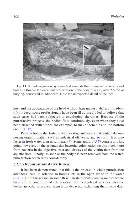
It has been demonstrated that this is the process in which putrefactionadvances least, in relation to bodies left in the open air or in the water(Fig. 11). For this reason, in some Brazilian states with scarce resources wherethere are no conditions of refrigeration, the medicolegal services bury thebodies in order to prevent them from decaying, exhuming them some days
Fig. 11. Buried corpses decay at much slower rate than immersed or air exposedbodies. Observe the excellent preservation of the body of a girl, after 2.5 mo ofburying, conserved in adipocere. Note the unexpected detail of the toes.
Decay Process of a Cadaver 101
later when the autopsy may be carried out. Thus, the soil functions as a kindof primitive refrigeration chamber.
This slower rate of decomposition is for obvious reasons: the absence ofair, inaccessibility to predators, and low temperature. The time taken beforeburial also affects the whole process. If putrefaction had not yet begun, thebody will remain in a better state of preservation than if the process had alreadygotten underway.
The kind of soil and depth at which a body is buried are also factors tobe taken into account. The process is faster in damp, porous soils and in bod-ies that are buried near the surface (5,7,8). The topography of the land is,however, more important than the type of terrain. If the body is buried in avalley or below the water table, the action of water will also be felt (5,8).
Bodies buried deeply in coffins decompose more slowly that when theyare in shallow graves—the temperatures are lower, there is less air, and theyare less affected by water (5,7,8). The type of coffin also influences the de-composition process. Laminated wooden coffins rot quickly, whereas thosemade of zinc or lead offer better protection.
2.2. Adipocere
The formation of adipocere is a natural preservation process that hasbeen known for centuries. Its name, attributed to Fourcroy in 1789, comesfrom the combination of the Latin adipo- (fat) and cera (wax) (5). This pro-cess, which some wrongly consider as a part of putrefaction (6,7), is knownas saponification.
It is a variable and irregular process, only occasionally involving thewhole body, which results from the hydrolysis and hydrogenation of the adi-pose tissue. This produces a waxy, fatty substance that is brittle; in color, it isyellowish off-white (see Figs. 11 and 12), although when stained by decayedmatter or blood, may acquire reddish, grayish, or gray-green tones (see Fig. 10).It also gives off a characteristic “earthy, cheesy, and ammoniacal” odor, whichmay be recognized by dogs trained to discover human remains (11).
Despite some points that are controversial and unclear, the biochemicalsequence of the formation of adipocere is, today, largely well established.The process begins immediately after death (10,11), with the hydrolysis (medi-ated by enzymes) of the triglycerides, which cleave the fatty acids from theglycerol molecules, giving rise to a mixture of unsaturated (palmitoleic acid,oleic acid, linoleic acid) and saturated (myristic acid, palmitic acid, stearicacid) fatty acids (16). As the process advances, the quantity of fatty acidsincreases, and the triglycerides diminish until they disappear completely (11).When there are sufficient enzymes and water, decomposition will continue
102 Pinheiro
with the hydrogenization of unsaturated fatty acids into saturated, after whichthe process is considered complete and stable. However, during hydrolysisand hydrogenization, some other products are formed. Free fatty acids mayattach to sodium and potassium ions of the interstitial liquid and cellular water, and later, to calcium ions, forming fatty acid salts. The subsequent actionof some microorganisms leads to the formation of 10-hydroxy fatty acids—themost common of which is 10-hydroxy stearic (11,16,17)—and, according toTakatori (17) of 10-oxo fatty acids. This author has shown that bacteria, such asPseudomonas, Staphylococcus aureus, and Clostridium perfringens, produce10-hydroxystearic acid from oleic acid, whereas Micrococcus luteus producesoxo fatty acids (17). These acids, and their respective soaps, as well as partici-pating in the formation of adipocere, also help to stabilize it. This stability maybe attributed to the action of ionic, covalent, hydrogen bonds between the car-boxyl terminal of the fatty acids and the hydroxyl groups (16). These substancesand glycerol form a matrix with fiber residues, nerves, and muscles, whichgives a degree of solidity to the saponified mixture (5).
At the moment of death, the body’s fatty acid content is 1%, but withadipocere, in the first month, it goes up to 20%, and at 3 mo is 70% (5). It isthought that the process is only superficial and therefore does not involve theviscera (7). However, whereas subcutaneous fat is the most affected, internalstructures containing adipose tissue, such as the mesentery, epiploon (omen-tum), perirenal fat, or organs with pathological processes involving a lipidicmetabolism, may also be involved.
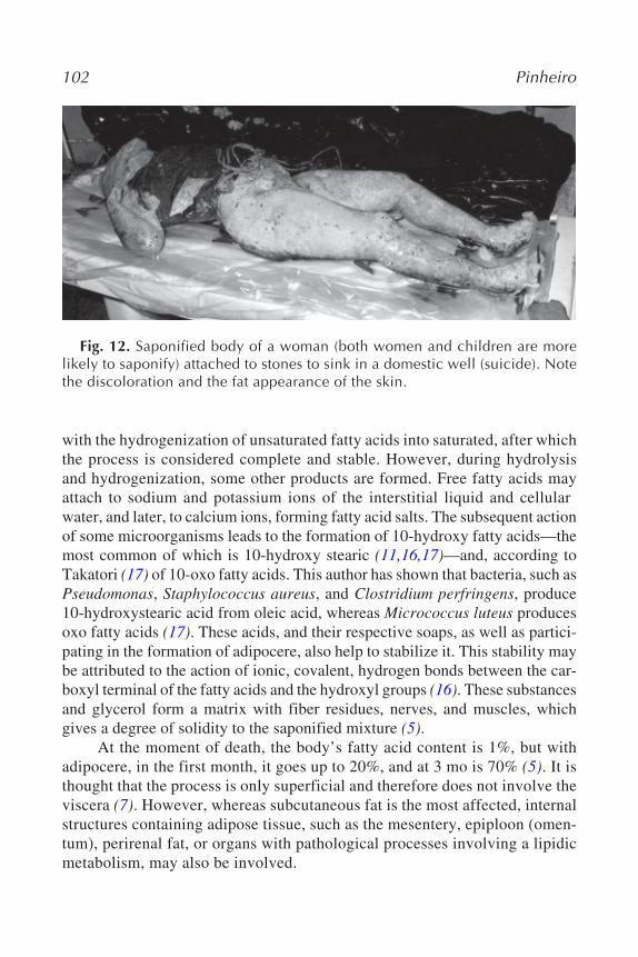
Fig. 12. Saponified body of a woman (both women and children are morelikely to saponify) attached to stones to sink in a domestic well (suicide). Notethe discoloration and the fat appearance of the skin.
Decay Process of a Cadaver 103
2.2.1. CONDITIONS
Saponification requires some heat, necessary for the development of themicrobes referred to in the previous subheading, as well as water, which maybe exogenous or from the organism itself (1,5). For this reason, it generallyappears in damp environments (5) in bodies immersed in cold water with alow oxygen content (1,10). Pfeiffer (16) associates the persistence of adipo-cere to the presence of Gram-negative microorganisms, which are known todevelop in anaerobic environments.
However, saponification is also found—much more often than isthought—in tombs, crypts, and graves, even dry ones, in a few days; the waterfrom the organism is enough to set in motion the chemical transformationsnecessary for the process. Adipocere benefits from a process of self-promotion,because it inhibits putrefaction, increasing acidity and dehydration, thus reduc-ing the growth and spread of putrefactive organisms.
Women (see Fig. 8) and children are much more likely to undergo thispreservation process because they have a greater fat content. For the samereasons, the parts of the body that tend most to saponification are the cheeks,eye sockets, chest, abdominal wall, and buttocks.
2.2.2. CHRONOLOGY
Adipocere may last for decades, even centuries. It can form between 3and 12 mo (5–7,11,10,18), although this is variable; the first signs may bevisible as early as the third week after death (5,11,10,19) or even earlier (8 d),as was the case of a victim of drowning in the central Atlantic Ocean (Portu-guese coast) autopsied by the author,* where atmospheric temperatures rangedfrom 16 to 30 C (average of 20 C), and the average of sea water temperaturewas 18 C (see Fig. 10).
Studies carried out in the area of marine taphonomy cited by Kahana(12) consider that, in cold waters (4 C), some 12–18 mo are required for saponi-fication, whereas in waters of between 15 and 22 C, only 2–3 mo are needed;very high temperatures are necessary for the process to be evident in 1–3 wk.However, other reports are highly contradictory. The same author reportsbodies recovered from a wreck of a Belgian ship in the China Sea, saponifiedat 38 d in water temperatures of between 10 and 12 C, among other discrep-ancies. It should be emphasized that in the same sample, three bodies were
* During the period this body was submersed, weather was similar, with the excep-tion of a day when meteorological conditions had suffered a sudden change, withan increase of both atmospheric temperature (to 35 C) and the temperature of thesea water, which ranged from 19 to 36 C.
104 Pinheiro
found later (after 433 d) with adipocere, but with some parts skeletonized.Studies of controlled decomposition in samples of pig carcasses (11) in shal-low graves have shown that there is no correlation between stages of saponi-fication and the period of decomposition, which once more confirms thevariability of the whole process and the probable intervention of other factorsas yet unstudied, such as temperature, humidity, pH, clothing, and soil type.
These findings reinforce the idea that has been argued consistently inthis text as to the lack of confidence in estimates of PMI based on states ofdecomposition of the body.
The decay of the adipocere is not completely clarified because it hasbeen alleged that soil microbiota including bacteria, fungus, and algae mayplay a role in this decomposition. Pfeiffer (16) suggests that the maintenanceof adipocere is associated with the development of Gram-negatives in a slightlyanaerobic environment, and that its decay has to do with exposure to condi-tions of aerobiosis and to the presence of Gram-positive bacteria. This facthas been confirmed for many by practical experience, because a saponifiedbody that has been removed from its environment for autopsy starts to decaymuch more rapidly than it did before its removal.
2.2.3. FORENSIC VALUE
The medicolegal interest of saponification lies not only in the possibili-ties it offers for identification because it conserves some bodily forms, butalso in determining the cause of death. It is, however, rare for a body con-served only through adipocere to be recognized by the face, given the physi-ognomic distortions that are common despite preservation (see Fig. 10).
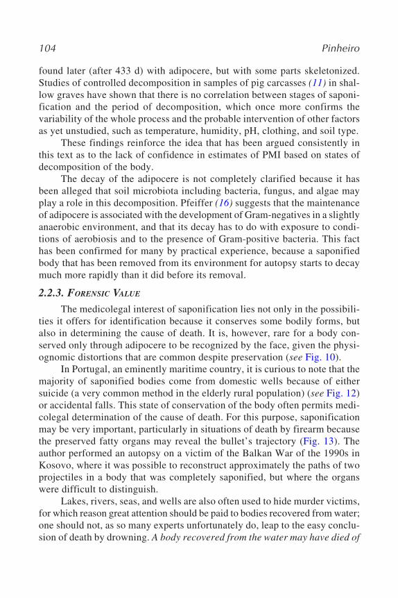
In Portugal, an eminently maritime country, it is curious to note that themajority of saponified bodies come from domestic wells because of eithersuicide (a very common method in the elderly rural population) (see Fig. 12)or accidental falls. This state of conservation of the body often permits medi-colegal determination of the cause of death. For this purpose, saponificationmay be very important, particularly in situations of death by firearm becausethe preserved fatty organs may reveal the bullet’s trajectory (Fig. 13). Theauthor performed an autopsy on a victim of the Balkan War of the 1990s inKosovo, where it was possible to reconstruct approximately the paths of twoprojectiles in a body that was completely saponified, but where the organswere difficult to distinguish.
Lakes, rivers, seas, and wells are also often used to hide murder victims,for which reason great attention should be paid to bodies recovered from water;one should not, as so many experts unfortunately do, leap to the easy conclu-sion of death by drowning. A body recovered from the water may have died of
Decay Process of a Cadaver 105
anything, even drowning. Among many examples present in the literature,Dix (19) tells of four separate cases of homicides, accidentally collected fromLake Missouri, all saponified.
2.3. Mummification
This is a process of natural or artificial conservation, which consists ofthe dehydration and exsiccation (the process of drying up) of tissues. It maybe partial and coexist with other forms of conservation and/or putrefaction(see Fig. 1). It extends more easily to the whole body than other processes,such as saponification (8).
It is characterized by dryness and brittle, torn skin on the prominences(cheeks, forehead, sides of the back, and hips), generally brown in color, thoughcoexisting with white, green, or black zones because of colonization by fun-gus (see Fig. 1), just as leather jackets look after they have been left for sometime in a musty wardrobe and start to become mildewed.
As for the internal organs, the process varies in relation to the time sincedeath, and they may be partially mummified, putrefied, with adipocere, or
Fig. 13. A saponified head of a homicide victim, found closed inside theback of his car, sunk in a lake to hide the body. The author could recover thefour bullets and determine its pathway. Two of the shots penetrated the bodythrough the same hole seen on the face (arrow). Another entrance hole can benoted below the ear (circle). Note the preserved aspect of the face that couldlead, but with some difficulty, to a positive identification. (See Fig. 3, Chapter 7to observe the X-ray.)
106 Pinheiro
even absent. Radanov et al. (20) describe an extreme and unique case of thenatural mummification of a brain, which was surprising, taking into accountthe softness of brain tissue, as well its fatty composition. The body had comefrom a mass grave with 39 other bodies and had been buried for 40–50 yr, ata shallow depth in a stony terrain, exposed to sunlight.
It is common for there to be slight adipocere in mummified bodies.Indeed, there is a close interconnection between these two processes; the useof the water from the body for hydrolysis of fats contributes also to the exsic-cation of tissues (5,8). This relationship is extendable to the biochemical level,as has been demonstrated by Makristathis et al. (21), who detected in mum-mies the same constituents of saponification: palmitic, oleic, and 10-hydroxy-stearic acid, among other substances.
2.3.1. CONDITIONS
As is to be expected, mummification is found in dry, ventilated environ-ments (1,5) and generally, though not always, in warm places where the bodyloses fluids through evaporation (5,6): closed rooms, attics, wardrobes and pan-tries, barns, stairwells, and so on. More extensive and complete mummificationoccurs in desert environments; indeed, this preservation process was practicedby the ancient Egyptians, who added spices and herbs to the heat (1,5,8).
Mummification also takes place in icy environments, not only becauseof the dryness of the air, but also because of the low growth of bacteria atsuch temperatures. A frozen mummy approx 5000 yr old (known as theTyrolean Iceman) discovered in the Alps in 1991 has become famous as averitable star of anthropology, like others from Peru that are also thousandsof years old (21). The former, however, raises the question of knowing whethermummification, which is by definition related to exposure to dry air, maytake place in the snow. Ambach and Ambach (22) justify this from a physicalpoint of view, given that evaporation may occur from a frozen body througha superficial covering of snow (porous and air-permeable), if the weather con-ditions establish a water vapor pressure gradient between the snow layers.
Makristathis et al. (21) compared the composition of the fat of mummi-fied bodies from different parts of the world: the Tyrolean Iceman; two bodiesfound in alpine glaciers near to this; a body that had been immersed for 50 yr inan Austrian mountain lake; two bodies buried in the permafrost of Siberia; twoPeruvian mummies, one from the Andes (500 yr old) and one from the Peru-vian desert (1000 yr old); and three fresh bodies as a control. The compositionof fatty acids was very similar between the samples of fresh bodies and thosefrom the dry mummification of Peru, in which no significant concentrations of10-hydroxystearic acid were found, with oleic acid predominating. These were
Decay Process of a Cadaver 107
also the best preserved bodies. In addition, all those conserved in ice or in con-tact with water showed a high concentration of 10-hydroxystearic acid, suggest-ing the association of this acid with conditions of conservation in water. TheTyrolean Iceman was situated somewhere between the mummies conserved dryand those from the ice, and was in a much better state of preservation than manymore recent bodies, only tens of years old, also from glaciers. This can be explainedby the rapid initial desiccation caused by the cold mountain winds, followed by aburial in ice with periods in water.
Dehydration before death may also favor this process. Indeed, this issimilar to an ancient Japanese practice of natural self-mummification, accord-ing to which bonzes (Buddhist monks), nearing the end of their lives, wouldprogressively reduce their solid food intake, then the liquid, so that they werepractically desiccated at the moment of death. They were buried, and thenexhumed 3 yr later, when they were found to be already mummified, withoutany other kind of intervention (23).
2.3.2. CHRONOLOGY
The time necessary for mummification to take place is not well docu-mented because of the long periods that usually occur before the body is dis-covered. It certainly takes some weeks (5,7,8) and, in the early stages, is mixedwith putrefactive alterations, especially in the internal organs. In the desertsof Arizona, corpses exposed to the air require between 11 d and 1 mo to mum-mify (15). After they are dry, they may last years, even centuries (5,7,8).
The action of predators (see Fig. 2) in this phase may accelerate thedisintegration of the exsiccated tissues, which are fragile and brittle; frag-ments of parchment-like skin, tendons, and ligaments attached to the bonesmay remain for much longer, however.
2.3.3. FORENSIC VALUE
Mummification can have significant medicolegal relevance for the twogreat objectives of forensic anthropology, identification of the body and estab-lishing cause of death. Concerning the former, mummies are often found in asurprising state of preservation (Fig. 14), and it is usually much easier toinvestigate the victim’s identity in these situations than with adipocere. Con-cerning the latter, large lesions may be preserved. However, the detection ofecchymosis or wounds may be made difficult or impossible because of dis-coloration, artifacts, and the action of fungus.
Cadavers in this state are sometimes the victims of homicides that havebeen left in a place propitious to mummification. It can also be found in casesof natural death of people that live alone. It is a very common process in
108 Pinheiro
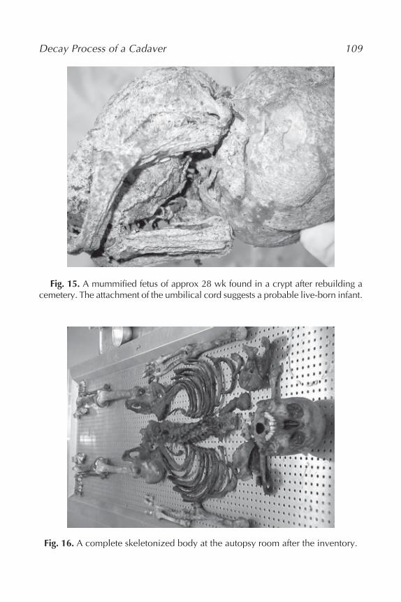
fetuses or newborn infants, who mummify much more rapidly and completely,often because of the types of graves or ventilated domestic locations wherethey are deposited (Fig. 15).
Autopsies in these cases require particular dexterity because the skin,which is brittle and disintegrates easily, is difficult to dissect. Some methodsof softening the tissues to permit better observation and histological study (5)have been described; one of these involves the use of a solution of 20% poly-ethylene glycol, with controlled pH of 8.0 and the addition of 1% Stericol toinhibit the growth of bacteria and fungus (24).
2.4. Skeletonization
As the name suggests, this consists of the removal of all soft tissue fromthe bone, and is the field par excellence of the forensic anthropologist.
A body that has been reduced merely to its bones may be, however,present in its totality, thus constituting a complete skeleton (Fig. 16). Differ-ent states of preservation may nonetheless coexist, as mentioned previously,of which one may be skeletonization, which will be, in this case, partial. Whenthis happens, the classic scenario is skeletonization of the cranium (which hasthe least soft tissues), mummification of the extremities, and saponificationof the back (see Fig. 1). The natural and most frequent tendency, if conditionsare propitious, is for complete skeletonization.
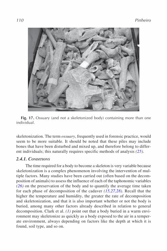
In the certain (and very frequent) case of a group of bones (Fig. 17)—sometimes already eroded, found in a church cemetery, obviously in a phasesubsequent to skeletonization, when the bones are completely disarticulatedand fragmented—it seems incorrect to designate this as a skeleton or body in
Fig. 14. The hands of a clerical mummified in a crypt (18th century). Theperfection of the details is surprising.
Decay Process of a Cadaver 109
Fig. 15. A mummified fetus of approx 28 wk found in a crypt after rebuilding acemetery. The attachment of the umbilical cord suggests a probable live-born infant.
Fig. 16. A complete skeletonized body at the autopsy room after the inventory.
110 Pinheiro
skeletonization. The term ossuary, frequently used in forensic practice, wouldseem to be more suitable. It should be noted that these piles may includebones that have been disturbed and mixed up, and therefore belong to differ-ent individuals; this naturally requires specific methods of analysis (25).
2.4.1. CONDITIONS
The time required for a body to become a skeleton is very variable becauseskeletonization is a complex phenomenon involving the intervention of mul-tiple factors. Many studies have been carried out (often based on the decom-position of animals) to assess the influence of each of the taphonomic variables(26) on the preservation of the body and to quantify the average time takenfor each phase of decomposition of the cadaver (15,27,28). Recall that thehigher the temperature and humidity, the greater the rate of decompositionand skeletonization, and that it is also important whether or not the body isburied, among many other factors already described in relation to generaldecomposition. Clark et al. (1) point out that a body buried in a warm envi-ronment may skeletonize as quickly as a body exposed to the air in a temper-ate environment, always depending on factors like the depth at which it isfound, soil type, and so on.
Fig. 17. Ossuary (and not a skeletonized body) containing more than oneindividual.
Decay Process of a Cadaver 111
As was mentioned with regard to mummification, the ligaments and sometendons are the soft tissues that most resist leaving the bone. Skin, soft tis-sues, and organs are lost much earlier. Disarticulation consists in the disap-pearance of the soft tissues, which, in living beings, hold bones together withina joint (3). Thus, even when each bone is in the right place, the skeleton isconsidered disarticulated whenever the soft tissues do not join the bonestogether.
Disarticulation is very common in skeletonization and, in unburied bod-ies, takes place in a cephalic–caudal direction, and from the center to theperiphery, that is to say, the head (without the jaw) usually separates firstfrom the spine and then, the other limbs (29). Dirkmaat and Sienickis (30)have proposed the following sequence for human disarticulation in bodiesexposed to the air: first the head, because of the accessibility of its cavities toinsects, followed by the sternum and clavicle; the upper limbs decomposemuch faster than the lower ones; the pelvis separates much later than the trunk,and the ribs do so in different degrees; the feet, often in socks and shoes, lastmuch longer than the rest. The vertebral column, although exposed early, isone of the last to break up because of the strong costovertebral and interverte-bral ligaments. Unprotected hands and feet are, however, the first to disar-ticulate, sometimes even before the head separates (3). For bodies that havecome out of water, Haglund (31) established that the areas that lose their softtissues first, leaving the bones visible, are those covered by soft layers oftissue like the head, hands, and front of legs. Disarticulation begins with thebones of the hand and wrists, bones of the feet and ankles, jaw, and cranium.Finally, the legs and arms separate.
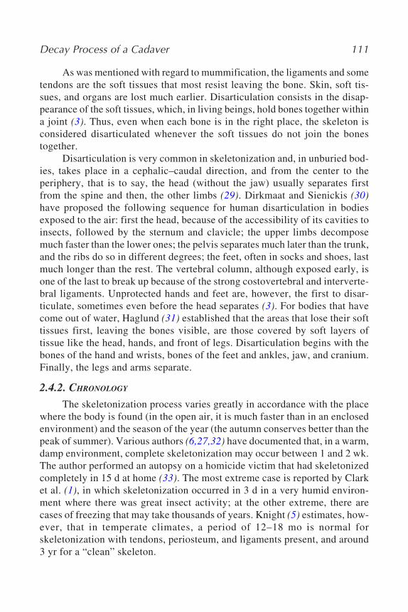
2.4.2. CHRONOLOGY
The skeletonization process varies greatly in accordance with the placewhere the body is found (in the open air, it is much faster than in an enclosedenvironment) and the season of the year (the autumn conserves better than thepeak of summer). Various authors (6,27,32) have documented that, in a warm,damp environment, complete skeletonization may occur between 1 and 2 wk.The author performed an autopsy on a homicide victim that had skeletonizedcompletely in 15 d at home (33). The most extreme case is reported by Clarket al. (1), in which skeletonization occurred in 3 d in a very humid environ-ment where there was great insect activity; at the other extreme, there arecases of freezing that may take thousands of years. Knight (5) estimates, how-ever, that in temperate climates, a period of 12–18 mo is normal forskeletonization with tendons, periosteum, and ligaments present, and around3 yr for a “clean” skeleton.
112 Pinheiro
The bone gradually wears away in the meantime, with fractures, decal-cification, and dissolution because of the combined action of various factors,such as acidic soils and water, in a process that begins, for some authors (15),after 9 mo of exposure. After the complete separation of the bony parts, thisdisaggregation accelerates markedly, until the body may even disappearcompletely.
2.4.3. FORENSIC VALUE
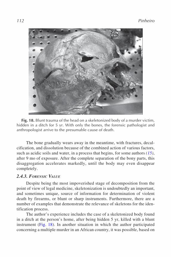
Despite being the most impoverished stage of decomposition from thepoint of view of legal medicine, skeletonization is undoubtedly an important,and sometimes unique, source of information for determination of violentdeath by firearms, or blunt or sharp instruments. Furthermore, there are anumber of examples that demonstrate the relevance of skeletons for the iden-tification process.
The author’s experience includes the case of a skeletonized body foundin a ditch at the person’s home, after being hidden 5 yr, killed with a bluntinstrument (Fig. 18). In another situation in which the author participatedconcerning a multiple murder in an African country, it was possible, based on
Fig. 18. Blunt trauma of the head on a skeletonized body of a murder victim,hidden in a ditch for 5 yr. With only the bones, the forensic pathologist andanthropologist arrive to the presumable cause of death.
Decay Process of a Cadaver 113
a multidisciplinary study of the mostly skeletonized remains, to solve ques-tions of identification and to determine the cause of death, which the case hadraised (34).
Finally, it is in this state of skeletonization that most victims of politicalgenocide and/or crimes against humanity across the world are found, such asin the Balkans, East Timor, Latin America, Africa, and Iraq. The Interna-tional Criminal Tribunal for the former Yugoslavia, in the trial at The Hagueof those presumed responsible for the abuse of human rights in the Balkanconflicts, made use of this kind of expertise in decomposed bodies. The inves-tigations were carried out by multidisciplinary teams under the auspices ofthe United Nations, which included forensic pathologists and anthropologistsfrom around the world, some from organizations like the Equipo Argentinode Antropologia Forense or Physicians for Human Rights, which have provenessential for the demonstration of these crimes. These missions have trulygalvanized this common adventure of forensic pathology and forensic anthro-pology—well documented in an article by Steadman (2)—which has not onlyefficiently resolved questions that were raised, but has also established somehighly stimulating challenges for the future, permitting a more effective admin-istration of justice and thus, the pacific cohabitation of peoples in a happier,healthier, and less violent world.
3. CONCLUSION
At the end of this chapter, the hope is to have given a perspective of themain alterations a human body might suffer until it is found or even com-pletely disappears. Forensic anthropologists as well as forensic pathologistsmust be familiarized with these processes in order to be prepared to getinvolved as experts in cases for which they are called. Nobody ever knows inwhich state a cadaver will be presented. Thus, it is good practice that immedi-ate and midputrefaction is not an unknown matter for forensic anthropolo-gists. In the same way, forensic pathologists should also know how to dealwith bare bones.
Interdisciplinarity is then essential. Referred everywhere (2,33,35–37)and permanently requested, it will be the issue of Chapter 7, and mentionedoften in other chapters of this book.
This type of multidisciplinary experience has been conducted in Portu-gal in the last 5 yr, using the knowledge of these concepts and processes, withsignificant success both in terms of civil purposes and even for the adminis-tration of the justice regarding the penal law. An example of the first is, amongsome cases of successful identification and distinction between more than an
114 Pinheiro
individual in cemeteries graves, the identification of an old female (whodisappeared after a family quarrel and was found decapitated in a river) par-tially by personal belongings, but confirmed trough exuberant pathologicalvascular disorders of the leg bones (38). Concerning criminal justice, the authorand forensic anthropologist Eugénia Cunha (two coeditors of this book) per-formed some successful cases on victims of homicides already presented(33,34).
This spirit, embodied by the whole philosophy of this book, must bekept and developed, not only in the international situations in which forensicprofessionals can be asked to participate, but also in routine cases in eachcountry, as is shown, in Chapter 7.
REFERENCES
1. Clark, M. A., Worrell, M. B., Pless, J. E. Postmortem changes in soft tissues. In:Haglund, W. D., Sorg, M. H., eds., Forensic Taphonomy: the Postmortem Fate ofHuman Remains. CRC Press, Boca Raton, FL, pp. 156–164, 1997.
2. Steadman, D. W., Haglund, W. D. The SCOPE of anthropological contributions tohuman rights investigations. J. Forensic Sci. 50:1–8, 2005.
3. Rocksandic, M. Position of skeletal remains as a key to understanding mortuarybehavior. In: Haglund, W. D., Sorg, M. H., eds., Advances in Forensic Taphonomy:Method, Theory and Archaeological Perspectives. CRC, Boca Raton, FL, pp. 99–113, 2002.
4. Bass, W. M. Anthropology. In: Siegel, J. A., Saukko, P. J., Knupfer, G. C., eds., Ency-clopedia of Forensic Sciences, Vol. 1. Academic, San Diego, CA, pp. 194–284, 2000.
5. Knight, B. Forensic Pathology, 2nd Ed. Arnold, London, pp. 51–94, 1996.6. Di Maio, V. J., Di Maio, D. Forensic Pathology, 2nd Ed. CRC Press, Boca Raton,
FL, pp. 21–41, 2001.7. Gordon, I., Shapiro, H. A., Berson, S. D. Forensic Medicine: a Guide to Principles,
3rd Ed. Churchill Livingstone, Edinburgh, pp. 1–62, 1988.8. Saukko, P., Knight, B. Knight’s Forensic Pathology, 3rd Ed. Arnold, London,
pp. 52–97, 2004.9. Prieto, J. L., Magaña, C., Ubelaker, D. H. Interpretation of postmortem change in
cadavers in Spain. J. Forensic Sci. 49:918–923, 2004.10. Yan, F., McNally, R., Kontanis, E. J., Sadik, O. A. Preliminary quantitative inves-
tigation of postmortem adipocere formation. J. Forensic Sci. 46:609–614, 2001.11. Forbes, S. L., Stuart, B. H., Dadour, I. R., Dent, B. B. A preliminary investigation
of the stages of adipocere formation. J. Forensic Sci. 49:1–9, 2004.12. Kahana, T., Almog, J., Levy, J., Shmeltzer, E., Spier, Y., Hiss, J. Marine taphonomy:
adipocere formation in a series of bodies recovered from a single shipwreck. J.Forensic Sci. 44:897–901, 1999.
13. Micozzi, M. S. Postmortem Change in Human and Animal Remains: a SystematicApproach. Charles C. Thomas, Springfield, IL, 1991.
Decay Process of a Cadaver 115
14. Shean, B. S., Messinger, L., Papworth, M. Observations of differential decomposi-tion on sun exposed v. shaded pig carrion in coastal Washington State. J. ForensicSci. 38:938–949, 1993.
15. Galloway, A., Birkby, W. H., Jones, A. M., Henry, T. E., Parks, B. O. Decay ratesof human remains in an arid environment. J. Forensic Sci. 34:607–616, 1989.
16. Pfeiffer, S., Milne, S., Stevenson, R. M. The natural decomposition of adipocere. J.Forensic Sci. 43:368–370, 1998.
17. Takatory, T. Investigations on the mechanism of adipocere formation and its relationto other biochemical reactions. Forensic Sci. Int. 80:49–61, 1996.
18. Mellen, P. F. M., Lowry, M. A., Micozzi, M. S. Experimental observations onadipocere formation. J. Forensic Sci. 38:91–93, 1993.
19. Dix, J. D. Missouri’s lakes and the disposal of homicide victims. J. Forensic Sci.32:806–809, 1987.
20. Radanov, S., Stoev, S., Davidov, M., Nachev, S., Stanchev, N., Kirova, E. A uniquecase of naturally occurring mummification of human brain tissue. Int. J. Legal Med.105:173–175, 1992.
21. Makristathis, A., Scharzmeier, J., Mader, R. M., et al. Fatty acid composition andpreservation of the Tyrolean Iceman and other mummies. J. Lipid Res. 43:2056–2061, 2002
22. Ambach E, Ambach W. Is mummification possible in snow? Forensic Sci. Int.54:191–192, 1992.
23. Hedouin, V., Laurier, E., Courtin, P., Gosset, D., Muller, P. H. Un cas demomification naturelle [in French]. J. Med. Legale Droit Med. 19:43–45, 1993.
24. Garrett, G., Green, M. A., Murray, L. A. Technical method—rapid softening ofadipocerous bodies. Med. Sci. Law 28:98–99, 1988.
25. Ubelaker D. Approaches to the study of commingling in human skeletal remains.In: Haglund, W. D., Sorg, M. H., eds., Advances in Forensic Taphonomy: Method,Theory and Archaeological Perspectives. CRC Press, Boca Raton, FL, pp. 331–351, 2002.
26. Henderson, J. Factors determining the state of preservation of human remains. In:Boddington, A., Garland, A. N., Janaway, R. C., eds., Death, Decay and Reconstruc-tion: Approaches to Archaeology and Forensic Science. Manchester UniversityPress, Manchester, pp. 42–53, 1997.
27. Mann, R. W., Bass, W. M., Meadows, L. Time since death and decomposition of thehuman body: variables and observations in case and experimental field studies. J.Forensic Sci. 35:103–111, 1990.
28. Sledzik P. Forensic taphonomy: postmortem decomposition and decay. In: Reichs,K, ed., Forensic Osteology: Advances in the Identification of Human Remains.Charles C. Thomas, Springfield IL, pp. 109–119, 1998.
29. Rodriguez, W. C., Bass, W. M. Decomposition of buried bodies and methods thatmay aid in their location. J. Forensic Sci. 30:836–852, 1985.
30. Dirkmaat, D. C., Sienicki, L. A. Taphonomy in the northeast woodlands: four casesfrom western Pennsylvania. Proceedings of the 47th Annual Meeting of the Ameri-can Academy of Forensic Sciences, Seattle, Washington. 1:10, 1998.
116 Pinheiro
31. Haglund, W. D. Disappearance of soft tissue and the disarticulation of human re-mains from aqueous environments. J. Forensic Sci. 38:806–815, 1993.
32. Galloway, A. The process of decomposition: a model from Arizona-Sonoran Desert.In: Haglund, W. D., Sorg, M. H., eds., Forensic Taphonomy: the Postmortem Fateof Human Remains. CRC Press, Boca Raton, FL, pp. 139–150, 1997.
33. Cunha, E., Pinheiro, J., Corte Real, F. Two Portuguese homicide cases: the impor-tance of interdisciplinarity in forensic anthropology. ERES (Arqueología yBioantropología) 15:65–72, 2005.
34. Cunha, E., Pinheiro, J., Ribeiro, I. P., Soares, J., Vieira, D. N. Severe traumaticinjuries: report of a complex multiple homicide case. Forensic Sci. Int. 136:164–165, 2003.
35. Symes, S. A., Woytash, J. J., Kroman, A. M., Wilson, A. C. Perimortem bonefracture distinguished from postmortem fire trauma: a case study with mixed sig-nals. Proceedings of the 54th American Academy of Forensic Sciences, Vol. 11.New Orleans, LA, 300, 2005.
37. Verano, J. Serial murder with dismemberment of victims in an attempt to hinderidentification: a case resolved trough multidisciplinary collaboration. Proceedingsof the 54th American Academy of Forensic Sciences, Vol. 11. New Orleans, LA,329, 2005.
38. Pinheiro, J., Cunha, E., Cordeiro, C., Vieira, D. N. Bridging the gap between forensicanthropology and osteoarchaeology—a case of vascular pathology. Int. J.Osteoarchaeol. 14:137–144, 2004.