18
Dept. of Medical Microbiology University of Sulaimani Sulaimani, KRG 24 th February, 2015
| Date post: | 15-Dec-2015 |
| Category: |
Documents |
| Upload: | august-saile |
| View: | 219 times |
| Download: | 3 times |

Dept. of Medical Microbiology
University of Sulaimani
Sulaimani, KRG
24th February, 2015
• Introduction of virology Introduction of virology
• Laboratory diagnosis of viral infectionLaboratory diagnosis of viral infection
• Specimen collection and transportSpecimen collection and transport
• Diagnostic Technique (rapid test)Diagnostic Technique (rapid test)

Virology: Study of viruses
Viruses: are obligatory intracellular parasites. It is composed of nucleic acid surrounded by a protein shell and/or a lipid layer.

Why are viruses important to vet?
Viruses cause disease in animals of economic and/or welfare importance.
Animal viruses may pose risk to human health (zoonosis).
Can act as important models for human disease.
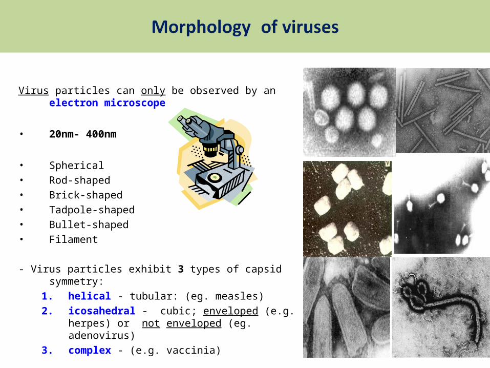
Virus particles can only be observed by an electron microscope
• 20nm- 400nm
• Spherical
• Rod-shaped
• Brick-shaped
• Tadpole-shaped
• Bullet-shaped
• Filament
- Virus particles exhibit 3 types of capsid symmetry:1. helical - tubular: (eg. measles)2. icosahedral - cubic; enveloped (e.g. herpes) or
not enveloped (eg. adenovirus)3. complex - (e.g. vaccinia)

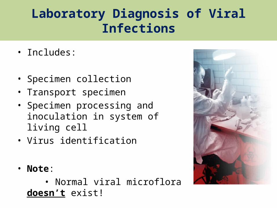
Laboratory Diagnosis of Viral Infections
• Includes:
• Specimen collection• Transport specimen• Specimen processing and inoculation in
system of living cell• Virus identification
• Note: • Normal viral microflora doesn’t exist!

1. Specimen collection
• Rules or Principle of successful viral sampling
1. At the correct time (stage of infection) 2. From the correct site (site of infection)3. In the correct way4. In the adequate volume (for all tests needed)5. In the proper containers (sterile and chemically
clean)6. Correctly labelled (name, date, type of
specimen) and with (age, sex, epidemiological data – vaccinations, etc.)

2. Transport specimen
• During transport specimen should be:
1. protected from breaking2. protected from light3. At adequate temperature: -48 hrs at +4°C (refrigerator, wet ice) -more than 48 hrs at -70°C (dry ice)
• NOTE// must not be frozen at -20°C!

Transport specimen
• * Viral Transport Media (VTM):
• (VTM) contains: -buffer (adequate pH) -saline (adequate ion concentration) - proteins (albumin or gelatine) - antibiotics and fungicides
• (VTM) is used to:• Preserve viral infectivity within the specimen• Prevent specimen from drying• Stop the growth of bacteria and fungi
• * MEM, Hank’s solution, Stuart’s• * EDTA, Sodium citrate, heparin for (blood).....


Common types of Specimen Collection
Specimen is taken from the viral lesion by:
Swab from the lesion
Aspiration from vesicles
o Both placed in a(container with VTM)

Swabs
• For diagnosis of viral infections;
Swabs should be: • made of rayon• Should not be made of cotton or calcium alginate
Swab’s shaft should be: • made of plastics or metal• Should not be made of wood
Swab from the lesion with rotation & place in (test-tube with VTM)

Tissue Biopsy or Autopsy
• Tissue samples collection: (Tissue Biopsy or Autopsy)
1. Sterile instruments should be used.2. Collect 3 x 5 mm and placed in tube with
VTM
• Storage • In the refrigerator (2 – 6.0° C) for up to one
week.• Can be stored frozen (≤ -70.0 ° C) indefinitely.

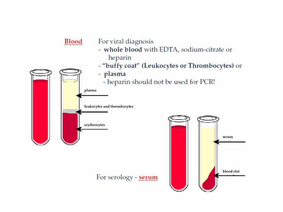
Blood (Serum) sample
• Whole Blood (serum) • Collected by venipuncture or venal catheter
(5-7 ml)• Place in a sterile tube (with EDTA, Sodium-
citrate or heparin)• Allow 15 minutes to clot to form. By Centrifuge
(serum)• Or by storing the whole blood sample,
overnight in the refrigerator (2– 6.0° C). (plasma)
• Urine, CSF, or other fluids can be used for diagnosis of viral infection
• Note// Sputum is not useful for viral detection
Diagnostic Techniques
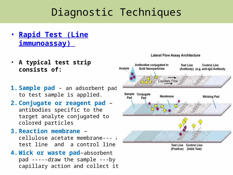
• Rapid Test (Line immunoassay)
• A typical test strip consists of:
1. Sample pad - an adsorbent pad to test sample is applied.
2. Conjugate or reagent pad –antibodies specific to the target analyte conjugated to colored particles
3. Reaction membrane – cellulose acetate membrane--- a test line and a control line
4. Wick or waste pad–absorbent pad -----draw the sample ---by capillary action and collect it
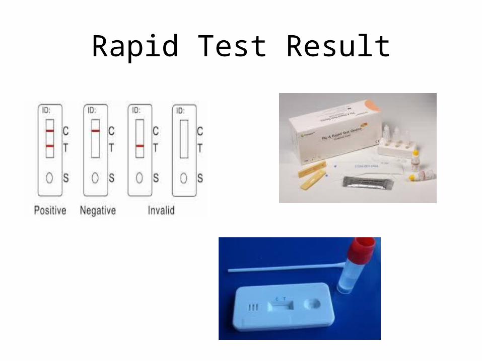
Rapid Test Result


