Journal of Systematic Palaeontology http://journals.cambridge.org/SYP Additional services for Journal of Systematic Palaeontology: Email alerts: Click here Subscriptions: Click here Commercial reprints: Click here Terms of use : Click here DESCRIPTION AND SYSTEMATIC RELATIONSHIPS OF †Tomognathus, AN ENIGMATIC FISH FROM THE ENGLISH CHALK Peter L. Forey and Colin Patterson Journal of Systematic Palaeontology / Volume 4 / Issue 02 / June 2006, pp 157 184 DOI: 10.1017/S1477201905001719, Published online: 16 May 2006 Link to this article: http://journals.cambridge.org/abstract_S1477201905001719 How to cite this article: Peter L. Forey and Colin Patterson (2006). DESCRIPTION AND SYSTEMATIC RELATIONSHIPS OF †Tomognathus, AN ENIGMATIC FISH FROM THE ENGLISH CHALK. Journal of Systematic Palaeontology, 4, pp 157184 doi:10.1017/ S1477201905001719 Request Permissions : Click here Downloaded from http://journals.cambridge.org/SYP, IP address: 128.233.210.97 on 04 Oct 2012
Transcript
Journal of Systematic Palaeontologyhttp://journals.cambridge.org/SYP
Additional services for Journal of Systematic Palaeontology:
Email alerts: Click hereSubscriptions: Click hereCommercial reprints: Click hereTerms of use : Click here
DESCRIPTION AND SYSTEMATIC RELATIONSHIPS OF †Tomognathus, AN ENIGMATIC FISH FROM THE ENGLISH CHALK
Peter L. Forey and Colin Patterson
Journal of Systematic Palaeontology / Volume 4 / Issue 02 / June 2006, pp 157 184DOI: 10.1017/S1477201905001719, Published online: 16 May 2006
Link to this article: http://journals.cambridge.org/abstract_S1477201905001719
How to cite this article:Peter L. Forey and Colin Patterson (2006). DESCRIPTION AND SYSTEMATIC RELATIONSHIPS OF †Tomognathus, AN ENIGMATIC FISH FROM THE ENGLISH CHALK. Journal of Systematic Palaeontology, 4, pp 157184 doi:10.1017/S1477201905001719
Request Permissions : Click here
Downloaded from http://journals.cambridge.org/SYP, IP address: 128.233.210.97 on 04 Oct 2012
Journal of Systematic Palaeontology 4 (2): 157–184 Issued 1 June 2006
Peter L. ForeyDepartment of Palaeontology, The Natural History Museum, Cromwell Road, London SW7 5BD, UK
Colin PattersonDeceased*
SYNOPSIS The Upper Cretaceous fish †Tomognathus mordax Dixon, 1850, is redescribed, based onnewly acid-prepared material that reveals many new structures and reinterpretations of some partsof anatomy previously described. The cranium shows the absence of a supraoccipital, a double jawjoint involving a symplectic and quadrate, a lower jaw incorporating a coronoid, prearticular and asurangular, concave shape of the posterior margin of the maxilla, a single supramaxiila, a ‘V’-shapedmedian rostral with lateral horns and an interopercle that lies in series with the subopercle. Thesefeatures suggest affinitieswith theHalecomorphi.WithinHalecomorphi, character conflictmeans that†Tomognathusmay only be placed as Superfamily Amioidea incertae sedis. It is highly apomorphic inshowing parietals, frontals, premaxillae and vomers all fused into median elements, a parasphenoidwith scythe-like processes developed along the ascending wings and an infraorbital series reducedto tube-like elements. †Tomognathus is one of only three halecomorphs recorded from the EnglishChalk.
KEY WORDS anatomy, Halecomorphi, Upper Cretaceous, Amioidea
* Note on authorship: During the early 1970s Colin Patterson (1933–1998) acid-prepared several specimens of †Tomognathus to reveal several wellpreserved braincases and disarticulated skull elements. He also produced several drawings (Figs 3C, 4, 5, 7, 8 & 9). However, no notes accompaniedthis work. The drawings are to his usual high standard and it seems an act of omission to leave them unrecorded in a drawer. Therefore, I haverecovered them, added to them and provided a description and discussion of the phylogenetic position of this hitherto enigmatic fish. P.L.F.
Contents
Introduction 158Methods 158Material 159Abbreviations used in text figures 159
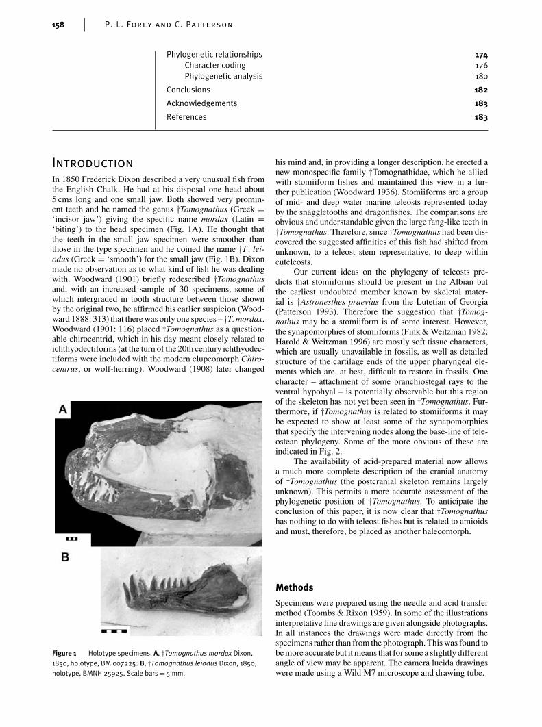
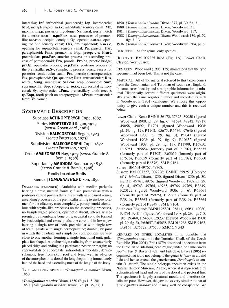
In 1850 Frederick Dixon described a very unusual fish fromthe English Chalk. He had at his disposal one head about5 cms long and one small jaw. Both showed very promin-ent teeth and he named the genus †Tomognathus (Greek =‘incisor jaw’) giving the specific name mordax (Latin =‘biting’) to the head specimen (Fig. 1A). He thought thatthe teeth in the small jaw specimen were smoother thanthose in the type specimen and he coined the name †T . lei-odus (Greek = ‘smooth’) for the small jaw (Fig. 1B). Dixonmade no observation as to what kind of fish he was dealingwith. Woodward (1901) briefly redescribed †Tomognathusand, with an increased sample of 30 specimens, some ofwhich intergraded in tooth structure between those shownby the original two, he affirmed his earlier suspicion (Wood-ward 1888: 313) that there was only one species – †T. mordax.Woodward (1901: 116) placed †Tomognathus as a question-able chirocentrid, which in his day meant closely related toichthyodectiforms (at the turn of the 20th century ichthyodec-tiforms were included with the modern clupeomorph Chiro-centrus, or wolf-herring). Woodward (1908) later changed
his mind and, in providing a longer description, he erected anew monospecific family †Tomognathidae, which he alliedwith stomiiform fishes and maintained this view in a fur-ther publication (Woodward 1936). Stomiiforms are a groupof mid- and deep water marine teleosts represented todayby the snaggletooths and dragonfishes. The comparisons areobvious and understandable given the large fang-like teeth in†Tomognathus. Therefore, since †Tomognathus had been dis-covered the suggested affinities of this fish had shifted fromunknown, to a teleost stem representative, to deep withineuteleosts.
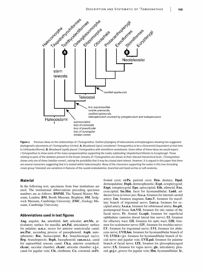
Our current ideas on the phylogeny of teleosts pre-dicts that stomiiforms should be present in the Albian butthe earliest undoubted member known by skeletal mater-ial is †Astronesthes praevius from the Lutetian of Georgia(Patterson 1993). Therefore the suggestion that †Tomog-nathus may be a stomiiform is of some interest. However,the synapomorphies of stomiiforms (Fink & Weitzman 1982;Harold & Weitzman 1996) are mostly soft tissue characters,which are usually unavailable in fossils, as well as detailedstructure of the cartilage ends of the upper pharyngeal ele-ments which are, at best, difficult to restore in fossils. Onecharacter – attachment of some branchiostegal rays to theventral hypohyal – is potentially observable but this regionof the skeleton has not yet been seen in †Tomognathus. Fur-thermore, if †Tomognathus is related to stomiiforms it maybe expected to show at least some of the synapomorphiesthat specify the intervening nodes along the base-line of tele-ostean phylogeny. Some of the more obvious of these areindicated in Fig. 2.
The availability of acid-prepared material now allowsa much more complete description of the cranial anatomyof †Tomognathus (the postcranial skeleton remains largelyunknown). This permits a more accurate assessment of thephylogenetic position of †Tomognathus. To anticipate theconclusion of this paper, it is now clear that †Tomognathushas nothing to do with teleost fishes but is related to amioidsand must, therefore, be placed as another halecomorph.
Methods
Specimens were prepared using the needle and acid transfermethod (Toombs & Rixon 1959). In some of the illustrationsinterpretative line drawings are given alongside photographs.In all instances the drawings were made directly from thespecimens rather than from the photograph. This was found tobe more accurate but it means that for some a slightly differentangle of view may be apparent. The camera lucida drawingswere made using a Wild M7 microscope and drawing tube.
Description and Systematic of†Tomognathus 159
Figure 2 Previous ideas on the relationships of †Tomognathus. Outline phylogeny of halecostome actinopterygians showing two suggestedphylogenetic placements of †Tomognathus (circles): A, Woodward (1901) considered †Tomognathus to be a chirocentrid (equivalent at that timeto ichthyodectiforms); B, Woodward (1908) placed †Tomognathus with stomiiform neoteleosts. Given either of these ideas we would expect†Tomognathus to show some of the many synapomorphies supporting the nodes subtending †Aspidorhynchifomes to Eurypterygii. Thoserelating to parts of the skeleton present in the known remains of †Tomognathus are shown at their relevant hierarchical level. †Tomognathusshows only one of these (median vomer), raising the possibility that it may be a basal stem teleost. However, it is argued in this paper that thereare several characters suggesting that it is nested within halecomorphs. Many of the characters supporting the nodes in this tree (includingcrown group Teleostei) are variations in features of the caudal endoskeleton, branchial and hyoid arches or soft anatomy.
Material
In the following text, specimens from four institutions areused. The institutional abbreviations preceding specimennumbers are as follows: BMNH, The Natural History Mu-seum, London; BM, Booth Museum, Brighton; SM, Sedg-wick Museum, Cambridge University; ZMC, Zoology Mu-seum, Cambridge University.
(sensu Patterson, 1973)Order AMIIFORMES Hay, 1929 (sensu Grande &
Bemis, 1998)Superfamily AMIOIDEA Bonaparte, 1838
(sensu Grande & Bemis, 1998)Family Incertae Sedis
Genus †TOMOGNATHUS Dixon, 1850
DIAGNOSIS (EMENDED). Amioidea with median parietalsbearing a crest, median frontals; fused premaxillae with aposterior ventral process that sutures with an unpaired vomer,ascending processes of the premaxilla failing to enclose fora-men for the olfactory tract completely, parasphenoid edentu-lous with scythe-like processes on the ascending processes,no basipterygoid process, opisthotic absent, intercalar rep-resented by membrane bone only, occipital condyle formedby basioccipital and exoccipitals; one coronoid in lower jawbearing a single row of teeth, prearticular with single rowof teeth; palate with single dermopalatine; double jaw jointin which the quadrate and symplectic contributions are veryclose to one another forming a single functional unit; gularplate fan-shaped, with fine ridges radiating from an anteriorlyplaced ridge and ending in a pectinated posterior margin; nosupraorbitals or suborbitals, infraorbitals tube-like, dermo-sphenotic free from skull roof and lying well in advanceof the autosphenotic; dorsal fin long, beginning immediatelybehind the head and extending most of the length of the body.
TYPE AND ONLY SPECIES. †Tomognathus mordax Dixon,1850.
figs 3–13.1936 †Tomognathus mordax Dixon; Woodward: 304, pl. 6.
DIAGNOSIS. As for genus, only species.
HOLOTYPE. BM 007225 head (Fig. 1A), Lower Chalk,Clayton, West Sussex.
REMARKS. Woodward (1908: 139) maintained that the typespecimen had been lost. This is not the case.
MATERIAL. All of the material referred to this taxon comesfrom the Cenomanian and Turonian of south east England.In some cases locality and stratigraphic information is min-imal. Historically, several different specimens were origin-ally given the same register number and recorded as suchin Woodward’s (1901) catalogue. We choose this oppor-tunity to give each a unique number and this is recordedbelow.
Lower Chalk, Kent: BMNH 36172, 37325, 39050 (figuredWoodward 1908: pl. 29, fig. 6), 41684, 47242, 47917,49058, 49092, P.1701 (figured Woodward 1908:pl. 29, fig. 12), P.1702, P.5675, P.5676, P.7646 (figuredWoodward 1908: pl. 29, fig. 3), P.9043 (figuredWoodward 1908: pl. 29, fig. 9), P.10632 (figuredWoodward 1908: pl. 29, fig. 13), P.11799, P.16950,P.16951, P.65654 (formerly part of P.1702), P.65655(formerly part of P.1702), P.65656 (formerly part ofP.7676), P.65659 (formerly part of P.1702), P.65660(formerly part of P.6576), SM B.9161.
of T. leiodus Dixon, 1850, figured Dixon 1850: pl. 30,fig. 31), 49761, 49762 (figured Woodward 1908: pl. 29,fig. 4), 49763, 49764, 49765, 49766, 49769, P.3849,P.20122 (figured Woodward 1936: pl. 6), P.65661(formerly part of 25925), P.65662 (formerly part ofP.3849), P.65663 (formerly part of P.3849), P.65664(formerly part of P.3849), SM B.9164.
South east England: BMNH 25801, 25813, 39051, 49080,P.4791, P.4844 (figured Woodward 1908: pl. 29, figs 7, 8,10), P.6460, P.6460a, P.9237 (figured Woodward 1908:pl. 29, fig. 5), P.65657, P.65658, BM 016968, SM B.9162,B.9163, B.75729, B75730, ZMC GN 543
REMARKS ON OTHER LOCALITIES. It is possible that†Tomognathus occurs in the Turonian Chalk of the CzechRepublic (Ekrt 2001). Fric (1879) described a specimen fromthe Turonian of Bıla hora, near Prague, under the name Istieusspottii. Fric & Bayer (1902) and Fritsch & Bayer (1905) re-cognised that it did not belong to the genus Istieus (an albulidfish) and hence erected the generic name Denticopsis to con-tain D. spottii. The single holotype specimen exists in theNatural History Museum, Prague, where it is represented bya disarticulated head and parts of the dorsal and pectoral fins.The specimen is largely a natural mould and therefore de-tails are poor. However, the jaw looks very similar to that of†Tomognathus mordax and it may well be conspecific. We
Description and Systematic of†Tomognathus 161
Figure 3 †Tomognathus mordax Dixon, 1850, skull roof. A, dorsal view of braincase of BMNH P.11799; B, interpretive drawing of BMNHP.11799; C, restoration of skull roof based on BMNH P.11799 with additional information from BMNH 49761 and 49765. Scale bar= 5 mm.
have not seen the specimen and therefore are reluctant toformally place it in synonymy.
Description
Braincase
The braincase is blunt-snouted, deep throughout its lengthwith a dorsally domed otic region that is keeled ventrally. Indorsal view (Fig. 3) the roof is parallel-sided above the orbit;it flares anteriorly towards the ethmoid region and posteriorlyat the rear limit of the skull. Unlike most members of theHalecomorphi the skull roofing bones are unornamented,except for minor rugosities and the skull roof is stronglyvaulted with a median crest. In most halecomorphs the skullroof is flat and the presence of ornament in most suggeststhat it was covered with a thin layer of skin only. We suspectthat, in life, the skull roof of †Tomognathus would have beendeeply buried within skin and muscle.
The frontals (see Figs 3, 7, 9 & 13: Fr) are fused in themid line and together make up the largest elements in theskull roof. Ornamentation is confined to a series of irregulargrooves that fan out anteriorly. The lateral margin is veryirregular but shows a minor flare at mid orbital level. Weare uncertain if the fused frontal condition is ontogenetic orphylogenetic; but all specimens showing the skull roof showfused frontals and this condition must be regarded as typicalfor this species. This is unlike the condition for Amia calvawhere Grande & Bemis (1998) found one specimen in 119examined that showed this condition. Fused frontals are rel-atively rare in actinopterygians but the condition is known in
eels: in fact Regan (1912) separated Recent eels into two ma-jor groups based on the presence of paired (e.g. anguilloids)or fused frontals (e.g. congroids and synaphobranchoids).The fused frontals are parallel-sided; that is, they are not pos-teriorly flared as is the usual teleostean condition (Arratia &Schultze 1987). Close to the lateral margins of the fusedfrontals there is a series of six pores (Fig. 3: so.s.c) openingfrom the supraorbital sensory canal, the latter opens at the ex-treme anterior end of the frontal through a large pore leadingfrom a prominent tube (Figs 3 & 4: o.so.s.c), somewhat sim-ilar to that seen in †Calamopleurus cylindricus (Grande &Bemis 1998: fig. 297). In some specimens the posterior open-ing is double. In the posterior half of the fused frontals amedian crest (see Figs 3, 4B, 6 & 8B: cr.Fr) continues thaton the parietals. This reaches to mid orbital level before itgives way to a shallow median depression.
The parietals are also fused into a median element (seeFigs 3 & 6: Pa) and developed as a prominent median crest(see Figs 3C, 6, 8A & 15: cr.Pa), particularly obvious inlateral view and, in life, no doubt provided a large area forthe insertion of epaxial musculature. The parietal is long andnarrow – the median width being equal to half of the length.Sutures with the frontal anteriorly, the pterotic laterally andthe epioccipital posteriorly are simple. The posterior mar-gin of the parietal is of complex shape. A small lacuna isleft between the parietal, epioccipital and the dorsal cartilagelined surface of the exoccipital (Fig. 3B). It is probable thatthis space was occupied with cartilage in life. A median pari-etal is rare but is a character of †Sinamiidae (†Ikechaoamiaand †Sinamia, see Grande & Bemis 1998: 583), where it isrepresented by an flat, equidimensional bone without a me-dian crest. A median parietal is also present in Amia as ananomaly, but this cannot be regarded as characteristic of this
162 P. L. Forey and C. Patterson
Figure 4 †Tomognathus mordax Dixon, 1850. Ethmoid region. A, reconstruction in posterior view; B, in anterior view. Based on BMNH 49761,49765 and P.11799. Approximately × 4 natural size.
species (contra Arratia 1999: table 1, character 3, state 1).Grande & Bemis (1998) discuss the occurrence of a medianparietal in Amia noting that in their sample of 119 speci-mens the condition occurred in one individual and, there-fore, was regarded as a teratological phenomenon. A medianparietal is found in most ichthyodectiforms, a group withwhich †Tomognathus was once associated. In ichthyodecti-forms there is an associated supraoccipital that divides mostof the median parietal into left and right limbs. The pterotic(see Figs 3, 6 & 15: Pto) is paired; it flanks the parietal andextends forward to suture with the frontal above the orbit. Itforms the roof of a shallow posttemporal fossa (see Fig. 8A:fos.ptt). Some specimens (e.g. BMNH P.4844) show smallpores leading from the otic sensory canal.
A note on previous identifications of the skull roof-ing bones is in order. Woodward (1936) identified the fusedparietals as the supraoccipital and the pterotics as the pari-etals. (Interestingly, Grande & Bemis (1998: 39) record thatBridge (1877) made the same identification for an anomalousspecimen of Amia calva.) Woodward’s (mis)identification isperfectly understandable in the context in which he was de-scribing †Tomognathus. In thinking that †Tomognathus wasa teleost he naturally likened a posteriorly placed, medianroofing bone to a supraoccipital (a teleost synapomorphy),a bone commonly developed as a crest. That this is not thecase in †Tomognathus is evidenced by four criteria: (1) themedian element in †Tomognathus is entirely dermal (in someteleosts the endochondral supraoccipital does have a dermalcomponent, usually thought to have come about by phylogen-etic/ontogenetic fusion of extrascapulars; the supraoccipitalwould therefore be a canal-bearing bone unlike the canal-freebone in †Tomognathus); (2) it fails to contribute to the pos-terior face of the braincase; (3) it has no association with thesemicircular canals (a supraoccipital is endochondral, con-
tributes to the posterior face of the braincase in at least lowerteleosts and is usually pierced by the posterior semicircularcanals); (4) the lateral elements carry the otic sensory canaland suture with the autosphenotics (attributes of a pterotic).There is no evidence of an ossified endochondral supraoticbone as occurs in some halecomorphs (Maisey 1999).
The anterior part of the braincase is formed by the eth-moid and the premaxilla, together with nasals, rostral andantorbital (these latter three bones are described later). In allspecimens the ethmoid (see Figs 3–6 & 15: Eth) is a com-plex single element that covers the territory occupied by thepaired lateral ethmoids and pre-ethmoids of Amia (Grande &Bemis 1998: fig. 22). Although the complex shape of thisbone suggests that it ossified from more than one centrethere is no indication of separate elements in any specimenavailable to us. The ethmoid forms the anterior wall of theorbit and, at the same time, the posterior wall of the nasalcapsules. It is developed laterally as two broad wings thathave small areas of unfinished bone both dorsally and vent-rally. Ventrally there are two small areas, both at the tips ofsmall projections, placed one behind the other and best seenin ventral view (Fig. 5). These provided articulation pointsfor the inturned head of the maxilla (Figs 5 & 6: art.Mx) andthe autopalatine (Figs 5 & 6 art.Pal) (see below). The sig-nificance of the unfinished area on the dorsal surface of theethmoid is unclear. The dorsal portion of the ethmoid con-tinues forward as a thin lamina that is ‘T’-shaped in anteriorview (see Figs 4B & 13) and terminates in unfinished bone,again suggesting that in life this would have been cappedwith cartilage. The posterior surface of the ethmoid is de-veloped as a thin, median interobital septum that sutures withthe orbitosphenoid posteriorly. This posterior lamina is oftenperforated by fenestrae, but these are variously developedfrom specimen to specimen, suggesting that they are without
Description and Systematic of†Tomognathus 163
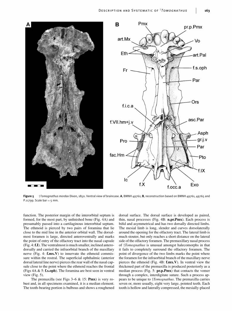
Figure 5 †Tomognathus mordax Dixon, 1850. Ventral view of braincase. A, BMNH 49761; B, reconstruction based on BMNH 49761, 49765 andP.11799. Scale bar= 5 mm.
function. The posterior margin of the interorbital septum isformed, for the most part, by unfinished bone (Fig. 4A) andpresumably passed into a cartilaginous interorbital septum.The ethmoid is pierced by two pairs of foramina that lieclose to the mid line in the anterior orbital wall. The dorsal-most foramen is large, directed anteroventrally and marksthe point of entry of the olfactory tract into the nasal capsule(Fig. 4: f.I). The ventralmost is much smaller, inclined antero-dorsally and carried the infraorbital branch of the maxillarynerve (Fig. 4: f.mx.V) to innervate the ethmoid commis-sure within the rostral. The superficial ophthalmic (anteriordorsal lateral line nerve) pierces the rear wall of the nasal cap-sule close to the point where the ethmoid reaches the frontal(Figs 4A & 5: f.s.oph). The foramina are best seen in ventralview (Fig. 5).
The premaxilla (see Figs 3–6 & 15: Pmx) is very ro-bust and, in all specimens examined, it is a median element.The tooth-bearing portion is bulbous and shows a roughened
dorsal surface. The dorsal surface is developed as paired,thin, nasal processes (Fig. 4B: n.pr.Pmx). Each process isbifid and asymmetrical and has two dorsally directed limbs.The mesial limb is long, slender and curves dorsolaterallyaround the opening for the olfactory tract. The lateral limb ismuch stouter, but only reaches a short distance on the lateralside of the olfactory foramen. The premaxillary nasal processof †Tomognathus is unusual amongst halecomorphs in thatit fails to completely surround the olfactory foramen. Thepoint of divergence of the two limbs marks the point wherethe foramen for the infraorbital branch of the maxillary nervepierces the ethmoid (Fig. 4B: f.mx.V). In ventral view thethickened part of the premaxilla is produced posteriorly as amedian process (Fig. 5: pr.p.Pmx) that contacts the vomerthrough a complex, interdigitate suture. Such a process ap-pears to be unique to †Tomognathus. The premaxilla carriesseven or, more usually, eight very large, pointed teeth. Eachtooth is hollow and laterally compressed, the mesially-placed
164 P. L. Forey and C. Patterson
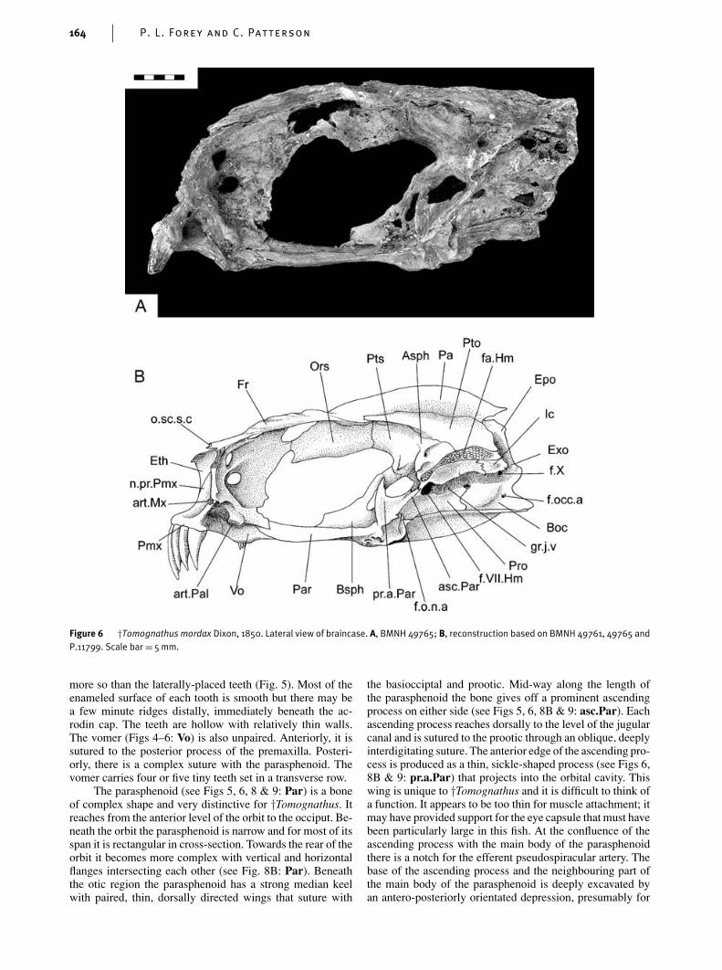
Figure 6 †Tomognathus mordax Dixon, 1850. Lateral view of braincase. A, BMNH 49765; B, reconstruction based on BMNH 49761, 49765 andP.11799. Scale bar = 5mm.
more so than the laterally-placed teeth (Fig. 5). Most of theenameled surface of each tooth is smooth but there may bea few minute ridges distally, immediately beneath the ac-rodin cap. The teeth are hollow with relatively thin walls.The vomer (Figs 4–6: Vo) is also unpaired. Anteriorly, it issutured to the posterior process of the premaxilla. Posteri-orly, there is a complex suture with the parasphenoid. Thevomer carries four or five tiny teeth set in a transverse row.
The parasphenoid (see Figs 5, 6, 8 & 9: Par) is a boneof complex shape and very distinctive for †Tomognathus. Itreaches from the anterior level of the orbit to the occiput. Be-neath the orbit the parasphenoid is narrow and for most of itsspan it is rectangular in cross-section. Towards the rear of theorbit it becomes more complex with vertical and horizontalflanges intersecting each other (see Fig. 8B: Par). Beneaththe otic region the parasphenoid has a strong median keelwith paired, thin, dorsally directed wings that suture with
the basiocciptal and prootic. Mid-way along the length ofthe parasphenoid the bone gives off a prominent ascendingprocess on either side (see Figs 5, 6, 8B & 9: asc.Par). Eachascending process reaches dorsally to the level of the jugularcanal and is sutured to the prootic through an oblique, deeplyinterdigitating suture. The anterior edge of the ascending pro-cess is produced as a thin, sickle-shaped process (see Figs 6,8B & 9: pr.a.Par) that projects into the orbital cavity. Thiswing is unique to †Tomognathus and it is difficult to think ofa function. It appears to be too thin for muscle attachment; itmay have provided support for the eye capsule that must havebeen particularly large in this fish. At the confluence of theascending process with the main body of the parasphenoidthere is a notch for the efferent pseudospiracular artery. Thebase of the ascending process and the neighbouring part ofthe main body of the parasphenoid is deeply excavated byan antero-posteriorly orientated depression, presumably for
Description and Systematic of†Tomognathus 165
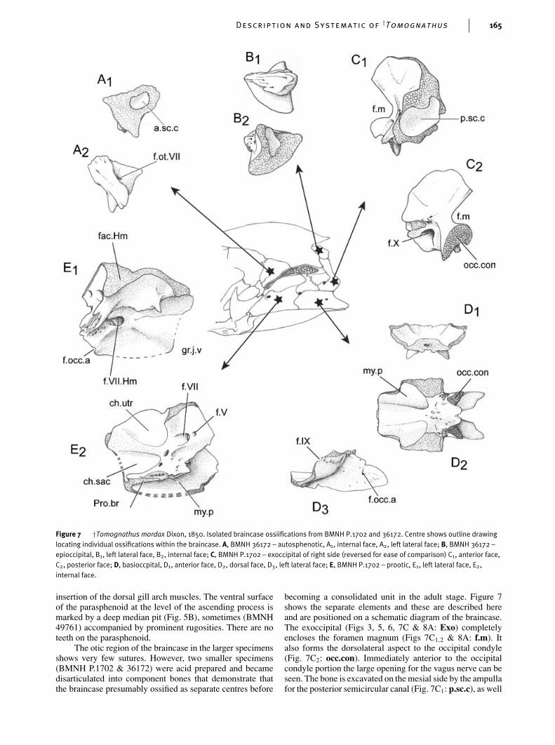
Figure 7 †Tomognathus mordax Dixon, 1850. Isolated braincase ossiifications from BMNH P.1702 and 36172. Centre shows outline drawinglocating individual ossifications within the braincase. A, BMNH 36172 – autosphenotic, A1, internal face, A2, left lateral face; B, BMNH 36172 –epioccipital, B1, left lateral face, B2, internal face; C, BMNH P.1702 – exoccipital of right side (reversed for ease of comparison) C1, anterior face,C2, posterior face; D, basioccpital, D1, anterior face, D2, dorsal face, D3, left lateral face; E, BMNH P.1702 – prootic, E1, left lateral face, E2,internal face.
insertion of the dorsal gill arch muscles. The ventral surfaceof the parasphenoid at the level of the ascending process ismarked by a deep median pit (Fig. 5B), sometimes (BMNH49761) accompanied by prominent rugosities. There are noteeth on the parasphenoid.
The otic region of the braincase in the larger specimensshows very few sutures. However, two smaller specimens(BMNH P.1702 & 36172) were acid prepared and becamedisarticulated into component bones that demonstrate thatthe braincase presumably ossified as separate centres before
becoming a consolidated unit in the adult stage. Figure 7shows the separate elements and these are described hereand are positioned on a schematic diagram of the braincase.The exoccipital (Figs 3, 5, 6, 7C & 8A: Exo) completelyencloses the foramen magnum (Figs 7C1,2 & 8A: f.m). Italso forms the dorsolateral aspect to the occipital condyle(Fig. 7C2: occ.con). Immediately anterior to the occipitalcondyle portion the large opening for the vagus nerve can beseen. The bone is excavated on the mesial side by the ampullafor the posterior semicircular canal (Fig. 7C1: p.sc.c), as well
166 P. L. Forey and C. Patterson
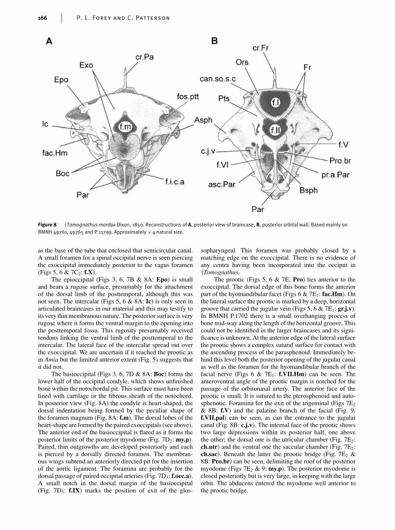
Figure 8 †Tomognathus mordax Dixon, 1850. Reconstructions of A, posterior view of braincase, B, posterior orbital wall. Based mainly onBMNH 49761, 49765 and P.11799. Approximately × 4 natural size.
as the base of the tube that enclosed that semicircular canal.A small foramen for a spinal occipital nerve is seen piercingthe exoccipital immediately posterior to the vagus foramen(Figs 5, 6 & 7C2: f.X).
The epioccipital (Figs 3, 6, 7B & 8A: Epo) is smalland bears a rugose surface, presumably for the attachmentof the dorsal limb of the posttemporal, although this wasnot seen. The intercalar (Figs 5, 6 & 8A: Ic) is only seen inarticulated braincases in our material and this may testify toits very thin membranous nature. The posterior surface is veryrugose where it forms the ventral margin to the opening intothe posttemporal fossa. This rugosity presumably receivedtendons linking the ventral limb of the posttemporal to theintercalar. The lateral face of the intercalar spread out overthe exoccipital. We are uncertain if it reached the prootic asin Amia but the limited anterior extent (Fig. 5) suggests thatit did not.
The basioccipital (Figs 3, 6, 7D & 8A: Boc) forms thelower half of the occipital condyle, which shows unfinishedbone within the notochordal pit. This surface must have beenlined with cartilage or the fibrous sheath of the notochord.In posterior view (Fig. 8A) the condyle is heart-shaped, thedorsal indentation being formed by the peculiar shape ofthe foramen magnum (Fig. 8A: f.m). The dorsal lobes of theheart-shape are formed by the paired exoccipitals (see above).The anterior end of the basioccipital is flared as it forms theposterior limits of the posterior myodome (Fig. 7D2: my.p).Paired, thin outgrowths are developed posteriorly and eachis pierced by a dorsally directed foramen. The membran-ous wings subtend an anteriorly directed pit for the insertionof the aortic ligament. The foramina are probably for thedorsal passage of paired occipital arteries (Fig. 7D3: f.occ.a).A small notch in the dorsal margin of the basioccipital(Fig. 7D3: f.IX) marks the position of exit of the glos-
sopharyngeal. This foramen was probably closed by amatching edge on the exoccipital. There is no evidence ofany centra having been incorporated into the occiput in†Tomognathus.
The prootic (Figs 5, 6 & 7E: Pro) lies anterior to theexoccipital. The dorsal edge of this bone forms the anteriorpart of the hyomandibular facet (Figs 6 & 7E1: fac.Hm). Onthe lateral surface the prootic is marked by a deep, horizontalgroove that carried the jugular vein (Figs 5, 6 & 7E1: gr.j.v).In BMNH P.1702 there is a small overhanging process ofbone mid-way along the length of the horizontal groove. Thiscould not be identified in the larger braincases and its signi-ficance is unknown. At the anterior edge of the lateral surfacethe prootic shows a complex sutural surface for contact withthe ascending process of the parasphenoid. Immediately be-hind this level both the posterior opening of the jugular canalas well as the foramen for the hyomandibular branch of thefacial nerve (Figs 6 & 7E1: f.VII.Hm) can be seen. Theanteroventral angle of the prootic margin is notched for thepassage of the orbitonasal artery. The anterior face of theprootic is small. It is sutured to the pterosphenoid and auto-sphenotic. Foramina for the exit of the trigeminal (Figs 7E2
& 8B: f.V) and the palatine branch of the facial (Fig. 9:f.VII.pal) can be seen, as can the entrance to the jugularcanal (Fig. 8B: c.j.v). The internal face of the prootic showstwo large depressions within its posterior half, one abovethe other; the dorsal one is the utricular chamber (Fig. 7E2:ch.utr) and the ventral one the saccular chamber (Fig. 7E2:ch.sac). Beneath the latter the prootic bridge (Fig. 7E2 &8B: Pro.br) can be seen, delimiting the roof of the posteriormyodome (Figs 7E2 & 9: my.p). The posterior myodome isclosed posteriorly but is very large, in keeping with the largeorbit. The abducens entered the myodome well anterior tothe prootic bridge.
Description and Systematic of†Tomognathus 167
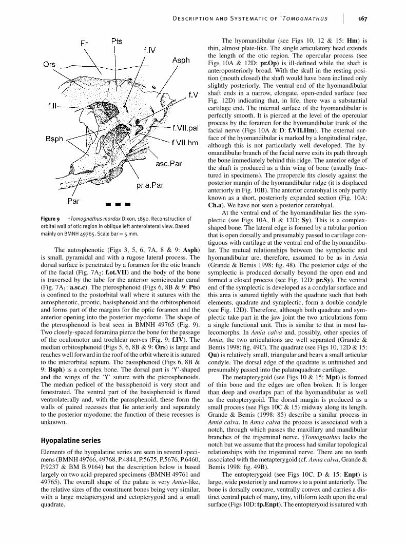
Figure 9 †Tomognathus mordax Dixon, 1850. Reconstruction oforbital wall of otic region in oblique left anterolateral view. Basedmainly on BMNH 49765. Scale bar= 5 mm.
The autosphenotic (Figs 3, 5, 6, 7A, 8 & 9: Asph)is small, pyramidal and with a rugose lateral process. Thedorsal surface is penetrated by a foramen for the otic branchof the facial (Fig. 7A2: f.ot.VII) and the body of the boneis traversed by the tube for the anterior semicircular canal(Fig. 7A1: a.sc.c). The pterosphenoid (Figs 6, 8B & 9: Pts)is confined to the postorbital wall where it sutures with theautosphenotic, prootic, basisphenoid and the orbitosphenoidand forms part of the margins for the optic foramen and theanterior opening into the posterior myodome. The shape ofthe pterosphenoid is best seen in BMNH 49765 (Fig. 9).Two closely-spaced foramina pierce the bone for the passageof the oculomotor and trochlear nerves (Fig. 9: f.IV). Themedian orbitosphenoid (Figs 5, 6, 8B & 9: Ors) is large andreaches well forward in the roof of the orbit where it is suturedto the interorbital septum. The basisphenoid (Figs 6, 8B &9: Bsph) is a complex bone. The dorsal part is ‘Y’-shapedand the wings of the ‘Y’ suture with the pterosphenoids.The median pedicel of the basisphenoid is very stout andfenestrated. The ventral part of the basisphenoid is flaredventrolaterally and, with the parasphenoid, these form thewalls of paired recesses that lie anteriorly and separatelyto the posterior myodome; the function of these recesses isunknown.
Hyopalatine series
Elements of the hyopalatine series are seen in several speci-mens (BMNH 49766, 49768, P.4844, P.5675, P.5676, P.6460,P.9237 & BM B.9164) but the description below is basedlargely on two acid-prepared specimens (BMNH 49761 and49765). The overall shape of the palate is very Amia-like,the relative sizes of the constituent bones being very similar,with a large metapterygoid and ectopterygoid and a smallquadrate.
The hyomandibular (see Figs 10, 12 & 15: Hm) isthin, almost plate-like. The single articulatory head extendsthe length of the otic region. The opercular process (seeFigs 10A & 12D: pr.Op) is ill-defined while the shaft isanteroposteriorly broad. With the skull in the resting posi-tion (mouth closed) the shaft would have been inclined onlyslightly posteriorly. The ventral end of the hyomandibularshaft ends in a narrow, elongate, open-ended surface (seeFig. 12D) indicating that, in life, there was a substantialcartilage end. The internal surface of the hyomandibular isperfectly smooth. It is pierced at the level of the opercularprocess by the foramen for the hyomandibular trunk of thefacial nerve (Figs 10A & D: f.VII.Hm). The external sur-face of the hyomandibular is marked by a longitudinal ridge,although this is not particularly well developed. The hy-omandibular branch of the facial nerve exits its path throughthe bone immediately behind this ridge. The anterior edge ofthe shaft is produced as a thin wing of bone (usually frac-tured in specimens). The preopercle fits closely against theposterior margin of the hyomandibular ridge (it is displacedanteriorly in Fig. 10B). The anterior ceratohyal is only partlyknown as a short, posteriorly expanded section (Fig. 10A:Ch.a). We have not seen a posterior ceratohyal.
At the ventral end of the hyomandibular lies the sym-plectic (see Figs 10A, B & 12D: Sy). This is a complex-shaped bone. The lateral edge is formed by a tubular portionthat is open dorsally and presumably passed to cartilage con-tiguous with cartilage at the ventral end of the hyomandibu-lar. The mutual relationships between the symplectic andhyomandibular are, therefore, assumed to be as in Amia(Grande & Bemis 1998: fig. 48). The posterior edge of thesymplectic is produced dorsally beyond the open end andformed a closed process (see Fig. 12D: pr.Sy). The ventralend of the symplectic is developed as a condylar surface andthis area is sutured tightly with the quadrate such that bothelements, quadrate and symplectic, form a double condyle(see Fig. 12D). Therefore, although both quadrate and sym-plectic take part in the jaw joint the two articulations forma single functional unit. This is similar to that in most ha-lecomorphs. In Amia calva and, possibly, other species ofAmia, the two articulations are well separated (Grande &Bemis 1998: fig. 49C). The quadrate (see Figs 10, 12D & 15:Qu) is relatively small, triangular and bears a small articularcondyle. The dorsal edge of the quadrate is unfinished andpresumably passed into the palatoquadrate cartilage.
The metapterygoid (see Figs 10 & 15: Mpt) is formedof thin bone and the edges are often broken. It is longerthan deep and overlaps part of the hyomandibular as wellas the entopterygoid. The dorsal margin is produced as asmall process (see Figs 10C & 15) midway along its length.Grande & Bemis (1998: 85) describe a similar process inAmia calva. In Amia calva the process is associated with anotch, through which passes the maxillary and mandibularbranches of the trigeminal nerve. †Tomognathus lacks thenotch but we assume that the process had similar topologicalrelationships with the trigeminal nerve. There are no teethassociated with the metapterygoid (cf. Amia calva, Grande &Bemis 1998: fig. 49B).
The entopterygoid (see Figs 10C, D & 15: Enpt) islarge, wide posteriorly and narrows to a point anteriorly. Thebone is dorsally concave, ventrally convex and carries a dis-tinct central patch of many, tiny, villiform teeth upon the oralsurface (Figs 10D: tp.Enpt). The entopteryoid is sutured with
168 P. L. Forey and C. Patterson
Figure 10 †Tomognathus mordax Dixon, 1850. Hyoid arch and palate. A, BMNH P.66297 mesial view of left hyoid arch, palate and lower jawwith interpretive drawing; B, BMNH P.66297 lateral view and interpretive drawing; C, BMNH 49765 isolated palatal bones in dorsal view withinterpretive drawing; D, BMNH 49765 ventral view with interpretive drawing. Scale bars= 5 mm.
Description and Systematic of†Tomognathus 169
the ectopterygoid laterally and the dermopalatine anteriorly.The ectopterygoid (see Figs 10A, C & 15: Ecpt) is very ro-bust. Posteriorly, it is slightly expanded and grooved alongthe dorsal (mesial) margin. Part of the quadrate lies in thisgroove. The ectopterygoid dentition consists of four large,conical and slightly recurved teeth situated near the anteriorend and lying in continuity with the palatine teeth. Behindthis level there is a small patch of tiny teeth, comparable insize with those upon the entopterygoid (Fig. 10D).
The palatine consists of separate auto- and dermopal-atine components. The dermopalatine (Fig. 10D: Dpal) issmall, triangular and sutured to the ectopterygoid and entop-terygoid. Three large, conical and slightly recurved teeth arepresent in BMNH 49765 (right side). †Tomognathus is veryunusual amongst halecomorphs in showing a single dermo-palatine (most have two). The autopalatine (Fig. 10C: Apal)is present as a thin perichondral shell of bone plastered ontothe dorsal surface of the dermopalatine. The posterior mar-gin of the autopalatine is very irregular and it is probablethat it passed into cartilage which must also have formed thecore of the autopalatine. In Recent teleosts the autopalatineis often the last of the palatal bones to ossify within the pal-atoquadrate cartilage and it often fails to ossify at all in manyfishes. Therefore, the slight ossification of this element in†Tomognathus is not unusual.
Maxilla and supramaxilla
The maxilla (see Figs 11 & 15: Mx) is a slender ele-ment with a narrow inturned head and a posteriorly ex-panded blade. There is a single supramaxilla (see Fig. 15:smx). No single specimen shows a perfectly preserved up-per jaw: therefore well preserved elements are taken fromthree specimens (Fig. 11). The outline of the maxilla isbest preserved in BMNH P.3849, Fig 11B). Here the trueposterior margin can be seen. The shape of the posteriormargin is of some importance in halecomorphs and in thisfeature †Tomognathus most closely resembles †Ionoscopus,†Amiopsis and †Solnhofenamia, rather than members of theAmiista, in which the posterior margin is deeply notched (seeGrande & Bemis 1998: fig. 243 for comparative outlines).The maxillary teeth are, on average, substantially smallerthan the premaxillary and dentary teeth. This differentiationis seen in members of the Amioidea and contrasts with thesituation in other halecomorphs, in which the teeth are of ap-proximately the same size. There are 26–37 teeth along theoral margin of the maxilla. The dentition is best displayedin SM B.9164 (34 teeth), which shows a gradation in toothsize along the margin (Fig. 11A). The anteriormost teethare nearly four times as long as the posterior teeth. In thelargest specimens at our disposal (BMNH P.20122) the sizedifferentiation between anteriormost and posteriormost teethis considerably less (Fig. 11C). We do not know if this isindividual variation or a growth phenomenon. All teeth bearacrodin caps and the larger anterior teeth are ridged, like theircounterparts on the premaxilla and the lower jaw. The head ofthe maxilla is strongly inturned as a rod-like process and thiswould have articulated with the anteriorly directed processon the ethmoid (see Figs 4B, 5 & 6: art.Mx). The ornamenton the maxilla consists of irregular shaped tubercles locatedposteriorly. The supramaxilla is rarely preserved, suggestingthat it was loosely associated with the maxilla. It is best seenin BMNH P.20122 (Fig. 11C). The shape of the supramaxilla
Figure 11 †Tomognathus mordax Dixon, 1850. Maxilla andsupramaxilla of right side – camera lucida drawings. A, SM B.9164; B,BMNH P.3849; C, BMNH P.20122. Scale bars= 5 mm.
mirrors that of the maxilla and it is about half the length ofthe maxilla.
Lower jaw
The structure of the lower jaw is one of the most obviousreasons to question Woodward’s view that †Tomognathus isa stomiiform. The lower jaw shows a coronoid, prearticularand surangular – all bones that are absent and presumedlost in teleosts (although Nelson (1973: 338) admitted thepossibility that coronoids had fused with the dentary and thesurangular with the angular).
The lower jaw is shallow anteriorly, it deepens to aprominent coronoid process and ends posteriorly in a robustcondylar area. The relative proportions of the dentary toothedarea to the total length of the jaw is 37–40% and this is morelike the proportions seen in parasemionotids and amioidsthan in caturids, in which the toothed area extends for about60% of the total jaw length.
The outer surface of the mandible is formed by the dent-ary, angular and surangular. From a shallow symphysis thedentary (see Figs 10A, B, 12A–C & 15: Den) deepens con-siderably and contacts the angular through an interdigitatesuture. There is a single row of nine or 10 teeth along theoral margin. Those anteriorly are substantially larger that theposterior teeth and the largest are comparable in size to thepremaxillary teeth and, like those teeth, they show anteropos-teriorly compressed bases. The size differentiation betweenanterior and posterior teeth is variable between individuals,as is the position of the largest tooth (which is either the third
170 P. L. Forey and C. Patterson
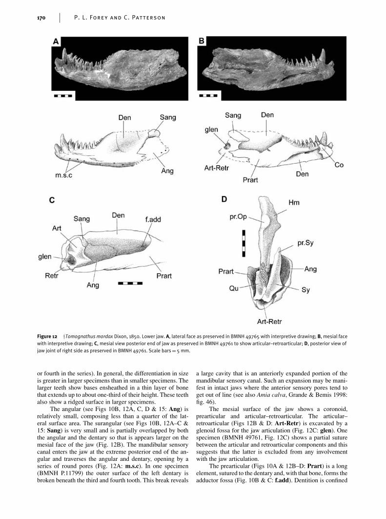
Figure 12 †Tomognathus mordax Dixon, 1850. Lower jaw. A, lateral face as preserved in BMNH 49765 with interpretive drawing; B, mesial facewith interpretive drawing; C, mesial view posterior end of jaw as preserved in BMNH 49761 to show articular–retroarticular; D, posterior view ofjaw joint of right side as preserved in BMNH 49761. Scale bars= 5 mm.
or fourth in the series). In general, the differentiation in sizeis greater in larger specimens than in smaller specimens. Thelarger teeth show bases ensheathed in a thin layer of bonethat extends up to about one-third of their height. These teethalso show a ridged surface in larger specimens.
The angular (see Figs 10B, 12A, C, D & 15: Ang) isrelatively small, composing less than a quarter of the lat-eral surface area. The surangular (see Figs 10B, 12A–C &15: Sang) is very small and is partially overlapped by boththe angular and the dentary so that is appears larger on themesial face of the jaw (Fig. 12B). The mandibular sensorycanal enters the jaw at the extreme posterior end of the an-gular and traverses the angular and dentary, opening by aseries of round pores (Fig. 12A: m.s.c). In one specimen(BMNH P.11799) the outer surface of the left dentary isbroken beneath the third and fourth tooth. This break reveals
a large cavity that is an anteriorly expanded portion of themandibular sensory canal. Such an expansion may be mani-fest in intact jaws where the anterior sensory pores tend toget out of line (see also Amia calva, Grande & Bemis 1998:fig. 46).
The mesial surface of the jaw shows a coronoid,prearticular and articular–retroarticular. The articular–retroarticular (Figs 12B & D: Art-Retr) is excavated by aglenoid fossa for the jaw articulation (Fig. 12C: glen). Onespecimen (BMNH 49761, Fig. 12C) shows a partial suturebetween the articular and retroarticular components and thissuggests that the latter is excluded from any involvementwith the jaw articulation.
The prearticular (Figs 10A & 12B–D: Prart) is a longelement, sutured to the dentary and, with that bone, forms theadductor fossa (Fig. 10B & C: f.add). Dentition is confined
Description and Systematic of†Tomognathus 171
to a single row of teeth. In others, an outer row of enlargedteeth is accompanied by a shagreen of villiform teeth spreadover the oral surface (e.g. see Amia calva, Grande & Be-mis 1998: fig. 45). The prearticular extends further forwardthan in other halecomorphs and covers territory occupied byseparate coronoids in other halecomorphs. In †Tomognathusthere is a single coronoid (Fig. 12B: Co) lying mesial to theenlarged teeth upon the dentary. This coronoid carries a singlerow of five or six incurved teeth that lie in series with therow upon the prearticular. †Tomognathus is unique in hav-ing only a single coronoid; most other halecomorphs havethree to five. †Tomognathus is also unusual in having only asingle row of coronoid teeth: however, this condition is alsoseen in †Calamopleurus cylindricus (Grande & Bemis 1998:fig. 310).
Circumorbitals, nasals and rostral ossicles
All of the circumorbital bones are very delicate and usuallypoorly preserved. The bones surrounding the eye are veryreduced; there is no suborbital or supraorbitals and a scler-otic ossicle is missing. The dermosphenotic (see Fig. 15:Dsph) is a characteristic ‘T’-shaped bone and this is locatedwell anterior to the level of the autosphenotic spine (BMNHP.4844, SM B.9164). The stem of the bone is tubular andlittle larger than the contained canal. The head is expandedand is pierced along its dorsal margin for the infraorbitalcanal that, at this level, lies opposite the anterior openingof the otic sensory canal. The dermosphenotic lies com-pletely free from the skull roof and this is an unusual featureseen only in parasemiontids and †Calamopleurus cylindri-cus among halecomorphs. Grande & Bemis (1998) considera free dermosphenotic to be a plesiomorphic character at thehalecomorph level but derived for †Calamopleurus cylindri-cus, which is nested deep within halecomorphs in which thedermosphenotic is an integral part of the skull roof. Its statusin †Tomognathus is similarly revealed by optimisation. Twomore tubular infraorbitals (see Fig. 15: Inf and Inf1) canbe seen reaching to mid-orbital level along the ventral rim.Infraorbital 1 (lachrymal) consists of a posterior tubular por-tion that expands to a rounded and plate-like anterior head(BMNH 49761). The tubular portion is perforated by twoor three pores leading from the infraorbital canal and thiscanal leaves infraorbital 1 through an anteroventrally placedopening.
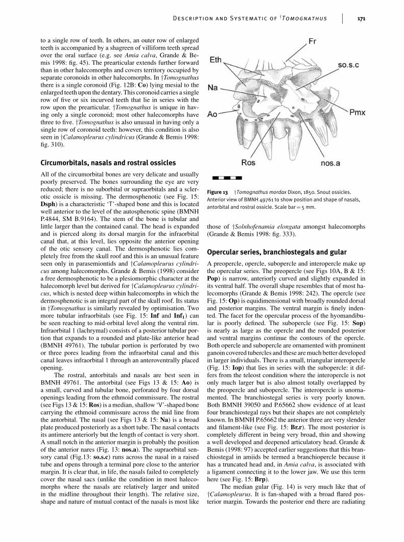
The rostral, antorbitals and nasals are best seen inBMNH 49761. The antorbital (see Figs 13 & 15: Ao) isa small, curved and tubular bone, perforated by four dorsalopenings leading from the ethmoid commissure. The rostral(see Figs 13 & 15: Ros) is a median, shallow ‘V’-shaped bonecarrying the ethmoid commissure across the mid line fromthe antorbital. The nasal (see Figs 13 & 15: Na) is a broadplate produced posteriorly as a short tube. The nasal contactsits antimere anteriorly but the length of contact is very short.A small notch in the anterior margin is probably the positionof the anterior nares (Fig. 13: nos.a). The supraorbital sen-sory canal (Fig.13: so.s.c) runs across the nasal in a raisedtube and opens through a terminal pore close to the anteriormargin. It is clear that, in life, the nasals failed to completelycover the nasal sacs (unlike the condition in most haleco-morphs where the nasals are relatively larger and unitedin the midline throughout their length). The relative size,shape and nature of mutual contact of the nasals is most like
Figure 13 †Tomognathus mordax Dixon, 1850. Snout ossicles.Anterior view of BMNH 49761 to show position and shape of nasals,antorbital and rostral ossicle. Scale bar= 5 mm.
those of †Solnhofenamia elongata amongst halecomorphs(Grande & Bemis 1998: fig. 333).
Opercular series, branchiostegals and gular
A preopercle, opercle, subopercle and interopercle make upthe opercular series. The preopercle (see Figs 10A, B & 15:Pop) is narrow, anteriorly curved and slightly expanded inits ventral half. The overall shape resembles that of most ha-lecomorphs (Grande & Bemis 1998: 242). The opercle (seeFig. 15: Op) is equidimensional with broadly rounded dorsaland posterior margins. The ventral margin is finely inden-ted. The facet for the opercular process of the hyomandibu-lar is poorly defined. The subopercle (see Fig. 15: Sop)is nearly as large as the opercle and the rounded posteriorand ventral margins continue the contours of the opercle.Both opercle and subopercle are ornamented with prominentganoin covered tubercles and these are much better developedin larger individuals. There is a small, triangular interopercle(Fig. 15: Iop) that lies in series with the subopercle: it dif-fers from the teleost condition where the interopercle is notonly much larger but is also almost totally overlapped bythe preopercle and subopercle. The interopercle is unorna-mented. The branchiostegal series is very poorly known.Both BMNH 39050 and P.65662 show evidence of at leastfour branchiostegal rays but their shapes are not completelyknown. In BMNH P.65662 the anterior three are very slenderand filament-like (see Fig. 15: Br.r). The most posterior iscompletely different in being very broad, thin and showinga well developed and deepened articulatory head. Grande &Bemis (1998: 97) accepted earlier suggestions that this bran-chiostegal in amiids be termed a branchiopercle because ithas a truncated head and, in Amia calva, is associated witha ligament connecting it to the lower jaw. We use this termhere (see Fig. 15: Brp).
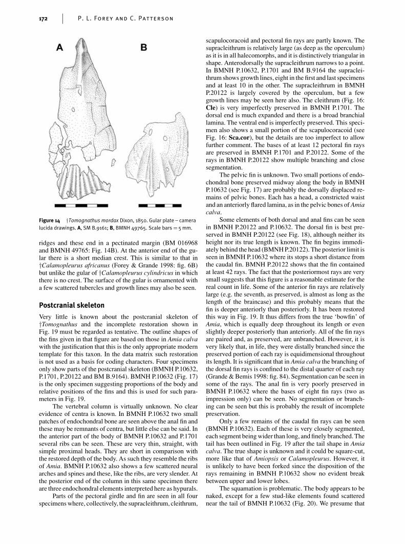
The median gular (Fig. 14) is very much like that of†Calamopleurus. It is fan-shaped with a broad flared pos-terior margin. Towards the posterior end there are radiating
ridges and these end in a pectinated margin (BM 016968and BMNH 49765: Fig. 14B). At the anterior end of the gu-lar there is a short median crest. This is similar to that in†Calamopleurus africanus (Forey & Grande 1998: fig. 6B)but unlike the gular of †Calamopleurus cylindricus in whichthere is no crest. The surface of the gular is ornamented witha few scattered tubercles and growth lines may also be seen.
Postcranial skeleton
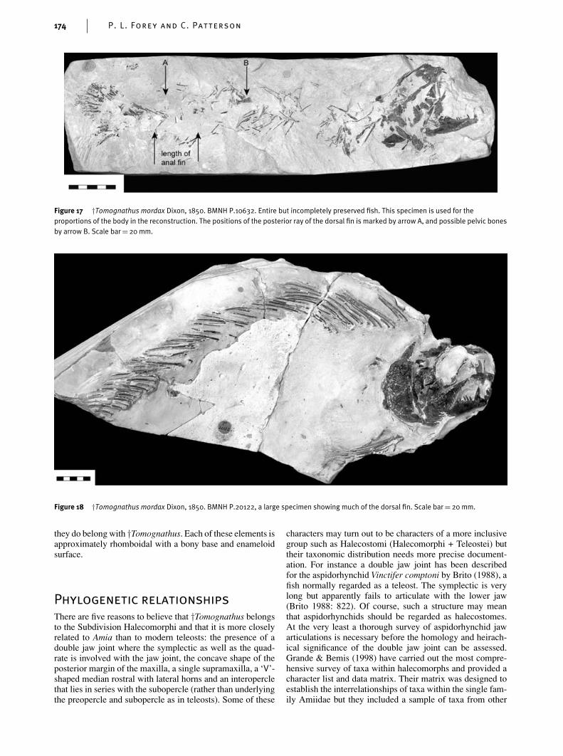
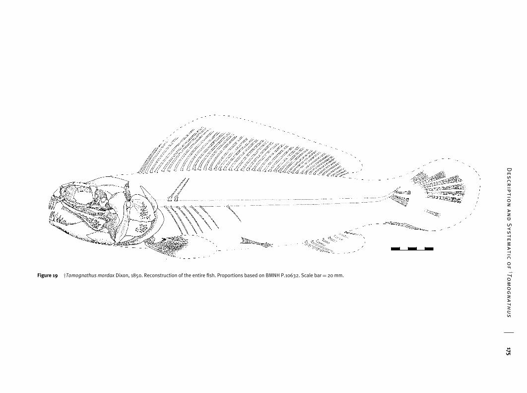
Very little is known about the postcranial skeleton of†Tomognathus and the incomplete restoration shown inFig. 19 must be regarded as tentative. The outline shapes ofthe fins given in that figure are based on those in Amia calvawith the justification that this is the only appropriate moderntemplate for this taxon. In the data matrix such restorationis not used as a basis for coding characters. Four specimensonly show parts of the postcranial skeleton (BMNH P.10632,P.1701, P.20122 and BM B.9164). BMNH P.10632 (Fig. 17)is the only specimen suggesting proportions of the body andrelative positions of the fins and this is used for such para-meters in Fig. 19.
The vertebral column is virtually unknown. No clearevidence of centra is known. In BMNH P.10632 two smallpatches of endochondral bone are seen above the anal fin andthese may be remnants of centra, but little else can be said. Inthe anterior part of the body of BMNH P.10632 and P.1701several ribs can be seen. These are very thin, straight, withsimple proximal heads. They are short in comparison withthe restored depth of the body. As such they resemble the ribsof Amia. BMNH P.10632 also shows a few scattered neuralarches and spines and these, like the ribs, are very slender. Atthe posterior end of the column in this same specimen thereare three endochondral elements interpreted here as hypurals.
Parts of the pectoral girdle and fin are seen in all fourspecimens where, collectively, the supracleithrum, cleithrum,
scapulocoracoid and pectoral fin rays are partly known. Thesupracleithrum is relatively large (as deep as the operculum)as it is in all halecomorphs, and it is distinctively triangular inshape. Anterodorsally the supracleithrum narrows to a point.In BMNH P.10632, P.1701 and BM B.9164 the supraclei-thrum shows growth lines, eight in the first and last specimensand at least 10 in the other. The supracleithrum in BMNHP.20122 is largely covered by the operculum, but a fewgrowth lines may be seen here also. The cleithrum (Fig. 16:Cle) is very imperfectly preserved in BMNH P.1701. Thedorsal end is much expanded and there is a broad branchiallamina. The ventral end is imperfectly preserved. This speci-men also shows a small portion of the scapulocoracoid (seeFig. 16: Sca.cor), but the details are too imperfect to allowfurther comment. The bases of at least 12 pectoral fin raysare preserved in BMNH P.1701 and P.20122. Some of therays in BMNH P.20122 show multiple branching and closesegmentation.
The pelvic fin is unknown. Two small portions of endo-chondral bone preserved midway along the body in BMNHP.10632 (see Fig. 17) are probably the dorsally displaced re-mains of pelvic bones. Each has a head, a constricted waistand an anteriorly flared lamina, as in the pelvic bones of Amiacalva.
Some elements of both dorsal and anal fins can be seenin BMNH P.20122 and P.10632. The dorsal fin is best pre-served in BMNH P.20122 (see Fig. 18), although neither itsheight nor its true length is known. The fin begins immedi-ately behind the head (BMNH P.20122). The posterior limit isseen in BMNH P.10632 where its stops a short distance fromthe caudal fin. BMNH P.20122 shows that the fin containedat least 42 rays. The fact that the posteriormost rays are verysmall suggests that this figure is a reasonable estimate for thereal count in life. Some of the anterior fin rays are relativelylarge (e.g. the seventh, as preserved, is almost as long as thelength of the braincase) and this probably means that thefin is deeper anteriorly than posteriorly. It has been restoredthis way in Fig. 19. It thus differs from the true ‘bowfin’ ofAmia, which is equally deep throughout its length or evenslightly deeper posteriorly than anteriorly. All of the fin raysare paired and, as preserved, are unbranched. However, it isvery likely that, in life, they were distally branched since thepreserved portion of each ray is equidimensional throughoutits length. It is significant that in Amia calva the branching ofthe dorsal fin rays is confined to the distal quarter of each ray(Grande & Bemis 1998: fig. 84). Segmentation can be seen insome of the rays. The anal fin is very poorly preserved inBMNH P.10632 where the bases of eight fin rays (two asimpression only) can be seen. No segmentation or branch-ing can be seen but this is probably the result of incompletepreservation.
Only a few remains of the caudal fin rays can be seen(BMNH P.10632). Each of these is very closely segmented,each segment being wider than long, and finely branched. Thetail has been outlined in Fig. 19 after the tail shape in Amiacalva. The true shape is unknown and it could be square-cut,more like that of Amiopsis or Calamopleurus. However, itis unlikely to have been forked since the disposition of therays remaining in BMNH P.10632 show no evident breakbetween upper and lower lobes.
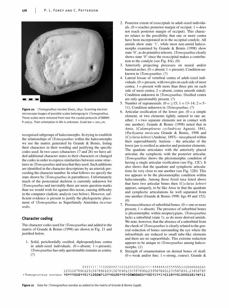
The squamation is problematic. The body appears to benaked, except for a few stud-like elements found scatterednear the tail of BMNH P.10632 (Fig. 20). We presume that
Description and Systematic of†Tomognathus 173
Figure 15 †Tomognathus mordax Dixon, 1850. Reconstruction of head in left lateral view.
Figure 16 †Tomognathus mordax Dixon, 1850. BMNH P.1701 showing partly preserved pectoral girdle and fin. Scale bar= 5 mm.
174 P. L. Forey and C. Patterson
Figure 17 †Tomognathus mordax Dixon, 1850. BMNH P.10632. Entire but incompletely preserved fish. This specimen is used for theproportions of the body in the reconstruction. The positions of the posterior ray of the dorsal fin is marked by arrow A, and possible pelvic bonesby arrow B. Scale bar= 20 mm.
Figure 18 †Tomognathus mordax Dixon, 1850. BMNH P.20122, a large specimen showing much of the dorsal fin. Scale bar= 20 mm.
they do belong with †Tomognathus. Each of these elements isapproximately rhomboidal with a bony base and enameloidsurface.
Phylogenetic relationships
There are five reasons to believe that †Tomognathus belongsto the Subdivision Halecomorphi and that it is more closelyrelated to Amia than to modern teleosts: the presence of adouble jaw joint where the symplectic as well as the quad-rate is involved with the jaw joint, the concave shape of theposterior margin of the maxilla, a single supramaxilla, a ‘V’-shaped median rostral with lateral horns and an interoperclethat lies in series with the subopercle (rather than underlyingthe preopercle and subopercle as in teleosts). Some of these
characters may turn out to be characters of a more inclusivegroup such as Halecostomi (Halecomorphi + Teleostei) buttheir taxonomic distribution needs more precise document-ation. For instance a double jaw joint has been describedfor the aspidorhynchid Vinctifer comptoni by Brito (1988), afish normally regarded as a teleost. The symplectic is verylong but apparently fails to articulate with the lower jaw(Brito 1988: 822). Of course, such a structure may meanthat aspidorhynchids should be regarded as halecostomes.At the very least a thorough survey of aspidorhynchid jawarticulations is necessary before the homology and heirach-ical significance of the double jaw joint can be assessed.Grande & Bemis (1998) have carried out the most compre-hensive survey of taxa within halecomorphs and provided acharacter list and data matrix. Their matrix was designed toestablish the interrelationships of taxa within the single fam-ily Amiidae but they included a sample of taxa from other
DescriptionandSystematicof
†Tomognathus
175
Figure 19 †Tomognathus mordax Dixon, 1850. Reconstruction of the entire fish. Proportions based on BMNH P.10632. Scale bar= 20 mm.
176 P. L. Forey and C. Patterson
Figure 20 †Tomognathus mordax Dixon, 1850. Scanning electronmicroscope images of possible scales belongong to †Tomognathus.These scales were removed from near the caudal peduncle of BMNHP.10632. Their orientation in life is unknown. Scale bar= 100 µm.
recognised subgroups of halecomorphs. In trying to establishthe relationships of †Tomognathus within the halecomorphswe use the matrix generated by Grande & Bemis, listingtheir characters in their wording and justifying the specificcodes used. In two cases (characters 17 and 26) we have ad-ded additional character states to their characters or changedthe codes in order to express similarities between some struc-tures in †Tomognathus and taxa that they used. Such additionsare identified in the character descriptions by an asterisk pre-ceeding the character number. In what follows we specify thestate shown by †Tomognathus in parentheses. Unfortunatelymuch of the postcranial skeleton is currently unknown for†Tomognathus and inevitably there are more question marksthan we would wish for against this taxon, causing difficultyin the computer cladistic analysis (see below). However, suf-ficient evidence is present to justify the phylogenetic place-ment of †Tomognathus as Superfamily Amioidea incertaesedis.
Character coding
The character codes used for †Tomognathus and added to thematrix of Grande & Bemis (1998) are shown in Fig. 21 andjustified below.
1. Solid, perichordally ossified, diplospondylous centrain adult-sized individuals. (0 = absent; 1 = present).†Tomognathus has only questionable remains as centra.(?)
Figure 21 Data for †Tomognathus mordax as added to the matrix of Grande & Bemis (1998).
2. Posterior extent of exocciptals in adult-sized individu-als. (0 = reaches posterior margin of occiput; 1 = doesnot reach posterior margin of occiput). This charac-ter relates to the possibility that one or more centrahave been incorporated in to the occipital condyle. Allamiids show state ‘1’, while most non-amiid haleco-morphs examined by Grande & Bemis (1998) showstate ‘0’, as do primitive teleosts. †Tomognathus clearlyshows state ‘0’ since the exoccipital makes a contribu-tion to the condyle (see Fig. 8A). (0)
3. Anteriorly projecting processes on neural and/orhaemal arches. (0 = absent; 1 = present). Condition un-known in †Tomognathus. (?)
4. Lateral fossae of vertebral centra of adult-sized indi-viduals. (0 = present, with two pits on each side of mostcentra; 1 = present with more than three pits on eachside of most centra; 2 = absent, centra smooth sided).Condition unknown in †Tomognathus. Ossified centraare only questionably present. (?)
5. Number of supraneurals. (0 =≥15; 1 = 13–14; 2 = 5–11). Condition unknown in †Tomognathus. (?)
6. Articular ossification of the lower jaw. (0 = a simpleelement, or two elements tightly sutured to one an-other; 1 = two separate elements not in contact withone another). Grande & Bemis (1998) noted that inAmia, †Calamopleurus cyclindricus Agassiz, 1841,†Pachyamia mexicana Grande & Bemis, 1998 and†Cyclurus kehreri (Andreae, 1893) – taxa placed withintheir supersubfamily Amiista – the articular of thelower jaw is ossified as anterior and posterior elements.The quadrate articulates with the anteriorly placedarticular, the symplectic with the posterior articular.†Tomognathus shows the plesiomorphic condition ofhaving a single articular ossification (see Fig. 12C). Italso shows that the quadrate and symplectic articula-tions lie very close to one another (see Fig. 12D). Thistoo appears to be the plesiomorphic condition withinhalecomorphs. Among those fossil taxa listed abovethat have two articular bones then †Cyclurus kehreriappears, uniquely, to be like Amia in that the quadrateand symplectic articulations lie well separated fromone another (Grande & Bemis 1998: figs 49 and 152).(0)
7. Presence/absence of suborbital bones. (0 = one or morepresent; 1 = absent). The presence of suborbital bonesis plesiomorphic within neopterygians. †Tomognathuslacks a suborbital (state 1), as do more derived amiids.We note, however, that the absence of a suborbital fromthe cheek of †Tomognathus is clearly related to the gen-eral reduction of bones surrounding the eye where theinfraorbitals are reduced to small tube-like elementsand there are no supraorbitals. This extreme reductionappears to be unique to †Tomognathus among haleco-morphs. (1)
8. Strength of ornamentation on dermal bones of skull.(0 = weak and/or fine; 1 = strong, coarse). Grande &
Description and Systematic of†Tomognathus 177
Bemis acknowledge the subjective nature of vari-ation expressed in this character but they do illus-trate their meaning, using operculae of various amiidtaxa. †Tomognathus shows dermal bones that are nearlysmooth (see Fig. 15). Only the maxilla, anteroventralportion of the dentary, angular and parts of the opercu-lum, subopercle and gular (see Fig. 14) are ornamentedwith discrete tubercles. Therefore, this corresponds tostate ‘0’. (0)
9. Hypural–ural centra fusion in adult-sized individuals.(0 = all hypurals autogenous (separate) from the uralcentra; 1 = all but the first hypural fused to correspond-ing centra). The caudal endoskeleton of †Tomognathusis too poorly known for comment or assessment ofcharacter states. (?)
10. Presence/absence of large parapophyses fused to mostof the abdominal centra. (0 = absent; 1 = present). Thevertebral column of †Tomognathus remains virtuallyunknown. (?)
11. Presence/absence of substantial scapulocoracoid ossi-fication in adult-sized individuals. (0 = one or moreelements present in the shoulder girdle; 1 = absent).Although the scapulocoracoid is very poorly known(see Fig. 16) sufficient remains to establish that somedegree of ossification was present. (0)
12. Presence/absence of supraorbital bones. (0 = two ormore present; 1 = absent). Grande & Bemis (1998:576) noted the two conditions listed here within theirhalecomorph sample, with the absence of supraorbit-als restricted to members of the subfamily Amiinae.†Tomognathus has no supraorbitals (see remarks undercharacter 7). (1)
13. Urodermals in the caudal skeleton. (0 = present;1 = absent). There are no specimens of †Tomognathussufficiently well-preserved to comment. (?)
14. Presence/absence of sclerotic ring ossification.(0 = present; 1 = absent). †Tomognathus lacks a scler-otic ring. (1)
15. Size and shape of the dorsal fin. (0 = short, with straightto falcate margin, 14–25 segmented rays and 14–25proximal radials; 1 = medium long, with bow-shapedor straight margin, 30–34 segmented rays and an es-timated 30–34 proximal radials; 2 = very long, withbow-shaped margin, 36–47 segmented rays and 37–48proximal radials; 3 = extremely long, with bow-shapedmargin, 48–53 segmented rays and 49–54 proximal ra-dials). Grande & Bemis (1998: 577) identified fourstates of variation of dorsal fin shape that, in combin-ation, describe shape and length. One of their states(‘0’) describes a short fin (14–25 segmented rays) witha straight to falcate margin. The other three states definemedium or long, straight or bow-shaped fins: the statesbeing distinguished by the number of rays. The dorsalfin of †Tomognathus corresponds most closely withtheir state ‘2’ (very long, with bow-shaped margin,36–47 segmented rays) and this is how we choose tocode it. In fact, the dorsal fin of †Tomognathus maybe considered autapomorphic in the sense that it be-gins immediately behind the head, there being littleor no gap between the occiput and its origin. Theremay therefore be a case for suggesting a fifth state,exclusive to †Tomognathus. Grande & Bemis (1998:577) also make the point that the length of the fin may
be decoupled from the number of supporting elements.In Amia calva the fin rays lie in series with the ptery-giophores that, in turn, lie in one-to-one series withthe neural spines. This is not the case in †Vidalamiacatalunica (Sauvage, 1903) as shown by Grande & Be-mis (1998: fig. 250). (2)
16. Morphology of teeth on anterior coronoid and vomer.(0 = conical, with pointed tips; 1 = styliform, withbroadly rounded or flattened tips; N = no anteriorcoronoid). †Tomognathus clearly has state ‘0’ (seeFig. 12B). (0)
*17. Anterior extent of parasphenoid tooth patch.(0 = extends well anterior to the lateral ascendingarms of the parasphenoid; 1 = short, does not extendanterior to the ascending arms of the parasphenoid;2 = parasphenoid tooth patch absent). Grande & Bemis(1998: 578) recognise three states. Two described theextent of teeth while the third they scored as ‘NotApplicable’ to describe the absence of teeth. In theirtaxon sampling this code is used only for the teleost†Pholidophorus macrocephalus Agassiz, 1833–1844.The presence of parasphenoid teeth is a plesiomorphiccondition for actinopterygians and we consider the re-cording of absence to have potential grouping signific-ance. Therefore we use this character and replace the‘N’ code with state ‘2’. (2)
18. Parietal length. (0 = relatively long, with a width-to-length ratio range not exceeding 0.90; relatively short,with a width-to-length ratio well exceeding 0.90).Grande & Bemis (1998: 578) recognise two states ofparietal dimension, using a Non-Applicable code formembers of the †Sinamiidae that have median parietals.We therefore code Non-Applicable for †Tomognathus.(N)
19. Number of ural centra. (0 = 10 or fewer; 1 = 11–22).Unknown for †Tomognathus. (?)
20. Shape of preopercle. (0 = L-shaped; 1 = crescent-shaped; 2 = crescent-shaped, wide in middle, taperingdorsally and ventrally; 3 = ovoid). Grande & Bemis(1998: fig. 242) illustrate two of the states of thisfour-state character. The preopercle of †Tomognathusmost closely corresponds with that of †Oshunia brevisWenz & Kellner, 1986 in being crescent-shaped with aslightly expanded ventral end (see Fig. 15). We there-fore use a coding similar to that for Oshunia. (1)
21. Morphology of caps of jaw teeth in adult-sized indi-viduals. (0 = round in cross-section, not sharply carin-ate; 1 = labiolingually compressed, sharply carinate(keeled)). †Tomognathus shows rounded, non-carinateteeth (e.g. Fig. 5B). (0)
22. Lateral edge of posttemporal in adult-sized individuals.(0 = shorter than length of anterior edge; 1 = elongate,about equal to or greater than width of anterior edge).The posttemporal is unknown in †Tomognathus. (?)
23. Shape of posterior margin of caudal fin. (0 = forked;1 = convexly rounded; 2 = straight and nearly vertical).The only specimens showing any of the caudal fin raysis BMNH P.10632 (see Fig. 17; Woodward 1908: pl. 29,fig. 13). Of this specimen Woodward writes (1908: 142)‘. . . . The caudal fin (c.) consists of very stout, closelyarticulated, and finely divided rays; and their remainsshown in fig. 13 suggest that was not forked.’ This isprobably true but it is impossible to identify whether
178 P. L. Forey and C. Patterson
it is convexly rounded or straight. In view of thevery incomplete preservation we leave it as question-able. (?)
24. Elongation of the opercular process of the hyomandibu-lar. (0 = absent; 1 = present). †Tomognathus shows abarely differentiated opercular process (see Figs 10C& 12D). (0)
25. Number of tooth rows on coronoids. (0 = two or morerows for at least part of one or more coronoids; 1 = onerow; N = no coronoids). †Tomognathus shows only asingle coronoid (we deal with this below), where theteeth are aligned in a single row and hence shows state1 with respect to tooth alignment. (1)
*26. Arrangement of vomerine teeth. (0 = tooth patch withtwo to several rows of teeth; 1 = tooth patch with onlya single marginal row, plus one or more teeth in a lon-gitudinal series perpendicular to the anterior marginalrow; 2 = tooth patch arranged as a single transverserow). Grande & Bemis (1998) established the first twostates of this character to differentiate a very specificpattern in †Calamopleurus and †Maliamia (Grande &Bemis 1998: figs 309, 321) as state ‘1’. †Tomognathusshows just a few teeth aligned in a single row approx-imately transversely (see Fig. 5B). We therefore addeda third state (coded as ‘2’) to this character since it haselements of the other two states. We also note here that†Tomognathus appears to be uniquely derived amongsthalecomorphs to show a median, unpaired vomer, acondition more usually seen in teleosts. (2)
27. Presence/absence of dermopterotic ribs. (0 = absent;1 = present). Such ribs are absent from †Tomognathus,as they are from most halecomorphs. (0)
28. Number of epurals. (0 = 2–8; 1 = 10–15). This regionof the body is unknown in †Tomognathus. (?)
29. Shape of basipterygium. (0 = proximal end flat andwidened anteriorly; 1 = proximal end long and rod-like, without significant widening anteriorly). This re-gion of the body is poorly known in †Tomognathus.(?)
30. Postmaxillary process under postmaxillary notch.(0 = tiny or absent; 1 = thick and elongate). Grande &Bemis (1998: fig 243) give outline drawings of themaxillae in a variety of halecomorphs and designatetwo states, one of which they assign to †Vidalamia and†Pachyamia in which the notch in the posterior mar-gin of the maxilla (characteristic of all halecomorphs)is particularly well developed and in which the lowerlimb demarcating the notch is thickened. There is con-siderable variation in the shapes designated as state ‘0’and that in †Tomognathus falls well within this vari-ation, being most similar to that of †Solnhofenamiaand †Amiopsis. (0)
31. Morphology of pleural ribs. (0 = distal ends pointedor without rounded points; 1 = distal ends flatly trun-cated, even in large adults). Ribs are poorly known in†Tomognathus. (?)
32. Shape of gular. (0 = subtriangular or subrectangularwith acute rounded anterior apex; 1 = broad, oval,without acute anterior apex). Grande & Bemis (1998:fig. 136) separate the rounded gular plate of †Vidalamiaand †Pachyamia as state ‘1’, all others being a variationon their description of state ‘0’. We note that the shapeof the gular in †Tomognathus is remarkably similar to
that of †Calamopleurus (see Fig. 14 and see below,character 64). (0)
33. Peculiar ornamentation of strongly defined, conver-ging lines on opercles in adult-sized individuals.(0 = absent; 1 = present). The ornament upon theopercle of †Tomognathus is best described under state‘0’. We note that the shape of the opercle appears to beunique among halecomorphs in showing an irregularand partly serrated ventral margin (see Fig. 15). (0)
34. Frontal width in adult-sized individuals. (0 = relativelywide, with a width-to-length ratio of 0.26–0.65;1 = relatively narrow, with a width-to-length ratio of0.13–0.21). The frontals are fused in †Tomognathus,hence this character is scored as ‘Not Applicable’. (N)
35. Shape of dermopterotic. (0 = greatly widened posteri-orly and tapered anteriorly; 1 = subrectangular, notsubstantially tapered anteriorly or expanded posteri-orly). The dermopterotic of †Tomognathus narrows an-teriorly (see Fig. 3), corresponding to state ‘0’. (0)
36. Width of opercle. (0 = narrow, with width-to-height ra-tio 0.56–1.06; 1 = wide, with width-to-height ratio inthe range of 1.07–1.39). The opercle of †Tomognathusis approximately equidimensional (see Fig. 15), mostlike that of †Cyclurus and Amia (Grande & Bemis 1998:fig. 180A-K). (0)
37. Presence of an interfrontal fontanelle in adult-sized in-dividuals. (0 = absent, frontals sutured to each othermedially for their entire length; 1 = frontals separatedfor about one half of their length or more by a fon-tanelle). The frontals of †Tomognathus are fused andtherefore this character must be scored as Not Applic-able’. (N)
38. Position of dermosphenotic relative to orbit in adult-sized individuals. (0 = anterior or anteroventral marginof dermosphenotic included in circumorbital margin,even in large adults of 200 mm standard length (SL)or more; 1 = dermosphenotic excluded from orbitalmargin in large individuals of 200 SL or more). Thedermosphenotic of †Tomognathus is included in the or-bital margin and lies anterior to the dermopterotic. (seeFig. 15 and Woodward 1908: fig. 7). (0)
39. Shape of supramaxilla. (0 = elongate; 1 = extremelydeep, shaped like a rounded triangle). †Tomognathushas an elongate supramaxilla (see Figs 11C & 15; cf.Grande & Bemis 1998: fig. 277 for state 0). (0)
40. Number of preural vertebrae (preural centra = abdo-minal plus preural caudal centra). (0 = 40–73; 1 = 75–82). Centra poorly known in †Tomognathus. (?)
41. Shape of posterior end of posttemporal in adult-sizedindividuals. (0 = elongate, with rounded apex or apices;1 = elongate but abruptly truncated). Posttemporal un-known in †Tomognathus. (?)
42. Ventral transverse ridge of gular. (0 = absent;1 = present). This character was identified as autapo-morphic for †Pachyamia mexicana by Grande & Bemis(1998). †Tomognathus lacks such a ridge. (0)
43. Shape of anterior subinfraorbital bone in adult-sized in-dividuals. (0 = short, subrectangular, longer than deep;1 = subrectangular, deeper than long; 2 = long, verythin, tube-like; 3 = posteriorly expansive, tapering an-teriorly). †Tomognathus shows a small tube-like an-terior subinfraorbital (called infraorbital 1 here – seeFig. 15). (2)
Description and Systematic of†Tomognathus 179
44. Number of epaxial procurrent caudal fin rays. (0 = 0–11; 1 = 12–15). The caudal fin of †Tomognathus is in-sufficiently known to code. (?)
45. Presence/absence of fringing fulcra on median fins.(0 = present; 1 = absent). Although the dorsal fin isonly partly known there appear to be no fringing fulcrain †Tomognathus. The other median fins are unknown.(1)
46. One-to-one arrangement of hypurals and caudal finrays. (0 = last few hypurals each articulate with thebases of several fin rays; 1 = each hypural nor-mally bears a single caudal ray). The caudal fin of†Tomognathus is insufficiently known to code. (?)
47. Number of ossified ural neural arches. (0 = normallyfour or more; 1 = normally two or fewer). The caudalfin of †Tomognathus is insufficiently known to code.(?)
48. Number of parietal bones. (0 = paired parietals nor-mally present; 1 = only a single median parietalpresent). †Tomognathus has a median parietal (seeFig. 3). (1)
49. Number of pairs of extrascapular bones. (0 = only onepair present; 1 = three pairs normally present). Extras-capulars are unknown in †Tomognathus. (?)
50. Dermopterotic length to parietal length. (0 =dermopterotic significantly longer; lengths aboutequivalent = 1). Grande & Bemis (1998) illustrate whatthey mean by these dimensions and †Tomognathus cor-responds with state ‘0’ (see Fig. 3). (0)
51. Presence/absence of opisthotic. (0 = present; 1 =absent). †Tomognathus lacks an opisthotic (seeFig. 6). (1)
52. Presence/absence of pterotic. (0 = present; 1 = absent).†Tomognathus lacks an (auto)pterotic (see Fig. 6). (1)
53. Shape of maxilla extremely slender and rod-like.(0 = no; 1 = yes). Based on illustrations specify-ing these states (Grande & Bemis 1998: fig. 243)†Tomognathus shows state ‘0’. (0)
54. Number of branchiostegal rays. (0 = 21 or fewer;1 = 22 or more). The branchiostegal ray series of†Tomognathus is very poorly known. It is known toshow a branchopercle, preceded by at least three fila-mentous branchiostegals but the exact number is notknown. (?)
55. Numerous paired block-like ural neural arch os-sifications. (0 = absent; 1 = present). The caudalfin of †Tomognathus is insufficiently known tocode. (?)
56. Dermosphenotic attachment to skull roof in adult-sizedindividuals. (0 = loosely attached on the skull roof orhinged to the side of skull roof; 1 = firmly sutured intothe skull roof and forming part of it). †Tomognathusshows a free dermosphenotic (see Fig. 15).(0)
57. Shape of rostral bone. (0 = plate-like or short tube-like, without lateral horns; 1 = roughly V-shaped, withlateral horns). †Tomognathus shows state ‘1’ (seeFig. 13). (1)
58. Lachrymal shape. (0 = longer than deep and smal-ler than orbit; deeper than long and massive aboutsize of orbit = 1)). The lachrymal (infraorbital 1) of†Tomognathus is small and therefore corresponds mostclosely to state ‘0’ (see Fig. 15). (0)
59. Posterior extent of maxilla. (0 = extends to below theposterior orbital margin; 1 = does not extend belowposterior orbital margin). In †Tomognathus the maxillaextends to a level posterior to the rear of the orbit (seeFig. 15). (0)
60. Presence/absence of lateral line canal in maxilla.(0 = absent; 1 = present). †Tomognathus lacks a lateralline in the maxilla. (0)
61. Symplectic involvement in jaw joint. (0 = does not ar-ticulate with lower jaw; 1 = distal end articulates witharticular bone of the lower jaw). The symplectic, quad-rate and the nature of the jaw joint of †Tomognathusis described (p. 11) and illustrated (see Fig. 12D) toshow the involvement of the symplectic in the lowerjaw joint. (1)
62. Shape of posterior margin of the maxilla. (0 = convexlyrounded or straight; 1 = excavated (concave or with aposterior maxillary notch present)). Shapes of the pos-terior end of the maxilla in a range of halecomorph taxa,all of which show state ‘1’, are illustrated by Grande& Bemis (1998: fig. 243). That of †Tomognathus (seeFig. 11B) most closely corresponds to that of †Amiopsisspp, the chief difference being that the margin is ser-rated in †Tomognathus. (1)
63. Innerorbital flange of dermosphenotic. (0 = smooth,without sensory canal; 1 = bearing sensory canal tube).The sensory canal runs within the main body of thedermosphenotic in †Tomognathus and this taxon there-fore lacks an innerorbital flange, corresponding to state‘0’. (0)
64. Posterior margin of gular. (0 = smooth; 1 = deeplyscalloped). In Grande & Bemis’ (1998) analysis a gu-lar with a scalloped margin was identified as a synapo-morphy of species of †Calamopleurus. †Tomognathusis now known to possess an almost identically shapedgular (see Fig. 14). (1)
65. Shape of the haemal spines. (0 = spine-like or rod-like; 1 = broadly spatulate in the transverse plane).†Tomognathus is insufficiently known for reliable cod-ing. (?)
66. Relative size of uppermost postinfraorbital in adult-sized individuals. (0 = short, much shorter than lower-most postinfraorbital; 1 = long, about equal in lengthto lowermost postinfraorbital). This character is prob-lematic for †Tomognathus. Grande & Bemis (1998)noted a clear difference between those taxa in whichthe broad plate-like postinfraorbitals were of equal size(e.g. Amia) and those in which they were not (e.g.†Calamopleurus). †Tomognathus differs from all hale-comorphs considered by them in that all infraorbitals(postinfraorbitals included) are reduced to small tube-like structures. Therefore, considering this character atface value we would say that †Tomognathus shows state‘1’. However, in view of the autapomorphic conditionof the infraorbitals in this taxon we prefer to code ‘Notapplicable’. (N)
67. Orientation of preural haemal and neural spinesnear caudal peduncle. (0 = positioned at about 25–45
◦from the horizontal; 1 = strongly inclined to
nearly horizontal). These structures are unknown for†Tomognathus. (?)
68. Presence/absence of interopercle. (0 = absent; 1 =present). †Tomognathus has an interopercle (see
180 P. L. Forey and C. Patterson
Fig. 15). We would also remark that as in allhalecomorphs this bone lies in series with the subo-percle and does not underlay the preopercle and subo-percle as in teleosts. (1)
69. Number of supramaxillae. (0 = none; 1 = one; 2 =two). †Tomognathus has a single supramaxilla (seeFigs 11C, 15). (1)
Phylogenetic analysis
†Tomognathus has 28 question marks against characters con-sidered here. These question marks denote missing data andnon-applicable data. This is a high percentage of questionmarks (40%) and causes trouble for computer generatedphylogenies because it leads to decay of the resolution givenby known data, although it needs to be stressed that the ques-tion marks do not lead to new topologies. In order to lessenthe effect of this high percentage of missing data it was de-cided that the taxon sampling be reduced to the minimumfollowing the advice of Grande & Bemis (1998: 567–568).This means that all taxa that showed redundancy (that is,their real codes did not add variation to the complement ofreal data against all taxa), were stripped out. As a result,only the 24 taxa used by Grande & Bemis were used, with†Tomognathus being added as a 25th. Nevertheless, it shouldbe pointed out that even with this reduced taxon samplingwe still ran into the perils of optimisation and spurious nodes(detailed below).
The data matrix was analysed using PAUP*4.0b10 (Swofford 2001) with the characters unordered;†Eurycormus, Atractosteus, †Pholidophorus bechei and †P.macrocephalus were used as the outgroup and the branch andbound tree-searching algorithm was employed.
The first analysis omitted †Tomognathus in order tocheck the results against those of Grande & Bemis (1998)since the coding of one character (see character number17 above) had been changed. Four trees were obtained at115 steps (one more than Grande & Bemis), the strict con-sensus of which is shown in Fig. 22 and the tree statistics aregiven in the legend. The results obtained were in topologicalagreement with those of Grande & Bemis, with the excep-tion that †Solnhofenamia elongata collapsed to a trichotomywith †Amiopsis lepidota and the remainder of the Amiidae(in Grande & Bemis’ analysis this taxon was resolved asthe sister taxon to all remaining Amiidae except †Amiopsislepidota).
The addition of †Tomognathus caused a major collapseof taxa within the Superfamily Amioidea to result in the strictconsensus of 24 trees showing an eight-fold multifurcation(Fig. 23). Even more surprising was the fact that the sister-group pairing of †Cyclurus gurleyi with the three includedspecies of †Amia was now included in a polytomy (previ-ously it had been calculated that the node †Cyclurus gurleyi+ Amia spp. had a Bremer support of 8). However, inspectionof alternative placements for †Tomognathus in the funda-mental trees demonstrate that the collapse is largely illusoryand must be viewed as having little significance. Within the24 fundamental trees, †Tomognathus is placed at five posi-tions as shown in Fig. 24. Two of these positions are sister-group pairings with terminal taxa, a third is as sistergroup toAmia spp. and a fourth is as sistergroup to the sinamiid taxa;hence, the collapse of the amioid clade in the strict consensustree when †Tomognathus is included. Closer inspection of
each of these alternatives shows very little support by realdata.
The node (†Tomognathus + †Cyclurus gurleyi) is sup-ported by character changes in three characters, namely 6(1 → 0, CI (consistency index) = 0.500) 17 (0 → 1, CI =0.667), 18 (0 → 1, CI = 0.200). †Tomognathus is coded asquestion marks for 6 and 18. Secondly, these two taxa differin the codings for character 17 and the node state is onlyresolved by ambiguous optimisation. This must be regardedas a spurious node.
The node (†Tomognathus (Amia spp.)) is only supportedby one character change – 40 (0 → 1, CI = 0.500); thischaracter is coded as a question mark for †Tomognathus andtherefore this node is spurious.
The node (†Tomognathus, †Amia pattersoni) is suppor-ted by two character changes – 6 (1 → 0, CI = 0.500) and25 (0 → 1, CI = 0.500), both of which characters are codedas question marks for †Amia pattersoni, so once again thismust be regarded as a spurious node.
The node (†Tomognathus (†Cyclurus gurleyi + Amiaspp.)) is supported by eight character changes – 5 (1 →2,CI = 0.667), 9 (0 → 1, CI = 1.000), 10 (0 → 1, CI =0.500), 12 (0 → 1, CI = 1.000), 13 (0 → 1, CI = 1.000),14 (0 → 1, CI = 0.500), 15 (0 → 2, CI = 0.750), 66 (0 → 1,CI = 1.000). Of these character changes, numbers 9, 12,13 and 66 are optimised as synapomorphies. However,†Tomognathus is coded as question marks for 9, 13 and 66(as well as 5 and 10). Therefore, the only unambiguous char-acter suggesting that †Tomognathus is more closely relatedto †Cyclurus gurleyi and Amia spp. is the ‘1’ state of charac-ter 12 – the shared absence of supraorbital bones. While thisis true it should be noted that the pattern of the remainingcheek bones in †Tomognathus is highly apomorphic amongthe halecomorphs considered here in being reduced to tinytube-like elements. Parenthetically it should be noted that thisis the result obtained when the characters were reweighted(using the rescaled consistency index with a baseweight of1000 applied).
The node (†Tomognathus (†Sinamia zdanskyi, †Ike-chaoamia meridionalis) is supported by seven characterchanges – 7 (0 → 1, CI = 0.500), 14 (0 → 1, CI = 0.333),17 (0 → 2, CI = 0.667), 22 (0 → 1, CI = 0.500), 26 (0→ 2, CI = 0.400), 48 (0 → 1, CI = 1.000), 49 (0 → 1,CI = 0.500). †Tomognathus shows question marks for twoof these characters (22 and 49), while †Sinamia zdanskyi and†Ikechaoamia meridionalis are coded as question marks forcharacters 7, 14, 17 and 26. There is one synapomorphy thatsuggests this sistergroup pairing: character 48 – fusion ofparietals to a median element.
Character changes supporting the SuperfamilyAmioidea are 23 (0 → 1, CI = 0.500), 45 (0 → 1, CI =1.000) and 46 (0 → 1, CI = 0.500). Characters 23 and 46 arecoded as question marks in †Tomognathus, leaving 45 (state1) – the absence of fringing fulcra on median fins – as the onlyevidence for the most inclusive position of †Tomognathus.
With the evidence before us there are two phylogen-etic positions that are recovered through the computer parsi-mony analysis that can be justified by real data; either†Tomognathus is the sistergroup to sinamiids, or the sistergroup to Cyclurus gurleyi + Amia spp.
Somewhat disturbingly, it should be noted that globalparsimony failed to identify two synapomorphies that place†Tomognathus as the sister group to †Calamopleurus.
Description and Systematic of†Tomognathus 181
Figure 22 Result of analysing the reduced data set of Grande & Bemis (1998) using PAUP∗ version 4.0b10 (Swofford 2001). The coding for onecharacter was changed (see discussion of character 17); all characters are unordered and of equal weight. Strict consensus tree of four trees:length= 115, consistency index (CI) excluding uninformative characters= 0.6667, retention index (RI)= 0.5884.
†Tomognathus and the two species of †Calamopleurus areunique among the taxa considered here in having a singlerow of teeth upon the coronoid (character 25, state 1) and inhaving the posterior edge of the gular plate deeply scalloped(character 64, state 1). A third character – dermosphenoticloosely attached to the skull roof (character 56, state 0) –is also shared with †Calamopleurus, but this is also foundin non-halecomorphs and †Watsonulus among the taxa con-sidered here. All of these observations are recorded in the
data matrix but any grouping criteria on these character-statesdisappears amongst global parsimony.
This case study is offered as a salutary reminderthat maximum parsimony analysis is fraught with inter-pretive problems. Care must be taken when interpreting aphylogenetic hypothesis. Of course it would have been pos-sible ‘load to the dice’ by differential weighting of characters.Without more information about †Tomognathus, especiallythe postcranial skeleton, then we are reluctant to overstretch
182 P. L. Forey and C. Patterson
Figure 23 Result of analysing the reduced data set of Grande & Bemis (1998) using PAUP∗ version 4.0b10 (Swofford 2001) and including†Tomognathus. Strict consensus of 24 trees: length= 125, CI excluding uninformative characters= 0.6303, RI= 0.5282.
the evidence and place this taxon as Superfamily Amioideaincertae sedis.
Conclusions
†Tomognathus is one of only three halecomorph fishesthat are found in the Upper Cretaceous of England; theother two being †Lophiostomus dixoni Egerton, 1852,†Neorhombolepis excelsus Woodward, 1888, and †N. (?)
punctatus Woodward, 1895 (Woodward 1909: 160 castdoubt on the generic identity as this species is only basedon a few isolated scales). Both †Lophiostomus dixoni and†Neorhombolepis excelsus are known from very few speci-mens (in both cases the holotype plus a few isolated pieces)and their relationships are in doubt. Both were originallyreferred to the †Eugnathidae (=†Caturidae = †Furidae) byWoodward 1909. It has been suggested that†Neorhombolepisis a member of the †Ophiopsidae (Grande & Bemis 1998:607) and that †Lophiostomus dixoni should also be removed
Description and Systematic of†Tomognathus 183
Figure 24 Alternative resolutions for †Tomognathus among the 24 trees obtained in Fig. 23. See the text for discussion of character supportfor each position.
from the family (Bartram 1975). Despite the fact that bothof these taxa are left in limbo pending a comprehensive re-vision of caturid fishes no-one has suggested that they arerelated to fishes outside the Halecomorphi. Halecomorphsare a clade of predaceous fishes that originated in the LowerTriassic and were very common in the Jurassic and LowerCretaceous. By the Upper Cretaceous, most were confined tofreshwater where they flourished until the Oligocene, fromwhen they declined to leave Amia calva (the bowfin of theeastern USA) as the sole representative. Among haleco-morphs, †Tomognathus is certainly one of the most unusualand among the last of the marine representatives. The highdegree of fusion of skull roof bones, the extreme reductionof the cheek bones and the naked body is unique within thisclade, as is the weakness, or absence of ossification of thecentra. The characters that are available place it as a memberof the Superfamily Amioidea and with these fishes it sharesthe overall form of the body.
Acknowledgements
We would like to thank John Cooper, Booth Museum,Brighton; Jenny Clack, Museum of Zoology, Cambridge Uni-versity and Rod Long, Sedgwick Museum, Cambridge Uni-versity for the loan of material. We also extend our thanksto Phil Crabb and Alison Longbottom, The Natural HistoryMuseum, for their skills in taking the photographs and com-posing the electronic figures, respectively. We also thank ourtwo reviewers, Dr John Maisey (American Museum of Nat-ural History) and Dr Paulo Brito (Universidade do Estado doRio de Janeiro, Brazil) for corrections and suggestions.
References
Agassiz, J. L. R. 1833–1844. Recherches sur les Poissons Fossiles,vol. 1. Petitpierre, Neuchatel, 188 pp.
184 P. L. Forey and C. Patterson
Andreae, A. 1893. Vorlaufige Mittheilung uber die Ganoiden (Lepidosteusund Amia) des Mainzer Beckens. Verhandlungen des Natuhistorisch-medizinischen Vereins Heidelberg 2 (ser 5): 7–15.
Arratia, G. 1999. The monophyly of Teleostei and stem-group teleosts.Consensus and disagreements. Pp. 265–334 in G. Arratia and H.-P.Schultze (eds) Mesozoic fishes 2 – systematics and the fossil record.Dr Friedrich Pfei l, Munchen.
— & Schultze, H.-P. 1987. A new halecostome fish (Actinopterygii,Osteichthyes) from the late Jurassic of Chile and its relationships.Dakoterra 3: 1–13.
Bartram, A. W. H. 1975. The holostean fish genus OphiopsisAgassiz. Zoological Journal of the Linnean Society of London 56:183–205.
Bonaparte, C. L. 1838. Selachorum tabula analytica. Nuovi Annali dellaScienze Naturali, Bologna 2: 195–214.
Bridge, T. W. 1877. The cranial osteology of Amia calva. Journal ofAnatomy 11: 605.
Brito, P. M. 1988. La structure du suspensorium de Vinctifer, pois-son actinopterygien Mesozoique: remarques sur les implicationsphylogenetiques. Geobios 21: 819–823.
Cope, E. D. 1872. Observations of the systematic relations of the fishes.Proceedings of the American Association for the Advancement of Sci-ence 20: 317–343.
— 1887. Geology and paleontology. American Naturalist 21: 1014–1019.Dixon, F. 1850. The geology and fossils of the Tertiary and Cretaceous
members of Sussex. Richard & John Edward Taylor, London, 442 pp.Egerton, P. G. 1852. Figures and descriptions illustrative of British or-
ganic remains. Lophiostomus dixoni. Memoirs of the Geological Surveyof the United Kingdom Decade 6: Article 10, 1–7, pls X, X*.
Ekrt, B. 2001. Revize Osteichthyes Ceske krıdove panve. MSc thesis;Charles University, Prague, 167 pp.
Fink, W. L. & Weitzman, S. H. 1982. Relationships of the stomiiformfishes (Teleostei), with a description of Diplophos. Bulletin of theMuseum of Comparative Zoology 150: 31–93.
Forey, P. L. & Grande, L. 1998. An African twin to the BrazilianCalamopleurus (Actinopterygii: Amiidae). Zoological Journal of theLinnean Society of London 123: 179–195.
Fric, A. 1879. Ueber einem neuen Fisch aus dem Planer des weissenBerges bei Prag. Sitzungsberichte der Koniglichen Bohmischen Gesell-schaft der Wissenschaften 1879: 3–4.
— & Bayer, F. 1902. Nove Ryby Ceskeho utvaru Krıdoveho. Palaeonto-graphica Bohemiae 7: 1–18, 13 pls.
Fritsch, A. & Bayer, F. 1905. Neue Fische und Reptilien aus derBohmischen Kreideformation. Selbstverlag, Prague, 34 pp.
Grande, L. & Bemis, W. E. 1998. A comprehensive phylogenetic studyof amiid fishes (Amiidae) based on comparative skeletal anatomy. Anempirical search for interconnected patterns of natural history. Journalof Vertebrate Paleontology Memoir 4: 1–690.
Harold, A. S. & Weitzman, S. H. 1996. Interrelationships of stomiiformfishes. Pp. 333–353 in M. L. J. Stiassny, L. R. Parenti & G. D. Johnson(eds) Interrelationships of fishes. Academic Press, San Diego.
Hay, O. P. 1929. Second bibliography and catalogue of the fossil vertebrataof North America. Publications of the Carnegie Institute of Washington390: 1–2003.
Maisey, J. G. 1999. The supraotic bone in neopterygian fishes(Osteichthyes, Actinopterygii). American Museum Novitates 3267:1–52.
Nelson, G. J. 1973. Relationships of clupeomorphs, with remarks on thestructure of the lower jaw in fishes. Pp. 333–349 in P. H. Greenwood,R. S. Miles & C. Patterson (eds) Interrelationships of Fishes. AcademicPress, London.
Patterson, C. 1973. Interrelationships of holosteans. Pp. 233–305 inP. H. Greenwood, R. S. Miles & C. Patterson (eds) Interrelationshipsof Fishes. Academic Press, London.
— 1993. Osteichthyes: Teleostei. Pp. 621–656 in M. J. Benton (ed.) TheFossil Record 2. Chapman and Hall, London.
Regan, C. T. 1912. The osteology and classification of the teleosteanfishes of the order Apodes. Annals and Magazine of Natural History10: 377–387.
— 1923. The skeleton of Lepidosteus, with remarks on the origin and evol-ution of the lower neopterygian fishes. Proceedings of the ZoologicalSociety of London 1923: 445–461.
Rosen, D. E., Forey, P. L., Gardiner, B. G. & Patterson, C. 1981.Lungfishes, tetrapods, paleontology, and plesiomorphy. Bulletin of theAmerican Museum of Natural History 167: 159–276.
Sauvage, H. E. 1903. Noticia sobre los peces de la caliza litografica dela provincia de Lerida. Memorias de la Real Academia de Ciencias yArtes de Barcelona, 3rd series 4: 467–481.
Swofford, D. L. 2001. PAUP*: phylogenetic analysis using parsimonyand other methods. (Software). Sinauer Associates, Sunderland, Mas-sachusetts.
Toombs, H. A. & Rixon, A. E. 1959. The use of acids in the preparationof vertebrate fossils. Curator 2: 304–312.
Wenz, S. & Kellner, A. W. A. 1986. Decouverte du premier Ionoscop-idae (Pisces, Halecomorphi) sud-americain, Oshunia brevis n.g, n.sp.,dans le Cretace de la Chapada do Araripe (nord-est du Bresil). Bul-letin Museum national d’Histoire naturelle, Paris 8(4) sect. C: 77–88.
Woodward, A. S. 1888. A synopsis of the vertebrate fossils from theEnglish Chalk. Proceedings of the Geologists Association 10: 273–338.
— 1895. Catalogue of the Fossil Fishes in the British Museum (Nat-ural History). Part 3. British Museum (Natural History), London,544 pp.
— 1901. Catalogue of the Fossil Fishes in the British Museum (NaturalHistory). Part 4. British Museum (Natural History), London, 636 pp.
— 1908. The fossil fishes of the English Chalk, Part 4. The Palaeonto-graphical Society, London, 23 pp.
— 1909. The fossil fishes of the English Chalk. Part 5. The Palaeonto-graphical Society, London, 31 pp.
— 1936. On Tomognathus, a teleostean fish from the English Chalk.Annals and Magazine of Natural History 17: 304–306.