408 Anal. Chem. 1991, 63, 408-412 Design of 3-(4-Carboxybenzoyl)-2-quinolinecarboxaldehyde as a Reagent for Ultrasensitive Determination of Primary Amines by Capillary Electrophoresis Using Laser Fluorescence Detection Jinping Liu, You-Zoung Hsieh,' Donald Wiesler, and Milos Novotny* Department of Chemistry, Indiana University, Bloomington, Indiana 47405 Amino acids and peptides, from both standard solutions and biological samples, were successfully reacted with 34 4- carboxybenroyi)-2-quinoiinecarboxaidehyde, at low concen- tration, to form highly fluorescent isoindole derivatives. The formed mixtures are effectively separated by high-perform- ance capillary electrophoresis and their constituents detected by their laser-induced fluorescence signals. The minimum detectable quantities in the low attomoie (lo-" mol) range are encountered. INTRODUCTION Numerous research problems of modern biochemistry ne- cessitate highly sensitive analytical methods for the deter- mination of amino acids and peptides. During the last 2 decades, the analytical methodologies in this area have been considerably strengthened through the rapid advances of modern liquid chromatography (LC) and the development of fluorogenic reagents for LC detection (1-7). Recently, capillary electrophoresis (CE) and laser fluorescence measurements have further enhanced prospects for investigations of peptides and proteins at trace levels (8-21). Since most amino acids and peptides are not readily de- tectable in their native forms by either LC or CE, their pre- column or postcolumn chemical treatment is a common rem- edy. For ultrasensitive applications, there is considerable advantage if the reagent is not fluorescent, while the products of its reaction with amino acids and peptides are. This ad- vantage is met through the reactions of primary amines with dialdehydes (5) or aroylaraldehydes (6) that result in the formation of highly fluorescent isoindoles. In order to form isoindoles that are easily excitable at the output wavelengths of the readily available helium/cadmium or argon ion lasers, we have recently synthesized 3-benzoyl- 2-quinolinecarboxaldehyde (7), 3-benzoyl-2-naphthaldehyde (22), and 3-(2-furoyl)quinoline-2-carboxaldehyde (13) as precolumn fluorogenic reagents. While such derivatization agents are quite versatile when used in conjunction with LC techniques, their utility in CE is limited, presumably due to a relatively hydrophobic nature of the resultant isoindoles. These reagents also failed to react with most peptides con- sisting of more than three amino acids. We report here a new fluorogenic reagent, 3-(4-carboxybenzoyl)-2-quinolinecarbox- aldehyde (CBQCA),that combines desirable properties of (a) the excitation spectrum coincidence of the formed isoindoles with the 442-nm (blue line) output of the helium/cadmium laser, (b) optimal migration behavior in CE and its variant, micellar electrokinetic capillary chromatography (MECC), (c) reactivity with a variety of peptides, and (d) adequate stability of the measured reaction products. Through the combination of CE with laser-induced fluorescence measurements, it will now be feasible to separate various mixtures of derivatized * To whom all correspondence should be addressed. Current address: Institute of Applied Chemistry, National Chiao Tung University, Hsinchem, Taiwan, ROC. 0003-2700/91/0363-0408$02.50/0 Scheme Io H C ~ C O O C H , a . . * - 2 - 0 b 3 - 0 4 - 1 CEOCA - (a) NaH, acetone; (b) 2-amino-N-(p-tolyI)benzaldimine; (c) KOH; (d) SeOz. amino acids, model peptides, and tryptic fragments and detect them at low attomole mol) quantities. While the sep- aration of amino acids has been maximized through the ad- dition of sodium dodecyl sulfate (SDS) to the buffer medium, the resolution of various peptide mixtures and their detection have been optimized through the use of cyclodextrin additives. MATERIALS AND METHODS Apparatus. All measurements carried out in this work em- ployed a CE/laser-induced fluorescence system assembled in- house, which is essentially a combination and modification of the instruments described by this laboratory previously (24-17). A schematic diagram of this is shown in Figure 1. Electromigration experiments are facilitated through the use of a high-voltage dc power supply (Spellman High Voltage Electronics Corporation, Plainview, NY) capable of delivering 0-60 kV. The separation columns were unmodified fused silica capillaries of 50-100 cm in length (with 50-pm i.d. and 187-pm o.d.), suspended between two electrodes that were immersed in the reservoirs filled with an appropriate operating buffer solution. The column and electrode reservoirs were enclosed in a Plexiglas box with an interlock safety system. On-column fluorescence measurements were performed with a Model 4112-50 helium/cadmium laser (Omnichrome, Chino, CA) as a light source (50-mW power at 442 nm). An on-column optical cell was made by removing the polyimide coating from a short section of the fused silica capillary. The incident laser beam was aligned to its optimum position on the flow cell by adjusting the positioner holding the capillary. Fluorescence emission at 550 nm was collected through a 600-pm fiber optic situated at a right angle to the incident beam. For optimum performance, positioners holding the column and the fiber were fine-adjusted by monitoring the fluorescent signal originated from a test sample. Signals isolated by a narrow band-pass interference filter (Oriel, Stamford, CT) featured a peak wavelength of 560 nm and bandwidth of 9 nm, while 50% peak transmission was monitored with an R928 photomultiplier tube and amplified through a Model 128A lock-in amplifier (EG&G Princeton Applied Research, Princeton, NJ). Static fluorescence spectral data were obtained on a Perkin- Elmer (Norwalk, CT) 650 spectrofluorimeter equipped with a xenon arc lamp, powered by a P-E 150 power supply. 0 1991 American Chemical Society
Transcript
408 Anal. Chem. 1991, 63, 408-412
Design of 3-(4-Carboxybenzoyl)-2-quinolinecarboxaldehyde as a Reagent for Ultrasensitive Determination of Primary Amines by Capillary Electrophoresis Using Laser Fluorescence Detection
Jinping Liu, You-Zoung Hsieh,' Donald Wiesler, and Milos Novotny*
Department of Chemistry, Indiana University, Bloomington, Indiana 47405
Amino acids and peptides, from both standard solutions and biological samples, were successfully reacted with 34 4- carboxybenroyi)-2-quinoiinecarboxaidehyde, at low concen- tration, to form highly fluorescent isoindole derivatives. The formed mixtures are effectively separated by high-perform- ance capillary electrophoresis and their constituents detected by their laser-induced fluorescence signals. The minimum detectable quantities in the low attomoie ( lo-" mol) range are encountered.
INTRODUCTION Numerous research problems of modern biochemistry ne-
cessitate highly sensitive analytical methods for the deter- mination of amino acids and peptides. During the last 2 decades, the analytical methodologies in this area have been considerably strengthened through the rapid advances of modern liquid chromatography (LC) and the development of fluorogenic reagents for LC detection (1-7). Recently, capillary electrophoresis (CE) and laser fluorescence measurements have further enhanced prospects for investigations of peptides and proteins at trace levels (8-21).
Since most amino acids and peptides are not readily de- tectable in their native forms by either LC or CE, their pre- column or postcolumn chemical treatment is a common rem- edy. For ultrasensitive applications, there is considerable advantage if the reagent is not fluorescent, while the products of its reaction with amino acids and peptides are. This ad- vantage is met through the reactions of primary amines with dialdehydes ( 5 ) or aroylaraldehydes (6) that result in the formation of highly fluorescent isoindoles.
In order to form isoindoles that are easily excitable a t the output wavelengths of the readily available helium/cadmium or argon ion lasers, we have recently synthesized 3-benzoyl- 2-quinolinecarboxaldehyde (7), 3-benzoyl-2-naphthaldehyde ( 2 2 ) , and 3-(2-furoyl)quinoline-2-carboxaldehyde (13) as precolumn fluorogenic reagents. While such derivatization agents are quite versatile when used in conjunction with LC techniques, their utility in CE is limited, presumably due to a relatively hydrophobic nature of the resultant isoindoles. These reagents also failed to react with most peptides con- sisting of more than three amino acids. We report here a new fluorogenic reagent, 3-(4-carboxybenzoyl)-2-quinolinecarbox- aldehyde (CBQCA), that combines desirable properties of (a) the excitation spectrum coincidence of the formed isoindoles with the 442-nm (blue line) output of the helium/cadmium laser, (b) optimal migration behavior in CE and its variant, micellar electrokinetic capillary chromatography (MECC), (c) reactivity with a variety of peptides, and (d) adequate stability of the measured reaction products. Through the combination of CE with laser-induced fluorescence measurements, it will now be feasible to separate various mixtures of derivatized
* To whom all correspondence should be addressed. Current address: Institute of Applied Chemistry, National Chiao
amino acids, model peptides, and tryptic fragments and detect them at low attomole mol) quantities. While the sep- aration of amino acids has been maximized through the ad- dition of sodium dodecyl sulfate (SDS) to the buffer medium, the resolution of various peptide mixtures and their detection have been optimized through the use of cyclodextrin additives.
MATERIALS AND METHODS Apparatus. All measurements carried out in this work em-
ployed a CE/laser-induced fluorescence system assembled in- house, which is essentially a combination and modification of the instruments described by this laboratory previously (24-17). A schematic diagram of this is shown in Figure 1. Electromigration experiments are facilitated through the use of a high-voltage dc power supply (Spellman High Voltage Electronics Corporation, Plainview, NY) capable of delivering 0-60 kV. The separation columns were unmodified fused silica capillaries of 50-100 cm in length (with 50-pm i.d. and 187-pm o.d.), suspended between two electrodes that were immersed in the reservoirs filled with an appropriate operating buffer solution. The column and electrode reservoirs were enclosed in a Plexiglas box with an interlock safety system.
On-column fluorescence measurements were performed with a Model 4112-50 helium/cadmium laser (Omnichrome, Chino, CA) as a light source (50-mW power at 442 nm). An on-column optical cell was made by removing the polyimide coating from a short section of the fused silica capillary. The incident laser beam was aligned to its optimum position on the flow cell by adjusting the positioner holding the capillary. Fluorescence emission at 550 nm was collected through a 600-pm fiber optic situated at a right angle to the incident beam. For optimum performance, positioners holding the column and the fiber were fine-adjusted by monitoring the fluorescent signal originated from a test sample. Signals isolated by a narrow band-pass interference filter (Oriel, Stamford, CT) featured a peak wavelength of 560 nm and bandwidth of 9 nm, while 50% peak transmission was monitored with an R928 photomultiplier tube and amplified through a Model 128A lock-in amplifier (EG&G Princeton Applied Research, Princeton, NJ).
Static fluorescence spectral data were obtained on a Perkin- Elmer (Norwalk, CT) 650 spectrofluorimeter equipped with a xenon arc lamp, powered by a P-E 150 power supply.
0 1991 American Chemical Society
ANALYTICAL CHEMISTRY, VOL. 63, NO. 5, MARCH 1, 1991 409
Synthesis of 3-(4-Carboxybenzoyl)-2-quinolinecarbox- aldehyde. The synthesis of CBQCA, 1, a modification of that described by us previously (7), is straightforward (Scheme I). The desired carboxyl group was generated in satisfactory yield by hydrolysis of a cyano group after the quinoline ring had been formed. All intermediates yielded the expected NMR and/or mass spectra.
(4-Cyanobenzoy1)acetone (2 ) . To 20 mmol of sodium hydride (obtained by washing 950 mg of a commercial 50% slurry with pentane) in 7 mL of THF (distilled from LiAlH,) was added 1.51 g (9.37 mmol) of methyl 4-cyanobenzoate (Aldrich, Milwaukee, WI) in 10 mL of THF, followed, dropwise, by 1.38 mL (1.09 g, 18.8 mmol) of acetone (distilled from CaCl,). The mixture was heated under reflux 1.5 h, cooled, and acidified with 3 M HC1. The organic layer was washed with brine and NaHC03 and dried (MgSO,). Removal of solvent left 1.16 g (6.21 mmol, 66%). 3-(4-Cyanobenzoyl)-2-methylquinoline (3). A mixture of
433 mg (2.32 mmol) of 2, 486 mg (2.32 mmol) of Z-amino-N-(p- toly1)benzaldimine (7), 69 mL of piperidine, and 9 mL of 95% ethanol was heated 18 h under reflux. Volatiles were removed by steam distillation and the residue divided between water and CH2C12. Concentration of the dried organic layer yielded 547 mg, 2.01 mmol, 87%. 3-(4-Carboxybenzoyl)-2-methylquinoline (4). To 547 mg
(2.01 mmol) of 3 suspended in 13 mL of 95% ethanol was added 500 mg of KOH. The mixture was heated under reflux 6 h, cooled, and concentrated. The residue was divided between ether and water and the aqueous portion acidified to pH 5 with tartaric acid, digested 15 min, and filtered. The precipitate was washed with water and dried in vacuo, yielding 266 mg, 0.914 mmol, 45%.
3- (4-Carboxy ben zoyl)-2-quinolinecarboxalde hyde (CBQCA) (1) . To a solution of 266 mg (0.914 mmol) of 4 in 6 mL of acetic acid was added 112 mg (1.01 mmol) of selenium dioxide. The mixture was stirred at 80 "C for 2 h and filtered through Celite. The precipitate, largely selenium, was washed with several portions of hot methanol. The product was isolated by dilution of the filtrate with water, digestion, filtration, washing with water, and drying in vacuo. The yield was nearly quanti- tat ive.
Chemicals Used. All amino acids, peptides, and protein standards were purchased from Sigma (St. Louis, MO). Sodium dodecyl sulfate (SDS), cy- and P-cyclodextrin (used as buffer additives): and 2-[N-[tris(hydroxymethyl)methyl]amino]- ethanesulfonic acid (TES) were also received from the same source. Sodium hydroxide and sodium cyanide were analytical-grade reagents, purchased from Mallinckrodt (Paris, KY). Operating buffer solutions were prepared by dissolving appropriate amounts of TES in water. SDS or other additives were used in appropriate concentrations, depending on the nature of the experiments. Samples were prepared in aqueous solutions and kept frozen prior to use. All water was purified by using a Milli-Q system from Millipore Corp. (Bedford, MA) and then filtered through Nylon 66 membranes from the Anspec Company, Inc. (Ann Arbor, MI).
Protein Hydrolysis. Lysozyme was acid-hydrolyzed in a 1-mL vacuum hydrolysis tube (Pierce Chemical, Rockford, IL) for 24
h at 110 "C. The solution was subsequently lyophilized and the residue dissolved in water just prior to derivatization.
Tryptic digestion of &casein was accomplished by using a small column of an enzyme immobilized on the agarose gel, as described recently by this laboratory (17).
Derivatization Procedure. CBQCA reagent solutions were prepared by dissolving the reagent in methanol (3 mg/mL). Potassium cyanide was dissolved in water to give a 10 mM so- lution. The derivatization of amino acids and peptides (typical concentration ranging from lo-, to lo4 M) was carried out by mixing their aliquots (usually 2-5 pL) with 10-20 pL of potassium cyanide solution and 5-10 pL of CBQCA solution. The mixture was then allowed to stand a t room temperature for a t least 1 h prior to the sample injection into the analytical system. Aliquots of protein hydrolysate and tryptic digest solutions were derivatized in the same manner. The samples were introduced into the capillary through either a hydrodynamic or electromigration technique (18).
RESULTS Optimum Reaction Conditions. The reaction scheme for
derivatizing primary amines with CBQCA is given in Scheme 11. As expected from our previous experience of using other aroylaraldehydes as derivatization agents (6, 7, 12, 13), all primary amino acids react readily and completely, under similar conditions, with CBQCA. The excitation and emission spectra for a CBQCA-amino acid derivative are shown in Figure 2. The excitation maximum for the glycine derivative has been found a t 450 nm, which closely matches the He-Cd laser 442-nm laser line. Its emission maximum is a t 550 nm. Somewhat surprising and highly beneficial, however, has been the easy reaction between CBQCA and various peptides that do not react readily with the previously used aroylaraldehydes, presumably due to their hydrophobic nature. For relatively small peptides (3-10 amino acid residues), derivatization by CBQCA under excess reagents was nearly complete. Pre- sumably, this is due to the presence of an ionic moiety in CBQCA and favorable "phase-transfer" circumstances during derivatization.
Similar to our previous work using aroylaraldehydes in conjunction with the LC analyses of amino acids, the effects of reagent concentration, cyanide concentration, and pH had to be assessed. With the model amino acids, the minimum molar excess of the reagent (6-fold) and the cyanide catalyst (5-fold) were found to be in agreement with the results ob- tained on the structurally similar 3-benzoyl-2-quinoline- carboxaldehyde (BQCA) reagent (7).
A representative result of pH studies is shown in Figure 3. A small peptide (Gly-Gly-Tyr-Arg) shows a distinct op- timum pH of around 8.5-9.5, which is also the range for the amino acid maximum fluorescence intensity, demonstrating a slight shift from the optimum value (pH = 8) measured for another quinoline substance (7). While the larger peptides, such as Des-Asp1-angiotensin I (a nine-residue peptide), do
410 ANALYTICAL CHEMISTRY, VOL. 63, NO. 5, MARCH 1, 1991
450 nm A
I I I
300 400 500 600
Wavelength (nm)
Figure 2. Excitation and emission spectra of CBQCAderivatized gly- cine.
2.0
1.0 ' 4.0 6 0 8 0 10 0
PH
Figure 3. Effect of pH on the yield of CBQCA-derivatized peptides. (0) Gly-Gly-Tyr-Arg; (0) Des-Asp1-angiotensin I .
react with CBQCA, no distinct reaction optimum is indicated (Figure 3). The stability of the formed isoindoles for both amino acids and peptides is quite satisfactory. Plotting relative fluorescence intensity against duration time indicated the stability for more than 24 h. When the derivatives are stored in a dry state (in a freezer), they do not decompose for a t least 2 weeks. The reaction reproducibility was assessed a t the levels similar to 3-benzoyl-2-quinolinecarboxaldehyde (7).
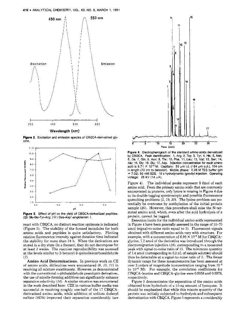
Amino Acid Determinations. In previous work on CE of amino acids, difficulties were encountered (8, 10, 11) in resolving all mixture constituents. However, as demonstrated with the conventional o-phthalaldehyde precolumn derivatives, the use of micelle-forming additives can significantly enhance separation selectivity (14). A similar situation was encountered in the work described here: CZE in various buffer media was successful in resolving roughly one-half of the 17 CBQCA- derivatized amino acids, while addition of sodium dodecyl sulfate (SDS) improved their separation considerably (see
1
11
I 0 5 10 15 20 25 30
Time (min)
Flgure 4. Electropherogram of the standard amino acids derivatized by CBQCA. Peak identification: 1, Arg; 2, Trp; 3, Tyr; 4, His; 5, Met; 6, Ile; 7, Gln; 8, Asn; 9, Thr; 10, Phe, 1 1 , Leu; 12, Val; 13, Ser; 14, Ala; 15, Gly; 16, Glu; 17, Asp. Injection concentration for each amino acid is 8.71 X lo-' M. Capillary: 50 pm i.d. (184 pm o.d.), 104 cm in length (73 cm to detector). Mobile phase: 0.05 M TES buffer (pH = 7.02), 50 mM SDS. 10 s hydrodynamic (gravity) injection. Operating voltage: 25 kV (14 FA).
Figure 4). The individual peaks represent 9 fmol of each amino acid. From the primary amino acids that are commonly encountered in proteins, only lysine is missing in Figure 4 due to its double-tagging spectroscopic and possible fluorescence quenching problems (5 , 19,20). The lysine problem can po- tentially be overcome by methylation of the initial protein sample (20). However, this procedure shall miss the N-ter- minal amino acid, which, even after the acid hydrolysis of a protein, cannot be tagged.
Detection limits for the individual amino acids represented in Figure 4 have been generally assessed in the range of 10-70 amol (signal-to-noise ratio equal to 3). Fluorescent signals obtained with different amino acids vary with structure. For example, with a concentration of 6.95 X M for CBQCA- glycine, 7.2 amol of the derivative was introduced through the electromigration injection ( I t ? ) , corresponding to a measured peak with signal-to-noise ratio of 15. The minimum quantity of 1.4 amol (corresponding to 0.2 nL of sample solutien) should thus be detectable a t a signal-to-noise ratio of 3. The linear dynamic range for these measurements has been assessed a t over 3 orders of magnitude (concentration ranging from to lo4 M). For example, the correlation coefficients for CBQCA-leucine and CBQCA-glycine were 0.9958 and 0.9978, respectively.
Figure 5 demonstrates the separation of the amino acids obtained from hydrolysis of a 15-ng amount of lysozyme. I t should be emphasized that while this minute quantity of the protein was initially subjected to hydrolysis and subsequent derivatization with CBQCA, Figure 5 represents a considerably
ANALYTICAL CHEMISTRY, VOL. 63. NO. 5, MARCH 1, 1991 411
3
4
0 5 10 15 20 25 30 Time (min)
Figure 5. Electropherogram of the amino acids representing 1.9 pg (134 fmoi) of the hydrolyzed lysozyme. Concentration of hydrolysate: 9.37 X lo-‘ pg/pL. Capillary: 50 pm i.d. X 97 cm (67-cm effective length). Electromigration injection: 5 kV, 9 s. Peak numbering and experimental conditions same as in Figure 4.
smaller aliquot of the analyzed solution (concentration of 9.4 X pcg/pL). By use of the electrophoretic injection method (181, the actual introduced amount of lysozyme is 1.9 pg, or 134 amol. In this particular application, some band dispersion was observed. According to our experience, such peak broadening and change in migration time sometimes occur with repeated use of capillaries for “real” samples. Washing capillaries with a potassium hydroxide solution usually rectifies the problem.
Separation of CBQCA-Derivatized Peptides. Initially, standard solutions of small (three to four residue) peptides and somewhat larger angiotensin derivatives were tagged with CBQCA and separated. In a pH = 9.50 borate buffer, the negatively charged peptide derivatives migrated in the system according to their predicted (21) mass-to-charge ratios, al- though the presence of electroosmotic flow (22) modified their migration rates somewhat. An electropherogram of 10 model peptides, separated in 15 min, is shown in Figure 6; since none of these standards contain lysine in their molecules, single peaks are observed. Although CBQCA was also found to react with proteins, formation of multiple peaks diminishes the value of precolumn derivatization.
Detection limits for the model peptides, Val-Ala-Ala-Phe and Gly-Gly-Tyr-Arg, were assessed at 4.6 and 13.8 amol, respectively, which is roughly in agreement with the amino acid results. For a more precise determination of the sample amount injected, the electromigration method was employed in this case. Plotting peak heights against concentration of peptide derivatives ( 10-8-10-5 M), the linear dynamic range was found to be at least 4 orders of magnitude; for example, the correlation coefficients for Gly-Leu-Tyr and Gly-Gly- Tyr-Arg were 0.9956 and 0.9984, respectively. We should point out that, during the peptide separations and sensitivity studies, 20 mM tu-cyclodextrin was used as a buffer additive. This addition resulted in (a) a several-fold increase of detection sensitivity and (b) narrower peptide peaks.
Following the encouraging results on the standard peptide mixtures, a preliminary exploration of CBQCA as a reagent for high-sensitivity peptide mapping was carried out. Figure 7 demonstrates a complex electropherogram obtained from a tryptic digest of a small amount of P-casein (17), with the final concentration of 0.33 pg/pL after derivatization. This recording represents 17 fmol (396 pg) of the digested protein.
1
L
2
1. J 0 5 10 15
Time (min)
Figure 6. Electropherogram of standard peptides as CBQCA deriva- tives. Capillary: 50 pm i.d. (184 pm o.d.), 90 cm in length (60 cm to detector). Mobile phase: 0.05 M borate buffer (pH = 9.50), 20 mM a-cyclodextrin. Peak identification: 1, Ile7-angiotensin I1 I; 2, Gly- Gly-Tyr-Arg; 3, Val5-angiotensin I I; 4, Gly-Leu-Tyr; 5, Met-Leu-Tyr; 6, VaCAla-Ala-!; 7, Vaffily-Ser-Glu; 8, GCGIy-Phe; 9, Glu-VaChe; 10, Val-Gly-Asp-Glu. 20-s hydrodynamic (gravity) injection. Operating voltage: 20 kV (7 FA).
0 10 20
Time (mid
Figure 7. Electropherogram of peptides representing 17 fmol(392 pg) of trypticdigested p-casein sample. Sample concentration for injection is 0.33 pgIpL. Electromigration injection: 3 kV, 5 s. Other conditions the same as in Figure 6.
Yet, the pattern was found highly reproducible in run-to-run and sample-to-sample comparisons.
DISCUSSION Design of appropriate fluorogenic agents for improved
detection of biological compounds has been an active research area for some time. The isoindole derivatives, formed from the amino acids and peptides with nonfluorescent reagents, are among the most fluorescent compounds known. The CBQCA reagent synthesized here forms such compounds, uniquely combining the structural properties needed in a most powerful combination of forefront bioanalytical techniques, that of capillary electrophoresis and laser-induced fluorometry.
412 ANALYTICAL CHEMISTRY, VOL. 63, NO. 5, MARCH 1, 1991
In the work reported here, very high sensitivities have been demonstrated for amino acids and peptides, although it is likely that applications to additional biologically active sub- stances (such as amino sugars, certain polar lipids, various drugs, or catechol amines) can further be developed.
Compared to the previously used reagents, the reactivity of CBQCA with peptides is viewed as particularly important. A need for highly sensitive procedures of peptide mapping has been felt, particularly in the area of protein characteri- zation a t trace levels (23), as well as in pursuing analyses a t the level of single biological cells (24, 25) . The preliminary result with the tryptic digest of @-casein (Figure 7 ) indicates that, together with the capability of low-level digestion ( I 7), characteristic “protein fingerprints” can be recorded from extremely small samples of proteins. Such an attractive ca- pability is currently somewhat limited by the appearance of multiple peaks due to tagging of lysine residues in some peptides. Work is currently in progress to overcome this difficulty.
Pending instrumental improvements a t the level of laser detection, further sensitivity gains are expected, since the instrumentation used here is considerably less sophisticated than the designs reported by other laboratories (8, 9, II) . Additional developments in the area of preconcentration techniques in CE can further improve small-sample capa- bilities. This is evident in considering Figures 4 and 5 (a run of standard amino acids and a protein hydrolysate, respec- tively) and the ways to prepare the respective samples. While 15-ng lysozyme, total, was the smallest amount that we could analyze without a serious volumetric overloading of the CE system, in our recent work with microcolumn LC/laser fluorescence detection (7), analyses at 1-ng levels appeared feasible because the entire sample could be utilized for chromatographic analysis.
With respect to the detection limits measured for the amino acids versus peptide derivatives, it must be pointed out that the peptide runs permitted more favorable detection condi- tions (conventional CE buffers and the use of a signal-en- hancing cyclodextrin) than the amino acids. Naturally, sen- sitivity normally drops for larger peptides due to fewer amino groups on a per-mole basis. The results obtained with amino acids represent a compromise between the separation and detection conditions: the presence of SDS micelles, while beneficial to the separation selectivity, increased the detection limits by approximately 1 order of magnitude, presumably due to light-scattering phenomena, as compared to “straight” buffers. Under the circumstances of this work, no such lim- itations were encountered with peptides. Overcoming this
“SDS problem” for the amino acids can yield even greater sensitivities in future work.
As an important extension of this work, CBQCA has re- cently been employed in derivatization of amino sugars (26) and the neutral, reducing oligosaccharides modified by re- ductive amination (27).
443-453. (17) Cobb, K. A.; Novotny, M. Anal. Chem. 1989, 67, 2226-2231. (18) Rose, D. J.; Jorgenson, J. W. Anal. Chem. 1988, 60, 642-648. (19) Matuszewski, B. K.; Givens, R. S.; Srinivasachar, K.; Carlson. R. G.;
Higuchi. T. Anal. Chem. 1987, 59, 1102-1 105. (20) Oates, M. D.; Jorgenson, J. W. Anal. Chem. 1990, 62 , 2056-2058. (21) Liu, J.; Cobb, K. A.; Novotny, M. J . Chromatogr. 1990, 519, 189-197. (22) Jorgenson, J. W.; Lukacs, K. D. Anal. Chem. 1981, 53, 1298-1302. (23) Novotny, M . J . Microcolumn Sep. 1990, 2 , 7-20. (24) Wallingford, R. A.; Ewing, A. G. Anal. Chem. 1988, 60, 1972-1975. (25) Kennedy, R. T.; Oates, M. D.; Bruce, R.; Nickerson, B.; Jorgenson, J.
W. Science 1989, 246. 57-63. (26) Liu, J.; Shirota, 0.; Novotny, M. Anal. Chem. 1990, following paper in
this issue. (27) Liu, J.; Shirota, 0.; Wiesler. D.; Novotny, M. Proc. Natl. Acad. Sci.
U.S.A ., in press.
RECEIVED for review June 28,1990. Accepted November 28, 1990. This research was supported by Grant No. GM 24349 from the National Institute of General Medical Sciences, U.S. Department of Human Health and Services.