Alma Mater Studiorum – Università di Bologna Dottorato Di Ricerca in Morfofisiologia veterinaria e applicazioni biotecnologiche Ciclo XXII SSD: VET 02 Detection and localization of GLUTs in spermatozoa from different domestic species Presentata da Diego Bucci Coordinatore Dottorato Docente Guida Prof. Eraldo Seren Prof. Carlo Tamanini Correlatore Dr.ssa Marcella Spinaci Esame finale anno 2010
Transcript
Alma Mater Studiorum – Università di Bologna
Dottorato Di Ricerca in
Morfofisiologia veterinaria e applicazioni biotecnologiche
Ciclo XXII
SSD: VET 02
Detection and localization of GLUTs in spermatozoa from different domestic species
Presentata da Diego Bucci
Coordinatore Dottorato Docente Guida
Prof. Eraldo Seren Prof. Carlo Tamanini Correlatore Dr.ssa Marcella Spinaci
Studying sperm: when it began .................................................................................. 5 Sperm cells ................................................................................................................. 6 Sperm Morphology ..................................................................................................... 7 Sperm maturation ....................................................................................................... 9 Capacitation .............................................................................................................. 13 Acrosome reaction .................................................................................................... 18 Sperm metabolism .................................................................................................... 21 Hexose transporters ................................................................................................. 31 GLUT in sperm cells ................................................................................................. 42
AIMS OF THE STUDY ............................................................................................. 47 MATERIALS AND METHODS ................................................................................. 48
Semen collection and preparation ............................................................................ 48 Sperm evaluation ...................................................................................................... 49 Induction of in vitro capacitation and acrosome reaction (AR) in boar, stallion and dog semen ....................................................................................................................... 50 IGF and Insulin-stimulated capacitation in boar semen ............................................ 51 Capacitation assessment ......................................................................................... 51 Acrosome reaction .................................................................................................... 52 Immunocytochemistry ............................................................................................... 52 Western blot analysis ............................................................................................... 53 Flow sorting .............................................................................................................. 54 Experimental design ................................................................................................. 54
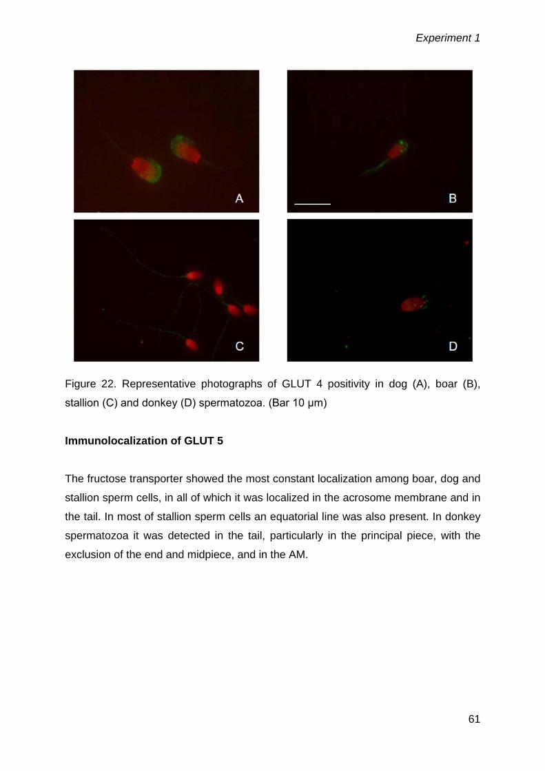
Detection of GLUTs 1, 2, 3, 4 and 5 by western blotting and their immunolocalization in boar, dog, stallion and donkey sperm cells ........................................................... 55
Evaluation of GLUTs re-localization after capacitation and acrosome reaction in boar, stallion and dog spermatozoa ................................................................................... 66
Sex sorting procedure and stimulation with either IGF or insulin does not affect GLUT localization in boar spermatozoa .............................................................................. 71
GENERAL DISCUSSION ......................................................................................... 74 CONCLUSIONS ....................................................................................................... 78 BIBLIOGRAPHY ...................................................................................................... 79
Abstract
3
Abstract
Sperm cells need hexoses as a substrate for their function, for both the maintenance
of membrane homeostasis and the movement of the tail. These cells have a peculiar
metabolism that has not yet been fully understood, but it is clear that they obtain
energy from hexoses through glycolisis and/or oxidative phosphorylation.
Spermatozoa are in contact with different external environments, beginning from the
testicular and epididymal fluid, passing to the seminal plasma and finally to the
female genital tract fluids; in addition, with the spread of reproductive
biotechnologies, sperm cells are diluted and stored in various media, containing
different energetic substrates. To utilize these energetic sources, sperm cells, as
other eukaryotic cells, have a well-constructed protein system, that is mainly
represented by the GLUT family proteins. These transporters have a membrane-
spanning α-helix structure and work as an enzymatic pump that permit a fast gradient
dependent passage of sugar molecules through the lipidic bilayer of sperm
membrane.
Many GLUTs have been studied in man, bull and rat spermatozoa; the presence of
some GLUTs has been also demonstrated in boar and dog spermatozoa.
The aims of the present study were
- to determine the presence of GLUTs 1, 2, 3, 4 and 5 in boar, horse, dog and
donkey spermatozoa and to describe their localization;
- to study eventual changes in GLUTs location after capacitation and acrosome
reaction in boar, stallion and dog spermatozoa;
- to determine possible changes in GLUTs localization after capacitation
induced by insulin and IGF stimulation in boar spermatozoa;
- to evaluate changes in GLUTs localization after flow-cytometric sex sorting in
boar sperm cells.
GLUTs 1, 2, 3 and 5 presence and localization have been demonstrated in boar,
stallion, dog and donkey spermatozoa by western blotting and immunofluorescence
analysis; a relocation in GLUTs after capacitation has been observed only in dog
sperm cells, while no changes have been observed in the other species examined.
As for boar, the stimulation of the capacitation with insulin and IGF didn’t cause any
change in GLUTs localization, as well as for the flow cytometric sorting procedure.
Abstract
4
In conclusion, this study confirms the presence of GLUTs 1, 2 ,3 and 5 in boar, dog,
stallion and donkey spermatozoa, while GLUT 4 seems to be absent, as a
confirmation of other studies. Only in dog sperm cells capacitating conditions induce
a change in GLUTs distribution, even if the physiological role of these changes
should be deepened.
Introduction
5
Introduction
Studying sperm: when it began
Sperm cell study, nowadays better known as spermatology, finds its roots in the first
observations, carried out by Antonius Leeuwenhoek, in 1677. This scientist, with the
help of the microscope, discovered “little animals” swimming in seminal fluid. The
fascination of the movement of these little animals has driven the science of sperm
study to develop and better understand the metabolic strategy undergoing sperm
function, as well as to study structural components of the cell from its inner part
(nucleus) to the outer one (cell membrane).
The studies in XIX and first part of XX century concentrated on fertilization and sperm
motility in invertebrates, as fertilization in most of these species is external and
spermatozoa could be observed easily. In 1919, in his famous book “Problems of
fertilization”, Lilie reported that spermatozoa are provided with all the energy sources
from their gonadal development, and that they are completely unable to find some
nutrients to support motility from the external environment. To that time, this was a
reasonable assessment, even if the subsequent studies focused on sperm cell
physiology have deepened these themes and contributed to form a more scientific
approach.
The first studies on sperm respiration were carried out in 1933 by Redenz and they
were subsequently followed, in 1941, by those by Lardy and Philips on bull
spermatozoa. They demonstrated that bull sperm cells can survive without seminal
plasma but only in presence of sugars, and that the main energy source is the
anaerobic glycolisis. In the same years, at the University of Pennsylvania, in the
bacteriology unit, Zittle and co-workers (1942) performed the first studies on flagellar
movements of spermatozoa, that were at that time still thought to be bacterial-like
organisms. They demonstrated a cytochrome oxidase activity in epididymal bull
spermatozoa, as well as an active glucose metabolism and oxygen consumption, but
their studies didn’t meet the interest of the scientific community. In 1945 Lardy and
coworkers deepened the studies on bull sperm metabolism, demonstrating an active
Krebs cycle metabolism in these cells, increased by bicarbonate and reduced by
cianure. Also ram sperm studies confirmed the presence of a functioning oxidative
Introduction
6
phosphorylation metabolism, accompanied by the anaerobic metabolism (Lardy,
1945b).
McLeod (1941-1943) performed, in the same period, the first studies on human
sperm metabolism, demonstrating their ability to use and metabolize sugars, even if
he couldn’t directly discriminate the activity of cytochrome oxidase, due to the low
density of the samples, if compared with bull, ram and boar ones. Mann (1946)
demonstrated that the main energy source for human spermatozoa is represented by
fructose and later studies (Albers and co-workers, 1961) showed that the various
mammalian spermatozoa present very different glucolitic/fructolitic rates.
Since that moment sperm cells have been considered as a morphological, structural
and functional unit and no distinctions were made on the basis of the various
compartments. Fawcett in 1957 initiated the morphological studies with the electron
microscopy and gave important inputs to the knowledge of the tail structures; in 1965
Mhori and coworkers set up a good method to isolate pure mid-pieces, that lasted in
studies focused on oxidative mitochondrial metabolism.
There are a lot of studies carried out later on sperm metabolism, which lead to a
better knowledge of this subject, and could be found in the interesting review by Ford
(2006) and Storey (2008).
When talking about sperm cell function, as movement and fertilization, we cannot
forget the work by Yanagimachi, who focused on fertilization in mammals, after he
began to work with two scientists who had previously discovered in vivo sperm
capacitation (Chang and Noyes).
His work led him to describe in vitro capacitation process, as well as to define
hyperactivated motility, with all that goes with it such as Calcium influxes, protein
phosphorylation, different energy metabolism and subsequent acrosome reaction.
The development of knowledge and application of in vitro capacitation and
fertilization was growing in the subsequent years, permitting a spread of the use of
reproductive biotechnology in human and veterinary medicine, as well as the
development of new research branches as germ cell research and cloning (for a
review see Yanagimachi, 2009).
Sperm cells
Introduction
7
Spermatozoa are the male gametes and they are highly specialized: in fact they have
the aim to transport the male genetic material through the female genital tract to the
other gamete, the oocyte. To do this, they undergo a maturation that starts in the
testis and ends in the female genital tract, where they reach the ability to fertilize the
oocyte.
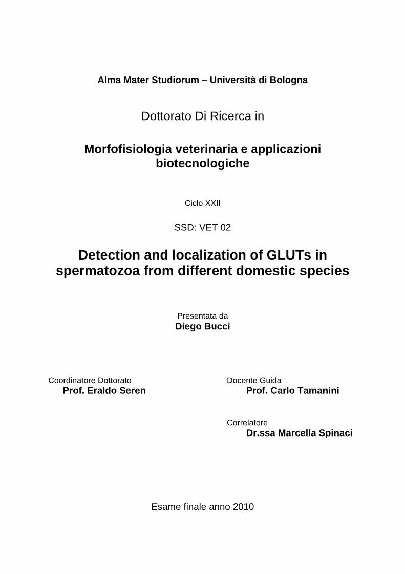
Sperm Morphology
Sperm cell anatomy is peculiar for every mammalian species and is different from all
the other cells of the animal body; there are anyway some common characteristics in
all mammalian species. The spermatozoon is constituted by a head, a connecting
piece, also called neck, and the tail, which is divided into three more parts: the
midpiece, where we find the mitochondrial sheath, the principal piece, where there
are the motility structures and the end piece. In the sperm head we can also
distinguish two different anatomical parts: the nucleus and the acrosome (Fig. 1).
Sperm cell nucleus is constituted by the haploid genomic male material and peculiar
proteins binding DNA, which are histones and protamines. Histones are the typical
chromatin-binding proteins of mammalian cells, but in spermatozoa they are replaced
in a large way by protamines, that render sperm nucleus structure more stable and
less active (Braun, 2001). The acrosome is an exocytotic vesicle deriving from Golgi
apparatus and containing some important enzymes such as hyaluronidase and
acrosine, which are hydrolytic enzymes fundamental to achieve zona pellucida
penetration. This vesicle is rounded by a peculiar membrane, the acrosomal
membrane, which is distinguished into the inner acrosomal membrane, that is nearer
to the nucleus, and the outer acrosomal membrane, or external, which is just below
the plasma membrane.
Introduction
8
Fig. 1 The spermatozoon head (from Pathways to Pregnancy and Parturition- Senger P. L.,
2003).
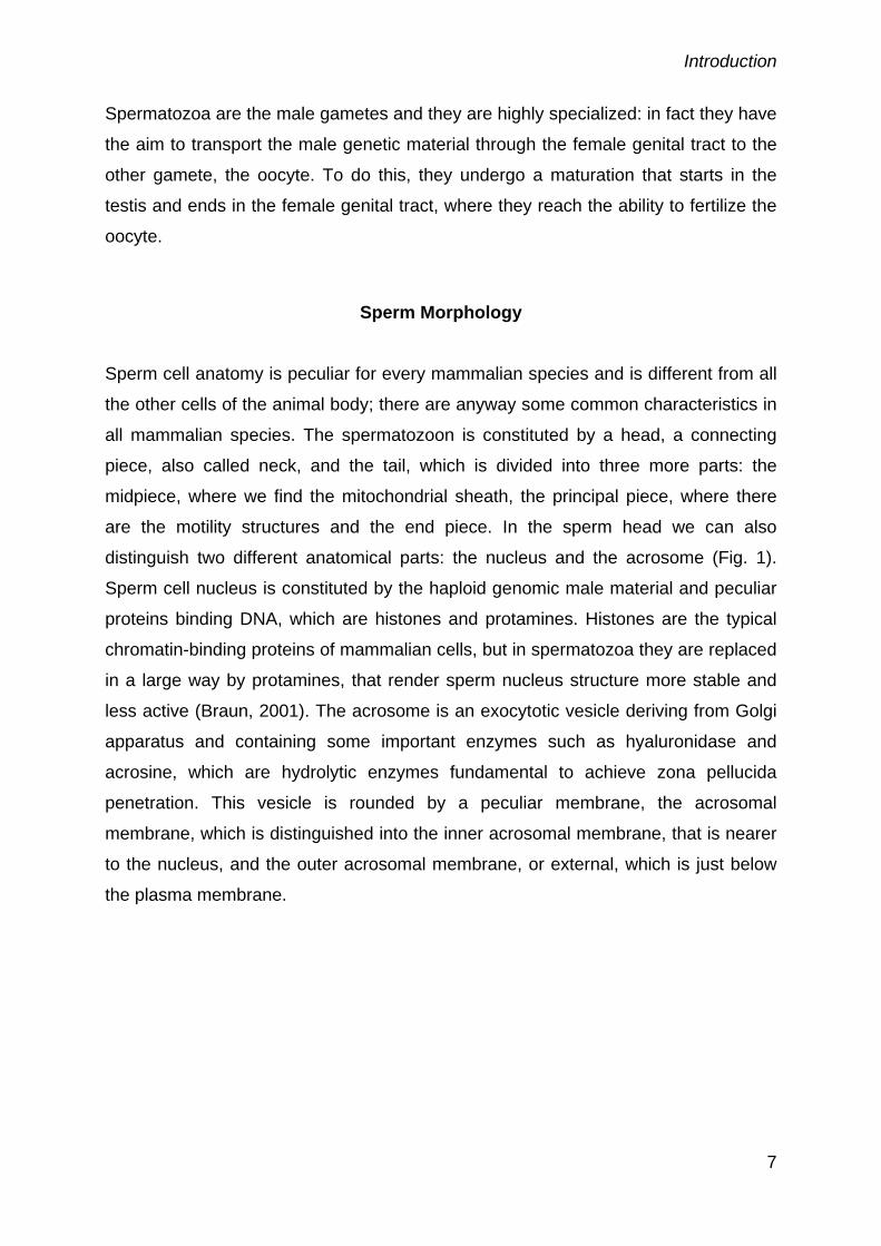
Spermatozoa tail (Fig. 2) is the main movement organ of the cell. In the neck we find
the centrioluses deriving from the paternal germinal cells, which have the function of
connecting the head with the other parts of the tail. The connection with the head is
achieved by a dense fibrous material that binds directly to the nucleus envelope and
condensed cytoplasmic material. On the other side, the segment structure changes
and the first structures dedicated to movement appears. In fact, the axoneme begins
in this point of the tail and goes through the whole length of this structure. The
axoneme is composed of 2 α and β tubuline microtubules surrounded by 9 couples of
microtubules; in the midpiece the axoneme is surrounded by 9 outer dense fibers,
and in the outer part we find the mitochondrial sheath (Eddy et al., 2003). Here about
100 mitochondria are localized (Hallap et al., 2005) and they are the unique source of
aerobic energy in the sperm cell through the Krebs cycle (Peña et al., 2009).
Introduction
9
Fig. 2 The spermatozoon tail (from Pathways to Pregnancy and Parturition- Senger P. L.,
2003).
Following the midpiece we find the principal piece, and the two structures are
separated by the annular ring, also known as Jensen ring. The main part of principal
piece is composed of the continuation of the axoneme, rounded by 7 outer dense
fibers and by a specialized cytoskeletal structure, known as fibrous sheath (Eddy et
al., 2003; Kim. et al., 2007), whose function has been identified as a protection for
the axoneme, as a scaffold for enzymes involved in signal transduction and as an
anchoring structure for glycolitic enzymes (Eddy et al., 2003; Kim et al., 2007). The
end of the tail, named endpiece, presents only the 9 + 2 microtubules couples that
are directly covered by the plasma membrane.
Sperm maturation
Sperm maturation is the process which spermatozoa undergo to reach their final
structure and functionality (França et al., 2005). The maturation itself takes place in
two distinct parts of the testicle, seminiferous epithelium and epididymis.
Introduction
10
In the testis we find spermatogonia, the primordial cells that undergo cellular division
to create a reserve of male gametes. Two subtypes of spermatogonia have been
individuated: spermatogonia A and B, even if some intermediate forms have been
discovered in mouse and pig (França et al., 2005)(Fig. 3).
Fig. 3 Typical sequence of spermatogenesis in mammals (from Pathways to Pregnancy and
Parturition-Senger P. L., 2003).
Spermatogonia A undergo several mitotic division, forming some intermediate cell
lines (Fig. 3)
After spermatogonia B undergo a mitotic division, they form the primary
spermatocytes, that undergo the first meiotic division forming the secondary
spermatocytes. These ones too undergo a meiotic division, the second, that hesitates
in the formation of the spermatid (Kretse and Kerr, 2008). This kind of cells still have
a round form, but their genetic material is haploid. In this first phase of
spermatogenesis the most important aim is to divide the genetic material of the cells,
while the rearrangement of this material and the expulsion of the cytoplasm take
place in a second phase called spermiogenesis.
During this process, 3 main things happen: the formation of the acrosomal vesicle
from Golgi apparatus, the rearrangement of the nuclear content and the formation of
the tail that also consists in the expulsion of the cytoplasm (Kretse and Kerr, 2008).
The acrosome formation consists in the transition of a part of Golgi apparatus to a
pole of the spermatid, with a contextual migration of some other intracytoplasmic
vacuoles containing hydrolytic enzymes. In the same time mitochondria migrate to
the neo-formed axoneme, still rounded by a quantity of cytoplasm, while in the
Introduction
11
principal piece the fibrous sheath surrounds the axoneme and changes its structure
in a most dense one. Finally, the nucleus undergoes very deep changes in its
structure, as there is a substitution of the main chromatin-binding protein, histones,
with another protein that will be the most important one in sperm cells, protamine.
These proteins make the genomic material more condensate and resistant to DNAse
action, and the process ends in a reorganization of the nuclear material that renders
the nuclear shape and volume peculiar for each species, as protamines are species
specific and each species presents a different grade of nuclear condensation (Kretse
and Kerr, 2008).
Maturation process is regulated and controlled by hypothalamus-pituitary axis and its
secretion of gonadotropins as well as testosterone-estrogens by Leydig or Sertoli
cells.
At this point of their lives, spermatozoa reach the end of the seminiferous epithelium
and are transferred into the lumen to reach the epididymis.
The epididymis is a long tubule, constituted of three parts, head, body and tail, and in
each one of these sperm cells continue their maturation, especially for what concerns
the expulsion of cytoplasm, nuclear condensation, acrosome organization and
membrane surface re-arrangement (França et al., 2005).
Nuclear DNA undergoes a more deep condensation, achieved by the formation of
disulphide bonds, as well as it happens in the fibrous sheath (Yanagimachi, 2008).
Sperm cells transport along epididymis is achieved by smooth muscles of the tubule
wall, that permit the fluid and cells to proceed. This transport is very active in the first
two parts of the epididymis, while in the tail spermatozoa are stored and
concentrated (França et al., 2005).
During this travel the spermatozoon loses the last part of its cytoplasm: in fact, when
sperm cells arrive to the epididymis head they still have a cytoplasmic droplet near
the connecting piece (proximal cytoplasmic droplet); passing through the epididymis
the droplet is moved to the end of the midpiece (distal droplet) and at the end it is
expulsed. The presence of a cytoplasmic droplet is a typical feature of epididymal
sperm cells and could be considered as an immaturity characteristic of ejaculated
spermatozoa (Harrison and Gadella, 2005).
The epididymal fluid plays an important role not only in sperm transport, but also as a
fundamental component in sperm maturation and survival: in fact it contains some
important factors, secreted by the epithelial cells, that adhere to the sperm surface
Introduction
12
avoiding a premature capacitation and/or acrosome reaction (Vadnais et al., 2007).
In particular these factors are proteins such as the 25 kDa protein, anti-agglutinin,
secreted by the porcine epididymal duct of the corpus epididymis that subsequently
binds to the acrosome of spermatozoon (Harayama et al. 1999). Another example is
Crisp-1, a glycoprotein secreted by rat epididymal tail where it associates with the
spermatozoon membrane (Xu et al. 1997); it is present also in man (Dacheux et al.
2006).
The secretion and adherence of protein goes on in the seminal plasma too.
Depending on the species, the spermatozoon acquires another very important
feature in different parts of the epididymis: motility. In fact, testicular spermatozoa are
normally less or no motile, while spermatozoa collected from the epididymal tail are
always motile (Yanagimachi, 2008).
Introduction
13
Capacitation
After the maturation process a spermatozoon is still unable to fertilize an oocyte,
because it has to undergo a series of processes known under the name of
“capacitation”.
Capacitation has been discovered in 1951 by two different researchers, Chang and
Austin, but the major studies began in the ‘60s and are due to Yanagimachi (2009).
Ejaculated spermatozoa have to go through the whole female genital tract (FGT) to
encounter the oocyte and during this passage they get capacitated to fertilize the egg
(Rodriguez-Martinez, 2007).
Spermatozoa transport through the FGT is predominantly a passive process,
accomplished by uterus movements and contractions (Brüssow et al., 2008), during
which sperm cells lose the “uncapacitating coat”, until they reach the sperm reservoir,
a particular structure near the utero-tubal junction (Rodriguez-Martinez, 2007;
Brüssow et al., 2008). In this part of the uterus spermatozoa are kept protected from
phagocytosis and the capacitation process is stopped, as soon as they are released
and reach the tubal environment, that seems to play an important role in “in vivo”
capacitation (Rodriguez-Martinez, 2007). In particular the tubal fluid composition
seem to be central in promoting sperm capacitation, especially for the basic pH it
reaches due to bicarbonate ion secretion.
The modifications the sperm cell undergoes during capacitation involve membrane
changes in fluidity and composition, activation of tyrosine phosphorylation, calcium
flux augmentation, activation of the hyperactivated motility pattern, changes in the
metabolic state of the cell.
It is noteworthy that the removal of all the non-capacitating (above mentioned) agents
the spermatozoon is enriched during its maturation in epididymis, is concretely an
important membrane change that occurs and starts capacitation.
As for membrane changes, it has been suggested that cholesterol could play a very
central role: in facts one of the molecules of which sperm plasma membrane is
enriched during its passage into the epididymal lumen is cholesterol, and it is one of
the molecules that are removed from its surface under the action of albumins and in
particular BSA “in vitro” (Flesh and Gadella, 2000; Gadella, 2008). In this way we can
identify cholesterol as a stabilizing factor of the membrane that, being removed,
permits the lipidic bilayer to flow more freely as well as some external proteins to
Introduction
14
move through the membrane (Yanagimachi, 2008). It is furthermore reported that
capacitation, in species in which the cholesterol content of the plasma membrane is
higher, occurs later than other species (Flesh and Gadella, 2000). The cholesterol
efflux seems however to be a consequence of the activation of protein kinases (in
particular PKA, via bicarbonate adenilate cyclase activation), and not to be the cause
of this process (Harrison and Gadella, 2005).
The membrane structure of sperm cells is organized in microdomain of
phospholipids, normally kept in a static and regulated manner (Harrison and Gadella,
2005); during capacitation, and under the action of bicarbonate, these microdomains
are “scrambled” by a series of enzymes (regulated by phospho/dephosphorylation)
called scramblases. This process seems to be of great importance in permitting
cholesterol efflux from the plasma membrane, as well as in rendering it “fusogenic”,
less rigid and ready to undergo the acrosome reaction (Harrison and Gadella, 2005).
HCO3- ion is fundamental in capacitation, in particular for what concerns cell
activation: in fact, it is stated that in the epididymis tail the concentration of
bicarbonate ion is lower than in normal serum (Flesh and Gadella, 2000; Harrison,
2004), but when spermatozoa are ejaculated in the FGT, they encounter higher
concentration of both CO2 and bicarbonate. The action of this ion is to initiate the
phosphorylation of some proteins (more of which still unknown) by augmenting the
activity of soluble adenilate cyclase, with an increase in cAMP and consequent
activation of Protein Kinases (Fig. 4) (Flesh and Gadella, 2000; Harrison and
Gadella, 2005).
Introduction
15
Fig. 4. A representation of capacitation phenomena (Flesh and Gadella, 2000). The role of
BSA and HCO3- are stressed in this picture in activating internal mechanisms.
The sperm plasma membrane contains a variety of Ca++ channels: voltage
dependent, Ca++-ATPase, Na/ Ca++ exchanger and probably others (Flesh and
Gadella, 2000). Capacitation, as well as acrosome reaction, is dependent on the
influx of calcium into the cell, even if it is not yet well established how this calcium
passes through the different regions of the cell. It is well known that calcium influx
induces an activation of adenilate cyclase, with a subsequent activation of protein
kinase A via cAMP, that reinforces the process of capacitation (Flesh and Gadella,
2000; Visconti, 2009). In addition, calcium is very important in membrane
hyperpolarization and inner cell pH augmentation during capacitation (Vadnais et al.,
2007).
Hyperactivated motility is another typical feature of capacitated sperm cells and
consists in a more rapid and also non-linear movement of the cell. This kind of
movement has been hypothesized and demonstrated to permit spermatozoa to
penetrate the mucus barrier they encounter in the tubal lumen, as well as in
trespassing cumulus oophorus cells (Suarez and Ho, 2003).
To better understand the importance and relevance of hyperactivated motility, it is
helpful to remember that sperm cells acquire the capacity of moving only after their
Introduction
16
testicular maturation, when they rich the epididymis; this kind of motility is the so
called activated motility (Yanagimachi, 2008) and its principal characteristic is to be
almost linear or at least to proceed in a straight trajectory. Hyperactivated sperm cells
swim in a very different manner, as tails beat in a most asymmetrical manner,
forming larger curves (Suarez and Ho, 2003) and resulting in a more disordered
trajectory.
Hyperactivated motility is regulated by many factors, the most important of which is
Ca++ availability. As stated above, Ca++ can enter the cell via the numerous Ca++
channels present in the membrane of the spermatozoon (Flesh and Gadella, 2000).
It is also stated that the activity of these channels can’t provide the whole amount of
Ca++ needed for the activation of the cell, so the intracellular reserve of Ca++
becomes of primary importance. Ca++ storage entering in the activation of
hyperactivated motility is that found in the neck of the sperm cell, and in particular in
the redundant nuclear envelope where the centrioluses anchor (Suarez and Ho,
2003; Suarez, 2008). Influx of Ca++ induces an activation of the adenilate cyclase
and a subsequent phosphorylation in tyrosine and serine residues of the tail proteins
(Suarez, 2008).
In “in vivo” trials it was tried to explain the possible role of some molecules (such as
progesterone) as well as chemotactic stimuli in controlling hyperactivated motility,
and relating this event to capacitation (for a review see Suarez, 2008), but the
precise mechanisms are not yet fully understood.
As mentioned above, an important event occurring during capacitation is protein
phosphorylation, and particularly protein tyrosine phosphorylation. This process is
finely controlled: the activation of the sperm specific Adenilate Cyclase (AC) via
various stimuli (Flesh and Gadella, 2000; Urner and Sakkas, 2003; Harrison and
Gadella, 2005; Suarez, 2008) induces an accumulation of cAMP that initiates the
activity of some protein kinases, in particular PKA, that initiates the threonine, serine,
tyrosine phosphorylation of some proteins. PKA activation drives to activate PKC,
that reinforces calcium entrance flux. At the same time some parallel pathways are
activated, such as the AKAP and ERK ones (see Fig. 5).
Introduction
17
Fig. 5 Intracellular pathways in capacitation (de Lamirande and O’Flaherty, 2008). The role of
Protein kinases is described in this picture, as well as the activity of ROS.
We should not think of these events as separated or independent, because it is well
demonstrated that they are interdependent and surely it’s possible to individuate
crosstalks between these pathways (Breitbart and Naor, 1999; Breitbart et al., 2006).
During capacitation there is also a translation of PLC (phospholipase C) to the
plasma membrane, via the activation of some substrates of PKA, that is an important
passage for the initiation of the subsequent acrosome reaction (Flesh and Gadella,
2000).
Another aspect that is fundamental in reaching the capacitated status is production of
reactive oxygen species (ROS): the more representative agents are H2O2, NO-, OH- .
These molecules are usually retained negative in cell survivor and function, as they
affect membrane integrity and nuclear function. If produced in small quantities they
have been discovered to be very important in capacitation (de Lamirande and
Gagnon, 1995; Dröge, 2001; de Lamirande and O’Flaherty, 2008). As stated in
Introduction
18
Figure 5, ROS enter the activation of many pathways regarding protein tyrosine
phosphorylation and contribute to the enhance of capacitation.
Acrosome reaction
Acrosome reaction is the last pre-fusion phase of the living spermatozoon. As some
sperm cells reach the ovum (a significantly lower number than that ejaculated, see
Brüssow et al., 2008), they are still unable to fertilize it, because mammalian ova are
surrounded by a glycoproteic coat, zona pellucida (Yanagimachi, 2008). To penetrate
this coat, a sperm cell must adhere it and then go through an exocytotic process,
known as acrosome reaction. The main enhancer of the acrosome reaction is zona
pellucida, even if also cumulus oophorus matrix could play some role (Yanagimachi,
2008).
It is well stated that ZP 3 (or C) and progesterone, via its non-genomic receptor
(Breitbart and Sungin, 1997; Flesh and Gadella, 2000) (Fig. 6) are the most important
inductors of acrosome reaction. Sperm surface is provided with adequate Zona
Binding Proteins, that are exposed during the capacitating process (Flesh and
Gadella, 2000; Gadella, 2008b), and activate these receptors via tyrosine
phosphorylation and aggregation. At the same time progesterone can bind to its non-
genomic receptor and the consequence of these two events are an increase of the
intracellular pH via G protein and a depolarization of plasma membrane. As a result,
Ca++ channels open and permit a massive influx of Ca++ ions that permit the
activation of the phospholipase C (PLC), already translocated to the plasma
membrane during capacitation. Activated PLC converts phosphor-inositol-di-
phosphate (PIP2) to diacilglycerol (DAG) and Inositol-3-phosphate (IP3). At the same
time, the high levels of calcium activate cAMP/PKA pathway, leading to a major
release of calcium from the acrosomal storage; this Ca++ concentration increase
leads to the activation of phospholipase A2 (PLA2) that acts in degradating
phosphocoline (PC) to lysophosphaditilcholine (LPC) and free fatty acids. All this
secondary products activate PKC (Flesh and Gadella, 2000), as well as
Ca++concentration augmentation and activation of PKA (Breitbart and Spungin, 1997;
Abou-Haila and Tulsiani, 2009; Breitbart et al., 2009). PKC migrates toward the
membrane and initiate fusion process, that will lead to the final hexocytosis.
Introduction
19
Fig. 6. Acrosome reaction mechanisms (Breitbart and Spungin, 1997; Flesh and Gadella,
2000). The importance of ZP and Ca++ is stressed in this figure, as well as the various
intracellular mechanisms leading to acrosome reaction.
In studing acrosome reaction a particular attention should be given to actin
cytoskeleton changes and especially to actin polymerization during capacitation and
its subsequent depolimerization just before acrosome reaction (Breitbart et al., 2005).
It is stated that actin is present in various mammalian species sperm head and tail,
thus suggesting that it could be involved in sperm motility as well as other functions
as capacitation and acrosome reaction (Breitbart et al., 2005). It is still unclear if it is
in the monomeric form (G actin) or in the polimerizated or filamentous form (F actin),
and the presence of the numerous actin-bound proteins (that are necessary for actin
polymerization/depolymerization) in sperm cells is a proof that the two forms are
present in spermatozoa (Breitbart et al., 2005). Some Authors described a
polymerization of actin during capacitation in various mammalian species (Castellani-
Cresa et al., 1993; Cabello-Aguero et al., 2003; Brener et al., 2003 ), and a
depolymerization of F actin right before acrosome reaction (Brener et al., 2003).
This could explain the actin role, as its polymerization is necessary to reach the
capacitated status as well as the fertilizing ability in many mammalian species
(Rogers et al., 1989; Castellani Cresa et al., 1993; Brener et al., 2003; Cohen et al.,
2004), and its breakdown is necessary to obtain an acrosome reaction (Spungin et
al, 1995; Breitbart et al., 2005) (Fig.7).
Introduction
20
Fig.7 Actin polymerization in capacitation and acrosome reaction (Breitbart et al., 2005). The
mechanism of polymerization during capacitation and depolymerization during acrosome
reaction of the actin cytoskeleton is described in this figure
.
Introduction
21
Sperm metabolism
When talking about sperm metabolism, we have to keep clear that the main goal of
sperm cells is taking male haploid DNA to the female one, and that, to do this, they
have to move across the female reproductive trait. Therefore, movement is the main
function of sperm cells and the main aim of energy obtainment. Anyway, we should
not forget that sperm cells undergo some functional changes that permit them to
acquire the fertilizing ability, such as capacitation and acrosome reaction; during this
functional moments energy requirements are not only dedicated to movements, but
also in activating internal cell functional pathways such as protein tyrosine
phosphorylation, calcium channel activation, hyperactivated motility and acrosome
reaction (Flesh and Gadella, 2000).
Sperm cells need energy for moving, as it is the main goal of their living condition.
They acquire the capacity of moving after the epididymal maturation (Yanagimachi,
2008), as in testis sperm cells are actually non-motile. Active movements are
necessary to pass through the female genital tract, even if in most of this journey the
most important “transport” of the male genomic material is performed by the female
genital tract itself, by movements of the smooth muscles of the uterus and tuba
(Rodriguez-Martinez, 2007; Brüssow et al., 2008).
Sperm cells use sugars as an energy source: they can use hexoses, such as
glucose, mannose, fructose, but they can also use some other sources, such as
lactate and citrate. The two metabolic pathways involved in sperm energy obtainment
are anaerobic glycolysis and oxidative phosphorylation. In fact, as already stated,
spermatozoa have a mitochondrial sheath in the midpiece, where the oxidative
processes may take place, and the glycolitic enzymes in the principal piece of the
tail, connected to the fibrous sheath (Eddy et al., 2003; Ford, 2006).
There are different opinions whether glycolysis or oxidative phosphorylation is the
major source of energy, in the form of ATP, for sperm cells. In particular Miki and co-
workers (2004; 2007) demonstrated the predominant role of glycolysis: in fact they
produced genetically modified mice, lacking the gene of glyceraldehyde-3-phosphate,
that is a very important enzyme in the glycolitic chain. These mice’s spermatozoa
have an ATP production 90% decreased than normal mice, and their motility is very
much decreased. These data support the hypothesis that the only way sperm cell
use to get energy from a sugar substrate is the glycolitic one, while ATP production
Introduction
22
from oxidative phosphorylation is not indispensable for motility (Miki, 2007). What’s
more, they affirm that glucose is indispensable for capacitation in mouse, while the
absence of pyruvate or lactate (that are metabolized directly in the oxidative
phosphorylation cycle) do not affect capacitation. As a last proof of the prominence of
glycolysis, they elaborated some trials with inhibitors of the phosphorylation chain,
not affecting the movement of the cells (Miki, 2007).
On the other hand Ruiz-Pesini and coworkers (2007) retain that oxidative
phosphorylation is central in sperm motility and sperm function: they report studies in
which it is stated that mitochondrial function and sperm movement are associated, or
that the use of mitochondrial activity enhancers results in a more relevant activation
of sperm motility and fertilizing rate in human. They also report other studies
demonstrating the increase of oxygen consumption related to an augmented motility
pattern, as well as many studies reporting that inhibitors of phosphorylation chain
hesitate in a impaired motility and fertilizing rate. Finally, when taking into account
Miki and coworkers’ results (2004), they state that glyceraldehydes-3-phosphate
dehydrogenase (G3PD) knockout mice, used as a model to demonstrate that
glycolysis is the main energy-producing pathway, are not the best to use or almost
the interpretation of the results is not correct. In fact they affirm that the lack of G3PD
makes the glycolitic process (that can only form 2 molecules of ATP, pyruvate and
NADH for each glucose molecule) an energy dispersive process, and not an energy
productive. In fact the positive balance of the entire glycolitic chain is achieved after
the G3PD step, where 4 ATP molecules are produced, while prior to this passage
ATP is used to phosphorylate glucose and fructose. From this point of view Ruiz-
Pesini and coworkers (2007) affirm that ATP is produced in oxidative
phosphorylation process and that it is used by sperm cells to metabolize glucose and
to move. This fact could also explain the other results by Miki et al. (2004) that
demonstrated that glucose rich medium negatively affect motility in knockout mice for
G3PD: in fact, having more glucose to process, energy balance is displaced toward
the ATP-consuming impaired glycolysis, and not toward motility pattern.
The presence of such different interpretations of sperm metabolism implies a great
difficulty in approaching this theme: is glycolysis the main ATP source for sperm
metabolism or is it oxidative phosphorylation? Probably the best approach to the
argument is furnished by Ruiz-Pesini et al. (2007): they simply define glycolysis and
oxidative phosphorylation as two interdependent and consequent pathways, that only
Introduction
23
depend on the presence of a carbon and oxygen source. Being glycolysis less
efficient in obtaining ATP, it is normal that sperm cells (like other cells) obtain energy
from the aerobic pathway, as they have the possibility to do that with their enzymatic
mitochondrial apparatus.
Another important theme regarding sperm metabolism is protein phosphorylation: this
event, occurring mainly during capacitation (Flesh and Gadella, 2000; Urner and
Sakkas, 2003; Suarez, 2008), is important to activate some protein functions and has
been demonstrated to be strictly related to the achievement of hyperactivated motility
(Flesh and Gadella, 2000; Suarez, 2008). These are ATP consuming events that are
regulated by the availability of hexoses (glucose primarily, as stated in mouse by
Urner et al., 2001) and by the activity of the catabolic pathways involved in sugar
metabolism (Mukai and Okuno, 2004). Tail proteins get phosphorylated as the
capacitation process proceeds: in fact at the beginning of capacitation in mouse
spermatozoa (Urner and Sakkas, 2003) we can find protein phosphorylation in the
principal piece of the tail, and subsequently even the midpiece is involved. In general,
a wide augmentation in protein phosphorylation is recognizable during capacitation,
and this could be thought as a major request to produce energy from the metabolic
apparatus.
When talking about capacitation and hyperactivated motility, we should remember
that the engine of the spermatozoon is the tail, and precisely the principal piece. At
the same time we should take into account that midpiece, where mitochondria are
set, is divided from the principal piece by the distal annular ring, and this, as the tail
length, could represent a problem in delivering energetic substrates to the dinein-
tubuline engine. This aspect has been described by Ford (2006) and it is stated that
diffusion of ATP is possible thanks to sperm movement itself, as well as to some
protein delivering ATP to other sites (Ford, 2006).
The compartmentalization of the spermatozoon is important in understanding some
metabolic pathways: in fact, it is demonstrated that glycolitic enzymes are located in
the tail of the cell (Eddy et al., 2003; Krifalusi et al., 2004), as well as some other
proteins involved in cell signaling as AKAP4, AKAP3, rhophilin and ropporin (Eddy et
al., 2003). Glycolitic enzyme products are taken to the mitochondria or secreted in
the external medium (as lactate), but ATP produced in the tail should, at least in part,
remain in this site to permit movement and phosphorylation of some proteins (Urner
and Sakkas, 2003). What’s more, in sperm head there are no glycolitic enzymes,
Introduction
24
except for hexose kinase 1, but it has been reported that glucose may play a role in
gamete fusion, and in particular that NADPH is very important during this functional
moment (Urner and Sakkas, 2005). In this item the pentose phosphate pathway is
another important metabolic strategy implied in sperm function: in fact it is important
to keep the reductive potential of the cell, as well as to fertilize the egg (almost in
mouse). This metabolic pathway could take place in the head or in sperm tail (Urner
and Sakkas, 2003; 2005) (See Fig. 8).
The possible role of pentose phosphate pathway in the midpiece is not yet well
defined, as it is possible that the isocitrate and malate pathways take place in the
mitochondria (Urner and Sakkas, 2003).
During capacitation and, preeminently, acrosome reaction there is an activation of
Ca++ channels and some of them are ATP-consuming channels (Flesh and Gadella,
2000; Harrison and Gadella, 2005; Miki, 2007). Therefore, during capacitation and
acrosome reaction there is another fount of ATP consumption, that contributes to the
rise in overall metabolism of sperm cells. ATP is required to undergo acrosome
reaction (Miki, 2007), but it is still unknown how ATP does reach the head of the cell;
Miki (2007) proposed that it could be generated by oxidative phosphorylation in
mitochondria, and could be transposed to the cell head more easily than what could
happen with glycolysis. The compartmentalization of sperm cell and the presence of
pentose phosphate pathway in the head (Urner and Sakkas, 2003, 2005); could be
important in limiting negative effects of ROS (Williams and Ford, 2004) or in
modulating their activity (Storey, 2008).
Introduction
25
Fig. 8. Compartmentalization of sugar metabolism and protein phosphorylation in sperm cell
(Urner and Sakkas, 2003). The figure describes the different metabolic ways implied in the
distinct parts of the spermatozoon.
The approach to be taken in studying sperm metabolism should be to analyze the
differences between the various species to avoid any generalization that could lead
to misunderstandings.
A study in boar metabolomic demonstrated the peculiar utilization of
monosaccharides by this species (Marin et al., 2003); in fact, it is stated that the
glycolitic pathway is the most important in freshly ejaculated boar spermatozoa, as
the production of lactate at this state is relatively high. On the other side, the pentose
phosphate shunt, that is very important for the maintenance of the reducing capacity
of the cells, is almost absent, in particular for what concerns the aerobic part of the
reaction. The gluconeogenesis does not seem to take place in boar sperm cell, while
a very little quantity of glycogen is produced. The incubation of sperm cell with lactate
induced production of CO2 that can be referred to the activation of the aerobic Krebs
cycle via PDH (pyruvate dehydrogenase), even if this could represent only a 5% of
the energy production. No fatty acids synthesis was demonstrated to occur in
presence of glucose (Marin et al., 2003).
In this metabolic activity glucose, as well as fructose, play an important role (Jones
and Connor, 2000), as they are the main substrates present in boar seminal plasma.
It was stated that glycolysis is the main pathway to obtain energy for freshly
ejaculated spermatozoa, and it was hypothesized that this could be the main energy
reserve for tail movements (Mukai and Okuno, 2004; Medrano et al., 2006), even if
others have different vision about this question (Folgero et al., 1993; Ruìz-Pesini et
al., 1998) in other species. Glucose plays the central role in boar sperm metabolism,
as it is the most “lactogenic” sugar, as well as the most phosphorylated one
(Medrano et al., 2006). This could be explained with the relative lack of fruttokinase
in boar (as well as in dog, Ballester et al., 2000), that ends in a forced passage of
fructose through hexokinase, that has a lower affinity for this sugar (Medrano et al.,
2006). Hexokinase itself works as a real control point in the entrance of substrates in
the glycolitic pathway. Another important control point in boar sperm metabolism is
Pyruvate Kinase (PK): in fact, being this enzyme ADP dependent, a high rate of ADP
Introduction
26
(achieved in highly ATP production) down regulates its activity (Medrano et al., 2006)
and doesn’t permit pyruvate to enter the Krebs cycle, as it seems to happen in boar
sperm. What’s more, tyrosine phosphorylation is not affected by the different sugars
present in the milieu, attesting the difference with other mammalian species as dog
(Rigau et al., 2002).
This control role achieved by hexokinase and PK coupled with that of lactate
dehydrogenase, has been referred by other Authors to the presence of two isoforms
of the enzymes or, at least, to the presence of two different substrate affinity of the
same enzyme (Medrano et al., 2006b); the high affinity function is carried out in
different conditions, as low concentrations of substrate and the original source of the
substrate itself. More, double affinity enzymes have been found in dog sperm
(Fernandez-Novell et al., 2004), thus indicating this could be a feature of mammalian
sperm cells.
Another important information about boar sperm metabolism is the possibility to use
exogenous lactate and citrate. In facts, boar spermatozoa utilize these non-hexose
sugars directly introducing them in the Krebs cycle (via LDH) or indirectly by
converting citrate into lactate. The final result of this process is the obtainment of
ATP and the production of CO2 (being this last the indicator of the involvement of the
aerobic part of glycolysis) (Medrano et al., 2006b). In boar, as well as in dog,
enzymatic activity is more effective at low concentration of substrate (physiologic-
like) than those found in normal sperm diluents.
Another different feature is found in dog spermatozoa (Ballester et al., 2000; Marin et
al., 2003; Rigau et al, 2002). It is in fact well known that dog spermatozoa can obtain
glycogen from monosaccharides, particularly glucose and fructose. More precisely,
there is a strict regulation of glycogen synthesis by the different hexoses, as fructose
strongly activates glycogen synthase, with no effect on glycogen phosphorylase,
while glucose activates the glycogen synthase in a fainter manner, but it contextually
down-regulates glycogen synthase activity. Another interesting difference between
fructose and glucose is that the first one is more effective in activating sperm
glycogen production, probably due to the higher capacity to produce a very important
substrate such as glucose 6 phosphate (G-6P). Glycogen and glycogen synthase are
localized in the sperm head and midpiece. It is not surprising to find this molecules in
sperm head, as some hexokinase activity has been described to take place in the
head, producing hexose phosphates, that are important for sperm-egg fusion
Introduction
27
(Ballester et al., 2000). What’s more, in dog spermatozoa the energy production in
form of ATP is highly activated by fructose, more than glucose, as well as lactate and
CO2 production, as a consequence of the major G 6-P production enhanced by
fructose (Rigau et al, 2002). In this way, even ribose phosphate production, that
could be considered a pentose-phosphate-pathway activation indicator, is higher with
fructose than glucose, as a consequence of the previously described glucose 6
phosphate production. Furthermore, fructose augments ATP consumption in dog
sperm cell via two different ways: the first one is to activate a more rapid and linear
velocity in sperm movements, and the second one is to activate a general
phosphorylation in the sperm cells, involving hexoses as well as tyrosine protein
residues (Rigau et al., 2002). A circling of glycolitic substrates could take place in dog
spermatozoa as it was described in bull sperm cells (Hammersted and Lardy, 1983)
and it exits in an activation of the cell function, even if not in a net ATP production
that explains the low difference in net ATP production between glucose and fructose
(Rigau et al., 2002).
For what concerns bull spermatozoa, it is stated that they can metabolize hexoses
via glycolitic and oxidative pathways (Hammersted and Lardy, 1983), and that
glucose is the favorite substrate to support motility and sperm function; in addiction,
they stated that pyruvate is metabolized to lactate and finally used in mitochondrial
oxidative phosphorylation. In the same study those Authors stated that bull
ejaculated spermatozoa usually do not produce the theoretical stoichiometric ATP
rate from glycolysis, as there is a strong circling between hexose phosphate
derivates, such as glucose phosphate, fructose biphosphate and monophosphate,
depending on the metabolites flux: the presence and activity of phosphatases as
control points in the metabolic processes of bull sperm is affirmed.
On the other hand, epididymal bull spermatozoa demonstrated the capacity to
produce lactate and to metabolize it through Krebs cycle to obtain ATP, obtaining a
theoretical production rate, thus indicating that accessory sex glands provide non-
activating molecules to the sperm. This observation strongly relates to the fact that
bull spermatozoa capacitation is inhibited by glucose (Storey, 2008). In the female
genital tract the low level of glucose (Storey, 2008) permits a regular achievement of
the capacitated state by spermatozoa, and this fact has also been demonstrated in
vitro: glucose added to capacitation media had the effect to reduce the capacitation
rate by lactate production (Galatino-Homer et al., 2004). In fact it is stated that bull
Introduction
28
sperm can reach the capacitation state in media containing Ca++ and heparin, and
that, after capacitation, internal cell pH is higher (Storey, 2008). This rise in
intracytoplasmic pH is achieved by the augmentation of cAMP via heparin receptor or
activation of PKA pathway. The hexoses circling could represent a regulatory
pathway of these events.
Data concerning horse spermatozoa are lacking: in fact, all the studies that have
been conducted till now are more interested in checking performance and
complexion of horses, more than reproductive performance. The lack of studies on
reproduction in horses is more and more deep in spermatology and in particular in
sperm metabolism.
It has been stated that horse spermatozoa, like other mammalian sperm cells, have
the enzymatic pool for glycolysis (Westhoff and Kamp, 1997), in particular the
presence of glyceraldehydes-3-phosphate dehydrogenase, and that glycolysis
products are delivered to mitochondria to be processed under oxidative
phosphorylation. Mitochondrial function has been assessed and demonstrated using
fluorescent probes (Gravance et al., 2000). These proofs were made on fresh
ejaculated stallion semen, so that it is likely predictable that both glycolysis and
oxidative phosphorylation participate in stallion sperm motility and overall
metabolism.
Some works by Mann (1974; 1975) report a good availability of glucose in stallion
seminal plasma, a lack of fructose, and the presence of inositol and citrate, thus
suggesting an ability to produce energy from hexoses.
It is not yet clear wherever glycolysis or oxidative phosphorylation plays the main role
in freshly ejaculated spermatozoa, even if it is likely that glycolysis is mainly involved
in furnishing energy for the high mobility of these spermatozoa. No data are shown
on energy metabolism during capacitation: recently, a strong protein tyrosine
phosphorylation has been demonstrated to occur in stallion spermatozoa under
capacitating condition (McPartlin et al., 2008) as well as an active control role carried
out by phosphatases (Gonzalez-Fernandez et al., 2009). Recent in vitro experiments
(McPartlin et al., 2008; 2009) focused on the importance of tyrosine phosphorylation
and hyperactivated motility in stallion sperm capacitation as well as in fertilization,
revealing a lack of information on the previous metabolic strategies used by horse
spermatozoa to achieve these functions.
Introduction
29
To conclude this argument, it is important to keep in mind that domestic mammalian
species have different physiologic reproductive strategies, with a strong impact on
sperm cell energy management and metabolism: as reported by Rogriduez-Gil
(2006) it is very different to talk about a dog sperm cell, that could have to survive in
the genital female tract for two weeks, or to talk about a bull spermatozoon, that has
to reach the oocyte in a very short time (as short is the estrous period of the cow).
Therefore, it is possible to define different sperm cells “metabolic phenotypes”
(Rodriguez-Gil, 2006), mainly represented by dog phenotype and boar phenotype.
Dog phenotype spermatozoa have a finely controlled hexose metabolism (Ballester
et al., 2000; Rigau et al., 2002; Fernandez.Novell et al., 2004) with the presence of
two distinct hexokinases, with an anabolic glicogenosinthetic pathway (Albarracin et
al., 2004), that play important roles in cell surviving and cell capacitation. In addition,
the supposed presence of a phospho-hexoses circling (Rigau et al., 2002) is another
control point in achievement of energy and its utilization and administration.
Boar spermatozoa phenotype is quite the opposite, as it is a strictly glycolitic cell
(Marin et al., 2003) that hasn’t an important anabolic energy management: the main
interest in hexose metabolism is the obtainment of energy to support motility and cell
homeostasis.
The other domestic species have their own “metabolic phenotype”, but we must
recognize that the main feature of mouse (Miki et al., 2004), stallion (Westhoff and
Kemp, 1997) and bull (Hammersted and Lardy, 1983) freshly ejaculated
spermatozoa is to have a mainly glycolytic energy management.
In different functional moments of sperm cell life, such as capacitation, acrosome
reaction, sperm-oocyte fusion, the cells’ requirement change inevitably (Rodriguez-
Gil, 2006); the strong activation of the motility, that occurs in every mammalian sperm
cell, the high rate of protein phosphorylation, that reflects a protein and functional
activation of the cell, are energy consuming events, that lead to major requirements
that should be supported by an augmented energy production. This subject has been
partially studied only in dog spermatozoa (Fernandez-Novell et al., 2004), while there
is a general lack of studies for the other species, in particular on the possible
activation (or hyper-activation) of the oxidative phosphorylation pattern in the
mitochondria.
Another very important argument that is not so well studied and deepened is the
physiological composition and role of seminal plasma: the last studies on “metabolic”
Introduction
30
composition of seminal plasma in mammalian species have been made in the ‘80s
(Gardner and Hafez, 2008) and now there is a clear discrepancy between these
studies and the “metabolomic” ones. There are certainly differences between bull
seminal plasma, rich in fructose and citric acid (like ram one) and horse or pig ones,
that are extremely poor in these substrates; in the same manner, the presence of
other sugars such as inositol and sorbitol is not yet fully understood.
The disposability of energetic substrates in physiologic liquid for sperm cells is well
known (Gardner and Hafez, 2008), but these sources are in the milieu and should be
up-taken by sperm cells to be utilized.
Introduction
31
Hexose transporters
Glucydes are polar molecules, very rich in –OH groups, that can pass through the
lipidic bilayer in a very slow and inefficient manner, so that cells need to include them
by carriers. The carriers presence was stated at the beginning of ‘50s, when some
researchers started to observe the dynamics of glucose uptake in erythrocytes
(Widbrand in Davson, 1964; Widdas et al., 1952), even if they didn’t discover the real
nature of these mechanisms. Widdas (1952), anyway, showed that the dynamic of
glucose transport into erythrocytes follows the typical enzymatic saturation kinetics,
applying the Michaelis-Menton law in the sugar transport of glucose in placental
tissues. Anyway other authors, such as Carruthers (1990) proposed a more complex
enzymatic transport for the red blood cell, that became the target cell to study
glucose transport.
Only in 1971 Jung and coworkers demonstrated the localization of glucose
transporters in the plasma membrane: this was an important issue, because it was
demonstrated that the lipidic bilayer itself cannot transport sugars, and needs some
particular molecules that possess an enzymatic-like capacity to transport sugars.
Two types of sugar transporters are known, and they correspond to two different
families of transport proteins, SGLTs (sodium dependent glucose transporters) and
GLUT.
SGLTs actuate an active transport of sugars, in particular glucose (Sheepers et al.,
2004): in fact they are sodium-glucose co-transporters or symporters, and they spend
energy to permit the passage of these molecules across plasma membrane. In
particular, this family is composed by six different proteins: first SGLT 1 and 2, that
are Na+ dependent glucose transporters, SGLT3, that is a glucose sensor. Then
other members of SGLT family do not transport only sugars: SGLT4 and 6, that are
widely diffused inositol and vitaminic transporters, and SGLT5 that is the thyroid
iodide transporter (Sheepers et al., 2004). The main characteristic these proteins
share is to have a secondary α-helix structure that crosses plasma membrane 14
times, and to have both N and C-terminus in the external face of the membrane.
In particular, SGLT1 and 2, the sodium dependent transporters, are those that more
fit the definition of co-transporters or symporters: in fact, the favorable concentration
gradient transport of sodium ions, that creates a membrane potential, is coupled to a
glucose transport across the membrane. On the other side of the cell membrane, an
Introduction
32
ATP-dependent Na+/K+ pump maintains the physiological state of the membrane
polarization.
SGLT1 is especially expressed in small intestine, kidneys and heart, and it is a high
affinity, low capacity glucose transporter, with a Na+/glucose ratio of 2:1 (Hediger et
al., 1989; Wright et al., 1994; Wright, 2001); SGLT2, on the contrary, is a low affinity,
high capacity glucose transporter with a Na+/glucose ratio of 1:1 widely expressed in
all the tissues, with a particular important role in kidney (Wright, 2001; Zhou, 2003).
SGLT3 role is not yet fully understood: in man it doesn’t transport sugar, but it
behaves as a neural sensor (Diez-Sampedro et al., 2003). It has been found in the
cholinergic fibers of small intestine and in the skeletal muscle, at the neural muscle
junction. In these positions it seems to work as a glucose sensor, that transmits the
information about glucose concentration of the external fluid directly inducing a
membrane depolarization or via a secondary effector as a G protein conjugated
molecule. On the other hand, in pig it has been demonstrated that SGLT3 perfectly
works as a Na+/glucose co-transporter with a lower glucose affinity, but a higher
sugar specificity (Diez-Sampedro et al., 2000; 2001).
The era of GLUTs studies began in the 1977, when GLUT1 was first isolated and
purified from its native cell, the red blood cell (Kasahara and Hinkle, 1977): this
transporter represents almost the 5% of all the red cells membrane, and it results to
fit with the kinetic properties observed later in the entire red blood cell (Wheeler and
Hinkle, 1981).
As for GLUTs’ structure and localization in erythrocytes membrane, they were
described firstly by Muekler and co-workers in 1985; in their work, those authors
stated for the first time the genomic sequence of a glucose transporter and its
hypothetical structure. They described a 55 kDa membrane protein, with 12 α-helix
spanning domains, an oligosaccharide binding site, and the N and C-terminus site in
the cytoplasmic domain (Fig. 9).
Introduction
33
Fig. 9. GLUT 1 hypothetical structure (Muekler et al., 1985).
Their hypothetical structure was confirmed by Lamieux et al. (2003), who could
crystallize a strictly related transporter of Drosophila.
The main feature of these transporters is that they are energy independent in their
function, and this represents the main difference with SGLT family. In addition, as
stated by Mueckler (1994), being these transporters passive, they can only facilitate
the transition of a substrate according to the concentration degree, and this kind of
transport is effective only in the case of a relative constant concentration of the
substrate. This fact can explain the importance of GLUTs in mammals and other
complex organisms, that are the only ones that can achieve a good regulation of
hexoses concentration. Following this hypothesis, it is easy to understand how a
complex organism could regulate its hexose levels in different compartments
depending on the necessities of the districts, i.e. to furnish glucose to brain or
muscles, or to regulate its blood concentration in post-prandial period. The
mechanism of transport was also studied by other groups, and the dynamics of
glucose transport was described very deeply by Carruthers (1990).
It is noteworthy that GLUTs family subdivides into 3 classes (Joost and Thorens,
2001; Sheepers et al., 2004), depending on tissue distribution, hexose specificity and
structural similarity.
Introduction
34
Fig. 10. GLUT family and classes division (Sheepers et al., 2004).
Studies carried out on the structure and genic differences into the members of the
family reported by Joost and Thorens (2001) show some peculiar characteristics of
GLUTs:
- 7 conserved glycine residues within the helices
- several acid and basic residues on the protein surface
- 2 conserved tryptophan residues
- 2 conserved tyrosine residues
The class I members are the most known and studied transporters: GLUTs 1, 2, 3
and 4, with the recently discovered GLUT 14. They all have in common a similar
structure and tissue distribution, as well as the hexose affinity.
GLUT 1 is, as reported above, the first discovered GLUT family member and
probably the most studied. It is also called red cell, brain or Hep2b glucose
transporter, as these tissues or cell cultures are the most studied. For what concerns
its kinetics, a lot of studies have been carried out (see for a review Muekler, 1994):
the transporter has been hypothesized to form an aqueous channel, by assembling
at least 5 bilayer spanning domains, probably of the same protein, but possibly even
in an inter-protein manner, as the presence of dimers and tetramers has been
described (Pessino et al., 1991; Herbert and Carruthers, 1991) in isolated cells.
Introduction
35
It is localized in almost all the body tissues, even if, in any case, it is low-expressed
and it is found in co-presence with a more highly tissue specific transporters
(Muekler, 1994); it was found in all embryo tissues of mouse, from the oocyte
onward. It has been also found in the basal membrane of hepatocytes surrounding
the central venule, thus evidencing a peculiar role of this transporter in body barriers
dividing blood flow from cell. In fact, another very important localization of GLUT 1 is
the blood-brain barrier, where it supports the energy basal maintenance of brain cells
(that are very sensible to glucose lacking). The regulation of GLUT 1 has been
studied in culture cells treated with various enhancers or depressive molecules
(Muekler, 1994), while in vivo studies have been performed in rat to look for the
variations in mRNA expression (Birnbaum et al., 1986). It is stated that low levels of
glucose stimulate this carrier expression, while high levels of the substrate do not
(Muekler, 1994).
GLUT 2 is another member of the class 2 subfamily: this interesting transporter has a
low affinity for glucose, can transport fructose (Wood and Trayhurn, 2003) and, with a
higher affinity, glucosamine (Uldry et al., 2002); its main distribution is in β-pancreatic
cells, in the small intestine epithelium (baso-lateral membrane), liver and kidneys
(Thorens et al., 1988; Thorens, 1992; Fukumoto et al., 1988). In these tissues
glucose uptake is not dependent on the presence of the different transporters, but on
glucose concentration in the fluids they are in contact (Sheepers et al., 2004).
GLUT 2 is involved in control of gluconeogenetic/glucolytic machinery of the body: in
fact, after a meal, blood glucose concentration increases and, as a response,
pancreatic β cells uptake glucose by GLUT 2 activity; this glucose, phosphorylated by
glucokinase into glucose 6 P, ends in a inhibition of the ATP sensible K+ channel,
leading to an increase in Ca++ intracellular concentration and to insulin secretion. In
liver cells glucose is up taken by GLUT 2 and metabolized through the anabolic
glicogenosyntetic pathway, under insulin stimulus (Sheepers et al., 2004; Fig. 11).
Introduction
36
Fig. 11. Glucose sensitive machinery (Scheepers et al., 2004). Description of the different
metabolic pathways of glucose utilization in insulin sensitive tissues.
This “sensitive” mechanism is therefore sustained by GLUT 2, low affinity – high
capacity glucose transporter, and by glucokinase, whose capacity to convert glucose
into glucose 6-P is lower than the transport capacity of the GLUT isoforms, and is
consequently the real control point of the fine mechanism.
A similar mechanism is also present in brain: as brain cells are very sensible to
glucose lack, there is a glucose-level controlling system that is supported by two
kinds of neurons. The first one, glucose sensitive neurons, are activated by low
glucose levels, while glucose responsive neurons are activated by high glucose
levels; in this case, glucose responsive neurons behave as pancreatic β cells and
induce the inactivation of the ATP sensible K+ channel, with an increase of
intracellular calcium and consequently an augmentation in neuronal firing. GLUT 2,
as well as GLUT 3, seems to be involved in this very important regulation system
(Sheepers et al., 2004).
In epithelial cells GLUT 2 is expressed only in basolateral membrane, that permits
the passage of intracellular glucose to the blood circle; on the other side of the cell
(Fig. 12), there is a SGLT transporter, where it acts by up-taking glucose from the
intestinal lumen or from the ultra-filtrate (Thorens et al., 1990; Orci et al., 1990;