67
Laboratory Issues – Oocytes, Spermatozoa and Embryos Jayne Mullen Scientific Director VIC
Laboratory Issues – Oocytes, Spermatozoa and Embryos
Jayne Mullen Scientific Director VIC
ART Lab
2
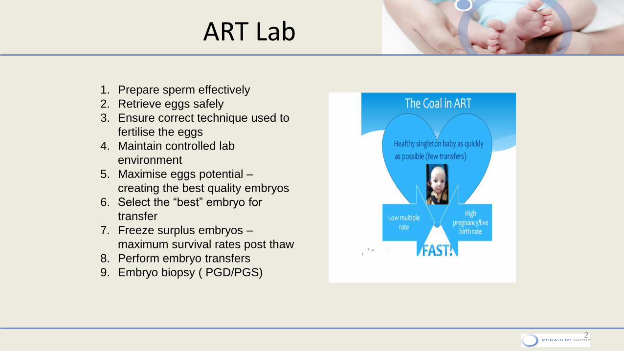
1. Prepare sperm effectively
2. Retrieve eggs safely
3. Ensure correct technique used to
fertilise the eggs
4. Maintain controlled lab
environment
5. Maximise eggs potential –
creating the best quality embryos
6. Select the “best” embryo for
transfer
7. Freeze surplus embryos –
maximum survival rates post thaw
8. Perform embryo transfers
9. Embryo biopsy ( PGD/PGS)
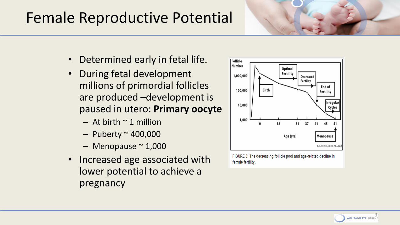
Female Reproductive Potential
• Determined early in fetal life.
• During fetal development millions of primordial follicles are produced –development is paused in utero: Primary oocyte – At birth ~ 1 million
– Puberty ~ 400,000
– Menopause ~ 1,000
• Increased age associated with lower potential to achieve a pregnancy
3
Meiosis
4
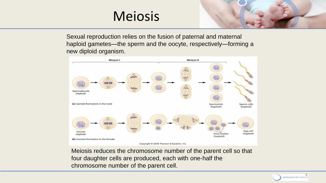
Meiosis reduces the chromosome number of the parent cell so that
four daughter cells are produced, each with one-half the
chromosome number of the parent cell.
Sexual reproduction relies on the fusion of paternal and maternal
haploid gametes—the sperm and the oocyte, respectively—forming a
new diploid organism.

Gametogenesis
5
• OOGENESIS
• X only + 23 chromosomes
• SPERMATOGENESIS
• X or Y + 23 chromosomes
Folliculogenesis
6
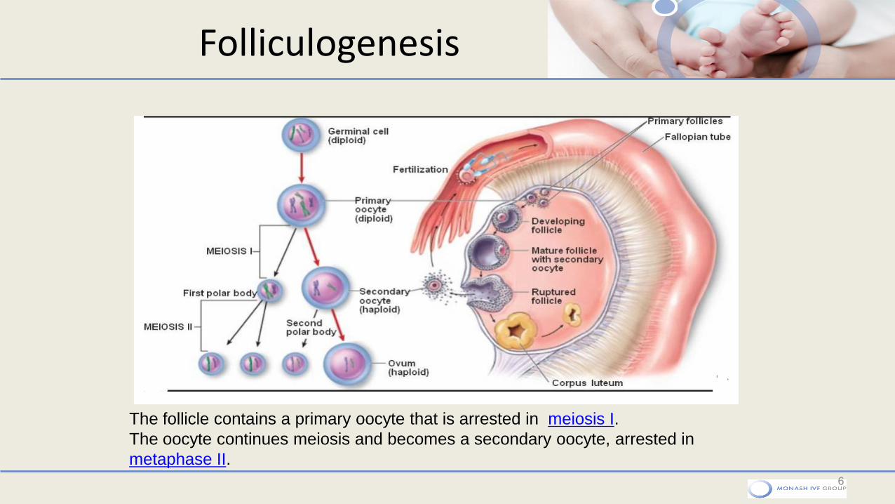
The follicle contains a primary oocyte that is arrested in meiosis I.
The oocyte continues meiosis and becomes a secondary oocyte, arrested in
metaphase II.
Mature egg (MII)
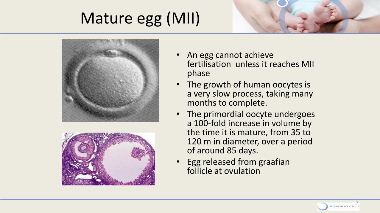
• An egg cannot achieve fertilisation unless it reaches MII phase
• The growth of human oocytes is a very slow process, taking many months to complete.
• The primordial oocyte undergoes a 100-fold increase in volume by the time it is mature, from 35 to 120 m in diameter, over a period of around 85 days.
• Egg released from graafian follicle at ovulation
7
Aneuploidy
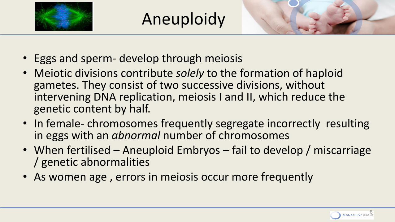
• Eggs and sperm- develop through meiosis • Meiotic divisions contribute solely to the formation of haploid
gametes. They consist of two successive divisions, without intervening DNA replication, meiosis I and II, which reduce the genetic content by half.
• In female- chromosomes frequently segregate incorrectly resulting in eggs with an abnormal number of chromosomes
• When fertilised – Aneuploid Embryos – fail to develop / miscarriage / genetic abnormalities
• As women age , errors in meiosis occur more frequently
8
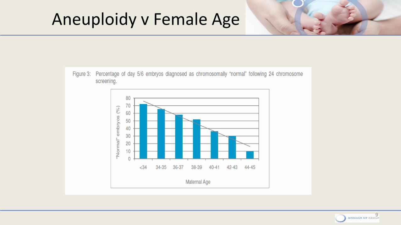
Aneuploidy v Female Age
9
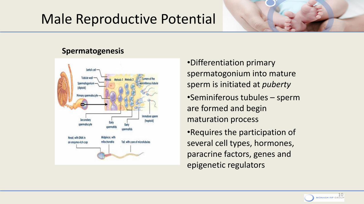
Male Reproductive Potential
•Differentiation primary spermatogonium into mature sperm is initiated at puberty
•Seminiferous tubules – sperm are formed and begin maturation process
•Requires the participation of several cell types, hormones, paracrine factors, genes and epigenetic regulators
10
Spermatogenesis
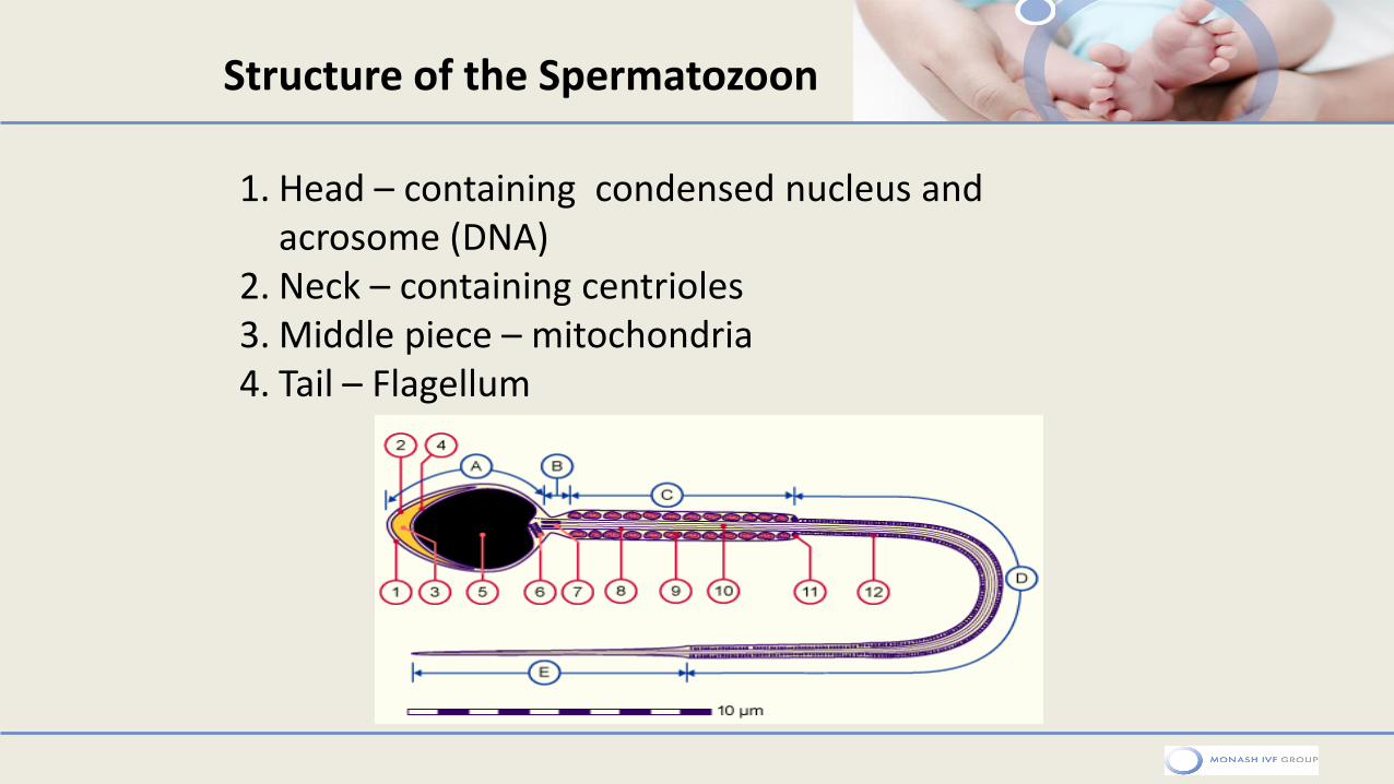
1. Head – containing condensed nucleus and acrosome (DNA)
2. Neck – containing centrioles 3. Middle piece – mitochondria 4. Tail – Flagellum
Structure of the Spermatozoon
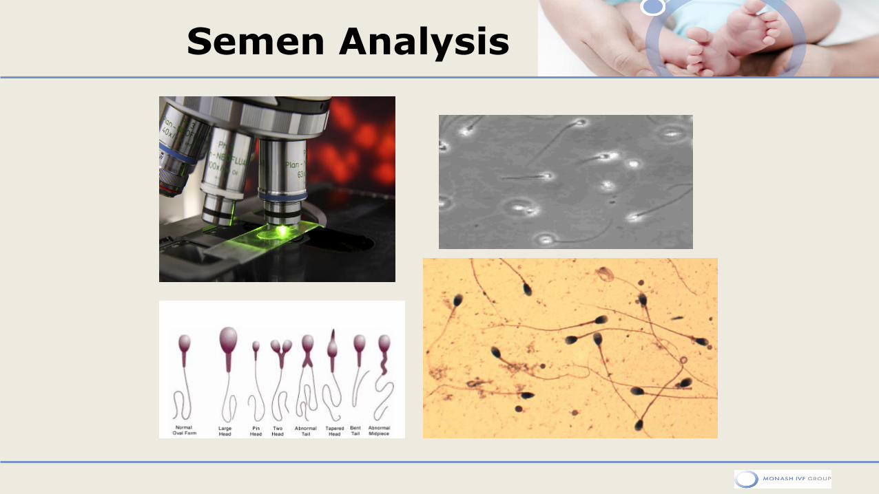
Semen Analysis
Production of Sperm sample
Most common form of sperm retrieval is ejaculation – 2-3 days abstinence Time between collection to lab < 1 hr; Protected against temperature, shock, light Sterile container Container properly labeled Identity check performed
13
Semen Analysis - Implications in Fertility Treatments
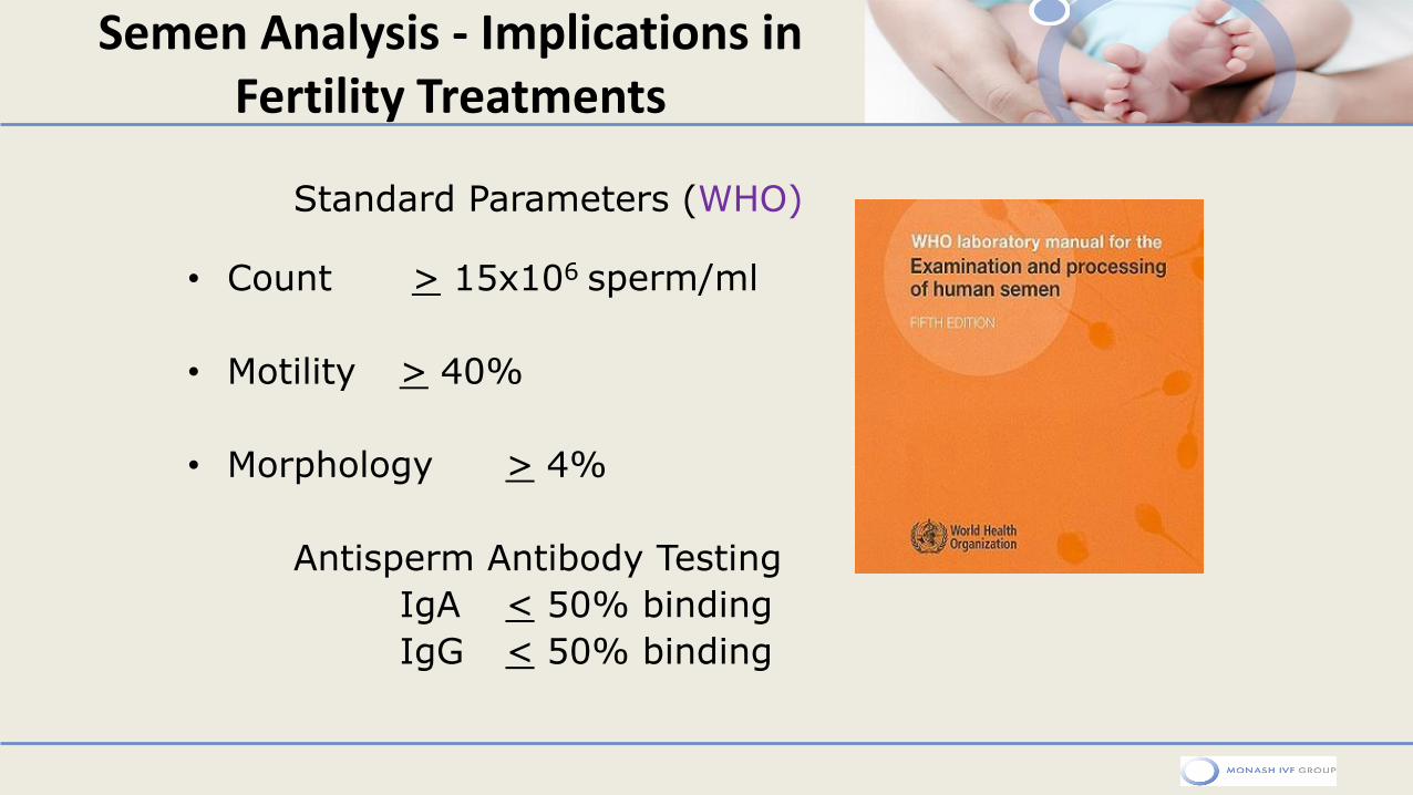
Standard Parameters (WHO)
• Count > 15x106 sperm/ml
• Motility > 40%
• Morphology > 4%
Antisperm Antibody Testing
IgA < 50% binding
IgG < 50% binding
15
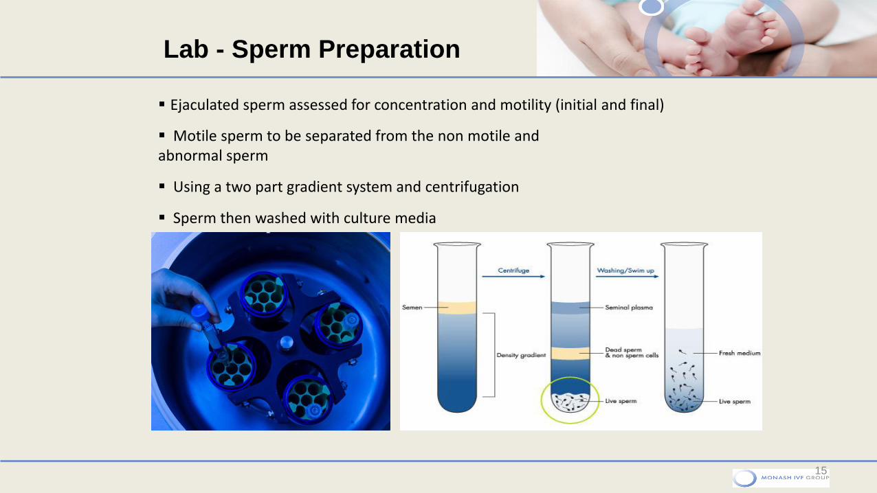
Ejaculated sperm assessed for concentration and motility (initial and final)
Motile sperm to be separated from the non motile and abnormal sperm
Using a two part gradient system and centrifugation
Sperm then washed with culture media
Lab - Sperm Preparation
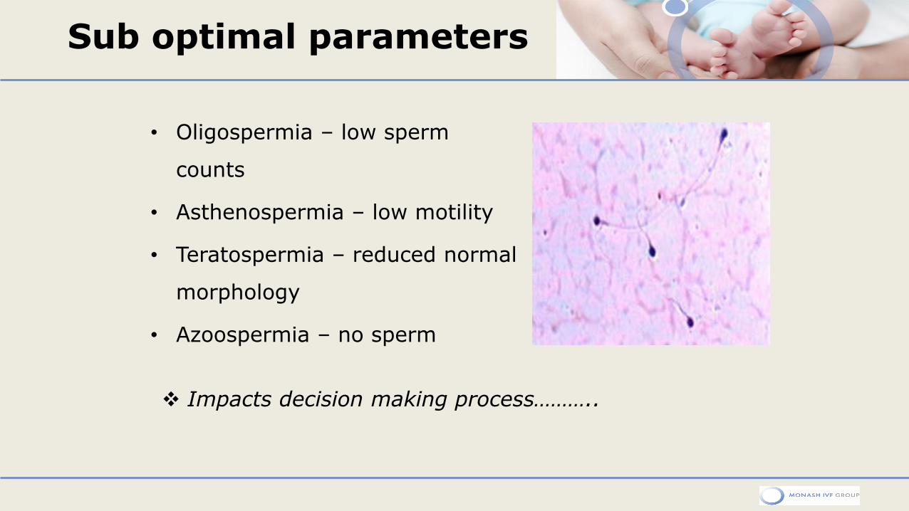
Sub optimal parameters
• Oligospermia – low sperm
counts
• Asthenospermia – low motility
• Teratospermia – reduced normal
morphology
• Azoospermia – no sperm
Impacts decision making process………..
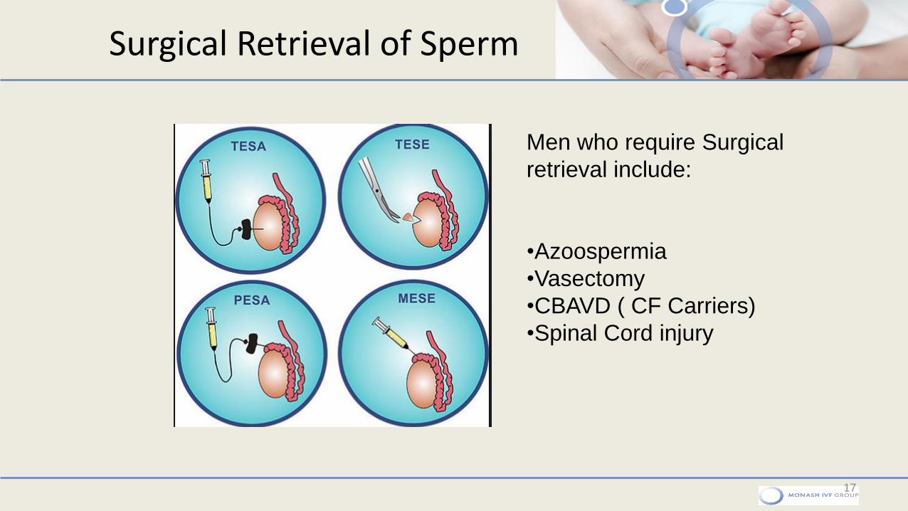
Surgical Retrieval of Sperm
17
Men who require Surgical
retrieval include:
•Azoospermia
•Vasectomy
•CBAVD ( CF Carriers)
•Spinal Cord injury
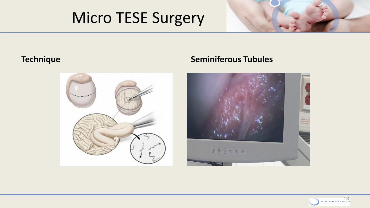
Micro TESE Surgery
Technique Seminiferous Tubules
18
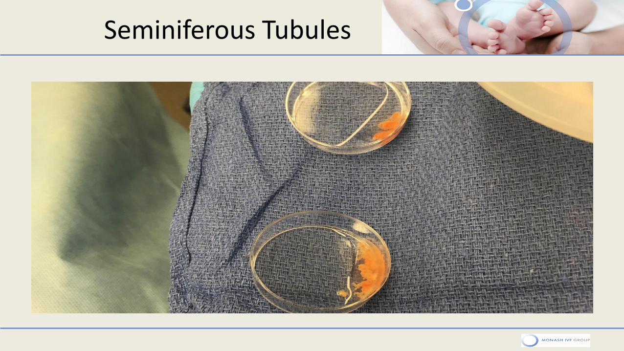
Seminiferous Tubules
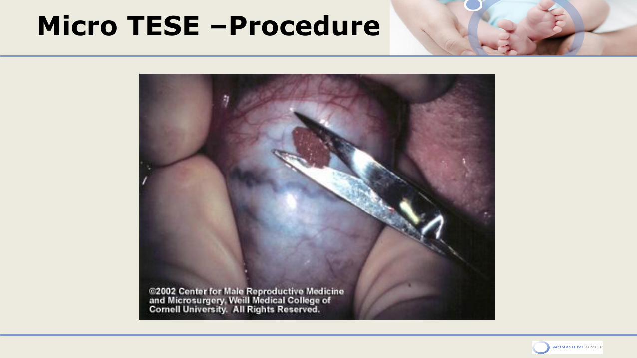
Micro TESE –Procedure

Theatre/ Lab
• Micro-TESE average time it takes for a single procedure varies from 3-4 hours in theatre
• The tissue processing time is approx 1.5 hours in the laboratory
• Time searching for sperm can be 3-4 hours with multiple embryologists searching
• When sperm found – cryopreservation or used on day egg collection
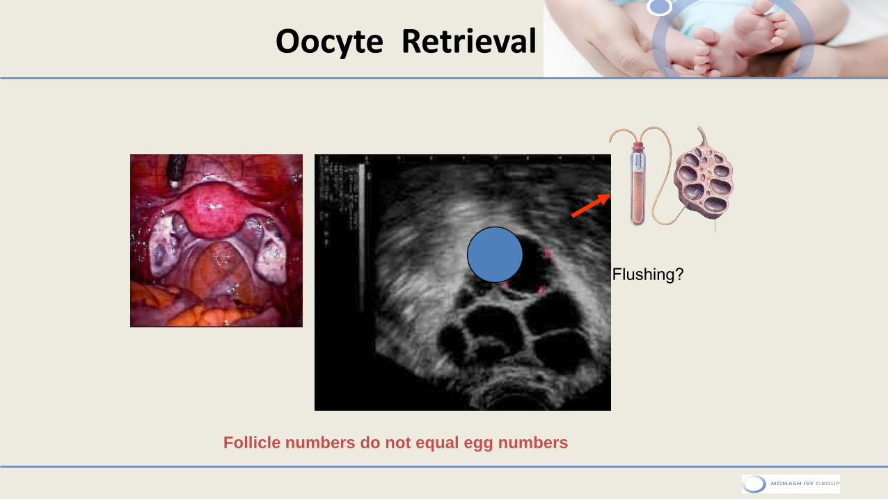
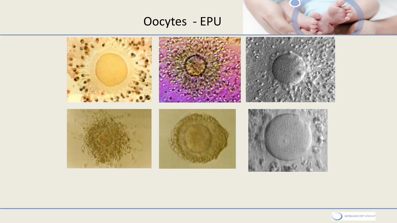
Oocyte Retrieval
Follicle numbers do not equal egg numbers
Flushing?
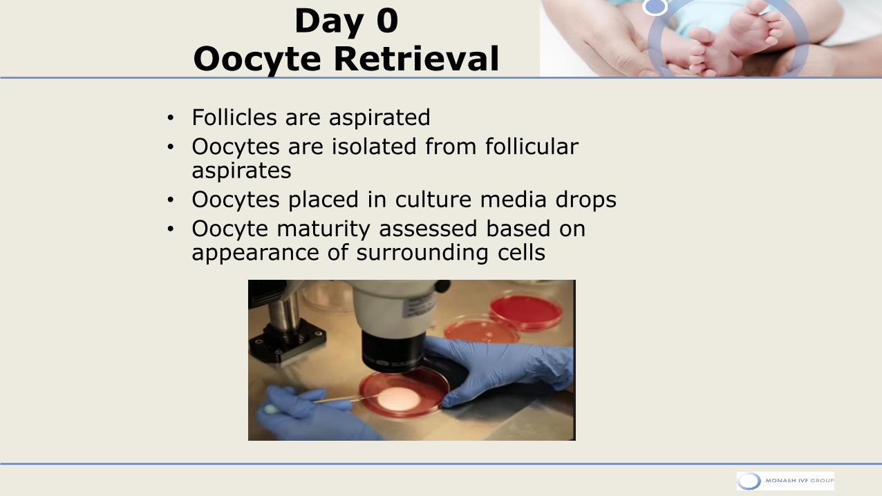
Day 0 Oocyte Retrieval
• Follicles are aspirated
• Oocytes are isolated from follicular aspirates
• Oocytes placed in culture media drops
• Oocyte maturity assessed based on appearance of surrounding cells
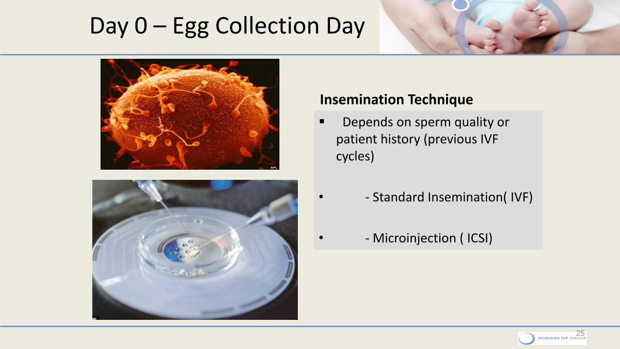
Day 0 – Egg Collection Day
Insemination Technique
25
Depends on sperm quality or patient history (previous IVF cycles)
• - Standard Insemination( IVF)
• - Microinjection ( ICSI)
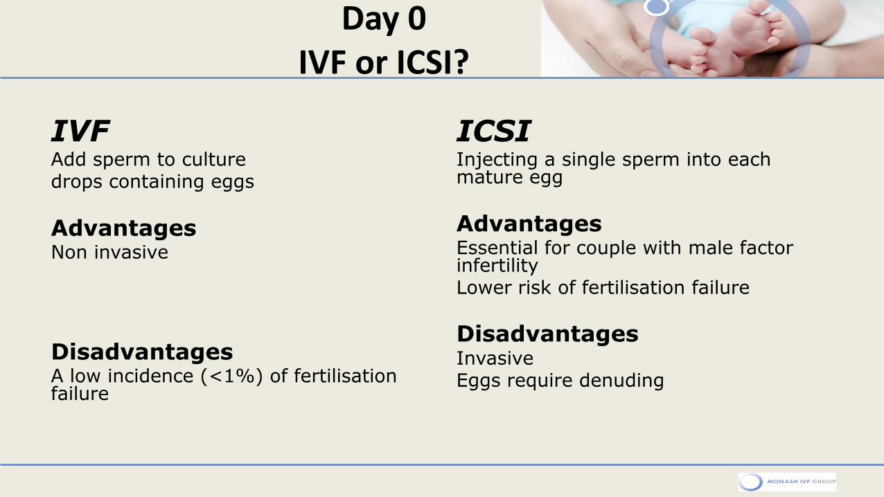
Day 0 IVF or ICSI?
IVF Add sperm to culture drops containing eggs
Advantages Non invasive
Disadvantages A low incidence (<1%) of fertilisation failure
ICSI Injecting a single sperm into each mature egg
Advantages Essential for couple with male factor infertility Lower risk of fertilisation failure
Disadvantages Invasive Eggs require denuding
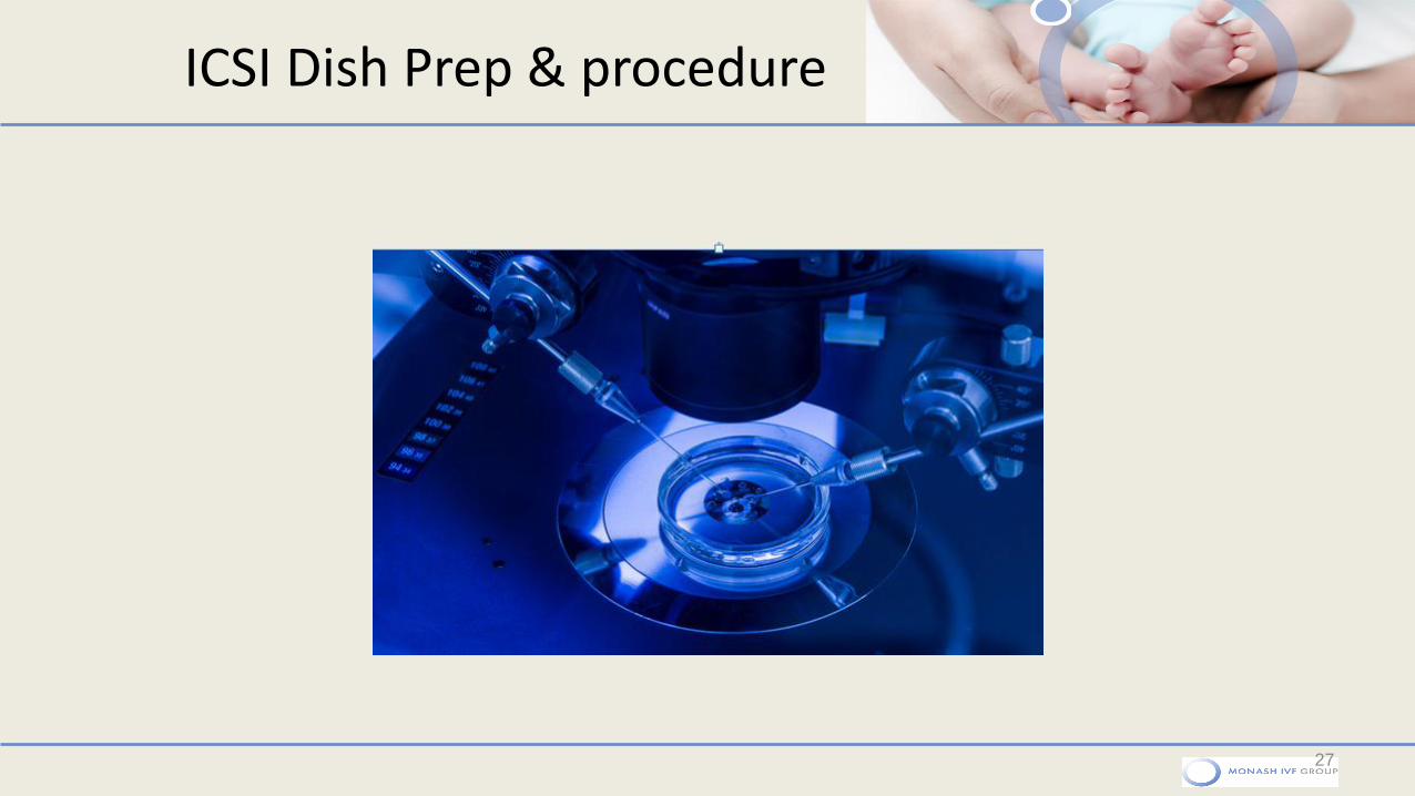
ICSI Dish Prep & procedure
27
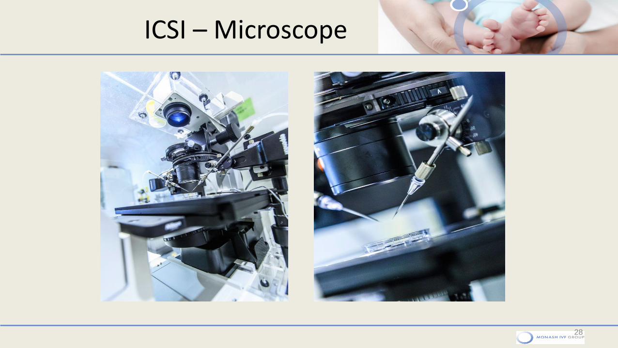
ICSI – Microscope
28
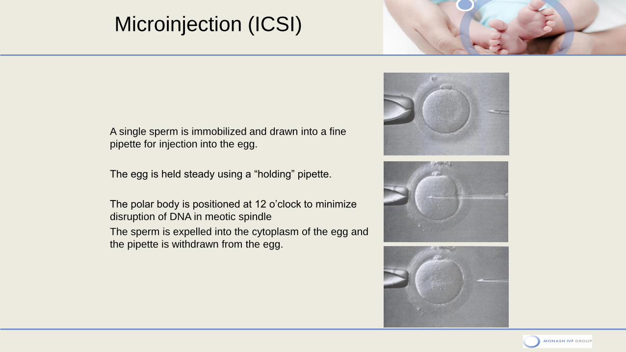
Microinjection (ICSI)
A single sperm is immobilized and drawn into a fine
pipette for injection into the egg.
The egg is held steady using a “holding” pipette.
The polar body is positioned at 12 o’clock to minimize
disruption of DNA in meotic spindle
The sperm is expelled into the cytoplasm of the egg and
the pipette is withdrawn from the egg.
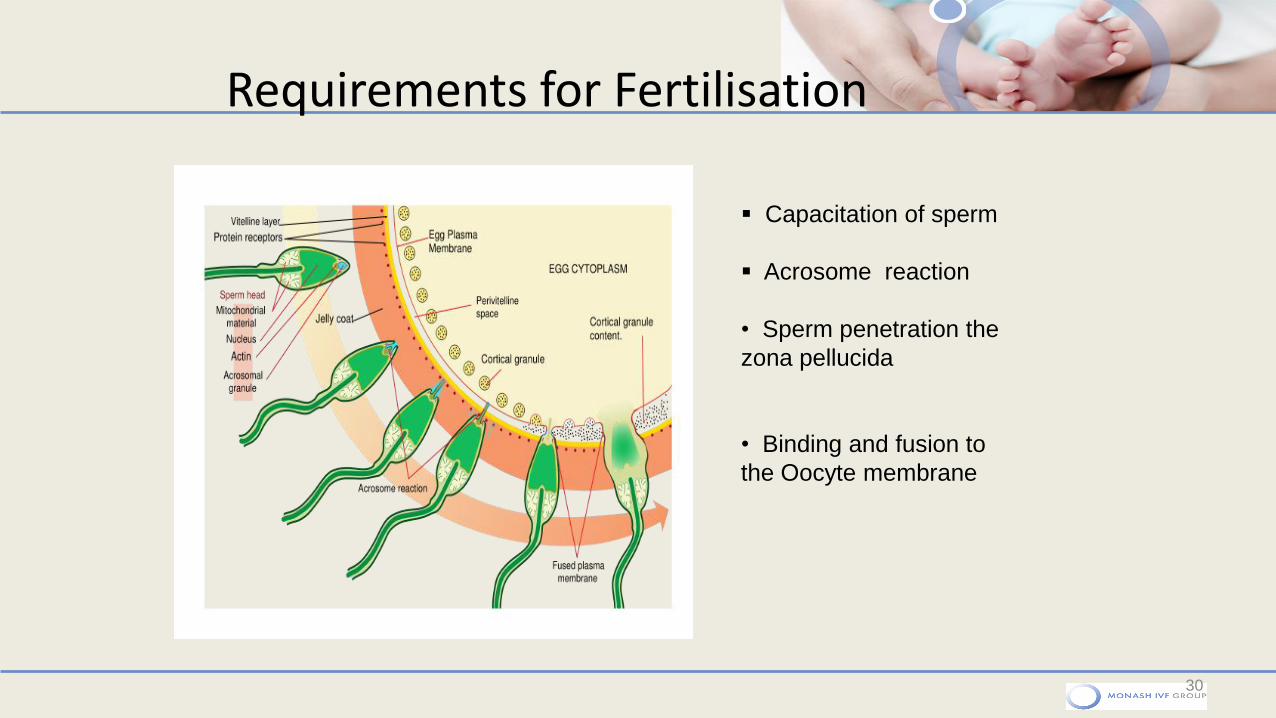
Requirements for Fertilisation
30
Capacitation of sperm
Acrosome reaction
• Sperm penetration the
zona pellucida
• Binding and fusion to
the Oocyte membrane
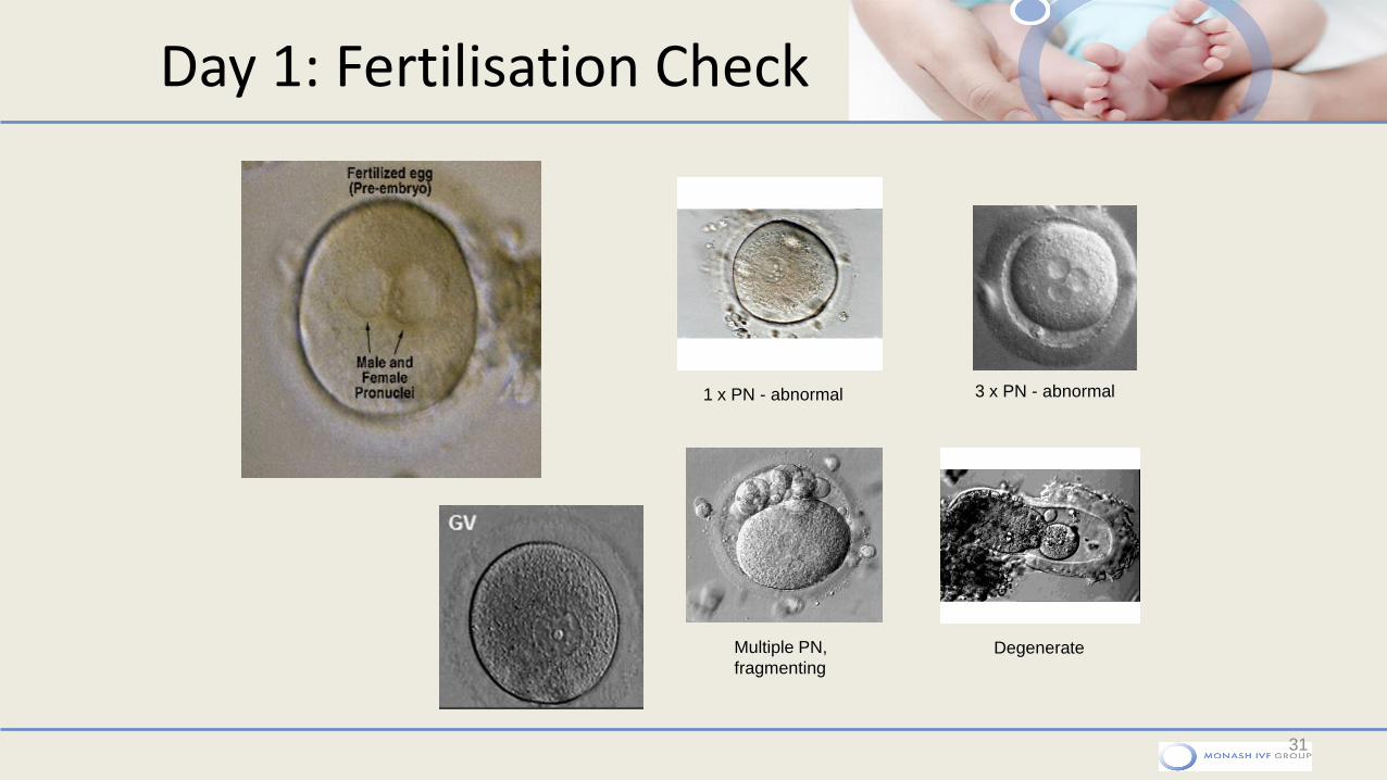
Day 1: Fertilisation Check
31
1 x PN - abnormal 3 x PN - abnormal
Multiple PN,
fragmenting Degenerate
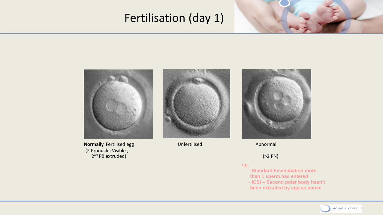
Fertilisation (day 1)
Normally Fertilised egg Unfertilised Abnormal (2 Pronuclei Visible ; 2nd PB extruded) (>2 PN)
eg.
- Standard Insemination more
than 1 sperm has entered
- ICSI – Second polar body hasn’t
been extruded by egg as above
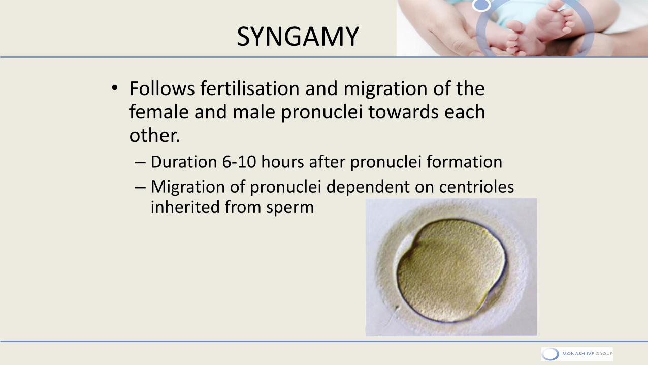
SYNGAMY
• Follows fertilisation and migration of the female and male pronuclei towards each other. – Duration 6-10 hours after pronuclei formation
– Migration of pronuclei dependent on centrioles inherited from sperm
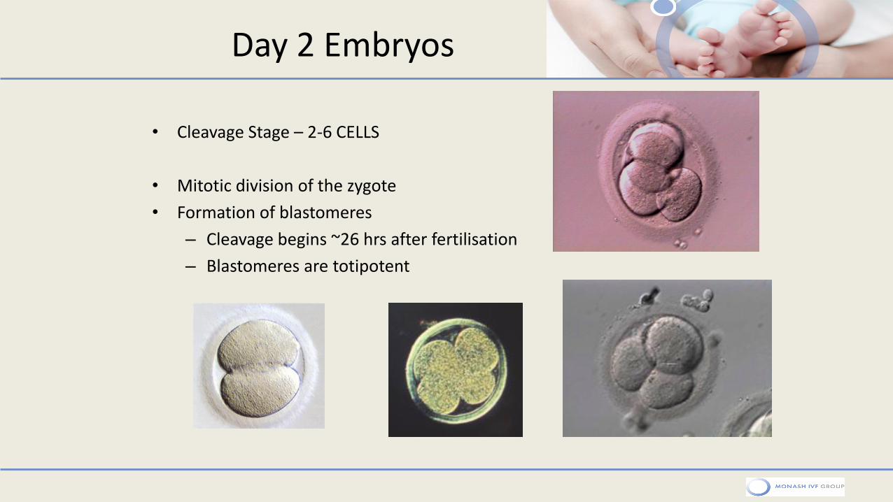
Day 2 Embryos
• Cleavage Stage – 2-6 CELLS
• Mitotic division of the zygote
• Formation of blastomeres
– Cleavage begins ~26 hrs after fertilisation
– Blastomeres are totipotent
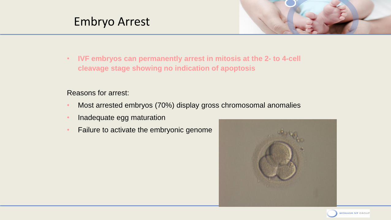
• IVF embryos can permanently arrest in mitosis at the 2- to 4-cell
cleavage stage showing no indication of apoptosis
Reasons for arrest:
• Most arrested embryos (70%) display gross chromosomal anomalies
• Inadequate egg maturation
• Failure to activate the embryonic genome
Embryo Arrest
Embryo Fragmentation
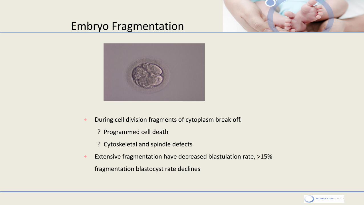
• During cell division fragments of cytoplasm break off.
? Programmed cell death
? Cytoskeletal and spindle defects
• Extensive fragmentation have decreased blastulation rate, >15%
fragmentation blastocyst rate declines
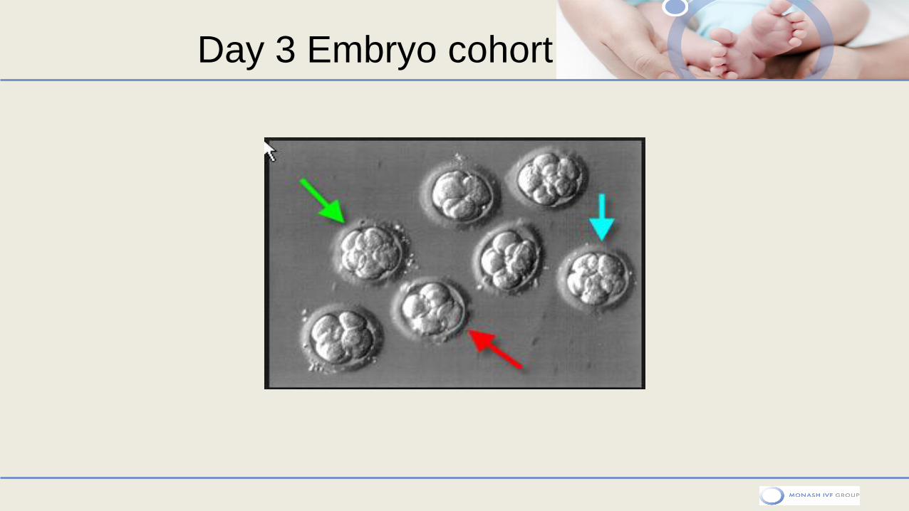
Day 3 Embryo cohort
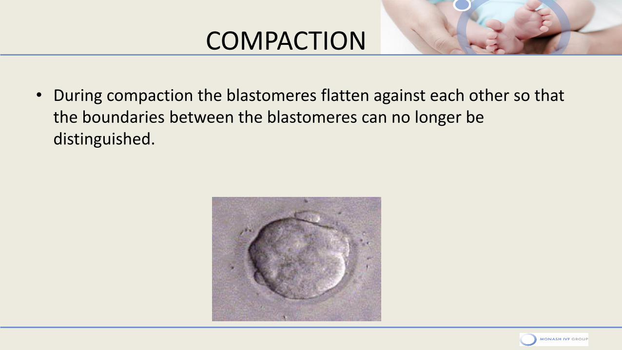
COMPACTION
• During compaction the blastomeres flatten against each other so that the boundaries between the blastomeres can no longer be distinguished.
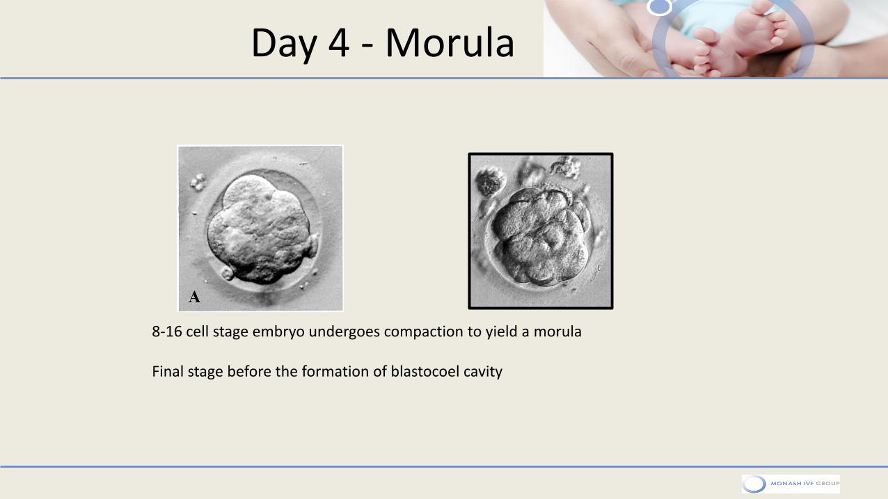
Day 4 - Morula
8-16 cell stage embryo undergoes compaction to yield a morula Final stage before the formation of blastocoel cavity
How does the cavitation occur?
• An osmotically driven movement of water into the embryo which forms the fluid filled cavity, called the Blastocoel.
• The movement of other ions (chloride, bicarbonate, Na+, K+) begin this energy dependant movement.
• As the fluid increases into the cavity, it then separates the cells into two parts.
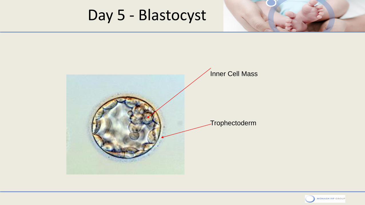
Day 5 - Blastocyst
Inner Cell Mass
Trophectoderm
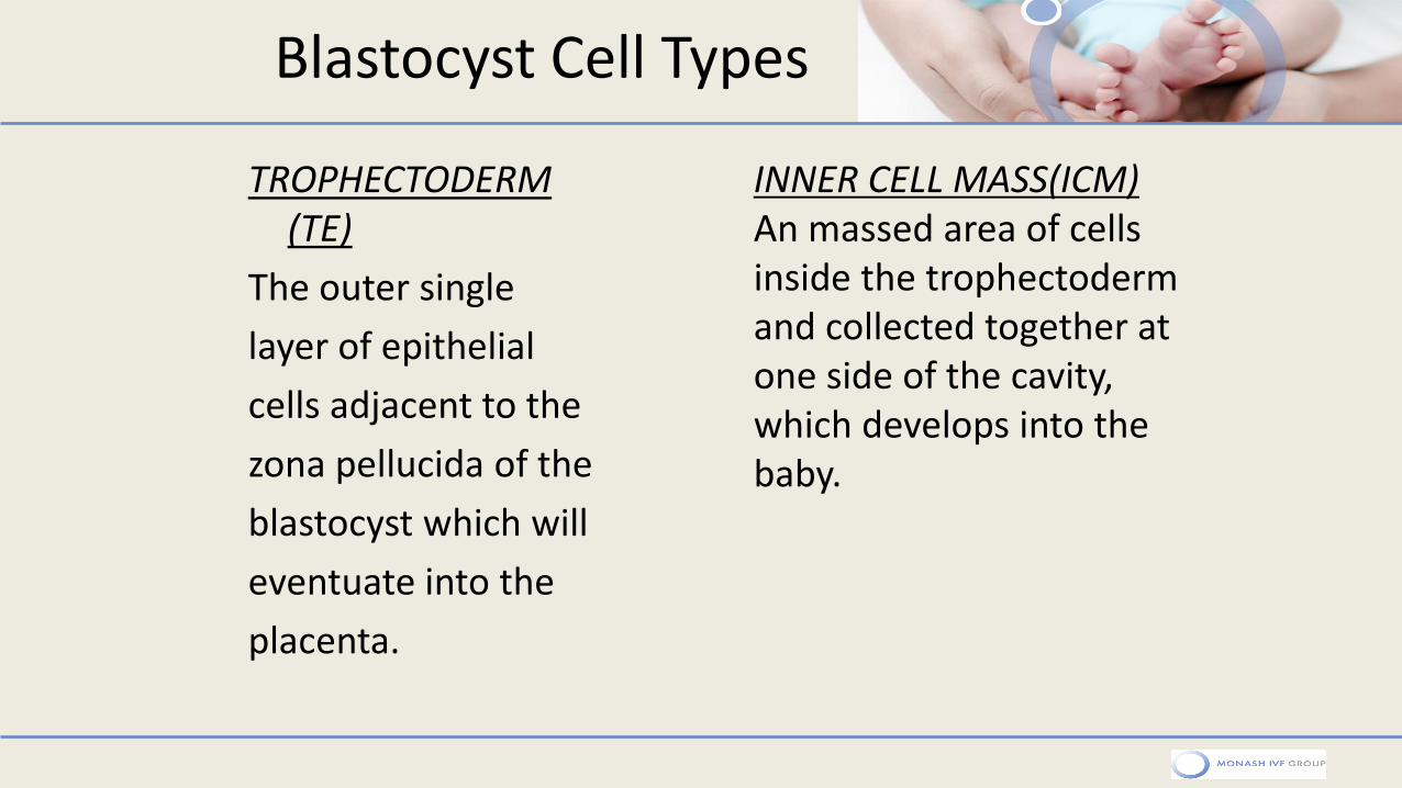
Blastocyst Cell Types
TROPHECTODERM (TE)
The outer single
layer of epithelial
cells adjacent to the
zona pellucida of the
blastocyst which will
eventuate into the
placenta.
INNER CELL MASS(ICM) An massed area of cells inside the trophectoderm and collected together at one side of the cavity, which develops into the baby.
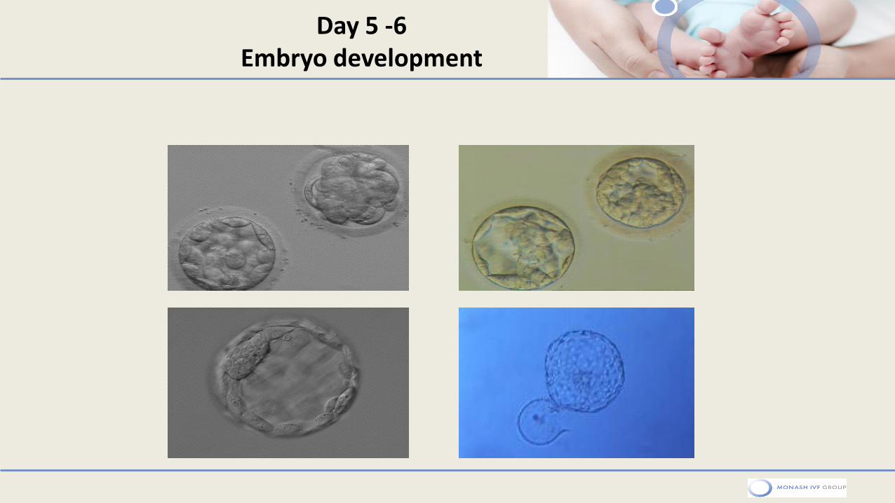
Day 5 -6 Embryo development
• •
• •
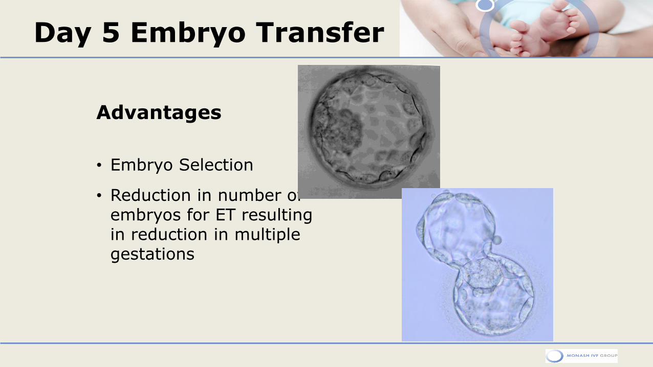
Day 5 Embryo Transfer
Advantages
• Embryo Selection
• Reduction in number of embryos for ET resulting in reduction in multiple gestations
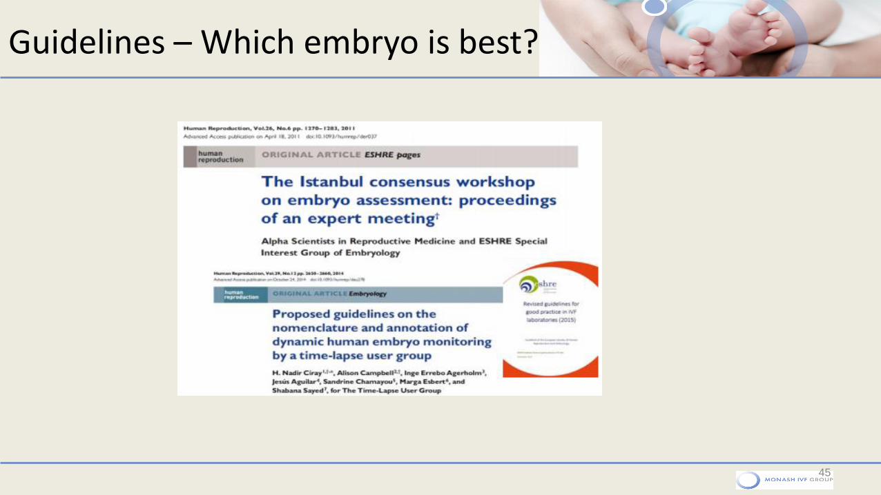
Guidelines – Which embryo is best?
45
Why Blastocyst ET?
Embryonic factors:
• 1. Better (self‐)selection
• 2. Higher implantation potential
• 3. Limitations of d2/3 (time lapse)
• 4. Better cryopreservation results
• 5. PGS
Clinically: • To reduce multiple‐pregnancy rates whilst maintaining pregnancy rates
•Higher twinning rates with blasts (33% vrs 16.5%) after equal number of embryos transferred (Scwärzler et al, 2004)
47
Day 3 ET v D5: Tough questions!
• Will we ”lose” embryos by culturing them to the blastocyst stage?
• Can any given day 2/3 embryo become a viable blast in vivo but not in vitro?
• Can an embryo of poor quality on d2/3 become a good quality blastocyst?
• Is poor/no blastocyst development evidence of poor development potential or a consequence of (poor) in vitro culture conditions?
• Health of children born?
48
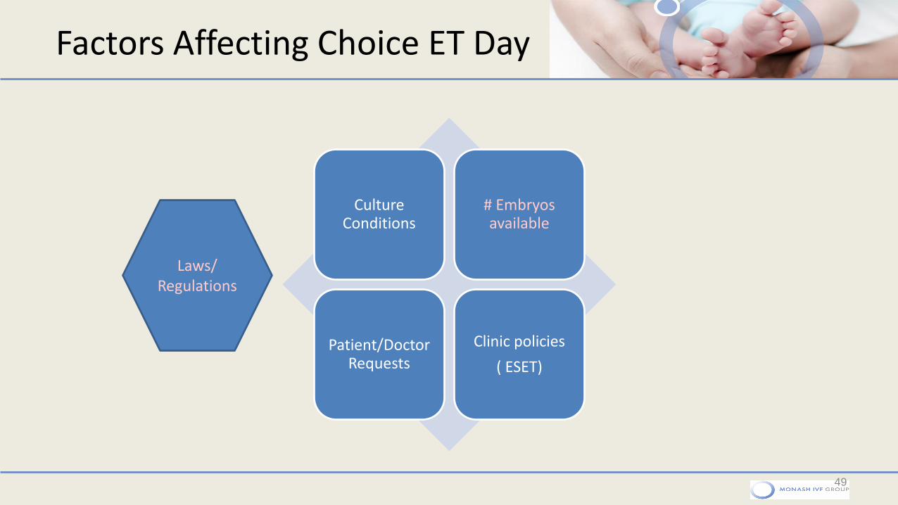
Culture Conditions
# Embryos available
Patient/Doctor Requests
Clinic policies
( ESET)
49
Laws/ Regulations
Factors Affecting Choice ET Day
50
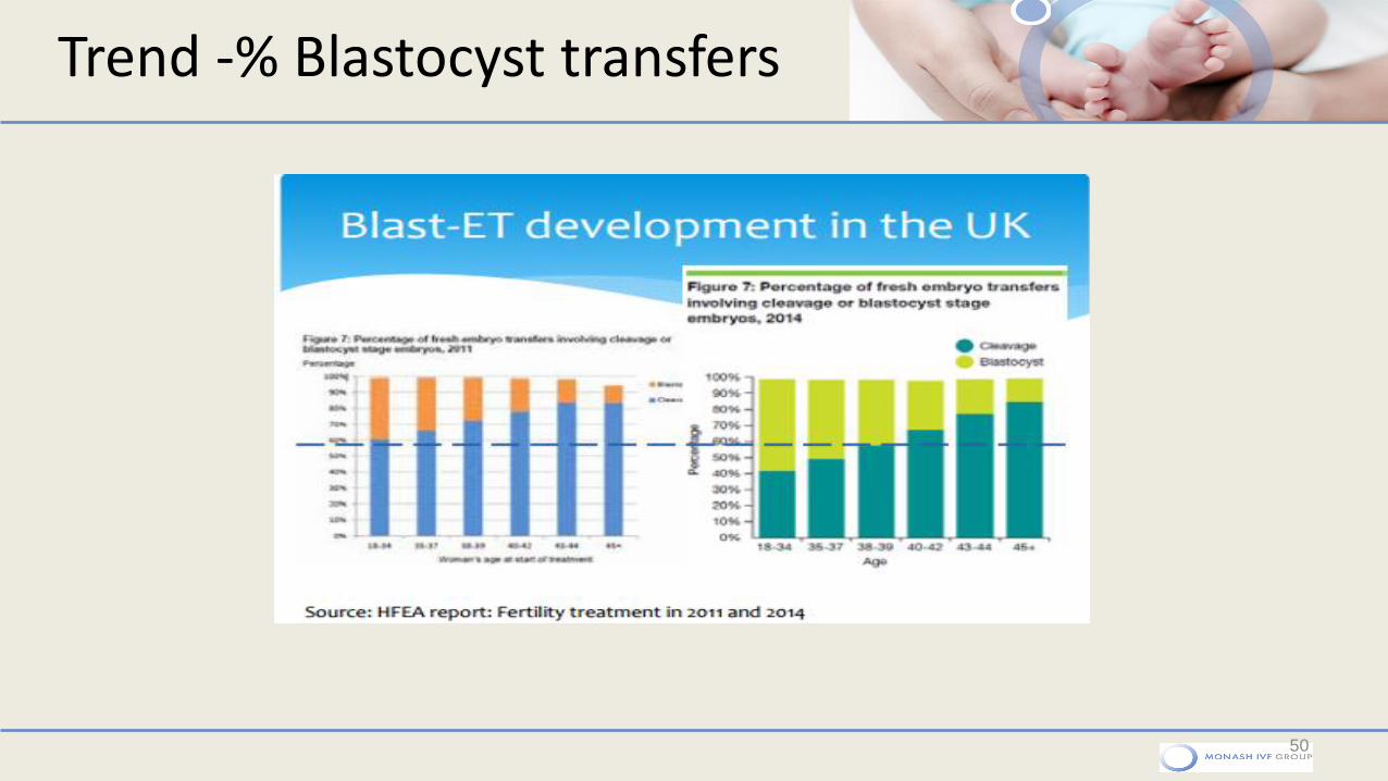
Trend -% Blastocyst transfers
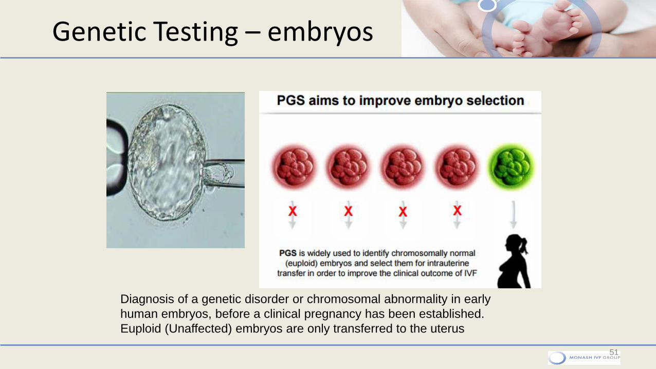
Genetic Testing – embryos
51
Diagnosis of a genetic disorder or chromosomal abnormality in early
human embryos, before a clinical pregnancy has been established.
Euploid (Unaffected) embryos are only transferred to the uterus
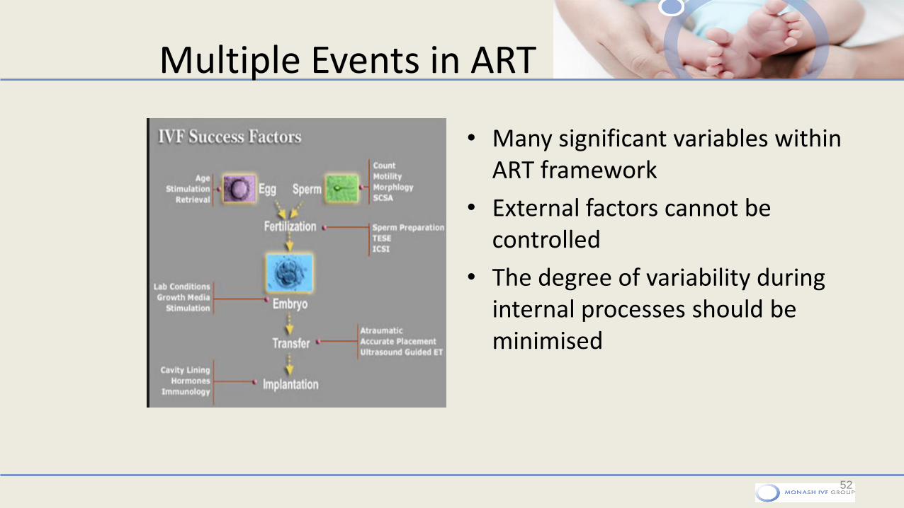
Multiple Events in ART
• Many significant variables within ART framework
• External factors cannot be controlled
• The degree of variability during internal processes should be minimised
52
Lab Environment
53
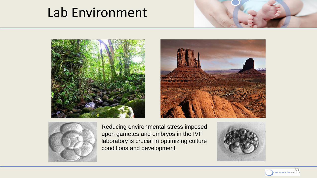
Reducing environmental stress imposed
upon gametes and embryos in the IVF
laboratory is crucial in optimizing culture
conditions and development
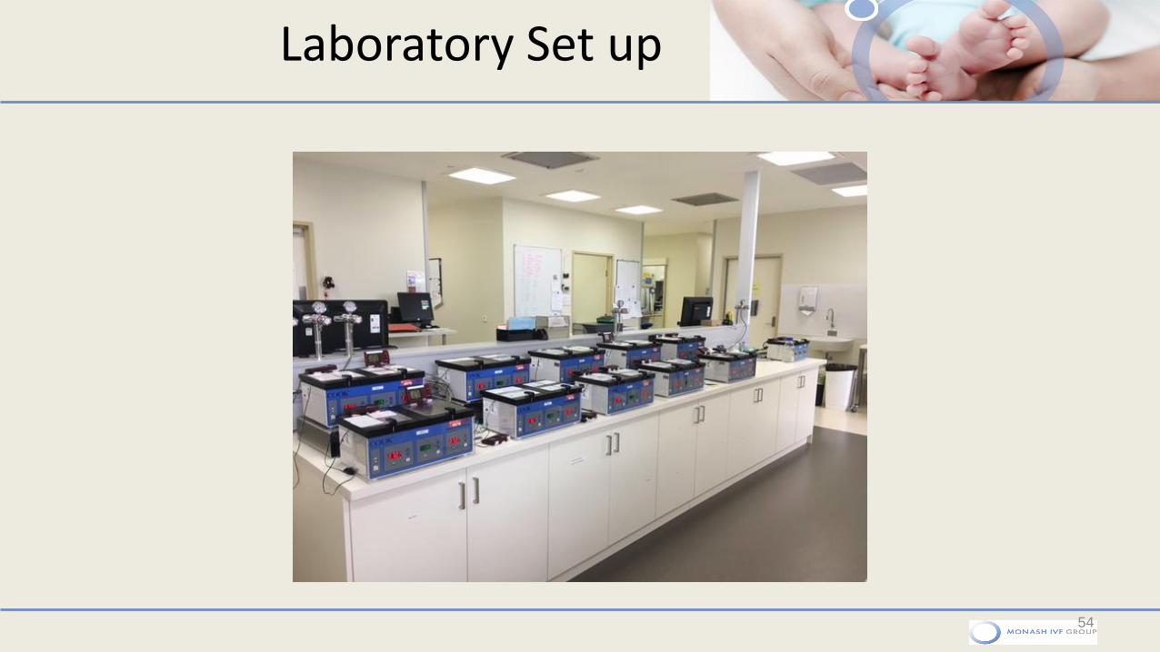
Laboratory Set up
54
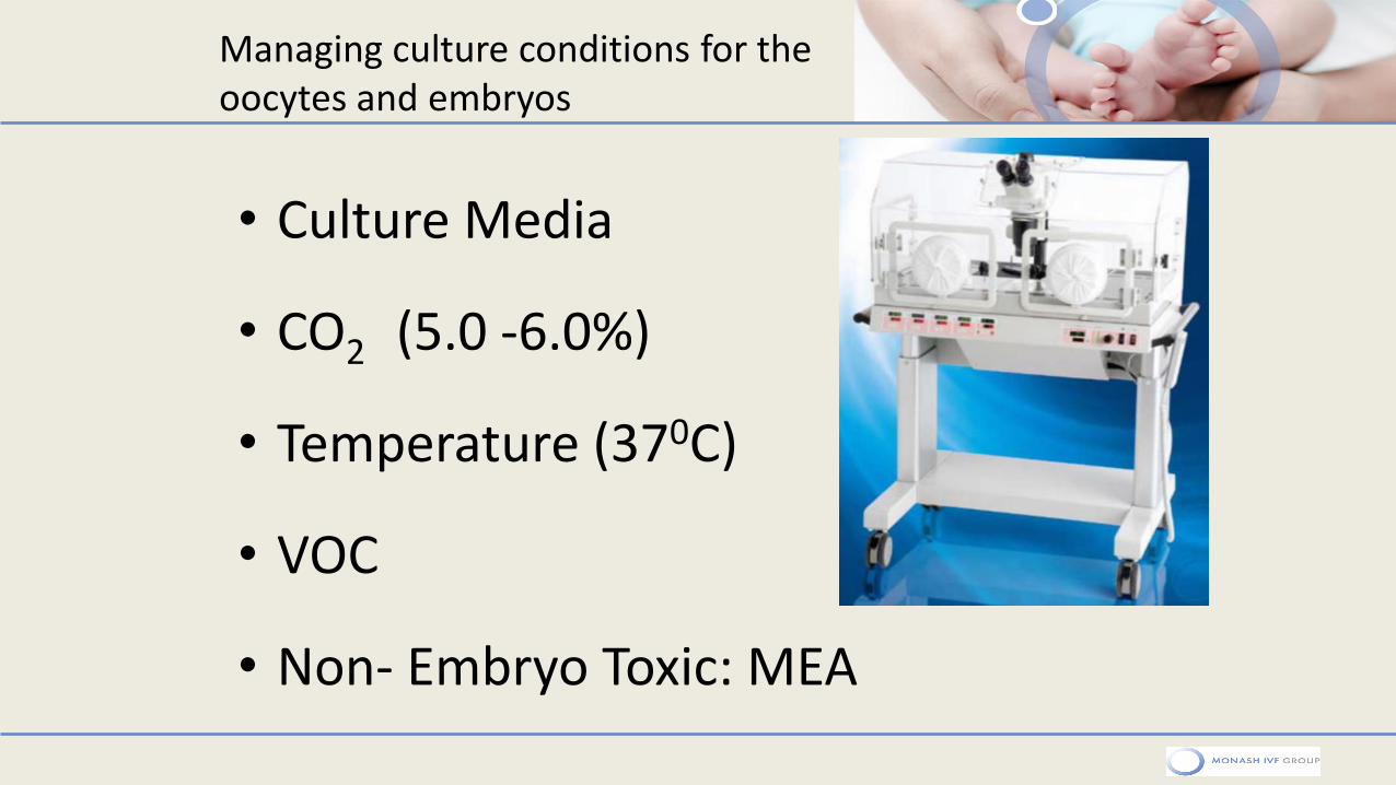
• Culture Media
• CO2 (5.0 -6.0%)
• Temperature (370C)
• VOC
• Non- Embryo Toxic: MEA
Managing culture conditions for the oocytes and embryos
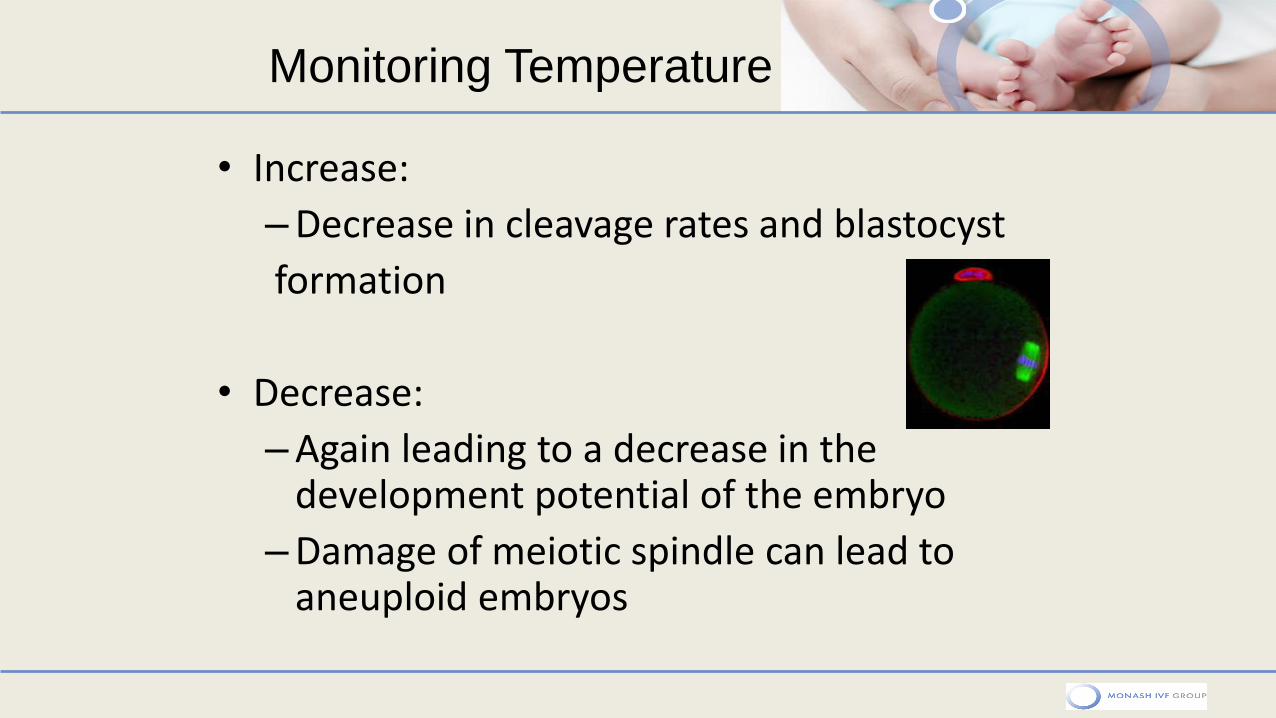
• Increase:
– Decrease in cleavage rates and blastocyst
formation
• Decrease:
– Again leading to a decrease in the development potential of the embryo
– Damage of meiotic spindle can lead to aneuploid embryos
Monitoring Temperature
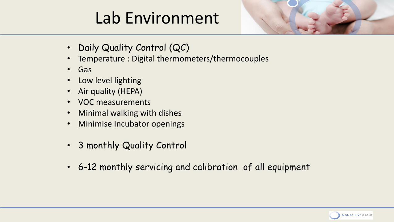
• Daily Quality Control (QC) • Temperature : Digital thermometers/thermocouples • Gas • Low level lighting • Air quality (HEPA) • VOC measurements • Minimal walking with dishes • Minimise Incubator openings
• 3 monthly Quality Control
• 6-12 monthly servicing and calibration of all equipment
Lab Environment
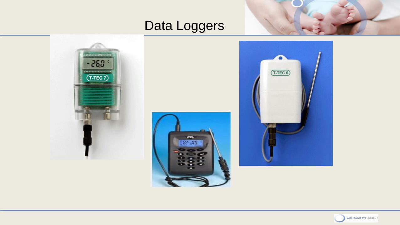
Data Loggers
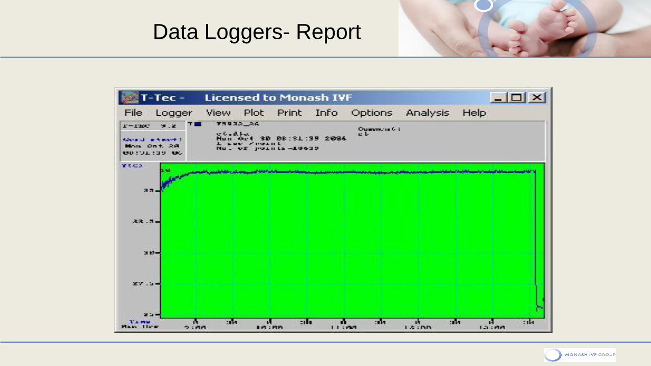
Data Loggers- Report
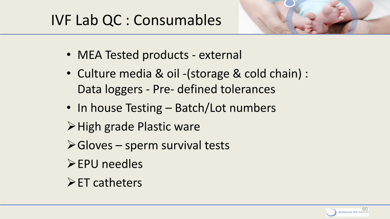
IVF Lab QC : Consumables
• MEA Tested products - external
• Culture media & oil -(storage & cold chain) : Data loggers - Pre- defined tolerances
• In house Testing – Batch/Lot numbers
High grade Plastic ware
Gloves – sperm survival tests
EPU needles
ET catheters
60
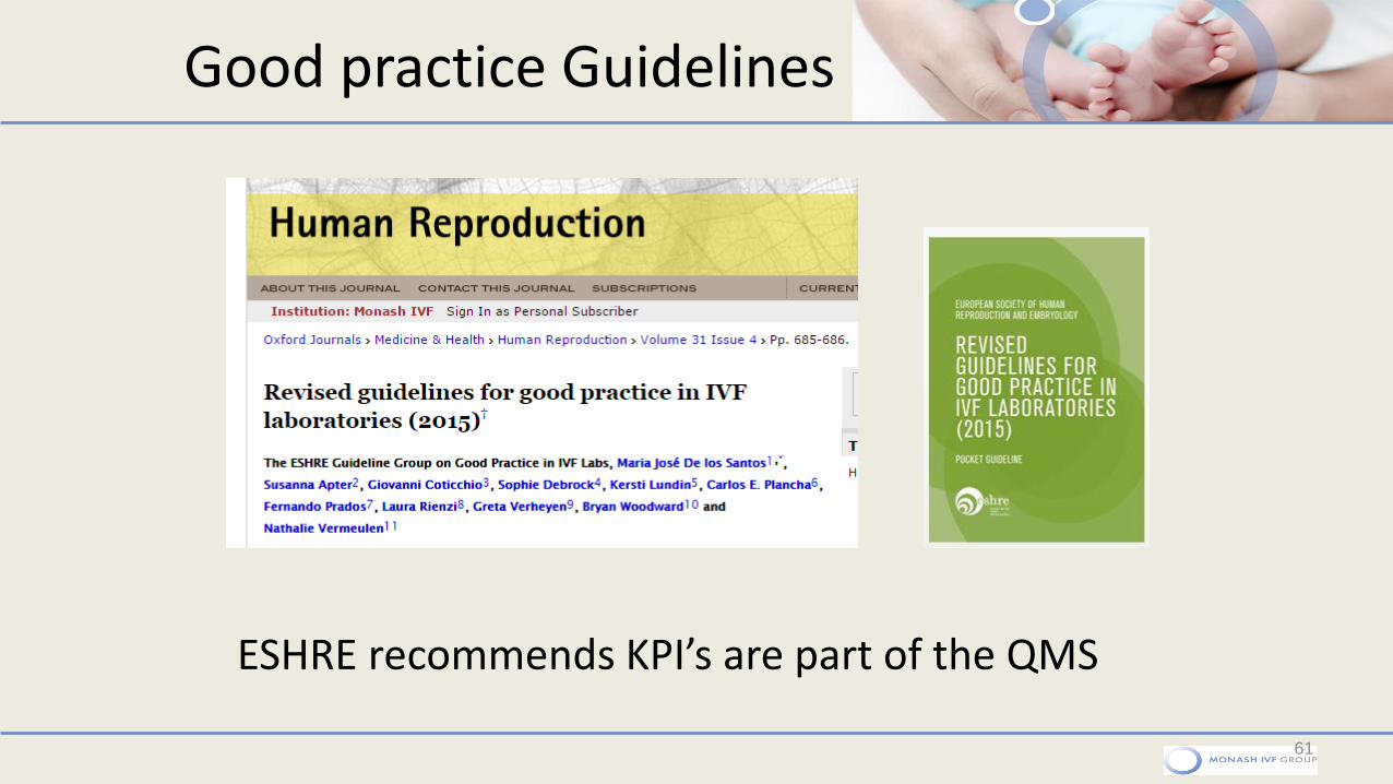
Good practice Guidelines
61
ESHRE recommends KPI’s are part of the QMS
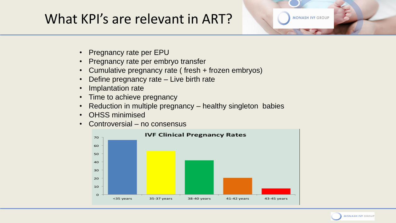
What KPI’s are relevant in ART?
• Pregnancy rate per EPU
• Pregnancy rate per embryo transfer
• Cumulative pregnancy rate ( fresh + frozen embryos)
• Define pregnancy rate – Live birth rate
• Implantation rate
• Time to achieve pregnancy
• Reduction in multiple pregnancy – healthy singleton babies
• OHSS minimised
• Controversial – no consensus
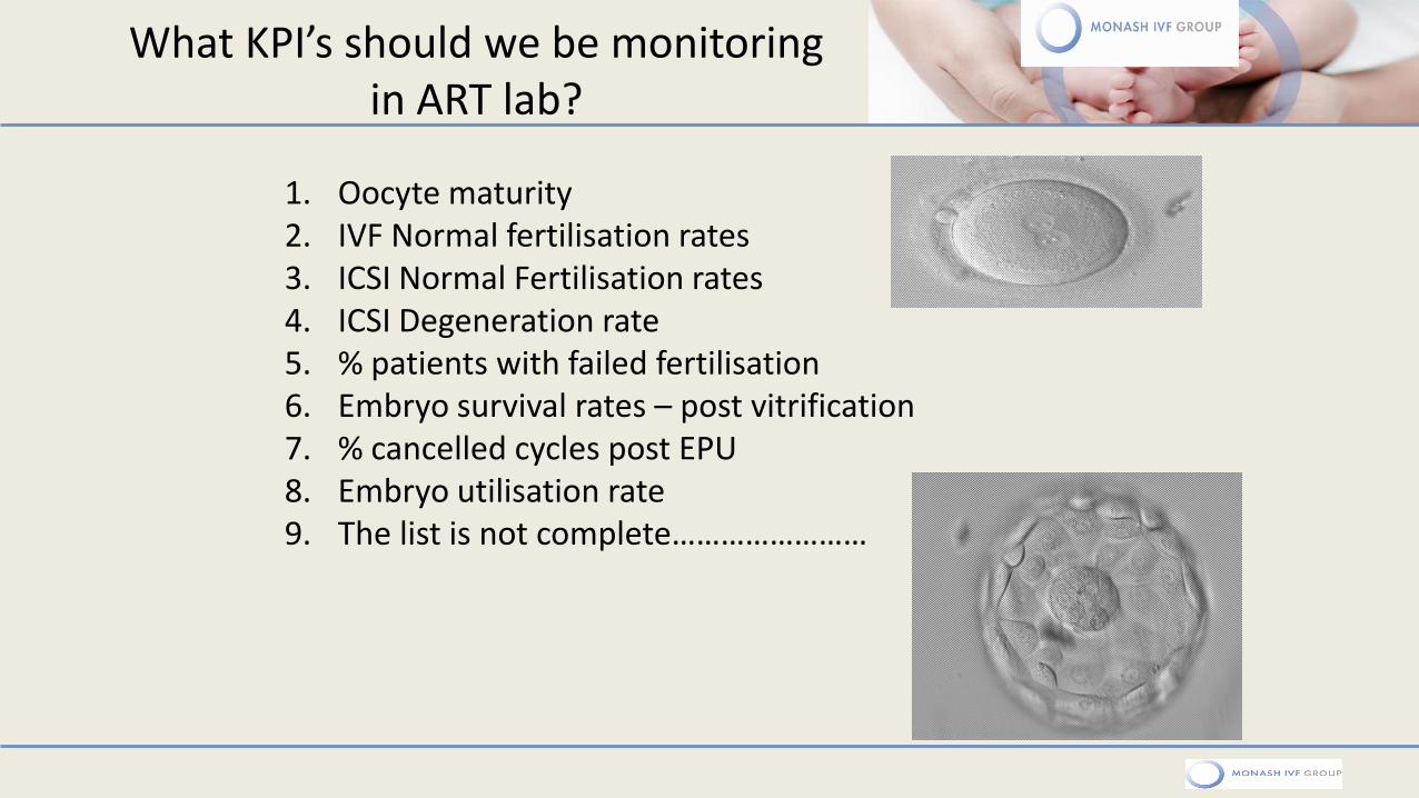
What KPI’s should we be monitoring in ART lab?
1. Oocyte maturity 2. IVF Normal fertilisation rates 3. ICSI Normal Fertilisation rates 4. ICSI Degeneration rate 5. % patients with failed fertilisation 6. Embryo survival rates – post vitrification 7. % cancelled cycles post EPU 8. Embryo utilisation rate 9. The list is not complete……………………
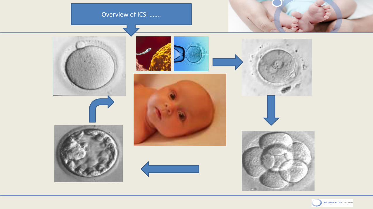
Overview of ICSI …….
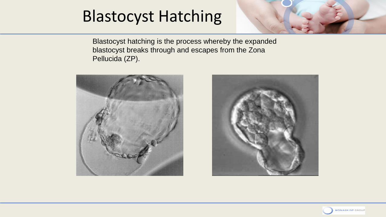
Blastocyst Hatching Blastocyst hatching is the process whereby the expanded
blastocyst breaks through and escapes from the Zona
Pellucida (ZP).
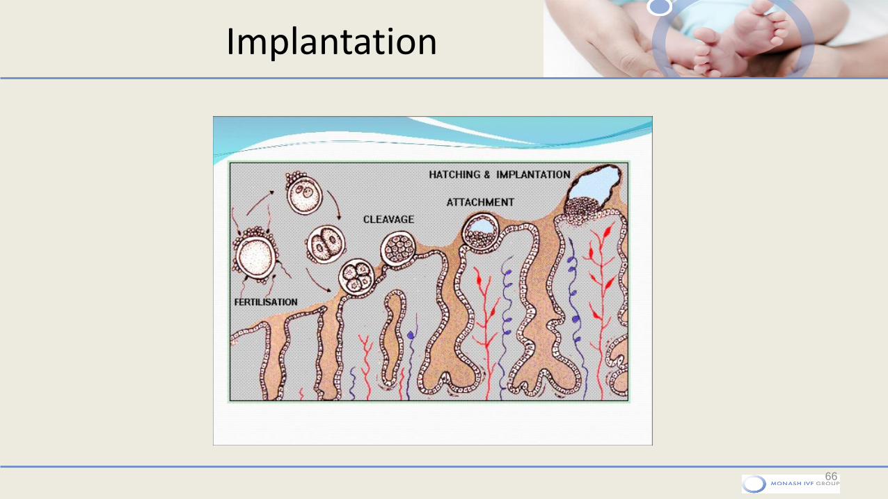
Implantation
66
Challenges faced ………
• Patients fail to develop follicles – cycle cancelled
• No eggs at egg collection
• Eggs collected are immature
• Discordant egg and follicle number
• Man fails to produce semen sample
• Azoospermic semen sample
• Eggs fail to fertilise
• Eggs fertilise abnormally
• Eggs may fertilise but not develop
• Eggs may develop to day 3 …….then do not progress
• Embryos are poor quality
• And many more things…
PERFECTING A PROCESS