60
The Big Book of Bovine Embryos a University of Florida Guide Version March 7, 2013

The Big Book of Bovine Embryos
a University of Florida Guide
Version March 7, 2013

The purpose of this collection of images of bovine oocytes and embryos is to assist new bovine embryologists with identification of structures encountered under the microscope. Unless otherwise stated, all images are of oocytes and embryos from in vitro maturation, fertilization and culture procedures performedin the laboratory of Peter J. Hansen, University of Florida.
The images are of oocytes and embryos viewed under inexpensive microscopes,usually dissecting scopes. By collecting typical views as might be seen in manylaboratories, it is hoped to facilitate the process of learning to identify structures.
Where known, the name of the person providing the image is on the lower rightcorner of each image.
Each image is shown twice – once without a label and a second time with importantfeatures identified. There is no particular order to the images and, in general, the magnification is not shown.
The Big Book of Bovine Embryos will be updated as more images are collected.

Let the Viewing Begin……………………………………………

First, oocytes and embryos in developmental sequence(special thanks to Firdous Khan)


Cumulus-Oocyte Complexes at the End of Maturation
Katherine Hendricks

Zona pellucida

Zona pellucida
Matured oocytes (after treatment with hyaluronidase)
Firdous Khan


Blastomeres
Cleavage furrow
Two-cell Embryos
Firdous Khan


3-4-cell embryos
Firdous Khan


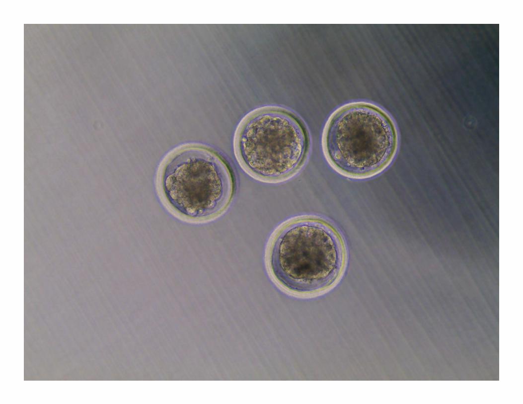
5-8 cell embryos
Firdous Khan


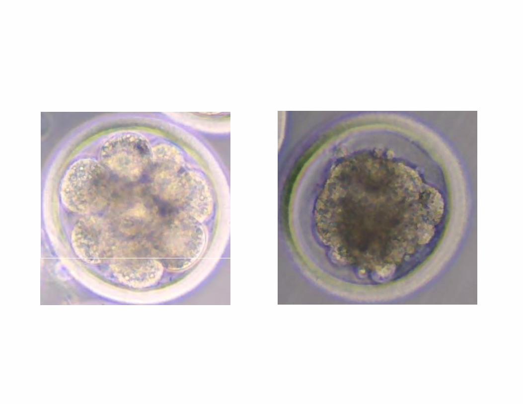
9-16 cell embryos
Firdous Khan

Morula
Compact morula
Morulae
Firdous Khan


Blastocoel
Blastocysts
Firdous Khan

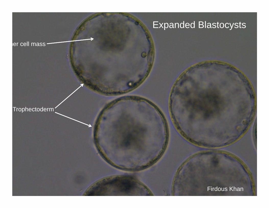
Trophectoderm
ner cell mass
Expanded Blastocysts
Firdous Khan


Expanded blastocysts
Firdous Khan


Hatching Blastocysts
Firdous Khan

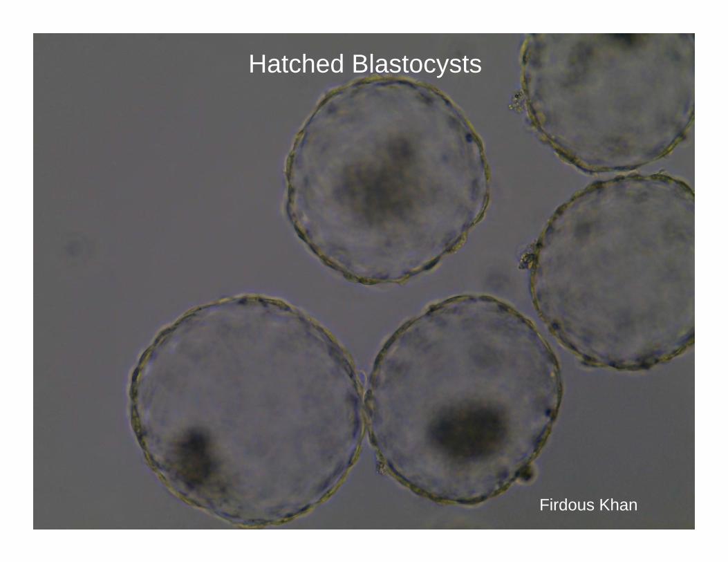
Hatched Blastocysts
Firdous Khan

Random images to help hone your skills at embryo identification

Embryo about 12-cells
Firdous Khan
Morula

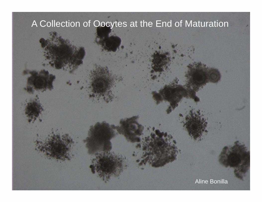
A Collection of Oocytes at the End of Maturation
Aline Bonilla


Amber Brad
Blastocysts
expanded
Non-expanded
Morula/early blastocyst


Blastocysts
Katherine Hendricks
hatched hatched
g
expanded
hatched


Hatching Blastocysts
Aline Bonilla
Hatching
Expanded and justbeginning to hatch


Two-cell embryo
Firdous Khan
Four-cell embryo

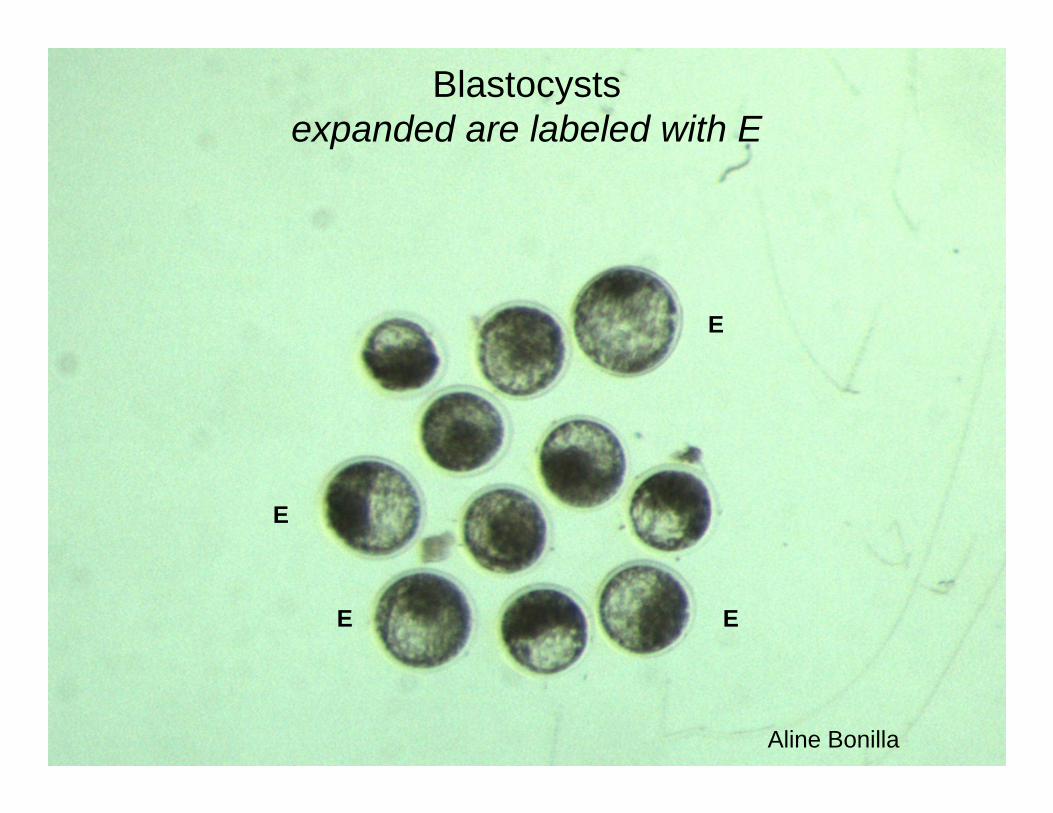
Blastocystsexpanded are labeled with E
Aline Bonilla
E
E E
E


Putative Zygotes following fertilization and removal of cumulus cells
Aline Bonilla


Eight-cell embryo
Firdous Khan


Expanded Blastocysts
Aline Bonilla


2-cell embryos
Firdous Khan


Hatched blastocysts
Aline Bonilla


Embryos at Day 7 after insemination
Anna Denicol
Abnormally-shaped morula
blast
blast
Two-cell
One cell or unfertilized
Degenerating/blocked development


Hatched blastocyst at Day 8 – this embryowas the abnormal morula in the Previous slide
Anna Denicol

And now, some embryos that have been labeled with specificmarkers…..

Differential immunostaining using FITC for TE (CDX2+) cells and Hoescht 33342 for all nuclei
Anna Denicol
Green cell – TE(note the green cells are blue alsobut it is difficult to visualize)
Blue cell with no green – ICM

ContributorsP.J. Hansen (text)
Aline BonillaAmber BradAnna DenicolKatherine HendricksFirdous Khan


