54
Detectors for imaging How PMT’s work Chris Power Product & Application Sales Specialist [email protected]

Detectors for imagingHow PMT’s work
Chris PowerProduct & Application Sales Specialist [email protected]

Confocal Point ScanningLaser – Scanning - Confocal
PMT (= Detector)
Laser
Focal Plane
Lasers are:• Monochromatic so better for specific fluorescence• Focusable to a single spot
HFT 488 orMBS 488HFT = Haupt-Farb-Teiler
Tran
smis
sion
Refle
ctio
n
500
600
700
MBS 488
Detectors for LSMCarl Zeiss Microscopy, Chris Power, BioSciences 2

Confocal
Widefield versus ConfocalLaser – Scanning - Confocal
Wide FieldWide Field
Confocal
high z-resolution3D via sectioning(haze removed)
limited z-resolutionthick sections(out-off-focus haze)
camera
scanning
z
z
Excitation
Plan-APOCHROMAT
40x /1,3 Oil
Emission
Plan-APOCHROMAT
40x /1,3 Oil
Excitation
Plan-APOCHROMAT
40x /1,3 Oil
Emission
Plan-APOCHROMAT
40x /1,3 Oil
Detectors for LSMCarl Zeiss Microscopy, Chris Power, BioSciences 3

Confocal Point ScanningLaser – Scanning - Confocal
PMT (= Detector)
Laser
Focal Plane
Pinhole Confocal Plane
A minute diaphragm, situated in a conjugated focal plane, prevents out of focus light from being detected.
Detectors for LSMCarl Zeiss Microscopy, Chris Power, BioSciences 4

How PMT’s workThe basic principles
PMT’s utilise 2 effects:
• Photoelectric effect, which is that electrons can be emitted from materials when they absorb energy from light. Discovered 1887 by Heinrich Heinz but used experimentally by Albert Einstein in 1905 leading to the 1921 Nobel prize for “The discovery of the law of the photoelectric effect”.
• Secondary emission – A high energy particle can induce the emission of secondary particles. Discovered in 1902 - Austin and Starke
• Both effects combined in 1934 by RCA –Harrison NJ.
Detectors for LSM 5Carl Zeiss Microscopy, Chris Power, BioSciences

How PMT’s workThe basic components
Photons hit the Photocathode which emits electrons by the photoelectric effect
The photoelectrons are electrostatically accelerated and focused (and sometimes shuttered) by focussing electrodes.
The electrons impact the dynode and liberate a number of secondary electrons which in turn are electrostatically accelerated and focussed onto the next dynode in the chain. The max gain per dynode is typically about 25x
The secondary electrons from the last dynode are collected at the anode where they can be measured.
Detectors for LSM 6Carl Zeiss Microscopy, Chris Power, BioSciences

How PMT’s workAdjustments
Typically only 2 adjustments can be made:
1. Gain (Actually voltage) - The ratio of secondary to primary electrons emitted at each dynode depends on the energy of the incident photons and is controlled by inter-electrode potentials.
Changing the gain in the middle ranges does not change sensitivity, it just changes amplification
2. Offset – A residual background current at the anode is usually always present but can be subtracted. Some modern LSM’s calibrate this automatically
Also:Analogue gain – Old fashioned multiplication of signal using analogue electronics which introduces noise. Phased out about 15 years ago.Digital gain – Multiplication of signal after signal digitised, useful in multi array detectors.
Detectors for LSM 7Carl Zeiss Microscopy, Chris Power, BioSciences

Key factors effecting performance of PMT’s
Factors effecting sensitivity and gain:• Size of Photocathode = Large is easy to hit so good for NDD’s but has a higher background• Fill factor – If light missed the detector, a factor in older multi array detectors
e.g. LSM 510 Meta only 80% versus 98% on the LSM 980• Window material = Usually borosilicate glass, only worth changing for deep UV work. • Photocathode compounds = (More detail on coming slide)• Window arrangement = ‘Head on’ versus ‘side on’. Side on designs are more robust and typically
used in LSM• Number and arrangement of dynodes• Age = (More detail on coming slide)
Contributors to noise:• Dynode design• Strong magnetic fields, or weak shielding – noise when mobile phones near• Absorbed Helium• High temperatures (More detail on coming slide)• Cosmic rays• Previous exposure to bright light
Detectors for LSM 8Carl Zeiss Microscopy, Chris Power, BioSciences

Key factors effecting performance of PMT’sPhotocathode compounds
• Photocathode compounds = Different combinations of materials have different band gaps and sensitivities
• In 1935 the peak QE was 0.4% at 800nm (silver Oxide-caesium)• In 1936 changing to caesium-antimony gave 12% at 400nm (1st commercial design)• 1953 Hamamatsu founded• Since 2003 GAllium ArSenide Phosphide (GaAsP) designs yield 45 to 56% QE
Detectors for LSM 9Carl Zeiss Microscopy, Chris Power, BioSciences

Key factors effecting performance of PMT’sTemperature
Detectors for LSM 10Carl Zeiss Microscopy, Chris Power, BioSciences
• At room temperature thermally generated electrons from the cathode dominate
• For all cathode types cooling below -25 gives no advantage
• For LSM work with a small photocathode area even working at room temperature will yield almost undetectable noise levels

Key factors effecting performance of PMT’sAge
Detectors for LSM 11Carl Zeiss Microscopy, Chris Power, BioSciences
• Its not so much the age as the mileage
• Illustrating long term stability (1 year) for SbCs dynodes as a function of mean anode current, under conditions of constant applied voltage and illumination.
• PMT sensitivity can be tested and checked.

Practical advice
Day to day• Protect the PMT from heavy oversaturation when in use – Some designs shut down to
protect themselves• Try to avoid very high gain levels, at least for prolonged periods• Protect the PMT from light when not in use• Keep room temperatures under 30 degrees C
Usually not a big concern:• Keep away from strong magnetic fields (be wary of phones)• Keep away from cosmic rays (deep in a building basement or underground) • Keep away from loose helium
Detectors for LSM 12Carl Zeiss Microscopy, Chris Power, BioSciences

Detector variants
Detectors for LSM 13Carl Zeiss Microscopy, Chris Power, BioSciences
• PMT: PhotoMultiplier Tube
• Most common used detector for confocal microscopy
• Good light collection• Integration of photons -> low shot noise• Difficult to calibrate (though possible)• Broad spectrum coverage• Many different variants including GaAsP• Best dynamic range options
• APD: Avalanche PhotoDetector
• Very fast responses• Typically used for photon counting• More narrow spectral coverage• Very high QE• Very limited count rate
• Hybrid detectors• Front end of a PMT, back end of an APD• The QE of a PMT – e.g. GaAsP• Very good time response for FLIM
PMT (photomultiplier, based on anode/cathode technology)APD (avalanche photodetector, based on avalanche diode)

SamplingPMT’s allow arbitrary pixel sizes – To Match Nyquist sampling
The graph illustrates the scanning of a two-point object with the minimum number of sampling points needed to avoid a loss of resolution (spacing of sampling points 0.25 AU).
System can automatically recommend the correct pixel size knowing the magnification, field of view (Zoom) and wavelengths used.
Detectors for LSMCarl Zeiss Microscopy, Chris Power, BioSciences 14

SamplingPMT’s allow arbitrary pixel formats
31x1 to 8192x9192 pixelsUp to 100’s of fps
Maximum frequency of 6830 lines Maximum frequency of 813,008 (1.23 microseconds)
Cardiomyocyte Cells Loaded with Fluo4Images and Samples Courtesy of Ben Prosser, UPENN
Detectors for LSMCarl Zeiss Microscopy, Chris Power, BioSciences 15

Quantitative measurement of protein dynamics and interactionFluorescence Correlation Spectroscopy
Excitation Volume
Objective Lens
1. Concentration 2. Diffusivity
3. Type of Movement
Detectors for LSMCarl Zeiss Microscopy, Chris Power, BioSciences 16

FLIMTime-Correlated Single Photon Counting
The quantum nature of light can be made visible in two ways:a) by reducing the intensity down to the order of
single photons
And
b) by shortening the observation time, despite high intensity.
The graph above illustrates case (b) – by cutting down the observation time, it is possible to resolve individual photons of the light flux in their irregular (statistical) succession.
The ability of PMT’s to count this quickly allows photon timings to be measured, just consider jitter, delays, after pulsing and count rate
Counting method -• Photon counting = Better for weak signals• Analogue integration = Better for brighter
signals• Some detectors have both modes
Detectors for LSMCarl Zeiss Microscopy, Chris Power, BioSciences 18

FLIMTime-Correlated Single Photon Counting
Requirements:• Pulsed laser: single or multi-photon• Single-photon detector (e.g. GaAsP PMT,
HyD, APD)• TCSPC electronics• Software
Detectors for LSMCarl Zeiss Microscopy, Chris Power, BioSciences 19

FLIMApplication example: single wavelength probe
Courtesy of D. Schweitzer, Augenklinik FSU Jena
Courtesy of Sandra Orthaus, former member of Leibniz Institute for Age
Research, Jena
long
short
Life
time
FLIM-FRET(intra- and intermolecular
interaction)
Environmental sensing(pH, ion, oxygen, lipid)
Medicine(anatomical structures,
disease-related markers)
Carsten Hille, Carsten Dosche, Potsdam University, Germany
Detectors for LSMCarl Zeiss Microscopy, Chris Power, BioSciences 20

LSM 980 with Quasar detectionVersatile GaAsP Spectral Array Overcomes Crosstalk with a Single Scan
Spectral Unmixing to Overcome Crosstalk
Detectors for LSMCarl Zeiss Microscopy, Chris Power, BioSciences 21

LSM 980 with Quasar detectionVersatile GaAsP Spectral Array Overcomes Crosstalk with a Single Scan
0
0.2
0.4
0.6
0.8
1
1.2
350 400 450 500 550 600 650 700 750
Emis
sion
(arb
itrar
y un
its)
Fluorophore Emission Spectrum
CFP eGFP eYFP
mOrange mCherry Alexa 647
Spectral Unmixing to Overcome Crosstalk
Detectors for LSMCarl Zeiss Microscopy, Chris Power, BioSciences 22

Simultaneous Spectral Imaging of Living Plant CellsRealtime Separation of 4 Fluorophores in Presence of Chlorophyll
Application Note: Spectral Imaging: a Powerful Tool for Confocal Multicolor Imaging in Living Plant Cells: www.zeiss.com/microscopy
Detectors for LSMCarl Zeiss Microscopy, Chris Power, BioSciences 23

PMT’s versus Cameras
Detectors for LSM 24Carl Zeiss Microscopy, Chris Power, BioSciences
Cameras PMTs APDQuantum Efficiency 60-95% 20-56% 80%Noise sources Read Noise – noise of reading the signal - fixed NA
Dark Current – noise from heat - exposure time and temperature dependent Very lowPhoton Shot Noise – square route of signal - signal dependent .--> Same
Excess Noise Factor – EMCCD NA Clock Induced Charge – All but mainly observed in EMCCD NA
Random Telegraph Noise - CMOS NA Gain 1 to 103 for EMCCD 106 to 108 100 to 1000Pixel formats Fixed, typically 0.5 to 16 Megapixels Freely configurable from 1 pixel to 64 MegapixelFrame rates large pixel format Typically 10s to 100's Single to 10's of frame per secondFrame rates small pixel format Up to Hundreds
Hundreds, thousands or more with line and pixel scanning
Bandwidth Mhz to Ghz 10s to 100s of Mhz Low, 1MhzDynamic range Medium High to very high depending on type Low Photon counting / FLIM? Modulation only TCSPCFill factor 70-95% 95 to 100%Used in Widefield, Spinnng Disk, Structured Illumination, TIRF, Lightsheet etc… Confocal, Multi Photon

PMT’s versus CamerasPMTs have much better SNR at the light levels in confocal than cameras, despite the QE difference
Detectors for LSM 25Carl Zeiss Microscopy, Chris Power, BioSciences
Cameras PMTs APDQuantum Efficiency 60-95% 20-56% 80%Noise sources Read Noise – noise of reading the signal - fixed NA
Dark Current – noise from heat - exposure time and temperature dependent Very lowPhoton Shot Noise – square route of signal - signal dependent .--> Same
Excess Noise Factor – EMCCD NA Clock Induced Charge – All but mainly observed in EMCCD NA
Random Telegraph Noise - CMOS NA Gain 1 to 103 for EMCCD 106 to 108 100 to 1000Pixel formats Fixed, typically 0.5 to 16 Megapixels Freely configurable from 1 pixel to 64 MegapixelFrame rates large pixel format Typically 10s to 100's Single to 10's of frame per secondFrame rates small pixel format Up to Hundreds
Hundreds, thousands or more with line and pixel scanning
Bandwidth Mhz to Ghz 10s to 100s of Mhz Low, 1MhzDynamic range Medium High to very high depending on type Low Photon counting / FLIM? Modulation only TCSPCFill factor 70-95% 95 to 100%Used in Widefield, Spinnng Disk, Structured Illumination, TIRF, Lightsheet etc… Confocal, Multi Photon
e.g. Image at 1024x1024 @ 1fps = 1,048,576 pixel per second
Camera read & dark noise = 1.6e- per pixel* + 0.15e-/p/s = 1.75e- per pixel
1.75 x 1,048,576 = 1,835,008 false counts, roughly the same as shot noise of 3 photons per pixel
PMT read noise = Multi Alkali – Typically 3,000e-/p/s and GaAsP 800e-/p/s The camera noise is 2,294x larger than a GaAsP
* = This is true for full frame but is much higher for cameras run in the Mhz range and used as a point detector

LSM 980 A scanhead build for efficiency
Carl Zeiss Microscopy, Chris Power, BioSciences
Laser: efficient input
Pinhole: apochromatic optics
Scanner: cooled, fast linear movement
TwinGate: low incident angle dichroics for highlaser rejection
QUASAR: Single-shot spectral detection. Cooled and improved electronics, higher data throughput
Recycling loop
Detectors for LSM 26

Importance of Gentle Imaging Sensitive Imaging System
Highest Signal to Noise Lowest Instrument Noise
Sensitive Imaging
Detectors for LSMCarl Zeiss Microscopy, Chris Power, BioSciences 27

28Carl Zeiss, Chris Power, Research Microscopy Solutions
LSM 980 – Airyscan DetectionSuperresolution, sensitivity and speed
Carl Zeiss Microscopy, Chris Power, BioSciences
Laser: efficient input
Pinhole: apochromatic optics
Scanner: cooled, fast linear movement
TwinGate: low incident angle dichroics for highlaser rejection
QUASAR: Single-shot spectral detection. Cooled and improved electronics, higher data throughput
Airyscan detectorfor superresolution and sensitivity
Hexagonal GaAsPdetection array
Recycling loop

29Carl Zeiss, Chris Power, Research Microscopy Solutions
AiryscanTake advantage of spatial information
The offset of individual detectors to the optical
axis provides additional spatial information in
Airyscan (detectors of a „conventional“ LSM just
integrate all light passing through its pinhole).
Linear deconvolution assigns all signals (and
frequencies) recorded by individual detector
elements to their appropriate locations.
Result:Isotropic 1.7-fold increase in resolving power!
(Further reading: White paper on Airyscan)
Airy pattern of a point-like emitter
Array detectorof Airyscan

30Carl Zeiss, Chris Power, Research Microscopy Solutions
170nm fluorescent beadsadsorbed on a glass slide,Imaged with 633nm laser
AiryscanTake advantage of spatial information

31Carl Zeiss, Chris Power, Research Microscopy Solutions
AiryscanTake advantage of spatial information
• 32 GaAsP detectors in hexagonal lattice
• Each detector approx. 0.2 AU in diameter
• Total detection area approx. 1.25 AU in diameter
• Simultaneous improvement in resolution and signal!

32Carl Zeiss, Chris Power, Research Microscopy Solutions
Airyscan detection1st ring of detectors

33Carl Zeiss, Chris Power, Research Microscopy Solutions
Airyscan detection2nd ring of detectors

34Carl Zeiss, Chris Power, Research Microscopy Solutions
Airyscan detection3rd ring of detectors

35Carl Zeiss, Chris Power, Research Microscopy Solutions
Pixel reassignmentSpatial reassignment of the signal

36Carl Zeiss, Chris Power, Research Microscopy Solutions
Airyscan processingIsotropic resolution improvement
Confocal microscopePlan-Apochromat 63x/1.4 Oil, 633nm illumination
Approx. resolution: 260nm
Pixel reassignment1.4x improved resolution
Approx. resolution: 185nm
170nm fluorescent beads
0.5 µm 0.5 µm
Airyscan processingUp to 2 x improved resolution
Approx. resolution: 153nm
0.5 µm

37Carl Zeiss, Chris Power, Research Microscopy Solutions
Airyscan 2 in Superresolution ModeMaximum Signal-to-Noise with Simultaneous Superresolution
Neuromuscular junction, bruchpilot, Drosophila melanogaster, Sample courtesy of J. Pielage, Basel, Switzerland
0.2 AU1 AU
Airyscan

38Carl Zeiss, Chris Power, Research Microscopy Solutions
Get better data fasterYour needs - our motivation
AiryscanGaAsP - PMT
HeLa cells stained for DNA (blue), microtubules (yellow) and F-actin (magenta). A. Politi, J. Jakobi and P. Lenart, MPI for Biophysical Chemistry, Göttingen, Germany.

39Carl Zeiss, Chris Power, Research Microscopy Solutions
AiryscanUp to 4x improvement in SNR over GaAsP PMT @1AU
Carl Zeiss Microscopy, Chris Power, BioSciences
GaAsP AiryscanSingle scan Average 4 Single scan Average 4
0.2% 488nm
0.02% 488nm
• Same sample – Stable and hard to bleach• Identical imaging parameters other than these stated above• All images scaled with best fit (0.4% top and bottom)

40Carl Zeiss, Chris Power, Research Microscopy Solutions
SNR Comparison on Microtubules
Carl Zeiss Microscopy, Chris Power, BioSciences
GaAsP AiryscanSingle scan Average 4 Single scan Average 4
0.2% 488nm
0.02% 488nm

41Carl Zeiss, Chris Power, Research Microscopy Solutions
Other Options for Improving Resolution and SNRPitfalls of Closing the Pinhole & Deconvolution
Carl Zeiss Microscopy, Chris Power, BioSciences
4x Avg 0.6 AU LSM+DCV 8x Avg of 0.6 AU LSM+DCV
1x Airyscan

42Carl Zeiss, Chris Power, Research Microscopy Solutions
Airyscan for Gentle Imaging Pitfalls of Closing the Pinhole & Deconvolution
Carl Zeiss Microscopy, Chris Power, BioSciences
4% Laser Power & DCV of 0.6 AU LSM 0.5% Laser Power & Airyscan
8x increase in laser power to match Airyscan SNR

Scaling Airyscan 2 for Todays Model Systems with Multiplex Mode

46Carl Zeiss, Chris Power, Research Microscopy Solutions
Airyscan 2 with Multiplex 8Y Mode8x Parallelization for High Signal-to-Noise and Simultaneous Superresolution

47Carl Zeiss, Chris Power, Research Microscopy Solutions
Recap LSM 880 with Airyscan FastIn 2016 Fast Mode Provided Usable Speed
Cardiomyocyte Cells with tubulin-EMTB to measure microtubule bucklingImages and Samples Courtesy of Ben Prosser, UPENN – “Detyrosinated microtubules buckle and bear load in contracting cardiomyocytes”, Science April 2016
Resonance Scanner – 80 FPS
Airyscan Fast Mode – 96 FPS

48Carl Zeiss, Chris Power, Research Microscopy Solutions
Multiplex Mode for Airyscan 2 Provides Larger Fields-of-ViewMaintain Resolution, SNR and Speed over Larger FOVs to Gain Context
Cardiomyocyte Cells with tubulin-EMTB to measure microtubule bucklingImages and Samples Courtesy of Ben Prosser, UPENN – “Detyrosinated microtubules buckle and bear load in contracting cardiomyocytes”, Science April 2016

49Carl Zeiss, Chris Power, Research Microscopy Solutions
Multiplex mode for ZEISS LSM 980 with Airyscan 2Fast and Gentle Confocal Superresolution Imaging of Large Model Systems
50Carl Zeiss, Chris Power, Research Microscopy Solutions
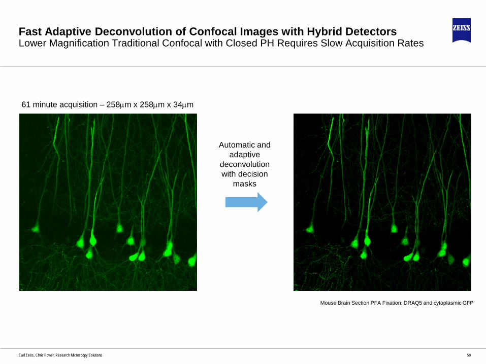
Fast Adaptive Deconvolution of Confocal Images with Hybrid DetectorsLower Magnification Traditional Confocal with Closed PH Requires Slow Acquisition Rates
61 minute acquisition – 258µm x 258µm x 34µm
Mouse Brain Section PFA Fixation; DRAQ5 and cytoplasmic GFP
Automatic and adaptive
deconvolution with decision
masks

51Carl Zeiss, Chris Power, Research Microscopy Solutions
Airyscan 2 with Multiplex ModeGentlest and Fastest Confocal Superresolution Imaging over large volumes
4 minute acquisition – 467µm x 467µm x 34µm
8Y-SR Multiplex mode
61 minute acquisition – 258µm x 258µm x 34µm
>15x faster over 3.3x larger volumewith 2 channels
Traditional Confocal with automatic and adaptive deconvolution with decision masks

52Carl Zeiss, Chris Power, Research Microscopy Solutions
Airyscan 2 with Multiplex ModeGentlest and Fastest Confocal Superresolution Imaging over large volumes
Dendritic spines accurately
represented
Dendritic spines NOT captured
61 minute acquisition – 258µm x 258µm x 34µm
50x increase in µm3/secRetaining all spatial information
4 minute acquisition – 467µm x 467µm x 34µm

53Carl Zeiss, Chris Power, Research Microscopy Solutions
Airyscan 2 with MultiplexParallelization – Gentle and Fast imaging
• Cells treated with cytotoxic drug
• Gentle superresolution imaging allows cells to
recover after the noxious substance is
removed
• Time lapse imaging for 20 hours every 10 min
Courtesy of Sarita Patnaik, PhD, Univ. of Mainz

54Carl Zeiss, Chris Power, Research Microscopy Solutions
Detectors used in the LSM 980

55Carl Zeiss, Chris Power, Research Microscopy Solutions
Detectors used in the LSM 980
Type Where Why Confocal NLO FLIM FCSDiode detector Transmitted light path Cheap and very robust X x
Traditional Multi Alkali Transmission PMT & Quasar channel 1
Robust to high light, very high dynamic range, better blue sensitivity X x
Multi alkali - Cooled Quasar channels 3 Better sensitivity in red. X x XGaAsP Quasar Better general sensitivity X x X
Multi Array Multi Alkali Quasar
32 channels of simultaneous spectral detection, used in the 510, 710 and 880, now obsolete X x X
Multi Array GaAsP Quasar
Better general sensitivity, 6 or 32 channels of simultaneous spectral detection X x X
Multi Array GaAsP AiryscanSuper resolution & super sensitive imaging X x
NDD Cooled Multi alkali NDD Reflected light or Transmitted light Better sensitivity in blue and red. XNosepiece GaAsP NDD Reflected light Best sensitivity in scattered light XBiG (Binary GaAsP) NDD Reflected light or Transmitted light Better general sensitivity X XBiG (Binary GaAsP) Confocal Direct couple port Better general sensitivity X x XGaAs or other Far red optimised PMT Confocal Direct couple port Best NIR detection X X XHybrid detectors Confocal Direct couple port Fast FLIM response x x X xMulti Channel Plate (MCP) Confocal Direct couple port Fastest FLIM response x x X
APD Dedicated FCS Unit
Used to be used for sensitivity, now obsolete as GaAsP internal more cost effective and much higher count X x X

56Carl Zeiss, Chris Power, Research Microscopy Solutions
Further reading
A good basic introduction (15 pages) and the source of several diagrams used todayhttp://www.et-enterprises.com/files/file/Understanding-photomultipliers.pdf
Complete handbook (323 pages)http://www.hamamatsu.com/resources/pdf/etd/PMT_handbook_v3aE.pdf
www.zeiss.com/airyscanWhitepapers on airyscan and Multiplex modes
http://zeiss-campus.magnet.fsu.edu/referencelibrary/pdfs/ZeissConfocalPrinciples.pdf
Detectors for LSM 56Carl Zeiss Microscopy, Chris Power, BioSciences

57Carl Zeiss, Chris Power, Research Microscopy Solutions Detectors for LSMCarl Zeiss Microscopy, Chris Power, BioSciences 57


