53
(a largely ectomesenchymally derived unit ) Dr.Jignesh R. Patel Dr. Shrikar Desai
| Date post: | 16-Jul-2015 |
| Category: |
Health & Medicine |
| Upload: | jignesh-patel |
| View: | 663 times |
| Download: | 4 times |

(a largely ectomesenchymally derived unit )
Dr.Jignesh R. PatelDr. Shrikar Desai

Contents :
•Introduction
•Development of teeth
•Stages of tooth growth
•Hertwig’s epithelial root sheath & root formation
•Development of cementum/ cementogenesis
•Development of PDL
•Development of alveolar bone
•Development of dentogingival unit
•conclusion
Dr.Jignesh

Introduction
The periodontium is simply defined as the tissues supporting
and investing the tooth - consists of cementum, PDL, bone
lining the alveolus & that part of the gingiva facing the tooth.
The tissues supporting the tooth are developmentally derived
from the dental follicle proper, whereas those investing the
tooth, that is the gingiva, are an adaptation of the oral mucosa.
- Richard Ten Cate
Dr.Jignesh

The widespread occurrence of periodontal diseases & the realization that
periodontal tissues lost to the disease can be repaired has resulted in
considerable effort to understand the factors & cells regulating the
formation, maintenance, & regeneration of the periodontium.
- Ten Cate et al Periodontology 2000, Vol. 13
Dr.Jignesh

Reciprocal induction
Dr.Jignesh
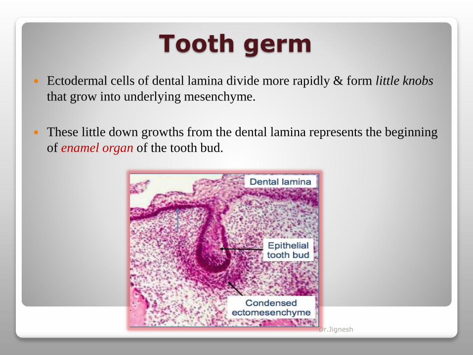
Tooth germ
Ectodermal cells of dental lamina divide more rapidly & form little knobs
that grow into underlying mesenchyme.
These little down growths from the dental lamina represents the beginning
of enamel organ of the tooth bud.
Dr.Jignesh

As cell proliferation continues...
Each enamel organ increase in size & sink deeper into the
ectomesenchyme, & due to differential growth shape also changes.
First it takes a shape that resembles a Cap, with an outer convex facing
the oral cavity & inner concavity.
Dr.Jignesh
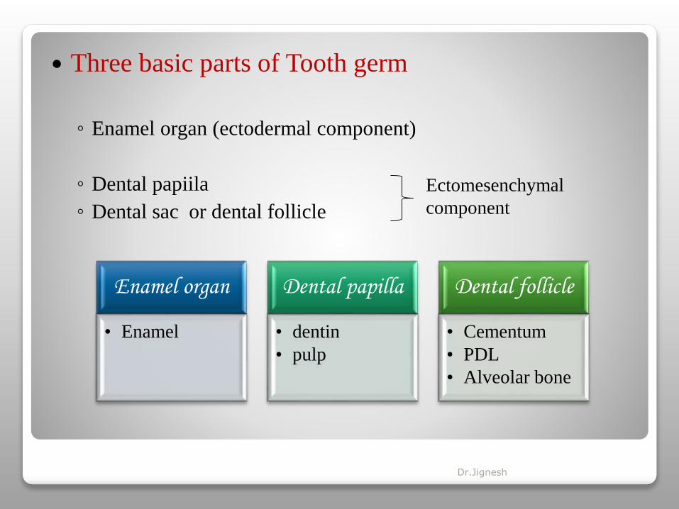
Three basic parts of Tooth germ
◦ Enamel organ (ectodermal component)
◦ Dental papiila
◦ Dental sac or dental follicle
Ectomesenchymal
component
Enamel organ
• Enamel
Dental papilla
• dentin
• pulp
Dental follicle
• Cementum
• PDL
• Alveolar bone
Dr.Jignesh
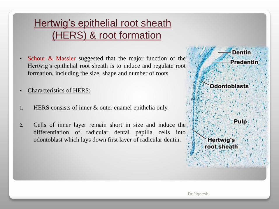
Hertwig’s epithelial root sheath
(HERS) & root formation
Schour & Massler suggested that the major function of the
Hertwig’s epithelial root sheath is to induce and regulate root
formation, including the size, shape and number of roots
Characteristics of HERS:
1. HERS consists of inner & outer enamel epithelia only.
2. Cells of inner layer remain short in size and induce the
differentiation of radicular dental papilla cells into
odontoblast which lays down first layer of radicular dentin.
Dr.Jignesh

3. Some outer layer cells in coronal root region induce cells of dental follicle to
differentiate into cementoblasts similar to osteoblasts which give rise to acellular
cementum.
4. Slavkin suggests, since the epithelial cells of the inner layer of Hertwig’s epithelial root
sheath are analogous to the preameloblasts, it is suggested that they might secrete
enamel matrix proteins over the newly deposited root dentin.
5. In addition to these matrix proteins there are also the components of the epithelial
basement membrane, such as laminin and collagen type IV are sectreted by root sheath.
Dr.Jignesh
Dr.Jignesh

Development of cementum
Process of cementum development is called as “cementogenesis”.
Cementum is calcified, avascular mesenchymal tissue that forms outer coverings of the
anatomic root.
It was first demonstrated microscopically in 1835 by two pupils of purkinje.
It is a specialized connective tissue that shares some physical, chemical & structural
characteristics with compact bone.
Dr.Jignesh

Cementogenesis (Briefly)
Dr.Jignesh

[Hertwig’s epithelial root sheath is broken up &
separated from root, and differentiation of
cementoblasts lead to formation of cementum] Dr.Jignesh

Varieties of cementum
Two basic types of cementum, hence they are usually classified on the basis of presence of
cementocyte (cellular cementum) or absence of it (acellular cementum).
It can also be classified on the basis of the types of fibers (extrinsic/intrinsic) presence or
their absence (afibrillar cementum).
acellular extrinsic fiber cementum is regarded as Primary cementum as it forms first.
Cellular cementum is regarded as secondary cementum because it forms later than
primary cementum.
Dr.Jignesh

Growth factor families involved in the differentiation of
cemetoblasts from dental follicle
TGFβ 1-5
BMP2-8
EGF & IGF
PGE2 & PGF2α enhance differentiation byactivating protein kinase cell signallingpathway
Fibroblast growth factor promotes proliferation, migration & angiogenesis
CAP, BSP and osteopontine helps inattachment of differentiated cells to newlyforming tissue
Dr.Jignesh
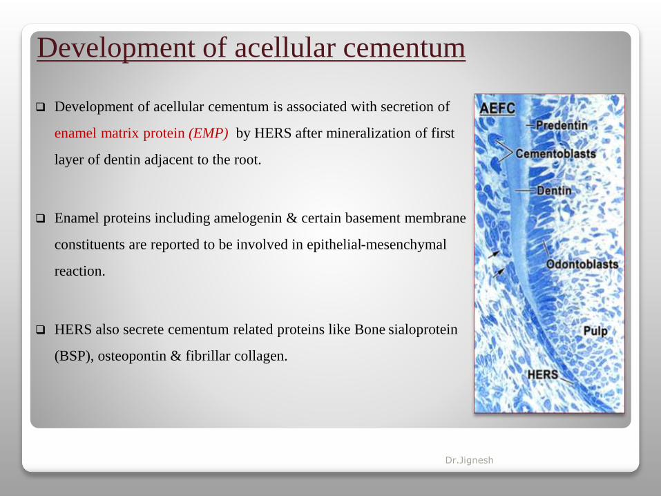
Development of acellular cementum
Development of acellular cementum is associated with secretion of
enamel matrix protein (EMP) by HERS after mineralization of first
layer of dentin adjacent to the root.
Enamel proteins including amelogenin & certain basement membrane
constituents are reported to be involved in epithelial-mesenchymal
reaction.
HERS also secrete cementum related proteins like Bone sialoprotein
(BSP), osteopontin & fibrillar collagen.
Dr.Jignesh

At the same time fibroblast precursors cells from dental follicle come in contact with
predentine matrix and start depositing bundle of collagen fibrils to form a thin layer of
perpendicularly oriented “Sharpey’s fibers” or “fringe fibers”.
Sharpey’s fibers interdigitate with unmineralized dentin at one end and into extracellular
compartment of acellular cementum at another end.
As the mineralization front advances, it contacts the sharpey’s fibers and they undergo slow
mineralization to complete the process of acellular extrinsic fiber cementum formation.
Dr.Jignesh

Development of cellular cementum(a more rapidly formed & less mineralized variety of cementum)
Formation occurs after at least half the root is formed.
Development can be divided into 2 stages:
An early stage in which extrinsic sharpey’s fibers produced by fibroblasts are few &
traces of intrinsic fibers produced by cementoblasts are randomly arranged
Later stage of cementogenesis- it closely resembles bone formation. Cementoblasts and
cementocytes are involved in the secretion of intrinsic fibers.
Dr.Jignesh
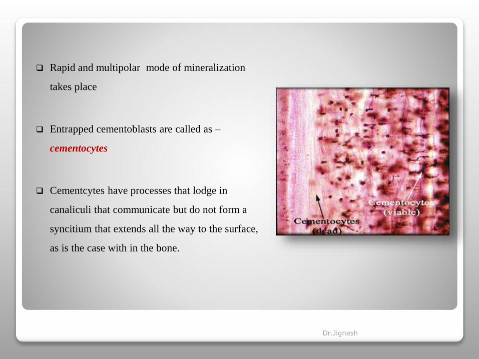
Rapid and multipolar mode of mineralization
takes place
Entrapped cementoblasts are called as –
cementocytes
Cementcytes have processes that lodge in
canaliculi that communicate but do not form a
syncitium that extends all the way to the surface,
as is the case with in the bone.
Dr.Jignesh

Cementoid tissue & calcification of matrix
The uncalcified matrix is called as cementoid.
the growth of cellular cementum is a rhythmic process, and as a new layer of cementoid is formed, the old calcifies.
Gla proteins – osteocalcin & osteonectin acts as neucleators for mineralization due to their strong affinity for calcium & BSP.
Alkaline phosphatase promotes mineralization.
Osteopontine regulates growth of apatite crystals.
Major proteoglycan located in non-mineralized cementum is keratan sulfates- lumican & fibromodulin.
Dr.Jignesh
Dr.Jignesh

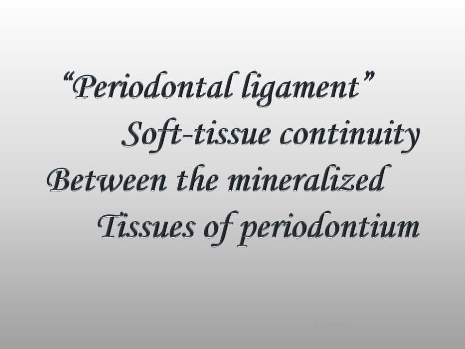
The periodontal ligamment (PDL) is composed of a complex
vascular & highly cellular connective tissue that surrounds the tooth
root & connects it to the inner wall of the alveolar bone.
Over the years it has been described by number of terms:
• Desmodont
• Gomphosis
• Pericementum
• Dental periosteum
• Alveodental ligament
• Periodontal membrane
Dr.Jignesh

Development
Development of PDL begins with root formation, prior to tooth eruption.
The dental follicle cells located between the alveolar bone & HERS are composed of two
subpopulations:
Mesenchymal cells of dental follicle proper
Perifollicular mesenchyme
Perifollicular mesenchymal cells bounded by mesenchymal cells of dental follicle proper.
Dr.Jignesh

As the root formation continues, cells in the perifollicular region and follicle proper are gain their
polarity & the cellular volume & synthetic activity increases.
These cells obtain long & thin, elongated cytoplasm with increased amount of mitochondria,
RER & active Golgi complex.
As a result, these cells actively synthesize & deposits collagen fibrils & glycoprotein in the
developing periodontal ligament.
progenitors for periodontal ligament, osteoblast and cementoblast cells adopt a paravascular
location in the periodontal ligament, and these cells, which exhibit some features of stem cells,
can regenerate functional tissues when the need arises.
Dr.Jignesh
Developmet of principal fibers
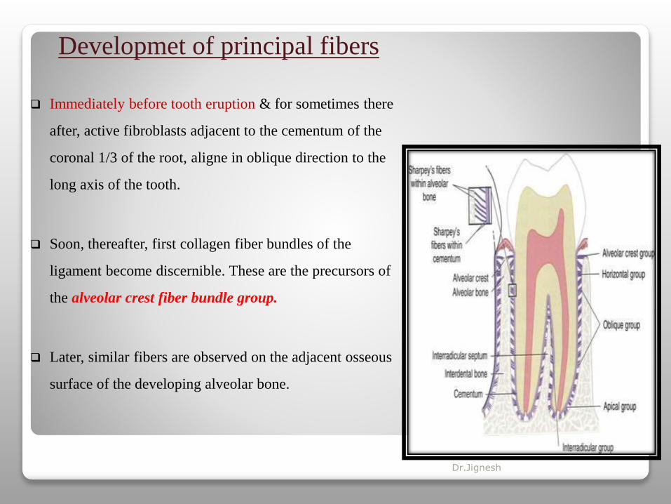
Immediately before tooth eruption & for sometimes there
after, active fibroblasts adjacent to the cementum of the
coronal 1/3 of the root, aligne in oblique direction to the
long axis of the tooth.
Soon, thereafter, first collagen fiber bundles of the
ligament become discernible. These are the precursors of
the alveolar crest fiber bundle group.
Later, similar fibers are observed on the adjacent osseous
surface of the developing alveolar bone.
Dr.Jignesh

Both set of fibers, alveolar & cemental, continue to elongate toward each other, ultimately to meet,
intertwine & fuse, & cross linking of individual collagen molecules occur.
By the time of first occlusal contact of the tooth with its antagonist, the principle fibers around the
coronal 1/3 of the root, the horizontal group are almost completely developed
Oblique fibers in middle third of the root are still being formed.
After complete root apex is formed, apical group of fibers are developed.
Dr.Jignesh

PDL homeostasis
A remarkable capacity of PDL is that it maintains its width more or less, despite the fact, it is
squeezed in between two hard tissues.
Various molecules have been proposed, which play a role in maintaining an unmineralized PDL.
Msx2
Bone sialoprotein
Matrix Gla proteins
(Inhibitors of mineralization)
• Inhibit mineralized bone tissue
• Prevents osteogeningdifferentiation of PDLfibroblasts by repressingcbfa1 activity
• osteopontin
Prostaglandins
Dr.Jignesh

Synthetic cells
Fibroblasts
Osteoblasts
cementoblasts
Resorportivecells
Fibroblasts
Osteoclasts
cementoclasts
Progenitor cells
Undifferentiated stem cells
Epithelial cells
Epithelial cell rests of
malassez
Defense cells
Mast cells
Macrophages
eiosinophils
Dr.Jignesh

Epithelial cell rests of malassez
Roles attributed to the Epithelial Rest of Malassez cells
range from bad to good.
Bad Role
Malassez cells are held responsible for the formation of
periodontal cysts and tumours as a result of peri-apical
inflammation associated with pulpal necrosis.
Epithelial Rest of Malassez cells contribute to the
formation of the periodontal pocket because of their
continuum with the junctional epithelium.
-Ohshima M, Nishiyama T, Tokunaga K, Sato S, Maeno M, Otsuka K.
Dr.Jignesh

Good Role
The cells of the Epithelial Rest of Malassez may protect the root from resorption
- Wallace JA, Vergona K.
Epithelial cells Rest of Malassez secrete hyaluronic acid, which contributes to the formation
of the loose connective tissue characteristics of the periodontal ligament & react to
mechanical stress, like that associated with orthodontic tooth movement, by increasing their
proliferation rate and cell size. - Brunette DM & Merrilees MJ, Sodek J, Aubin JE
Epithelial Rest of Malassez - help in cementum repair because of their ability to activate
matrix proteins, such as amelogenin, which are also expressed during tooth development -
Hamamoto Y, Nakajima T, Ozawa H, Uchida T.
Dr.Jignesh
Dr.Jignesh
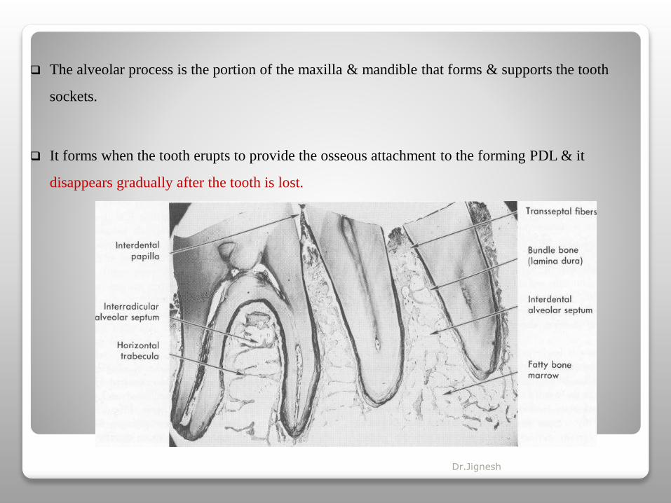
The alveolar process is the portion of the maxilla & mandible that forms & supports the tooth
sockets.
It forms when the tooth erupts to provide the osseous attachment to the forming PDL & it
disappears gradually after the tooth is lost.
Dr.Jignesh

Intramembranous ossification
Formation of bone matrix
Formation of woven bone
Appositional growth & formation of harvesian
system (osteon)
Endochondral bone formation
Formation of cartiagenous model
Dr.Jignesh

Development of alveolar process
An alveolar bone in the strict sense of words develops only during the eruption of the teeth.
As the root & its covering of primary cementum form, new bone is deposited against the
crypt wall.
Dr.Jignesh

Crystal form coalescing bone nodules with fast
growing, non oriented collagen fibers- is the
substructure of woven bone, first bone formed in the
alveolus.
Later, through bone deposition, remodelling &
secretion of oriented collagen fibers in sheets, mature
lamellar bone is formed.
Subsequently, a tissue may develop at alveolar crest
that combines characteristics of cartilage & bone.
It is called as chondroid bone.
Dr.Jignesh

Structure of alveolar bone
(a thin lamella of the bone that surrounds the rootof the tooth & gives attachment to the
principle fibers of the PDL)
• Inner socket wall of thin compact bone
• Bundle bone
• Cribriform plate
• Lamina Dura (radiographically)
supporting alveolar bone
(the bone that surrounds the alveolar bone proper
& gives support to the socket)
• External plate of cortical bone
• Spongy bone/ cancellous trabeculae
Dr.Jignesh

Bone remodeling
Bone is a highly dynamic connective tissue with continuous remodeling.
Process of bone formation & bone breakdown go on simultaneously, thus the bone represents
the net results of a balance between the two processes
This phenomena is called as coupling of bone resorption & bone formation.
The main function of the remodeling are to prevent the accumulation of damaged & fatigued
bone by regenerating new bone & to facilitate mineral homeostasis.
Dr.Jignesh

Mediators of bone remodeling
Mechanical factors : when stress is applied on the alveolar bone, two sites are formed, bone is
resorbed at compression site & bone is deposited at tension site.
Parathyroid hormone
Vita. D metabolites
Growth factors
Bacterial products
Dr.Jignesh

Markers of bone turnover
• Alkaline phosphatase
• osteocalcin
• Procollagen I extension peptide
• Urine calcium
• Urine hydroxyproline
• Collagen crosslink fragments
• Urine N-telopeptides
• Urine C-telopeptides
• Urine pyridinoline
• Urine free deoxypyridinoline
Dr.Jignesh

Dr.Jignesh
Gingiva is an adaptation of oral epithelium in areas involved in mastication of food
The gingiva is a part of the oral mucosa that covers the alveolar processes of the jaws &
surrounds the neck of the teeth.- McCall
Dr.Jignesh

Dr.Jignesh

Dentogingival junction
(junctional epithelium)
The epithelium of the gingiva which gets
attached to the tooth is called as junctional or
attachment epithelium.
It consists of collar like band of stratified
squamous non keratinizing epithelium, located at
CEJ in healthy tissue
Dr.Jignesh
JE resembles reduced enamel epithelium (RER) in its structure in that they have a basal
layer & few layers of flattened cells & express CK 5, 14, 19, which is typical of
nondifferentiating tissue like RER.
Highest turnover rate of 5-6 days
JE is highly permeable & it has large intracellular spaces, so that neutrophils can easily pass
in & out of the epithelium.
Permits easy flow of GCF
Dr.Jignesh

Development of dentoginval junction &
gingival sulcus
Dr.Jignesh

Formation of JE & Gingival sulcus
Dr.Jignesh
Shift of dentogingival junction
A. The actual movement of crown towards the occlusal plane is called as a active eruption
B. The separation of primary attachment epithelium from the enamel is termed as passive
eruption
crown exposure involving passive eruption & further recession has been described in
four stages
firsr two stages may be physiologic but last two are probably pathologic.
Dr.Jignesh

Shift of dentogingival junction
Dr.Jignesh

Dr.Jignesh
Reciprocal induction between oral ectoderm & mesenchymal cells derived from neural crest
cells form the major pathway for the development of periodontal tissues.
Various histochemical molecules favours the differentiation of fibroblasts, cementoblasts &
osteoblasts from the inner cells of the dental sac, which are also secreted at the time of
periodontal regeneration or repair by PDL
PDL contains both formative & resorptive cells for cementum, A.bone & PDL itself.
Dr.Jignesh

Based on the information presented, it appears that the developed or adult
periodontium retains its potential for repair/regeneration in the form of
cells of the Epithelial Rest of Malassez, progenitor cells and stem cells,
which can be induced to differentiate into cementoblast, osteoblast or
periodontal ligament cells to regenerate periodontal tissues.
Dr.Jignesh

References:
1. Textbook of Orban’s Oral histology & Embryology, 12th Ed.
2. Textbook of TenCate’s Oral histolgy & Embryology, 8th Ed.
3. Margarita zeichner-david, Regeneration of periodontal tissues: cementogenesis revisited,
Periodontology 2000, Vol. 41, 2006, 196–217.
4. A. Richard ten cate, The development of the periodontium - a largely ectomesenchymally derived
unit, Periodontology 2000, Vol. 13, 1997, 9-19.
5. Thomas HF, Kollar EJ. Differentiation of odontoblasts in grafted recombinants of murine epithelial
root sheath and dental mesenchyme. Arch Oral Biol 1989; 34: 27-35.
6. Textbook of clinical periodontology, F.A.Carranza, 10th Ed.
Dr.Jignesh


