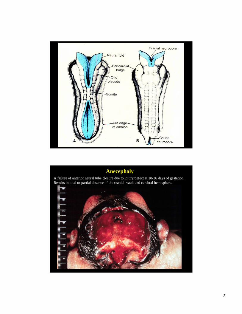
AnecephalyA failure of anterior neural tube closure due to injury/defect at 18-26 days of gestation. Results in total or partial absence of the cranial vault and cerebral hemisphere.
3
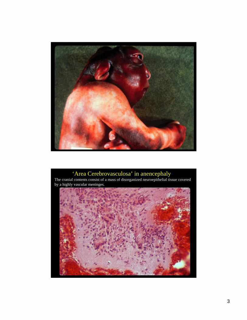
‘Area Cerebrovasculosa’ in anencephalyThe cranial contents consist of a mass of disorganized neuroepithelial tissue coveredby a highly vascular meninges.
4
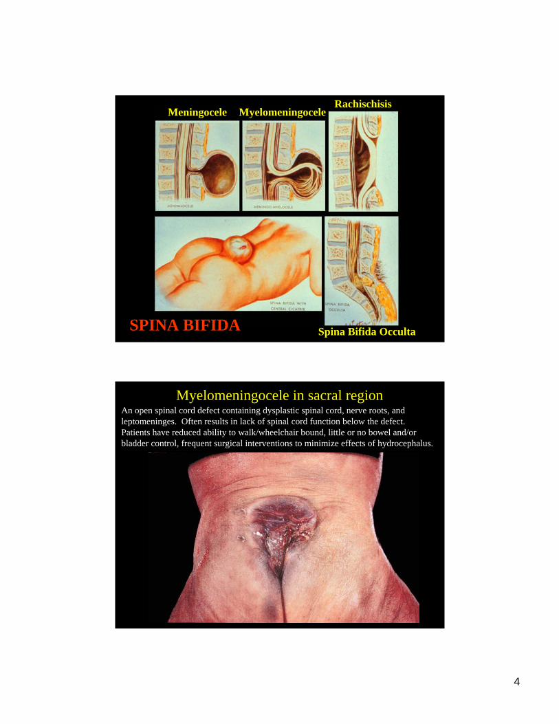
Meningocele MyelomeningoceleRachischisis
Spina Bifida OccultaSPINA BIFIDA
Myelomeningocele in sacral regionAn open spinal cord defect containing dysplastic spinal cord, nerve roots, and leptomeninges. Often results in lack of spinal cord function below the defect. Patients have reduced ability to walk/wheelchair bound, little or no bowel and/or bladder control, frequent surgical interventions to minimize effects of hydrocephalus.
5
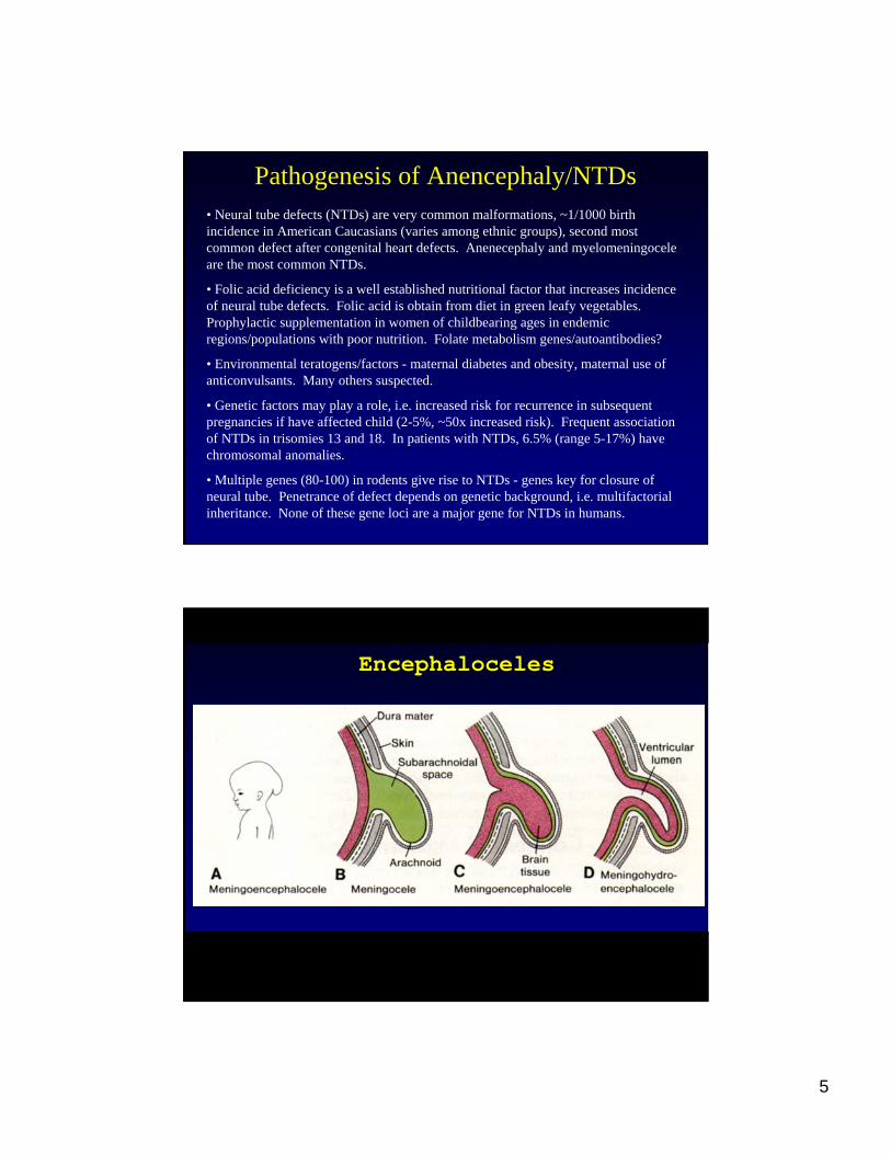
Pathogenesis of Anencephaly/NTDs• Neural tube defects (NTDs) are very common malformations, ~1/1000 birth incidence in American Caucasians (varies among ethnic groups), second most common defect after congenital heart defects. Anenecephaly and myelomeningoceleare the most common NTDs.
• Folic acid deficiency is a well established nutritional factor that increases incidence of neural tube defects. Folic acid is obtain from diet in green leafy vegetables. Prophylactic supplementation in women of childbearing ages in endemic regions/populations with poor nutrition. Folate metabolism genes/autoantibodies?
• Environmental teratogens/factors - maternal diabetes and obesity, maternal use of anticonvulsants. Many others suspected.
• Genetic factors may play a role, i.e. increased risk for recurrence in subsequent pregnancies if have affected child (2-5%, ~50x increased risk). Frequent association of NTDs in trisomies 13 and 18. In patients with NTDs, 6.5% (range 5-17%) have chromosomal anomalies.
• Multiple genes (80-100) in rodents give rise to NTDs - genes key for closure of neural tube. Penetrance of defect depends on genetic background, i.e. multifactorialinheritance. None of these gene loci are a major gene for NTDs in humans.
EncephalocelesEncephaloceles
6
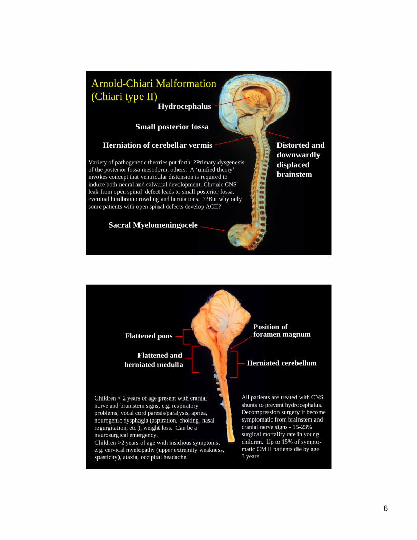
Hydrocephalus
Small posterior fossa
Herniation of cerebellar vermis
Sacral Myelomeningocele
Distorted and downwardly displaced brainstem
Variety of pathogenetic theories put forth: ?Primary dysgenesisof the posterior fossa mesoderm, others. A ‘unified theory’invokes concept that ventricular distension is required to induce both neural and calvarial development. Chronic CNS leak from open spinal defect leads to small posterior fossa,eventual hindbrain crowding and herniations. ??But why onlysome patients with open spinal defects develop ACII?
Arnold-Chiari Malformation(Chiari type II)
Flattened pons
Flattened and herniated medulla
Position offoramen magnum
Herniated cerebellum
Children < 2 years of age present with cranialnerve and brainstem signs, e.g. respiratory problems, vocal cord paresis/paralysis, apnea,neurogenic dysphagia (aspiration, choking, nasalregurgitation, etc.), weight loss. Can be a neurosurgical emergency.Children >2 years of age with insidious symptoms,e.g. cervical myelopathy (upper extremity weakness,spasticity), ataxia, occipital headache.
All patients are treated with CNSshunts to prevent hydrocephalus.Decompression surgery if becomesymptomatic from brainstem andcranial nerve signs - 15-23%surgical mortality rate in youngchildren. Up to 15% of sympto-matic CM II patients die by age3 years.
7
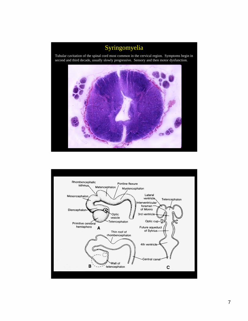
SyringomyeliaTubular cavitation of the spinal cord most common in the cervical region. Symptoms begin in second and third decade, usually slowly progressive. Sensory and then motor dysfunction.
8
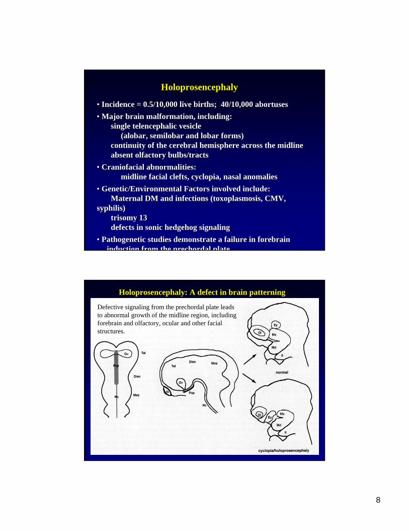
HoloprosencephalyHoloprosencephaly
•• Incidence = 0.5/10,000 live births; 40/10,000 Incidence = 0.5/10,000 live births; 40/10,000 abortusesabortuses•• Major brain malformation, including:Major brain malformation, including:
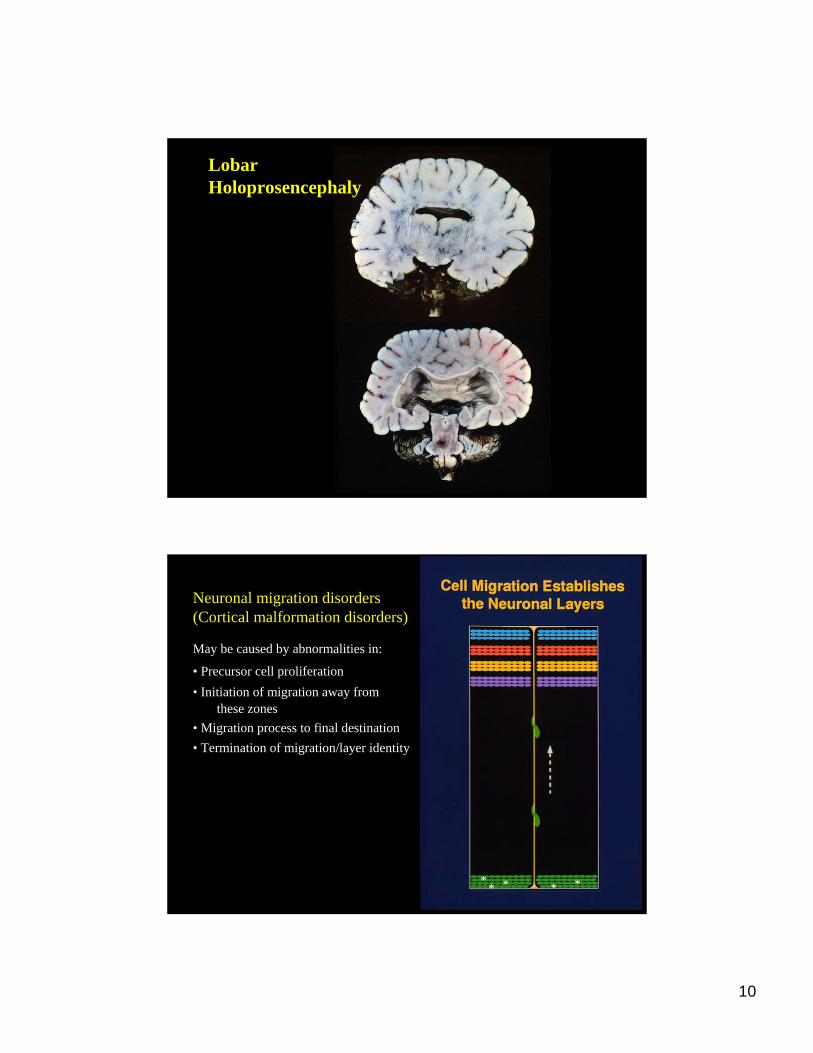
single single telencephalictelencephalic vesicle vesicle ((alobaralobar, , semilobarsemilobar and lobar forms)and lobar forms)
continuity of the cerebral hemisphere across the midlinecontinuity of the cerebral hemisphere across the midlineabsent olfactory bulbs/tracts absent olfactory bulbs/tracts
•• Genetic/Environmental Factors involved include:Genetic/Environmental Factors involved include:Maternal DM and infections (toxoplasmosis, CMV, Maternal DM and infections (toxoplasmosis, CMV,
syphilis)syphilis)trisomytrisomy 1313defects in sonic hedgehog signalingdefects in sonic hedgehog signaling
•• PathogeneticPathogenetic studies demonstrate a failure in forebrain studies demonstrate a failure in forebrain induction frominduction from thethe prechordalprechordal plateplate
HoloprosencephalyHoloprosencephaly: A defect in brain patterning: A defect in brain patterning
Defective signaling from the prechordal plate leads to abnormal growth of the midline region, including forebrain and olfactory, ocular and other facial structures.
9
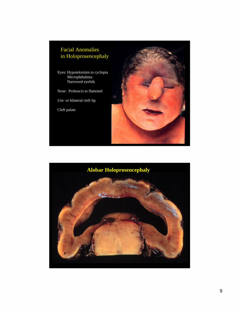
Facial Anomaliesin Holoprosencephaly
Eyes: Hypotelorisim to cyclopiaMicrophthalmiaNarrowed eyelids
Polymicrogyria ** Associated with disruptive/destructive lesions, in utero infections (e.g. CMV), other; Bilateral, partial (frontal, perisylvian, parietal or posterior)
Diffuse Heterotopia Leptomeningeal heterotopia Periventricular nodular heterotopia (unilateral or bilateral) Subcortical band heterotopia ("double cortex")
Focal Heterotopia ** Subcortical ** Subependymal ** = most common
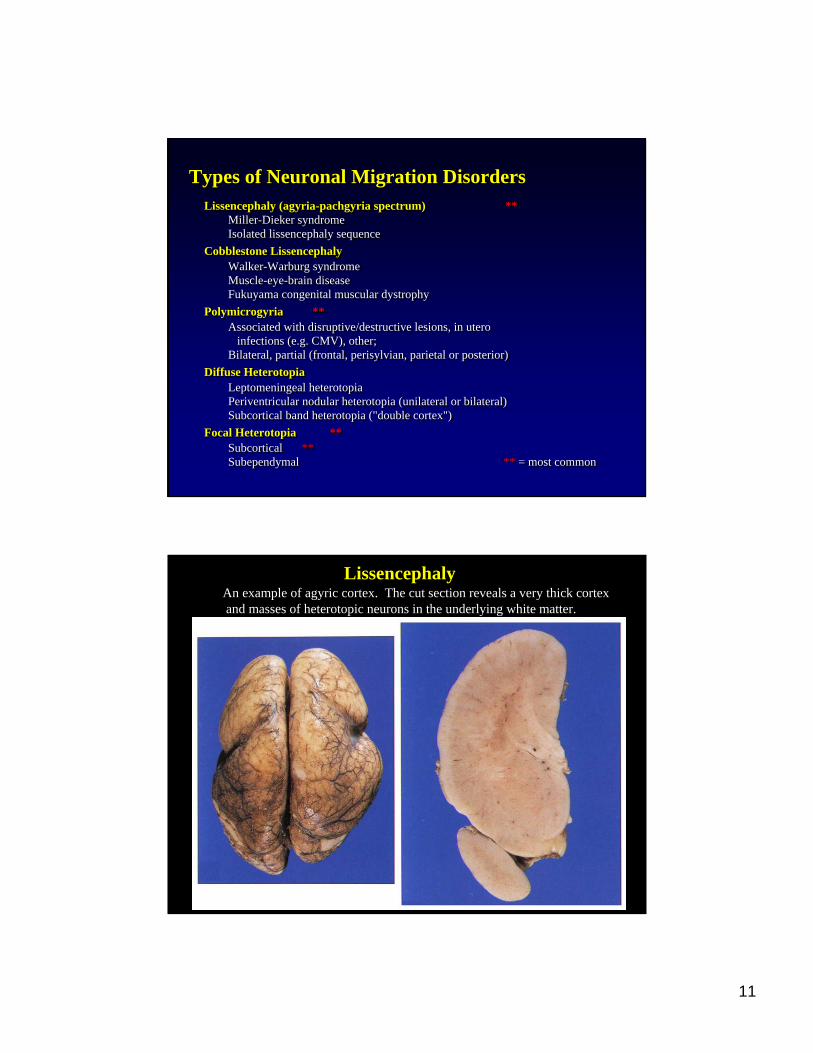
LissencephalyAn example of agyric cortex. The cut section reveals a very thick cortexand masses of heterotopic neurons in the underlying white matter.
12
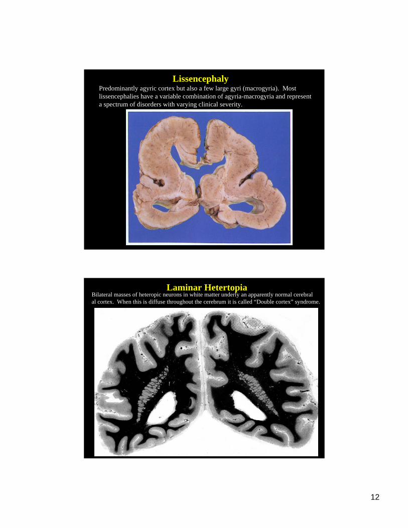
LissencephalyPredominantly agyric cortex but also a few large gyri (macrogyria). Most lissencephalies have a variable combination of agyria-macrogyria and representa spectrum of disorders with varying clinical severity.
Laminar HetertopiaBilateral masses of heteropic neurons in white matter underly an apparently normal cerebralal cortex. When this is diffuse throughout the cerebrum it is called “Double cortex” syndrome.
13
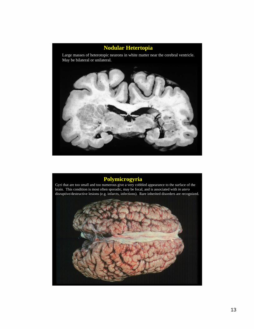
Nodular HetertopiaLarge masses of heterotopic neurons in white matter near the cerebral ventricle. May be bilateral or unilateral.
PolymicrogyriaGyri that are too small and too numerous give a very cobbled appearance to the surface of thebrain. This condition is most often sporadic, may be focal, and is associated with in uterodisruptive/destructive lesions (e.g. infarcts, infections). Rare inherited disorders are recognized.
14
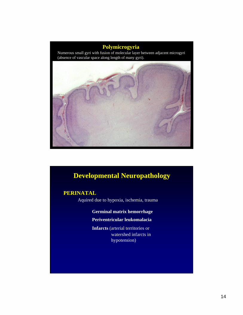
PolymicrogyriaNumerous small gyri with fusion of molecular layer between adjacent microgyri(absence of vascular space along length of many gyri).
PERINATALAquired due to hypoxia, ischemia, trauma
Germinal matrix hemorrhage
Periventricular leukomalacia
Infarcts (arterial territories or watershed infarcts inhypotension)
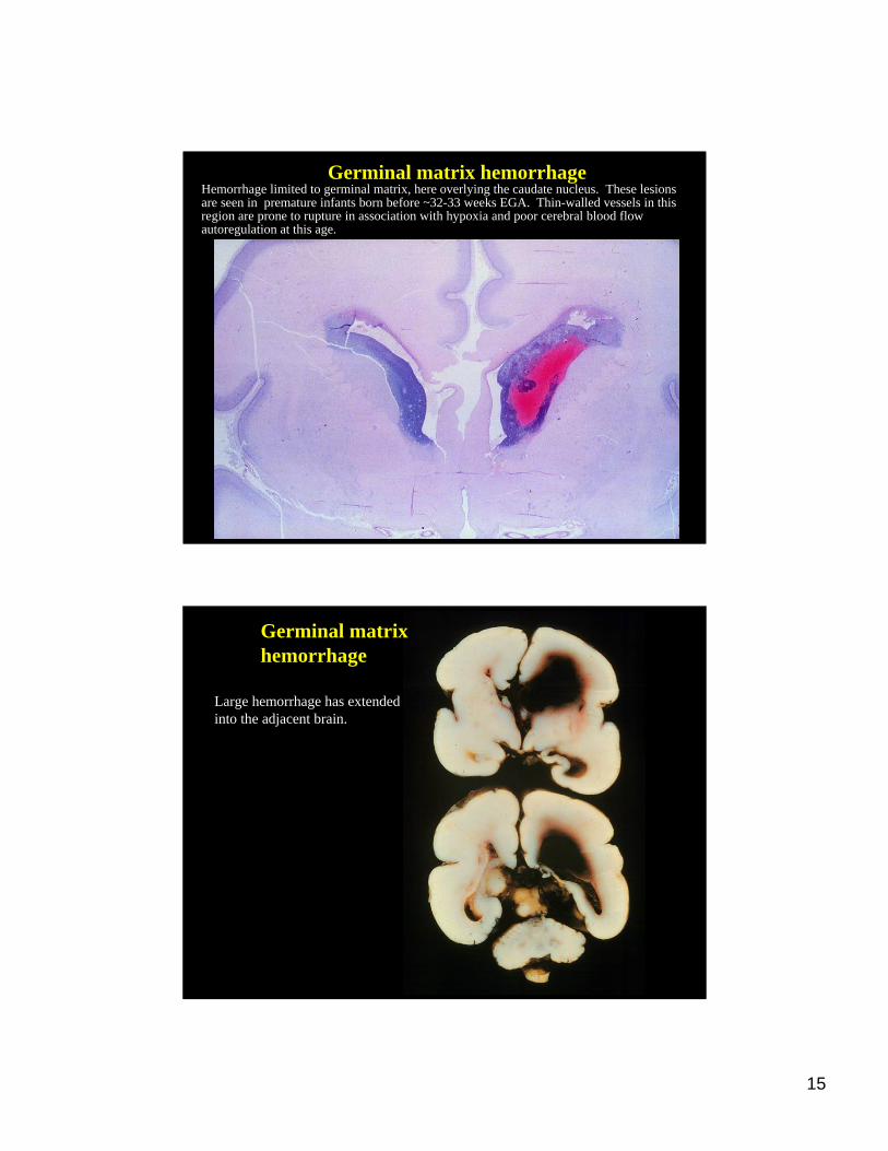
Germinal matrix hemorrhageHemorrhage limited to germinal matrix, here overlying the caudate nucleus. These lesions are seen in premature infants born before ~32-33 weeks EGA. Thin-walled vessels in this region are prone to rupture in association with hypoxia and poor cerebral blood flow autoregulation at this age.
Germinal matrix hemorrhage
Large hemorrhage has extendedinto the adjacent brain.
16
Germinal matrix hemorrhage
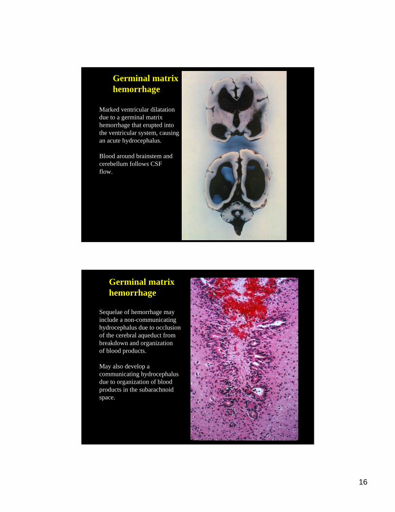
Marked ventricular dilatationdue to a germinal matrix hemorrhage that erupted intothe ventricular system, causingan acute hydrocephalus.
Blood around brainstem and cerebellum follows CSFflow.
Germinal matrix hemorrhage
Sequelae of hemorrhage mayinclude a non-communicatinghydrocephalus due to occlusionof the cerebral aqueduct frombreakdown and organization of blood products.
May also develop a communicating hydrocephalus due to organization of blood products in the subarachnoidspace.
17
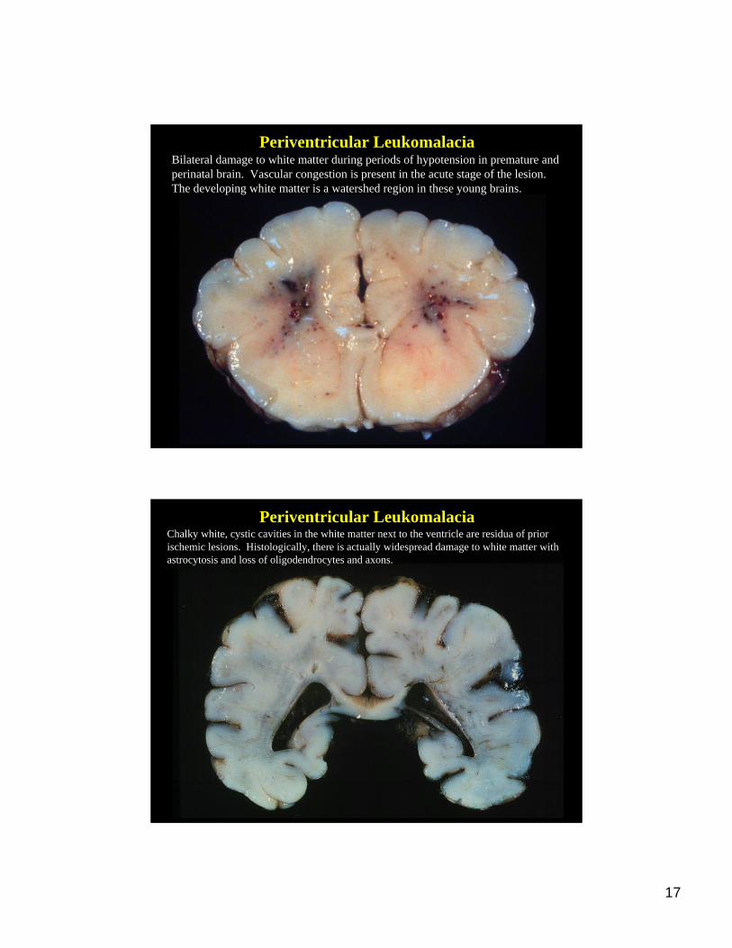
Periventricular LeukomalaciaBilateral damage to white matter during periods of hypotension in premature andperinatal brain. Vascular congestion is present in the acute stage of the lesion. The developing white matter is a watershed region in these young brains.
Periventricular LeukomalaciaChalky white, cystic cavities in the white matter next to the ventricle are residua of prior ischemic lesions. Histologically, there is actually widespread damage to white matter with astrocytosis and loss of oligodendrocytes and axons.
18
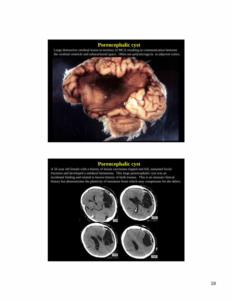
Porencephalic cystLarge destructive cerebral lesion in territory of MCA resulting in communication betweenthe cerebral ventricle and subarachnoid space. Often see polymicrogyria in adjacent cortex.
Porencephalic cystA 56 year old female with a history of breast carcinoma tripped and fell, sustained facialfractures and developed a subdural hematoma. This large porencephalic cyst was anincidental finding and related to known history of birth trauma. This is an unusual clinicalhistory but demonstrates the plasticity of immature brain which may compensate for the defect.
19
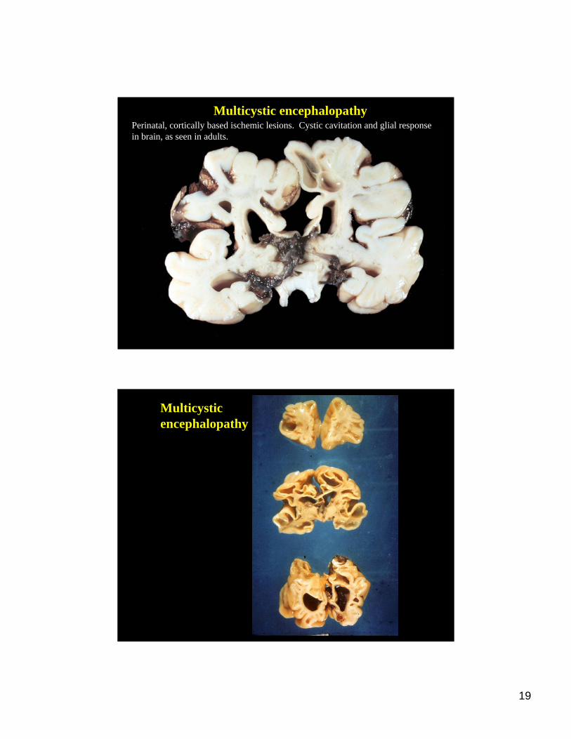
Multicystic encephalopathyPerinatal, cortically based ischemic lesions. Cystic cavitation and glial responsein brain, as seen in adults.
Cellular proliferations (Cellular proliferations (hyperplastichyperplastic or or neoplasticneoplastic) which) whichoccur in association with malformationsoccur in association with malformations
Affect the nervous system and skinAffect the nervous system and skin
Neurofibromatosis type INeurofibromatosis type I autosomalautosomal dominantdominant
Neurofibromatosis type IINeurofibromatosis type II autosomalautosomal dominantdominant
Von Recklinghausen NeurofibromatosisVon Recklinghausen NeurofibromatosisFirst described in 1882. Most commonly known form is Neurofibromatosistype I (NFI) with several variant forms having different clinical features (NF2-NFVII).
Characteristics of NFI:1) Autosomal dominant inheritance with variable expressivity and high
penetrance. Prevalence of 1 in 2500 - 3000.
2) 50% of patients have an affected family member; the remainingrepresent new mutations.
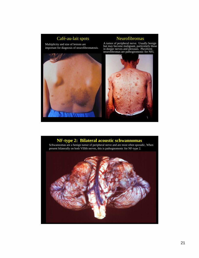
3) Common lesions: Café-au-lait spots (>6,>0.5 cm), 90% of patientsNeurofibromas (cutaneous, deep, plexiform)Pigmented iris hamartomas (Lisch nodules)Axillary or groin frecklingSkeletal abnormalities (e.g. scoliosis)Learning disordersIncreased risk of malignancy/other tumors
21
Café-au-lait spots NeurofibromasMultiplicity and size of lesions are important for diagnosis of neurofibromatosis.
A tumor of peripheral nerve. Usually benignbut may become malignant, particularly thosein deeper nerves and plexuses. Plexiformneurofibromas are pathognomonic for NFI.
NF-type 2: Bilateral acoustic schwannomasSchwannomas are a benign tumor of peripheral nerve and are most often sporadic. Whenpresent bilaterally on both VIIIth nerves, this is pathognomonic for NF-type 2.
22
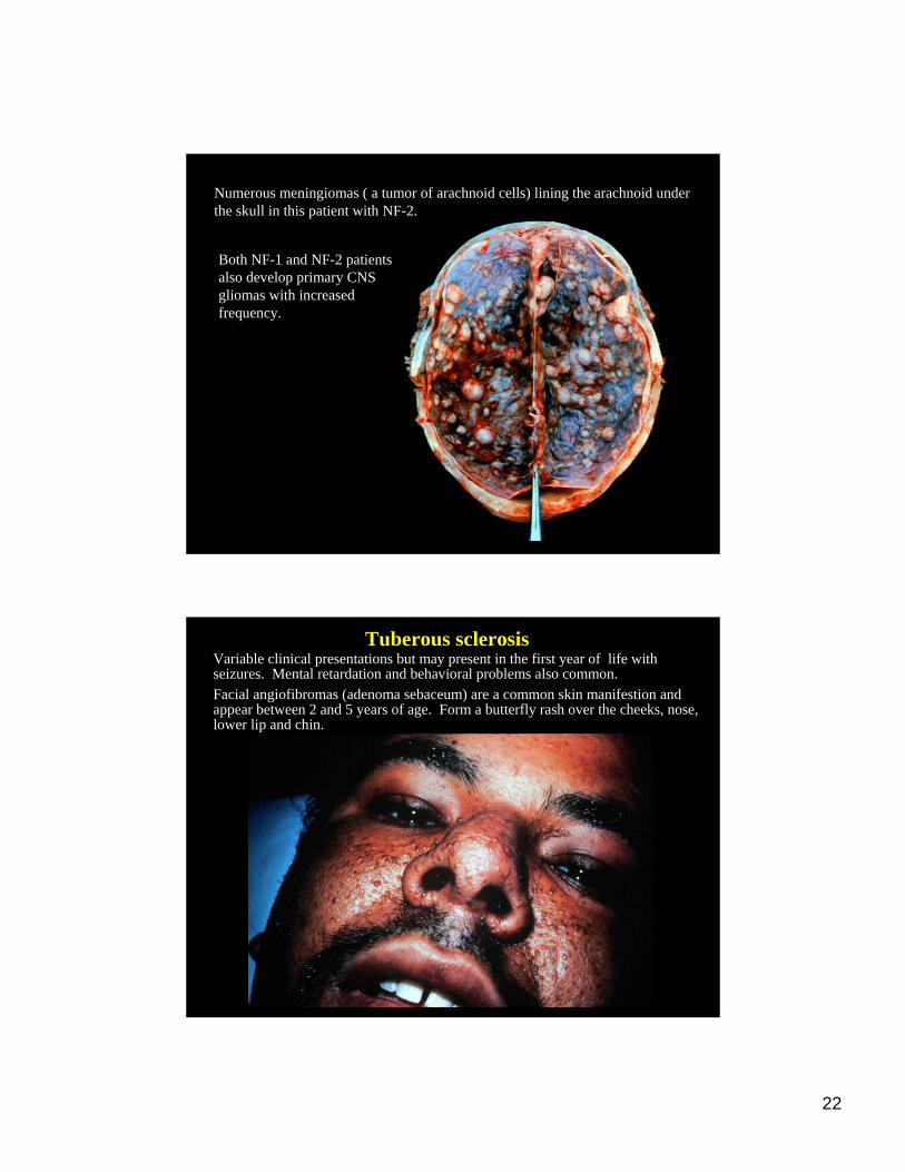
Numerous meningiomas ( a tumor of arachnoid cells) lining the arachnoid under the skull in this patient with NF-2.
Both NF-1 and NF-2 patients also develop primary CNS gliomas with increased frequency.
Tuberous sclerosisVariable clinical presentations but may present in the first year of life withseizures. Mental retardation and behavioral problems also common.Facial angiofibromas (adenoma sebaceum) are a common skin manifestion and appear between 2 and 5 years of age. Form a butterfly rash over the cheeks, nose,lower lip and chin.
23
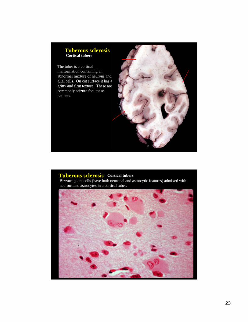
Tuberous sclerosisCortical tubers
The tuber is a cortical malformation containing an abnormal mixture of neurons and glial cells. On cut surface it has a gritty and firm texture. These are commonly seizure foci these patients.
Tuberous sclerosis Cortical tubersBizzarre giant cells (have both neuronal and astrocytic features) admixed withneurons and astrocytes in a cortical tuber.
24
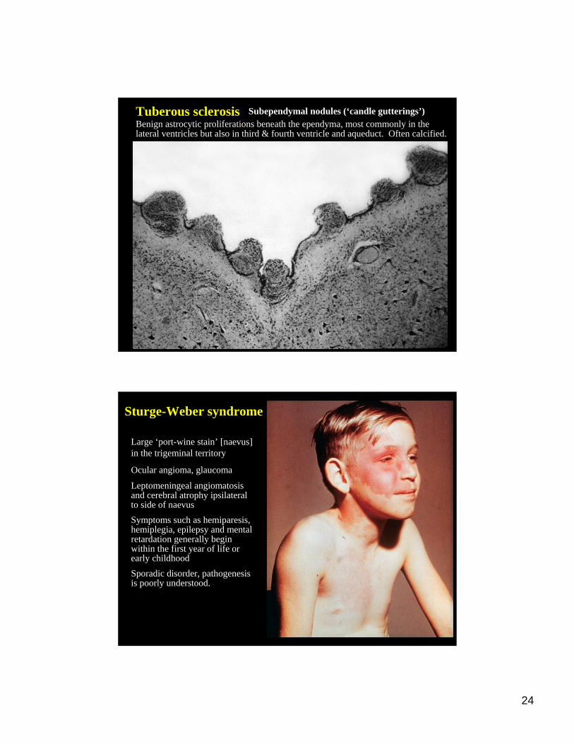
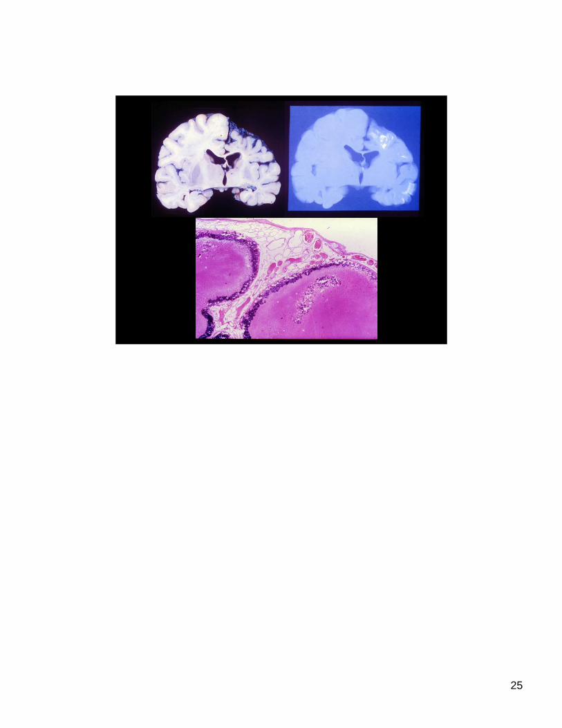
Tuberous sclerosis Subependymal nodules (‘candle gutterings’)Benign astrocytic proliferations beneath the ependyma, most commonly in the lateral ventricles but also in third & fourth ventricle and aqueduct. Often calcified.
Sturge-Weber syndrome
Large ‘port-wine stain’ [naevus] in the trigeminal territory
Ocular angioma, glaucoma
Leptomeningeal angiomatosisand cerebral atrophy ipsilateralto side of naevusSymptoms such as hemiparesis, hemiplegia, epilepsy and mental retardation generally begin within the first year of life or early childhoodSporadic disorder, pathogenesis is poorly understood.