2 Diagnostic Challenges in Acute Appendicitis Sanjay Harrison 1 and Harrison Benziger 2 1 Sunderland Royal Hospital 2 Queen Elizabeth the Queen Mother Hospital United Kingdom 1. Introduction Acute appendicitis is one of the commonest surgical problems (Humes et al, 2007) and though less common at the extremes of age, it can present classically or in an atypical manner in the very young and the elderly. Making a diagnosis of appendicitis is not always easy and it has led to the development of clinical scoring systems and a wider use of imaging techniques. A raised white cell count and C-reactive protein level reflect inflammation and raise the probability that a patient with right iliac fossa pain has appendicitis. These markers have been joined by serum levels of novel markers such as interleukin-6 and procalcitonin (Erkasap, 2000; Paajanen, 2002). Importantly, it should be realised that no test has a 100% sensitivity or specificity. The diagnosis of acute appendicitis can be missed leading to morbidity and mortality especially at the extremes of age sometimes with medico-legal consequences. It is estimated that a missed or delayed diagnosis of acute appendicitis is among the top five most frequent malpractice claims made against emergency department clinicians (Chung, 2000). This highlights the need for improving our ability to diagnose this condition. Missed or delayed diagnoses sometimes follow classical presentations and reveal an education gap in training. Missed or delayed diagnoses following atypical presentations are well documented (Alloo, 2004; Paulson, 2003; Rusnak, 1994). All clinicians involved in the initial assessment of patients with abdominal pain need a good understanding of the pathophysiology of appendicitis, appropriate investigations and imaging. 2. Methods A Pubmed and Medline search of papers utilising the keywords appendicitis, atypical appendicitis, appendicitis in pregnancy, appendicitis in the elderly, pathophysiology appendicitis, scoring systems appendicitis, imaging appendicitis, ultrasound appendicitis, computerised tomography appendicitis, causes of appendicitis were used for this review. The bibliographies of relevant articles were also searched. 3. The classical presentation The typical history usually attributed to an underlying inflamed appendix is a central abdominal pain which gets progressively worse and localises to the right iliac fossa. This www.intechopen.com
Transcript
2
Diagnostic Challenges in Acute Appendicitis
Sanjay Harrison1 and Harrison Benziger2 1Sunderland Royal Hospital
2Queen Elizabeth the Queen Mother Hospital United Kingdom
1. Introduction
Acute appendicitis is one of the commonest surgical problems (Humes et al, 2007) and though less common at the extremes of age, it can present classically or in an atypical manner in the very young and the elderly. Making a diagnosis of appendicitis is not always easy and it has led to the development of clinical scoring systems and a wider use of imaging techniques. A raised white cell count and C-reactive protein level reflect inflammation and raise the probability that a patient with right iliac fossa pain has appendicitis. These markers have been joined by serum levels of novel markers such as interleukin-6 and procalcitonin (Erkasap, 2000; Paajanen, 2002). Importantly, it should be realised that no test has a 100% sensitivity or specificity. The diagnosis of acute appendicitis can be missed leading to morbidity and mortality especially at the extremes of age sometimes with medico-legal consequences. It is estimated that a missed or delayed diagnosis of acute appendicitis is among the top five most frequent malpractice claims made against emergency department clinicians (Chung, 2000). This highlights the need for improving our ability to diagnose this condition. Missed or delayed diagnoses sometimes follow classical presentations and reveal an education gap in training. Missed or delayed diagnoses following atypical presentations are well documented (Alloo, 2004; Paulson, 2003; Rusnak, 1994). All clinicians involved in the initial assessment of patients with abdominal pain need a good understanding of the pathophysiology of appendicitis, appropriate investigations and imaging.
2. Methods
A Pubmed and Medline search of papers utilising the keywords appendicitis, atypical appendicitis, appendicitis in pregnancy, appendicitis in the elderly, pathophysiology appendicitis, scoring systems appendicitis, imaging appendicitis, ultrasound appendicitis, computerised tomography appendicitis, causes of appendicitis were used for this review. The bibliographies of relevant articles were also searched.
3. The classical presentation
The typical history usually attributed to an underlying inflamed appendix is a central abdominal pain which gets progressively worse and localises to the right iliac fossa. This
www.intechopen.com
Appendicitis – A Collection of Essays from Around the World
22
is usually associated with anorexia, nausea, vomiting and a low grade pyrexia. The initial central abdominal pain is described as dull and becomes sharp once localised to the right iliac fossa where it is exacerbated by movement, coughing or sneezing. There may be a history of one or two episodes of loose stools. The vomiting occurs once the abdominal pain has been noticed. Vomiting preceding the pain should raise suspicion of the symptoms being due to gastroenteritis. The frequencies with which these occur is illustrated in table 1. On physical examination, the patient may appear slightly dehydrated and in pain. A low grade pyrexia with an accompanying tachycardia might be noted. Tenderness is elicited on palpation of the right lower abdomen. Signs of localised peritonism include guarding and rebound tenderness. Other signs that have been described are a characteristic oral fetor and facial expression (Terry, 1983). Occasionally, there might be an increased urinary frequency and urinalysis may demonstrate the presence of white blood cells in the urine (Puskar, 1995). The signs and symptoms of the classical presentation of acute appendicitis are understood in terms of the underlying pathophysiology. The initial triggering factor is often the obstruction of the appendiceal lumen by a faecolith. This results in the accumulation of appendiceal secretions. Continued distension of the lumen stimulates the nerve endings of visceral afferent pain fibres which accounts for the initial central pain. As the distension progresses, the pressure within the appendix exceeds the venous pressure but not the arterial pressure. This results is a continued inflow of blood to the appendix but a very limited or no outflow. This leads to engorgement and vascular congestion and distension of this magnitude triggers a reflex nausea and vomiting. This distension exacerbates the diffuse visceral abdominal pain. Bacterial overgrowth occurs and an inflammatory reaction is triggered which spreads across the mucosa of the appendix through to the serosa and the overlying peritoneum. This is perceived by the patient as the initial central abdominal pain migrating to the right lower abdomen. The clinical presentation can vary with the position of the appendix (Ahmed, 2007). An inflamed appendix lying in the pelvis can sometimes irritate the rectum causing loose stools. If the inflamed appendix comes in contact with the right ureter or bladder, it causes a localised inflammatory response which can result in a urinalysis positive for white cells and sometimes blood (Puskar, 1995). Irritation of the bladder by the inflamed appendix can result in an increased urinary frequency. Classically, the point of maximal tenderness is described as being at McBurney’s point but this is very variable (Karim, 1990).
Sign or Symptom Frequency (%)
Abdominal pain 99 – 100
Right lower quadrant pain/tenderness 96
Anorexia 24 – 99
Nausea 62 – 90
Low grade pyrexia 67 – 69
Vomiting 32 – 75
Migration of pain to right iliac fossa 50
Rebound tenderness 26
Right lower quadrant guarding 21
Table 1. The frequencies with which the ‘classical signs’ present in acute appendicitis (Old et al, 2005)
www.intechopen.com
Diagnostic Challenges in Acute Appendicitis
23
4. Anatomical variations of the vermiform appendix
Clinicians involved in the assessment of patients suspected of having acute appendicitis
should be familiar with the variations that can occur in the anatomy of the appendix. The
variable position of the appendix is attributed to developmental processes (Schumpelick,
2000; Devlin, 1971). The structure that develops into the caecum and appendix is called the
caecal diverticulum or the ‘bud of the caecum’. This structure lies in the distal segment of
the umbilical loop. The appendix appears as a distinct structure and becomes visible only in
the eighth week of gestation. The embryonic gut undergoes rotations that bring the
duodenal curve to its classic “C” shape and a rotation that brings the early caecum to the
right. The caecum descends into the right iliac fossa as described below.
As a result of these morphological movements, the caecal diverticulum comes to occupy a
region in the right half of the abdominal cavity. This is in close apposition to the liver. The
liver in the developing fetus takes up a large proportion of the abdominal cavity. During
subsequent development, the liver migrates cranially and separates from the caecum and
appendix. The intervening bowel elongates to form the ascending colon. The appendix
along with the caecum is pushed downwards and occupies the right lower quadrant. At this
stage, the position of the appendix is determined by chance (Kozar, 1999). During the post
partum period, the caecum grows in the lateral direction which results in the vermiform
appendix being displaced medially. This movement of the appendix in the medial direction
and its acquisition of a position that borders the caecum is believed to play a major role in
placing the appendix in a retrocolic position, especially in later life (Herrinton, 1991).
Results of studies in humans looking at the relative frequencies with which the appendix
occupies various anatomical positions demonstrate considerable variability (Nayak, 2010).
Given the complexity of the underlying embryological processes this finding should not be
surprising. In a post mortem study of ten thousand cases, Wakeley (1933) found the
retrocaecal position to be the most common. Given our current understanding of the
embryological processes this is to be expected. However, several other studies indicate that
there might be other factors involved and that the final position of the appendix is not the
result of purely embryological development as suggested by Wakeley.
Most authors follow the variations described by Sir Frederich Treaves but alternative
descriptions have been suggested (Sahana’s Human Anatomy, 1994). In his paper, Treaves
considers the caecum to be analogous to the dial of a clock and the appendix as the hour
arm. Therefore, the positions of the appendix are described as in Figure 1.
11 O’clock position or para colic or para caecal. The appendix is directed upwards and lies to the right side of the caecum in close apposition. In this position, the appendix can even lie in front of the right kidney. In this position, a long appendix can irritate the ureter resulting in leucocytes detected on urinalysis or may even mimic the presentation of pyelonephritis (Jones, 1988).
12 O’clock position or retro caecal. The appendix lies behind the caecum or the ascending colon and may be intraperitoneal or lie behind the peritoneum.
2 O’clock position or splenic. The appendix lies directed towards the spleen or towards the left upper quadrant and may lie in front of the terminal ileum (pre-ileal) or behind the terminal ileum (post-ileal).
3 O’clock position or promonteric. The appendix is directed transversly in a medial direction towards the sacral promontory.
www.intechopen.com
Appendicitis – A Collection of Essays from Around the World
24
4 O’clock position or pelvic. The appendix hangs just at the brim of the pelvis and projects into the pelvic cavity. This position is of clinical importance as the tip of the appendix lies on the psoas muscle. Irritation of the psoas muscle by an inflamed appendix on flexion of the hip is the basis of the ‘psoas test’ (Sharma, 2005, Smith, 1965).
6 O’clock or midiguinal. The appendix passes inferiorly towards the midpoint of the inguinal ligament. This is also referred to as the sub-caecal position. In this position, the appendix lies in the iliac fossa, separated from the iliacus muscle only by the intervening peritoneum. This is of clinical importance as an inflamed appendix in this position can irritate the iliacus muscle which would be indicated by worsening pain when the right hip is flexed (Smith, 1965).
Fig. 1. A diagrammatic representation of the different positions of the appendix as described by Treaves
www.intechopen.com
Diagnostic Challenges in Acute Appendicitis
25
Based on his analysis of ten thousand cases, Wakeley (1933) also noted the presence of ectopic appendices. These are rare and accounted for only five cases out of the ten thousand. Of these five, two were found to be pre-hepatic in position. Another two were noted to be with the caecum in the umbilical region just below the stomach and transverse colon. The remaining one was in the left side of the abdomen and was the result of a complete transposition of the abdominal viscera. The results from various studies summarising their results on the different positions of the vermiform appendix are given in table 2. In addition to the considerable variability of the eventual position of the appendix, various
authors have also noted that there is a corresponding heterogeneity in the length of the
appendix as well (Alzaraa, 2009). In a classic study by Collins (1932) of 4680 specimens, the
length was found to vary from 0.3 cm to 24.5 cm with around 61% of the specimens having a
length between 6 cm and 9 cm. The length of the appendix is just as important as its position
in influencing the clinical presentation of acute appendicitis. This is because, a long
appendix in the paracolic position can abut the right kidney and sometimes even the
duodenum giving the clinical impression of cholecystitis or a pathology related to the
duodenum (Hsu, 2011). Likewise, an inflamed pelvic appendix which extends deep enough
into the pelvis can irritate the rectum causing the loose stool which is sometimes observed in
acute appendicitis (Codon & Telford, 1991). These kind of symptoms that appear to localise
in areas far removed from the’ traditional’ or ‘classical’ position of the appendix can mislead
the clinician who is not aware of the anatomical variations.
Authors Year Country Number Retro caecal
Pelvic Para caecal
Pre ileal Post ileal
Liertz 1909 Germany 2092 35.0 42.1 9.0 13.9
Smith 1911 USA 882 24.2 19.4 2.9 50.9
Ajmani & Ajmani
1983 India 100 58.0 23.0 7.0 2.0 10.0
Ojeifo 1989 Nigeria 548 44.5 25.0 8.7 1.8 1.6
Wakeley 1933 UK 10,000 65.3 31.0 12.3 1.4
Peterson 1934 Finland 373 31.0 42.2 0.0 26.8
Shah & Shah
1945 India 405 61.2 3.7 5.4 26.9
Waas 1960 South Africa
103 26.7 58.0 5.0 28.0
Solanke 1970 Nigeria 203 38.4 31.2 11.2 29.2
Bakheit & Warille
1996 Sudan 60 58.3 21.7 11.7 11.7
Delic 2002 Croatia 50 52.0 32.0 8.0 10.0
Table 2. The above table shows the relative frequencies (%) of the different positions of the appendix (see Old et al, 2005)
The results of various studies that have reported on the length of the vermiform appendix are summarised below (table 3). Although most anatomical studies of the appendix were done several decades ago, their results are still relevant today as it is the knowledge gained from such studies that guides our diagnostic reasoning to a very large extent.
www.intechopen.com
Appendicitis – A Collection of Essays from Around the World
26
Author Shortest Longest Average
Monks & Blake 1.0 24.0 7.9
Deaver 1.0 23.0 8.9
Lewis 2.0 20.0 8.3
Robinson 1.8 23.0 9.2
Royster 2.5 29.4 7.5
Hafferl 2.5 20.0 9.0
Solanke 4.0 20.0 9.6
Table 3. Lengths of the appendix (in cm) (Thyagaraj, 2005)
5. The pathophysiological mechanisms of acute appendicitis
It is widely believed that acute appendicitis is the result of an obstruction of the appendiceal lumen, usually by a faecolith (Larner, 1988) but some have found a faecolith only in a minority of cases of acute appendicitis (Chang, 1981). Furthermore, faecoliths need not always cause appendicitis since they have been demonstrated to be present in the appendix in the absence of any inflammation (Fraser, 2004). Obstruction can also be the result of lymphoid hyperplasia (Humes, 2007; Walker, 1990), or rarely caecal tumours can obstruct the lumen (Sieren, 2010). A number of authors find little convincing evidence that appendiceal obstruction is the principle cause of acute appendicitis (Carr, 2000; Andreou, 1990). In a small series by Horton (1997) the lumen was empty in 25% of 44 cases and faecoliths were only found in 9% and in the rest, only purulent material or soft faeces were found within the lumen of the appendix. Other important possible factors include: the role of infection, hygiene, genetics and diet. Interestingly Chang found lymphoid hyperplasia to be more common in non inflamed appendices and he estimated it to occur in only 6% of cases of acute appendicitis (Chang, 1981). Experimental work by Arnbjornsson and Bengmark (1983, 1984) provided evidence for an etiology other than just luminal obstruction. In their study, the pressure within the lumen of the appendix was measured with a U-tube manometer. Their results showed that there was no increase in intraluminal pressure in 19 out of the 21 cases of phlegmonous appendicitis. The other two cases of phlegmonous appendicitis along with all six cases of gangrenous appendicitis however, had intraluminal pressures of over 20 cm saline. No increase in intraluminal pressure was noted in the normal appendices. The results of this study are not consistent with luminal obstruction as a major cause of acute appendicitis. The finding of increased intraluminal pressure in the gangrenous appendices suggests that increases in pressures within the appendiceal lumen might be a late change and could result from an inflammatory process. There has been much speculation about the cause of the early mucosal ulceration seen in acute appendicitis. A study by Sisson et al (1971) demonstrated that mucosal ulceration happens before any distension of the appendix. Moreover, it has been observed in several studies that cases of acute appendicitis tend to cluster in manner that is suggestive of a transmissible infective agent (Anderson, 1995). Such investigations led to the idea of the mucosal ulceration being the result of a viral infection which is then followed by a secondary bacterial infection which aides the perpetuation of the inflammatory response. Various infectious agents have been implicated in the etiology of acute appendicitis (Lamps, 2010). This theory would to a certain degree explain the seasonal variations of the incidence of acute appendicitis reported by some (Sulu, 2010). Periods of high incidence might be
www.intechopen.com
Diagnostic Challenges in Acute Appendicitis
27
because certain associated gut infections are more common during these periods. While there is substantial evidence demonstrating the role played by different infectious agents in the pathogenesis of acute appendicitis, the effects of season, altitude and temperature are yet to be conclusively established. Closely connected with the role of a possible infectious etiology of acute appendicitis is the
‘hygiene hypothesis’ which also proposes that enteric infections during childhood and early
adulthood trigger acute appendicitis (Raynor, 2010). The appeal of this hypothesis is that it
provides an explanation for certain epidemiological characteristics of appendicitis. The
epidemiology of acute appendicitis shows a rising incidence accompanies increasing
industrialisation (Barker et al, 1988). This is believed to be because as increasing
industrialisation and improved socio-economic conditions arise there is a concomitant
improvement in hygiene. This in turn leads to a reduced immunity in adult life as infections
rates during childhood decrease. This decreased resistance predisposes to appendicitis. The
theory remains controversial (Coggon et al, 1991).
The observation that the incidence of acute appendicitis is a lot less common in developing
countries compared to the Western world led to the suggestion that diet may play an
important role in the etiology (Burkitt 1971). The initial suggestion was that a diet relatively
low in fibre and high in unrefined food predisposed to the development of acute
appendicitis (Larner, 1988). This was inferred primarily from the observation that there was
a decrease in the incidence of acute appendicitis during the second world war when there
was an increase in the use of high fibre and unrefined food (Burkitt, 1971). There has
however, been evidence to suggest the contrary as the decrease in incidence of acute
appendicitis in the United States and Western Europe in the last few decades has not been
associated with any significant alteration in diet (Larner, 1988). Moreover, studies in South
Africa demonstrate a lower incidence of acute appendicitis among the urban black
population despite their diet being relatively low in fibre (Walker & Segal, 1995). High fibre
diets decrease the stool transit times and also have the effect of reducing faecal viscosity.
This impairs the formation of faecoliths and could therefore provide a theoretical basis for
the association with appendicitis (Burkitt et al, 1972). Epidemiological studies have
suggested a protective role for green vegetables and tomatoes which might be partly due to
alterations in the gut flora (Barker et al, 1986). It is possible that diet works in conjunction
with other predisposing factors and exerts a modulatory role.
Vascular compromise is a factor that might play a role in triggering acute appendicitis.
Histological examinations of certain appendicectomy specimens have noted an association
between an obstructed blood supply and morphological changes resembling that of
ischaemic colitis (Carr, 2000). The role of vascular compromise in the pathogenesis of acute
appendicitis is also illustrated by the fact that appendicitis appears to progress more rapidly
to perforation in patients with sickle cell anaemia (Al Salem et al 1998). It has been
suggested that this is due to the ischaemia that results when the blood vessels get blocked
by the sickled red blood cells. Abdominal trauma has also been seen to result in acute
appendicitis (Toumi et al, 2010). This is believed to be mediated by processes that result in
the obstruction of the appendiceal lumen. However, it must also be noted that bruising,
edema and rupture of the mesoappendix are very often seen with these cases and can occur
without any luminal obstruction. The result would be compromise to the vascular supply of
the appendix which would then promote bacterial invasion of the appendiceal wall and
subsequent inflammation.
www.intechopen.com
Appendicitis – A Collection of Essays from Around the World
28
Some authors have suggested a genetic component in the pathogenesis of acute appendicitis (Azodi et al, 2009). It has been suggested that certain individuals could be more susceptible to developing appendicitis. Some studies demonstrate differences in the incidence of acute appendicitis between different races and also on the observation of a familial tendency (Hiraiwa et al, 1995). Some studies report that for individuals with a close family member who has had appendicitis, their likelihood of developing the condition is higher than average (Ergul, 2007). While it is possible that genetic factors play a role in determining an individual’s response to inflammation and other pathogenic factors such as bacteria, it is very difficult to isolate the effects of genetics from that of the environment. Rare causes of obstruction include foreign bodies with the following reported: nails, pins, screws, shot gun pellets, condoms and teeth (Klinger et al, 1997). Barium has been implicated in appendicitis following its use for investigation for other bowel pathology (Fang et al, 2009). ‘Barium appendicitis’ is more common in children as they tend to retain the barium for longer periods than adults.
6. The role of imaging
Studies have demonstrated that history and examination alone has an accuracy for diagnosis of between 78 to 92 percent in males and between 58 to 85 percent in females (Birnbaum & Wilson, 2000). Imaging has improved diagnostic accuracy to over 95% (Old, 2005). Imaging should be interpreted in conjunction with the history, clinical examination and blood test results.
6.1 Plain radiography
The use of plain radiography is generally not indicated when appendicitis is suspected unless there is clinical evidence to suspect a co-existing pathology such as an obstruction or perforation. Plain radiography is not cost effective and can be misleading (Rao et al, 1999). An incidental finding of faecal loading in the right hemicolon might mistakenly be labelled as the cause of the right iliac fossa pain. Faecoliths are visible on plain radiography in less than 5% of cases of appendicitis (Rao et al, 1999). Therefore, the routine use of abdominal films is not indicated as part of the initial assessment.
6.2 Ultrasonography (USS)
Diagnostic accuracy with ultrasonography in identifying an inflamed appendix varies between 71% and 97% (Rao et al, 1998a, Wilson et al, 2001). This large variation is due in part to the operator dependency of this imaging modality. While the biggest advantage of ultrasonography is the absence of any ionising radiation, its use is limited by the requirement for well trained staff out of normal working hours. Importantly, ultrasound can identify other causes of right iliac fossa pain such as ovarian cysts, ectopic pregnancies and tubo-ovarian abscesses. USS is also ideal for assessment during pregnancy. The features suggesting a diagnosis of acute appendicitis on ultrasonography are well established and when observed, are very reliable (Puylaert, 1986). One highly suggestive feature is the presence of a distended appendix with an outer diameter of 6 mm or greater when viewed in the cross sectional plane (Kessler et al, 2004). Other features include evidence of inflammatory changes in the vicinity of the appendix especially in the surrounding fat. Ultrasonography can also be used to identify a perforated appendicitis with evidence of a loculated fluid collection around the caecum, prominent pericaecal fat or
www.intechopen.com
Diagnostic Challenges in Acute Appendicitis
29
the presence of an abscess. Loss of the submucosal layer in a circumferential manner apparent on ultrasonography has also been associated with a perforated appendicitis. The presence of a phlegmon is identified as an ill defined structure very closely apposed to the appendiceal wall (Rumack et al, 1998). The pressure exerted by the transducer can cause discomfort for the patient (Wise et al, 2001). Visualisation of the appendix can be limited by a variety of factors which include a co-existent ileus or in the obese (Fefferman et al, 2001). Bowel gas in the overlying dilated small bowel loops can cast an ultrasonographic shadow which can obscure the appendix. Also, if the appendix occupies a retrocaecal position, it can be difficult to identify. Inflammatory bowel disease, caecal diverticulitis, pelvic inflammatory disease and endometriosis can not only mimic the clinical presentation of acute appendicitis but also its ultrasonographical findings.
6.3 Computed Tomography (CT)
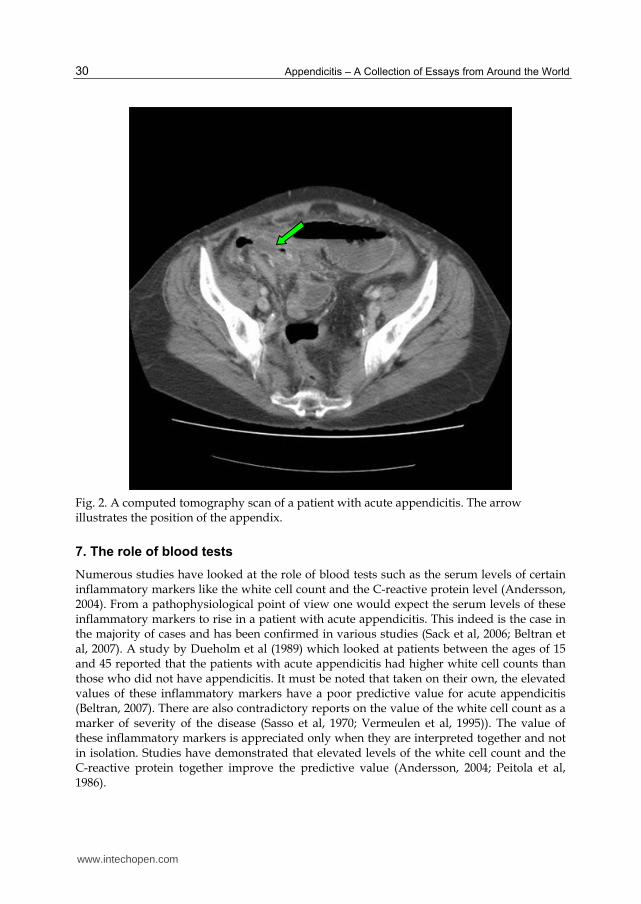
CT has an accuracy between 93% and 98% (Rao et al, 1998b) which is superior to that of ultrasonography in cases of suspected acute appendicitis (Terasawa et al, 2004). There has been a shift towards the use of CT in clinical practice with ultrasonography usually being reserved for cases where exposure to ionising radiation is contra indicated. The success of CT is due largely to its ability to visualise the appendix (Friedland & Siegel, 1997). An inflamed appendix usually appears to be larger than 6 mm in diameter and there will also tend to be evidence of inflammatory changes in the surrounding tissues (Choi et al, 2003; Haaga et al, 2003). These changes include the presence of a phlegmon, inflammatory fat stranding, free fluid, abscess formation and in the case of a perforated appendicitis even the presence of small amounts of free air (Haaga et al, 2003). CT also has the advantage of being able to detect other pathologies, for instance, luminal obstruction leading to acute appendicitis due to a caecal malignancy should be identifiable on computed tomography. Also, any associated adenopathy or metastasis could be detected as well. Figure 2 is a computed tomography scan that demonstrates acute appendicitis. The main disadvantage of CT is that it involves ionising radiation and its use in children has to be weighed against the potential risks. Cost is also an issue however, at present the cost of a CT scan is less than the costs associated with performing a negative laparotomy or diagnostic laparoscopy. Another disadvantage of using computed tomography scans is the need for administering a contrast agent to which there may be a reaction. The administration of rectal contrast is also associated with patient discomfort The presence of intra abdominal fat can be used as a contrast agent and therefore even very subtle inflammatory changes can be detected.. One factor that might lead to a diagnosis of acute appendicitis being missed on a CT scan is the lack of intra abdominal fat. This is usually encountered in children and very slim patients (Levine et al, 2005). In such cases an ultrasonography scan would seem to be more beneficial.
6.4 Magnetic Resonance Imaging (MRI)
There are no large studies conclusively demonstrating a superiority for MRI over other imaging modalities (Rothrock & Pagane, 2000). A recent study by Pedrosa et al (2009) studied 148 pregnant patients with suspected acute appendicitis. Their results showed that the use of magnetic resonance imaging did lower the incidence of negative laparotomies and perforation rates and thereby suggested a potential benefit. MRI however, is time consuming and its cost effectiveness is yet to be evaluated.
www.intechopen.com
Appendicitis – A Collection of Essays from Around the World
30
Fig. 2. A computed tomography scan of a patient with acute appendicitis. The arrow illustrates the position of the appendix.
7. The role of blood tests
Numerous studies have looked at the role of blood tests such as the serum levels of certain inflammatory markers like the white cell count and the C-reactive protein level (Andersson, 2004). From a pathophysiological point of view one would expect the serum levels of these inflammatory markers to rise in a patient with acute appendicitis. This indeed is the case in the majority of cases and has been confirmed in various studies (Sack et al, 2006; Beltran et al, 2007). A study by Dueholm et al (1989) which looked at patients between the ages of 15 and 45 reported that the patients with acute appendicitis had higher white cell counts than those who did not have appendicitis. It must be noted that taken on their own, the elevated values of these inflammatory markers have a poor predictive value for acute appendicitis (Beltran, 2007). There are also contradictory reports on the value of the white cell count as a marker of severity of the disease (Sasso et al, 1970; Vermeulen et al, 1995)). The value of these inflammatory markers is appreciated only when they are interpreted together and not in isolation. Studies have demonstrated that elevated levels of the white cell count and the C-reactive protein together improve the predictive value (Andersson, 2004; Peitola et al, 1986).
www.intechopen.com
Diagnostic Challenges in Acute Appendicitis
31
The converse however which is if the patient does not have elevated serum levels of the relevant inflammatory markers then they are unlikely to have acute appendicitis needs to be interpreted with caution. While there are studies that suggest the absence of raised inflammatory markers indicates a low likelihood of acute appendicitis (Sengupta, 2009), one must be wary when encountering a patient with a suggestive history but normal blood tests. There are reports in the literature suggesting that 21% to 65% of patients with acute appendicitis have a normal white cell count (Merlin et al, 2010). Similar results have been demonstrated for the serum levels of the C-reactive protein (Old, 2011). Studies indicate that it is a poor predictor of acute appendicitis when looked at in isolation but in conjunction with the white cell count it increases the likelihood ratio considerably (Vissers & Lennarz, 2010). Other studies have also indicated that normal levels of the white cell count, C-reactive protein and the neutrophil count have a high negative predictive value (Dueholm et al, 1989). One reason for the varying results between studies could be the heterogeneity of the sample population. Factors known to influence the levels of these inflammatory markers include the patient’s age and the duration of symptoms (Paajanen et al, 1997). The temporal relation that the serum levels of the inflammatory markers bear with the onset of symptoms is that the white cell count appears to rise early on in acute appendicitis with the C-reactive protein levels following later on. Persistently rising or high C-reactive levels could indicate the possibility of an appendiceal perforation (Chung et al, 1996, Sanjuan et al, 1999). This temporal relation does not seem to be so clear cut in children and therefore making a diagnosis in children poses additional problems (Kharbanda et al, 2011). As always, it should be emphasised that blood tests alone cannot be used to diagnose acute appendicitis and therefore what should be employed is a holistic approach which takes into account the patients history and also the results of any other investigations.
8. Appendicitis scoring systems
The need to collate information obtained from the patients history, clinical examination and investigation results, which should then be interpreted as a coherent whole when assessing a patient with suspected appendicitis, has been recognised by many investigators (Zimmermann, 2008). Attempts to simplify the process and quantify the likelihood of a positive diagnosis for acute appendicitis led to the development of various scoring systems. All these systems allocate particular numerical values to a feature. These are then added to give a final number which should give the clinician an idea of the likelihood of the patient having acute appendicitis. The most commonly used in clinical practice is the Alvarado score or the MANTRELS (Migratory pain, Anorexia, Nausea/vomiting, Tenderness in right iliac fossa, Rebound tenderness, Elevated temperature, Leucocytosis shift to the left) score (Alvarado, 1986). The scoring system is illustrated in table 4. Currently there are many variants of the Alvarado score, specifically modified to address certain groups of patients like children (Macklin et al, 1997). It must be noted that none of these modified scoring systems have demonstrated an increased accuracy in diagnosing acute appendicitis. A multicentre prospective study conducted by Ohmann et al (1995) looked at 1254 patients with acute abdominal pain and assessed various appendicitis scoring systems such as the Alvarado score, the Lindberg score, the Fenyo score and the Christian score against certain pre-set criteria. The pre-set standardised criteria included a negative laparotomy rate of 15% or less, a potential perforation rate of 35% or less, an initial missed perforation rate of 15% or
www.intechopen.com
Appendicitis – A Collection of Essays from Around the World
32
less and a missed appendicitis rate of 5% or less. The results of this study indicated that there were marked differences between the different scoring systems and that none of them satisfied the standardised criteria. However, when the published data was evaluated, many of the scoring systems demonstrated better agreement with the standardised criteria. The authors conclude that the original published data is ‘optimistically biased’ and that further large scale studies are required to validate these scoring systems.
Symptoms Score
Migratory right iliac fossa pain 1
Nausea/vomiting 1
Anorexia 1
Signs
Tenderness in right iliac fossa 2
Rebound tenderness in right iliac fossa 1
Pyrexia 1
Blood Test Results
Elevated white cell count 2
Shift to the left of neutrophils 1
TOTAL 10
Table 4. The Alvarado score (Alvarado, 1986; Malik et al, 2000)
A more recent study by Horzic et al (2005) looked at the value of appendicitis scoring
systems in diagnosing acute appendicitis in women. This study involved 126 female patients
admitted with abdominal pain and looked at the Alvarado score, the Ohmann score and the
Eskelinen score. This study too gave variable results in terms of accuracy but did report that
the scoring systems, when used in conjunction with each other can be used to determine
whether the patient would require surgery immediately or if observation would suffice
initially. This potentially has the advantage of avoiding delays in operation and further
investigations. It must be noted that the relatively small number of patients in this study
does mean that further studies are required before such a strategy is adopted in clinical
practice.
While clinical scoring systems appear to be useful adjuncts in diagnosing acute appendicitis,
one should always bear in mind the variable diagnostic accuracy for each scoring system
and therefore the limitations associated with them. As with all investigations, the different
scoring systems should only play an ancillary role in the diagnosis. Clinical decisions should
not be made solely on the basis of the value obtained on the application of a single scoring
system.
9. Appendicitis at the extremes of age
While a diagnosis of acute appendicitis is considered rare in the elderly population, it is still common enough to be included in the differential diagnosis in an elderly patient presenting with an acute abdomen. It is estimated that 7% of elderly patients with acute abdominal pain have acute appendicitis (Doria et al 2006; Vissers & Lennarz, 2010). The diagnosis of acute appendicitis in the elderly population is particularly difficult as the clinical picture is often complicated by comorbidities. These include conditions which can mask or suppress
www.intechopen.com
Diagnostic Challenges in Acute Appendicitis
33
the normal inflammatory reaction, such as diabetes or immunosuppression (Binderow & Shaked, 1991; Tsai et al, 2008). In such cases, the symptoms tend to be rather non specific. The damped inflammatory reaction demonstrated by certain elderly patients often complicates the clinical picture as palpation of the abdomen may not elicit marked tenderness or signs of peritonism. The physical examination can be complicated, and for the unwary clinician even misleading
at times , by the fact that many elderly patients have a weak abdominal musculature. This
would mean that the expected signs of rigidity and guarding in a patient with peritonitis
may not be present. It should also be noted that the rates of perforation are much higher in
the elderly population and so many of the patients can also present with diffuse abdominal
tenderness and peritonism (Korner et al, 1997; Hiu et al, 2002). The differential diagnosis for
elderly patients presenting with abdominal pain is much broader and therefore when such a
patient presents, especially with a non specific history, a computed tomography scan is the
imaging modality of choice (Paranjape et al, 2007). In such circumstances, the presence of
any other pathology can be assessed with a computed tomography scan and the appropriate
management can be planned accordingly. Also, the benefits of the scan outweigh the risks
associated with exposure to ionising radiation.
Appendicitis in the very young child can pose considerable challenges. The signs and
symptoms of acute appendicitis are age dependent (Blab et al, 2004). From the age of around
six and over the signs and symptoms are more reliable and tend to conform to the classical
presentation. In younger children, nausea and vomiting seem to be constant features.
Rothrock et al (1991) report that vomiting tends to precede the abdominal pain, and very
often it is the vomiting that is noticed first by parents before a history of abdominal pain. In
the 2 to 5 age group, the pain tends to be more localised which is in contrast to the children
of ages 2 and younger where the abdominal pain tends to be more diffuse. Localised
tenderness has been reported to be present in less that 50% in this age group (Barker &
Davey, 1988; Horwitz et al, 1997). Patients in this age group or even younger tend to also
exhibit lethargy, abdominal distension, diarrhoea and fever. Acute appendicitis has a higher
rate of perforation with subsequent diffuse peritonism in the very young patient. This is
partly due to the fact that in such young patients, the omentum has not reached sufficient
maturity and therefore is unable to wall off any leakage that occurs during a perforation.
This leads to the enteric contents being spread throughout the abdominal cavity. Other
misleading signs in the very young patient include irritability and pain or stiffness in the
right hip (Daehlin, 1982; Rothrock & Pagane, 2000).
Misdiagnosing acute appendicitis in young children can result in considerable morbidity
and has been the subject of numerous medicolegal investigations. An understanding of the
variations that occur in the very young should alert the astute clinician to the possibility of
an underlying acute appendicitis when encountering a child with non specific symptoms.
10. Acute appendicitis in pregnancy
The pathophysiology and presentation of acute appendicitis in the pregnant patient can be very similar to that of the non pregnant patient (Mourad et al, 2000). The appendix can be displaced upwards as the uterus enlarges and therefore it may give rise to pain and tenderness in unexpected positions. There are however, studies that report the location of pain in pregnant patients with acute appendicitis to be predominantly in the right iliac fossa
www.intechopen.com
Appendicitis – A Collection of Essays from Around the World
34
(Hodjati et al, 2003; Oto et al 2006). White cell counts can be misleading as they are raised as a consequence of the pregnancy. A delay in the diagnosis of acute appendicitis can put the fetus at risk and a ruptured appendicitis is associated with a fetal loss rate of between 20% and 25% (Kilpatrick et al, 2007). It is therefore essential to confirm the diagnosis by ultrasonography. This avoids ionising radiation but can be difficult due to the displaced anatomy. Not visualising the appendix does not rule appendicitis out and therefore the diagnosis would still be in question. In such cases the use of computed tomography would need to be considered with due consideration given to the associated risks and benefits. Magnetic resonance imaging is another possibility however, large scale studies demonstrating its accuracy in pregnant patients are lacking and therefore no definitive conclusion can be made regarding its usefulness (Blumenfeld et al, 2011).
11. Atypical presentations of acute appendicitis
The literature detail numerous atypical presentations. Akbulut et al (2010) report a case of a 21 year old lady with congenital situs inversus presenting with acute appendicitis. She had a one day history of mid epigastric pain which migrated to the left iliac fossa. Her white cell count was normal on admission although clinical examination revealed tenderness and guarding in the left iliac fossa. The diagnosis was confirmed on ultrasonography and she subsequently underwent an open appendicectomy. What is notable about this case is that the patient’s history was ‘classical’ apart from the migration of the pain to the left iliac fossa. Given her situs inversus, the reason for her left sided migratory pain is obvious. It is worthy of note that her chest radiograph demonstrated dextrocardia and a right sided gastric bubble. In patients presenting with such histories, dextrocardia can be deduced either from plain radiographs or from the clinical examination of the cardio-respiratory system. One should bear in mind that as the vermiform appendix can end up in the left side of the abdomen as a result of midgut malrotation, dextrocardia may not always be present. Chae et al (2007) report a case of a 49 year old lady who underwent a colonoscopy as part of a bowel screening programme. The colonoscopy was uneventful and all caecal landmarks were identified. The lady was well post procedure and discharged home the same day. She developed progressively worsening right sided abdominal pain and was seen in the outpatient department four days later. Her blood tests were within normal limits but physical examination revealed tenderness in her right iliac fossa. Appendicitis was confirmed on ultrasonography and the patient subsequently underwent an appendicectomy and was discharged 3 days later. The potential mechanisms suggested for this rare complication of colonoscopy are increased intra-luminal pressure due to gas insufflation and possible displacement of faecoliths into the appendiceal lumen. The authors also suggest that ulceration of the appendiceal mucosa by the colonoscope was a possibility. Another unusual presentation of acute appendicitis is as a small bowel obstruction with raised inflammatory markers. Harrison et al (2009) describe two elderly patients presenting with small bowel obstruction diagnosed on computed tomography scans to be due to an acute appendicitis (Figure 3). Often the inflammatory process that accompanies acute appendicitis can result in small bowel obstruction. A similar case has been reported by Assenza et al (2005) who suggested that adherence of the inflamed tip of the appendix to the posterior peritoneum across the terminal ileum resulted in bowel compression.
www.intechopen.com
Diagnostic Challenges in Acute Appendicitis
35
Fig. 3. A computed tomographic image illustrating an inflamed appendix (arrow) with a co-existent small bowel obstruction. (Harrison et al, 2009)
D’Ambrosio et al (2006) report a case of a 71 year old lady who presented with an inflamed tender lump on her right proximal thigh which had been progressively increasing in size over the preceding two weeks. On examination she was noted to have a tender erythematous and indurated mass near her inguinal region and laboratory investigations demonstrated a marked leucocytosis. Subsequent computed tomography demonstrated the inferior portion of the caecum to be thickened and in contact with an inflammatory mass which was contained in a femoral hernia. The patient underwent a laparotomy and right hemicolectomy. The appendix was found to be perforated and abscess formation was also noted and drained at surgery. While inflamed appendices within a hernia is rare, it should be noted that the perforation of the appendix into a limited space prevents the spillage of enteric contents into the abdominal cavity. In this case the perforated appendix was contained within the femoral hernia and so rather than developing diffuse peritonitis, the patient developed superficial signs of erythema, tenderness and induration.
12. Conclusion
Diagnosing appendicitis can pose considerable challenges even to the experienced clinician. A delayed or missed diagnosis can have complications which can result in morbidity and
www.intechopen.com
Appendicitis – A Collection of Essays from Around the World
36
medicolegal claims. To minimise the risks of missing the diagnosis, it is important for clinicians involved in the initial assessment of patients to have a good understanding of factors that can influence the clinical presentation. The clinician should be aware of atypical presentations especially at extremes of age and in pregnancy. No single test nor a combination of tests can distinguish all cases of acute appendicitis from other conditions. An awareness of the limitations of imaging, blood tests and scoring systems is essential. A thorough history and repeated clinical examinations if necessary in conjunction with the results of the appropriate investigations are essential to diagnose acute appendicitis. Sometimes, in equivocal cases, a period of clinical observation is necessary. Despite the advances made in diagnostic modalities, patience may prove to be the most valuable. Often, time and expert clinical review are key to successfully diagnosing acute appendicitis.
13. Acknowledgements
The authors would like to thank Mrs Sindhu Harrison for her help with the artwork for this article.
14. References
Ahmed I, Asgeirsson KS, Beckingham IJ et al (2007). The position of the vermiform
appendix at laparoscopy. Surgical and Radiological Anatomy, 29(2):165-8.
Akbulut S, Ulku A, Senol A et al (2010). Left-sided appendicitis: review of 95 published
cases and a case report. World Journal of Gastroenterology, 16(44):5598-602
Al-Salem AH, Qureshi ZS, Qaisarudin S et al (1998). Is acute appendicitis different in
patients with sickle cell disease? Pediatric Surgery International, 13(4):265-7
Alloo J, Gerstle T, Shilyansky J et al (2004). Appendicitis in children less than 3 years of age:
a 28 year review. Paediatric Surgery International, 19:777-779
Alvarado A (1986). A practical score for the early diagnosis of acute appendicitis. Annals of
Emergency Medicine, 15:557-564
Alzaraa A, Chaudhry S (2009). An unusually long appendix in a child: a case report. Cases
Journal, 11;2:7398.
Andersson R, Hugander A, Thulin A et al (1995). Clusters of acute appendicitis. Further
evidence of an infectious aetiology. International Journal of Epidemiology, 24:829-833
Andersson RE (2004), Meta-analysis of the clinical and laboratory diagnosis of appendicitis.
British Journal of Surgery, 91(1):28-37
Andreou P, Blain S, du Boulay EH (1990). A histopathological study of the appendix at
autopsy and after surgical resection. Histopatholoy, 17:427-431
Arnbjornsson E, Bengmark S (1983). Obstruction of the appendix lumen in relation to
pathogenesis of acute appendicitis. Acta Chirugica Scandinavica, 149:789-791
Arnbjornsson E, Bengmark S (1984). Role of obstruction in the pathogenesis of acute
appendicitis. American Journal of Surgery, 147:390-392
Assenza M, Ricci G, Bartolucci P (2005). Mechanical small bowel obstruction due to an
inflamed appendix wrapping around the last loop of ileum. Giornale di Chirugia,
26(6-7):261-6.
Azodi SO, Sandberg AA, Larsson H (2009). Genetic and environmental influences on the
risk of acute appendicitis in twins. British Journal of Surgery, 96(11):1336-40
www.intechopen.com
Diagnostic Challenges in Acute Appendicitis
37
Barker AP, Davey RB (1988). Appendicitis in the first three years of life. Australian and New
Zealand Journal of Surgery, 58:491-494
Barker JP, Morris J, Nelson M (1986). Vegetable consumption and acute appendicitis in 59
areas in England and Wales. British Medical Journal, 292:927-930
Beltrán MA, Almonacid J, Vicencio A et al (2007). Predictive value of white blood cell count
and C-reactive protein in children with appendicitis. Journal of Paediatric Surgery,
42(7):1208-1214
Binderow SR, Shaked AA (1991). Acute appendicitis in patients with AIDS/HIV infection.
American Journal of Surgery, 162(1):9-12.
Birnbaum BA, Wilson SR (2000). Appendicitis at the Millennium. Radiology, 215:337-48
Blab E, Kohlhuber U, Tillawi S et al (2004). Advancements in the diagnosis of acute
appendicitis in children and adolescents. European Journal of Paediatric Surgery,
14:404-409
Blumenfeld YJ, Wong AE, Jafari A et al (2011). MR imaging in cases of antenatal suspected
appendicitis--a meta-analysis. Journal of Maternal, Fetal and Neonatal Medicine,
24(3):485-8.
Burkitt DP (1971). The etiology of appendicitis. British Journal of Surgery,58:695-699
Burkitt DP, Walker RP, Painter NS (1972). Effect of dietary fibre on stools and transit times,
and its role in the causation of disease. Lancet, 2:1408-1412
Carr NJ (2000). The pathology of acute appendicitis. Annals of Diagnostic Pathology, 4(1):46-58
Chae HS, Jeon SY, Nam WS (2007). Acute appendicitis caused by colonoscopy. Korean
Journal of Internal Medicine, 22(4):308-11
Chang AR (1981). An analysis of the pathology of 3003 appendices. Australian and New
Zealand Journal of Surgery,51:169-178
Choi D, Park H, Lee YR et al (2003). The most useful findings for diagnosing acute
appendicitis on contrast enhances helical CT. Acta Radiologica, 44:574-82
Chung CH, Ng CP, Lai KK (2000). Delays by patients, emergency physicians and surgeons
in the management of acute appendicitis: retrospective study. Hong Kong Medical
Journal, 6:254-259
Chung JL, Kong MS, Lin SL et al (1996). Diagnostic value of C-reactive protein in children
with perforated appendicitis. European Journal of Paediatrics, 155:529-531
Codon RE, Telford GL (1991). Appendicitis; In:Townsend CM (eds). Sabiston Text book of
Surgery: The biological basis of modern surgical practice. 14th ed, Philadelphia,
Pa:WB Saunders and Co; pp 884-898
Coggon D, Barker JP, Cruddas et al (1991). Housing and appendicitis in Anglesey. Journal of
Epidemiology and Community Health, 45:244-246
Collins DC (1932). The Length and Position of the Vermiform Appendix: A Study of 4,680
Specimens. Annals of Surgery, 96(6):1044-8
Daehlin L (1982). Acute appendicitis during the first three years of life. Acta Chirugica
Scandinavic, 148:291-294
D'Ambrosio N, Katz D, Hines J (2006). Perforated appendix within a femoral hernia. AJR
American Journal of Roentgenology, 186(3):906-7
Devlin B (1971). Midgut malrotation causing intestinal obstruction. Annals of the Royal
College of Surgeons of England, 48:227-237
www.intechopen.com
Appendicitis – A Collection of Essays from Around the World
38
Doria AS, Moineddin R, Kellenberger CJ et al (2006). US or CT for diagnosis of appendicitis
in children and adults? A meta analysis. Radiology, 241:83-94
Dueholm S, Bagi P, Bud M (1989). Laboratory aid in the diagnosis of acute appendicitis. A
blinded, prospective trial concerning diagnostic value of leukocyte count,
neutrophil differential count, and C-reactive protein. Diseases of the Colon and
Rectum, 32(10):855-9.
Ergul E (2007). Heredity and familial tendency of acute appendicitis. Scandinavian Journal of
Surgery, 96(4):290-2
Erkasap S, Ates E, Ustuner Z et al (2000). Diagnostic value of interleukin-6 and C-reactive
protein in acute appendicitis. Swiss Surgery.6(4):169-72
Fang YJ, Wang HP, Ho CM et al (2009). Barium appendicitis. Surgery, 146(5):957-8
Fefferman NR, Roche KJ, Pinkney LP et al (2001). Suspected appendicitis in children:
focussed CT technique for evaluation. Radiology, 220:691-5
Fraser N, Gannon C, Stringer MD (2004). Appendicular colic and the non-inflamed
appendix: fact or fiction? European Journal of Paediatric Surgery, 14(1):21-4.
Friedland JA, Siegel MJ (1997). CT appearance of acute appendicitis in childhood. AJR
American Journal of Roentgenology, 168:439-442
Haaga JR, Lanzieri CF, Gilkeson RC et al (2003). CT and MR imaging of the whole body. 4th
edition, St Louis: Mosby: 2061
Harrison S, Mahawar K, Brown D et al (2009). Acute appendicitis presenting as small bowel
obstruction: two case reports. Cases Journal, Nov 28;2:9106.
Herrinton JL (1991). The vermiform appendix: its surgical history. Contemporary Surgery,
39:36-44
Hiraiwa H, Umemoto M, Take H (1995). Prevalence of appendectomy in Japanese families.
Acta Paediatrica Japan, 37:691-693
Hiu TT, Major KM, Avital I et al (2002). Outcome of elderly patients with appendicitis.
Archives of Surgery, 135:479-88
Hodjati H, Kazerooni T (2003). Location of the appendix in the gravid patient: a re-
evaluation of the established concept. International Journal of Gynaecology and
Obstetrics, 81(3):245-7
Horton WL (1977). Pathogenesis of acute appendicitis. British Medical Journal,2:1672-1673
Horwitz JR, Gursoy M, Jaksic et al (1997). Importance of diarrhoea as a presenting symptom
of appendicitis in very young children. American Journal of Surgery, 173:80-82
Horzić M, Salamon A, Kopljar M et al (2005). Analysis of scores in diagnosis of acute
appendicitis in women. Collegium Anthropologicum, 29(1):133-8
Hsu KF, Yu JC, Chan DC et al (2011). Atypical acute appendicitis and its complications: a
rare location of the appendix in the periduodenum. Acta Gastroenterologica Belgica,
InTech ChinaUnit 405, Office Block, Hotel Equatorial Shanghai No.65, Yan An Road (West), Shanghai, 200040, China
Phone: +86-21-62489820 Fax: +86-21-62489821
This book is a collection of essays and papers from around the world, written by surgeons who look afterpatients of all ages with abdominal pain, many of whom have appendicitis. All general surgeons maintain afascination with this important condition because it is so common and yet so easy to miss. All surgeons have aview on the literature and any gathering of surgeons embraces a spectrum of opinion on management options.Many aspects of the disease and its presentation and management remain controversial. This book does notanswer those controversies, but should prove food for thought. The reflections of these surgeons arepresented in many cases with novel data. The chapters encourage us to consider new epidemiological viewsand explore clinical scoring systems and the literature on imaging. Appendicitis is discussed in patients of allages and in all manner of presentations.
How to referenceIn order to correctly reference this scholarly work, feel free to copy and paste the following:
Sanjay Harrison and Harrison Benziger (2012). Diagnostic Challenges in Acute Appendicitis, Appendicitis - ACollection of Essays from Around the World, Dr. Anthony Lander (Ed.), ISBN: 978-953-307-814-4, InTech,Available from: http://www.intechopen.com/books/appendicitis-a-collection-of-essays-from-around-the-world/diagnostic-challenges-in-acute-appendicitis


![Acute Appendicitis[1]](https://static.documents.pub/doc/80x56/577cd3341a28ab9e7896e8e0/acute-appendicitis1.jpg)


