46
Diagnostic Microbiology 320 MIC Lecture: 10 Diagnosis of Virus Infections 1 2014 Manal AlKulaifi - Amal Alghamdi King Saud University Dept. of Bot. & Microbiology

Diagnostic Microbiology
320 MIC
Lecture: 10 Diagnosis of Virus
Infections
12014 Manal AlKulaifi - Amal Alghamdi
King Saud UniversityDept. of Bot. & Microbiology

Specimens for viral diagnosis
22014 Manal AlKulaifi - Amal Alghamdi

Diagnostic Methods in Virology
B. Direct Examination: Direct demonstration of the virus, its antigens or nucleic acid. • widely used• fast method of virus diagnosis
Virus or virus antigen is detected in lesions, fluids, tissues or excretions from the patient and a result can be obtained within an 1 or 2 hour of receipt specimen.
B. Indirect Examination:Virus Propagation Isolation - Chick embryo - Cell culture ( cell line)
C. Animal inoculationD. Serology
32014 Manal AlKulaifi - Amal Alghamdi

42014 Manal AlKulaifi - Amal Alghamdi
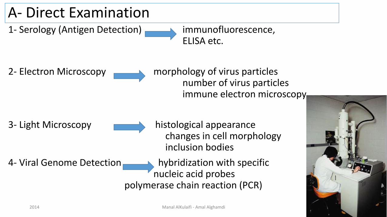
A- Direct Examination 1- Serology (Antigen Detection) immunofluorescence,
ELISA etc.
2- Electron Microscopy morphology of virus particles number of virus particles immune electron microscopy
3- Light Microscopy histological appearance changes in cell morphology inclusion bodies
4- Viral Genome Detection hybridization with specific nucleic acid probes
polymerase chain reaction (PCR)
52014 Manal AlKulaifi - Amal Alghamdi

B- Indirect Examination
1- Cell Culture cytopathic effect (CPE) haemabsorptionimmunofluorescence
2- Eggs pocks on chorioallantoic membrane (CAM) haemagglutinationinclusion bodies
3- Animals disease or death
62014 Manal AlKulaifi - Amal Alghamdi

Electron Microscopy (EM)
• 106 virus particles per ml required for visualization. 50,000 - 60,000 magnification normally used.
Viruses may be detected in the following specimens:
• Faeces Rotavirus, Adenovirus
• Vesicle Fluid HSV
• Skin scrapings papillomavirus
2014 Manal AlKulaifi - Amal Alghamdi 7
Immune Electron Microscopy The sensitivity and specificity of EM may be enhanced by immune electron microscopy.
There are two variants:-
• Classical Immune electron microscopy (IEM) - the sample is treated with specific anti-sera before being put up for EM. Viral particles present will be agglutinated and thus congregate together by the antibody.
• Solid phase immune electron microscopy (SPIEM) - the grid is coated with specific anti-sera. Virus particles present in the sample will be absorbed onto the grid by the antibody
Problems with Electron Microscopy
• Expensive equipment
• Expensive maintenance
• Require experienced observer
• Sensitivity often low 2014 Manal AlKulaifi - Amal Alghamdi 8

Epithelial-like (human lung carcinoma, A549)
Fibroblast like
(baby hamster kidney, BHK)
www.freelivedoctor.com
2014 Manal AlKulaifi - Amal Alghamdi 9
Measles
on human lung carcinoma

Vaccinia on monkey kidney
Low multiplicity of infection
2014 Manal AlKulaifi - Amal Alghamdi 10
High

Virus Isolation Cell Cultures are most widely used for virus isolation.
There are 3 types of cell cultures:
1. Primary cells - Monkey Kidney2. Semi-continuous cells - Human embryonic kidney and skin fibroblasts 3. Continuous cells (Hela)
Primary cell culture are widely acknowledged as the best cell culture systems available since they support the widest range of viruses. However, they are very expensive and it is often difficult to obtain a reliable supply. Continuous cells are the most easy to handle but the range of viruses supported is often limited.
112014 Manal AlKulaifi - Amal Alghamdi

Virus Isolation
• In the laboratory, solid samples are minced, homogenized, centrifuged at low speed to remove cellular debris that may be toxic to the cultured cells, and sterilized by 0.2 µm filter.
• A backup aliquot is stored at 4 or -70 C.
• Next step is the inoculation of a system supporting virus replication:
cell cultures, embryonating eggs, organ cultures, laboratory animals or host animals.
2014 Manal AlKulaifi - Amal Alghamdi 12

Virus Isolation (chick embryo)
132014 Manal AlKulaifi - Amal Alghamdi

Cell Cultures Growing virus may produce:
1. Cytopathic Effect (CPE):
such as the ballooning of cells or syncytia formation, may be specific or non-specific.
2. Haemadsorption:
cells acquire the ability to stick to mammalian red blood cells.
142014 Manal AlKulaifi - Amal Alghamdi

Types of Cytopathic Effects (CPE):
Cytopathic Effect (1)
Cytopathic effect of enterovirus and HSV in cell culture: note the ballooning of cells.
(Virology Laboratory, Yale-New Haven Hospital, Linda Stannard, University of Cape Town).
15
Cytopathic Effect (2) Syncytium formation in cell culture caused by RSV and measles virus.(courtesy of Linda Stannard, University of Cape Town, S.A.)
2014 Manal AlKulaifi - Amal Alghamdi

Primary cell culture
162014 Manal AlKulaifi - Amal Alghamdi

Subculture
Growth of cells in culture. A primary culture is defined as the original plating of cells from a tissue, grown to a confluent monolayer, without subculturing. A cell strain (solid line) is defined as a euploid population of cells subcultivated more than once in vitro, lacking the property of indefinite serial passage. Cell strains ultimately undergo degeneration and death, also called crisis or senescence. A cell line (dashed line) is an aneuploid population of cells that can be grown in culture indefinitely. Spontaneous transformation or alteration of a cell strain to an immortal cell line can occur at any time during cultivation of the cell strain. The time in culture and corresponding number of subcultivations or passages are shown on the abscissas. The ordinate shows the total number of cells that would 172014 Manal AlKulaifi - Amal Alghamdi

Cultured cells
a) Primary
• Heterogeneous – many cell types
• Closest to animal
• Technical hassle
b) Diploid cell strain
• Relatively homogeneous – fewer cell types
• Further from animal
• Technically less hassle
c) Continuous cell line
Immortal • Most homogeneous • Genetically weird – furthest from animal • Hassle free • Suspension or monolayer
182014 Manal AlKulaifi - Amal Alghamdi

Problems with cell culture
• Long period (up to 4 weeks) required for result.
• Often very poor sensitivity, sensitivity depends on a large extent on the condition of the specimen.
• Susceptible to bacterial contamination.
• Susceptible to toxic substances which may be present in the specimen.
• Many viruses will not grow in cell culture e.g. Hepatitis B, Diarrheal viruses.
2014 Manal AlKulaifi - Amal Alghamdi 19

Haemadsorption:Syncytial formation caused by mumps virus and haemadsorption of erythrocytes onto the surface of the cell sheet.
(courtesy of Linda Stannard, University of Cape Town, S.A.)
202014 Manal AlKulaifi - Amal Alghamdi

21
Haemadsorption:
Ebola virus infection,
inclusion bodies (arrows) 2014 Manal AlKulaifi - Amal Alghamdi

Haemadsorbtion
2014 Manal AlKulaifi - Amal Alghamdi 22

Haemadsorbtion
• Hemadsorption of erythrocytes to cells infected with influenza viruses. This virus express a hemagglutinin, which bind erythrocytes of selected animal species.
2014 Manal AlKulaifi - Amal Alghamdi 23

Haemagglutination: to Detect Viral Antigen
2014 Manal AlKulaifi - Amal Alghamdi 24

2014 Manal AlKulaifi - Amal Alghamdi 25
Haemagglutination: to Detect Viral Antigen

Serology
• Detection of rising titers of antibody between acute and convalescent stages of infection, or the detection of IgM in primary infection.
262014 Manal AlKulaifi - Amal Alghamdi
Serology
Criteria for diagnosing Primary Infection
• 4 fold or more increase in titre of IgG or total antibody between acute and convalescent sera
• Presence of IgM
Criteria for diagnosing Reinfection
• fold or more increase in titre of IgG or total antibody between acute and convalescent sera
• Absence or slight increase in IgM
2014 Manal AlKulaifi - Amal Alghamdi 27

Serology
• Neutralization
• Hemagglutination inhibition
• Western blot
• ELISA, radioimmune assay (RIA)
2014 Manal AlKulaifi - Amal Alghamdi 28

Antibody Detection: Neutralization & Haemagglutination
In the assay shown, 10 fold dilutions of serum were incubated with virus.Aliquots of the mixture were then added to cell cultures or erythrocytes. In the absence of antibody, the virus infected the monolayer (indicated by CPE) and caused hemagglutination.
In the presence of the antibody, infection was blocked (neutralization), and hemagglutination was inhibited, allowing the erythrocytes to pellet. 2014 Manal AlKulaifi - Amal Alghamdi 29

Western blot analysis of HIV antigens and antibody.
HIV protein antigens are separated by electrophoresis and blotted onto nitrocellulose paper strips.
The strip is incubated with patient antibody, washed to remove the unbound antibody, and then reacted with enzyme-conjugated antihuman antibody.
Serum from an HIV- infected person binds and identifies the major antigenic proteins of HIV.
2014 Manal AlKulaifi - Amal Alghamdi 30
Antibody Detection: Western Blot

2014 Manal AlKulaifi - Amal Alghamdi 31
HIV Antigen Detection: ELIZA

Microplate ELISA for HIV: coloured wells indicate reactivity
2014 Manal AlKulaifi - Amal Alghamdi 32

Virus Antigen Detection: Immunofluorescense
Antibody binds to antigen in fixed cells;
●fluorescein-labeled anti-IgG binds;
●fluorescens by UV microscopy
2014 Manal AlKulaifi - Amal Alghamdi 33

2014 Manal AlKulaifi - Amal Alghamdi 34
Virus Antigen Detection: Immunofluorescense
BHV-1 antigensin neuron in trigeminal ganglion
Positive immunofluorescence test for rabies
virus antigen. (Source: CDC)

Usefulness of Serological Results
• How useful a serological result is depends on the individual virus.
• For example, for viruses such as rubella and hepatitis A, the onset of clinical symptoms coincide with the development of antibodies. The detection of IgMor rising titres of IgG in the serum of the patient would indicate active disease.
• However, many viruses often produce clinical disease before the appearance of antibodies such as respiratory and diarrheal viruses. So in this case, any serological diagnosis would be retrospective and therefore will not be that useful.
• There are also viruses which produce clinical disease months or years. e.g. HIV. In this case, the presence of antibody is sufficient to make a definitive diagnosis.
2014 Manal AlKulaifi - Amal Alghamdi 35

Problems with Serology
• Long period of time required for diagnosis for paired acute and convalescent sera.
• Mild local infections may not produce a detectable humoral immune response.
• Extensive antigenic cross-reactivity between related viruses e.g. HSV may lead to false positive results.
• immunocompromised patients often give a reduced or absent humoral immune response.
• Patients given blood or blood products may give a false positive result due to the transfer of antibody.
2014 Manal AlKulaifi - Amal Alghamdi 36

Assay of viruses
– Biological• Plaque assay
• Transformation
• Endpoint Method
– Physical and biochemical.• Hemagglutination.
• Direct particle count.
• Immunological tests for proteins.
• Assay for nucleic acid (PCR).
• Enzymatic (reverse transcriptase for retroviruses).
2014 Manal AlKulaifi - Amal Alghamdi 37

Plaque Assay: Method
2014 Manal AlKulaifi - Amal Alghamdi 38

Plaque Assay : Result
2014 Manal AlKulaifi - Amal Alghamdi 39

Direct Particle Count
2014 Manal AlKulaifi - Amal Alghamdi 40

Assay for Viral Protein & Nucleic Acid
2014 Manal AlKulaifi - Amal Alghamdi 41

Molecular Methods
• Methods based on the detection of viral genome are also commonly known as molecular methods. It is often said that molecular methods is the future direction of viral diagnosis.
• However in practice, although the use of these methods is indeed increasing, the role played by molecular methods in a routine diagnostic virus laboratory is still small compared to conventional methods.
Classical Molecular Techniques
• hydridization are examples of classical techniques. They depend on the use of specific DNA/RNA probes for hybridization.
• The specificity of the reaction depends on the conditions used for hybridization. However, the sensitivity of these techniques is not better than conventional viral diagnostic methods.
• However, since they are usually expensive than conventional techniques, they never found widespread acceptance.2014 Manal AlKulaifi - Amal Alghamdi 42

Polymerase Chain Reaction
PCR allows the in vitro amplification of specific target DNA sequences in an extremely sensitive technique.
• it is based on an enzymatic reaction involving the use of synthetic oligonucleotides flanking the target nucleic sequence of interest.
• These oligonucleotides act as primers for the thermostable Taq polymerase. Repeated cycles (usually 25 to 40) of denaturation of the template DNA (at 94
oC), annealing of primers to their complementary sequences (50
oC), and
primer extension (72oC) result in the exponential production of the specific
target fragment.
• Detection and identification of the PCR product is usually carried out by agarose gel electrophoresis, restriction enzyme analysis, or DNA sequencing.2014 Manal AlKulaifi - Amal Alghamdi 43
Advantages of PCR:
Extremely high sensitivity, may detect down to one viral genome per sample volume - Easy to set up - Fast turnaround time
Disadvantages of PCR :
Extremely liable to contamination - High degree of operator skill required - Not easy to set up a quantitative assay- A positive result may be difficult to interpret, especially with latent viruses, where any seropositive person will have virus present in their blood irrespective whether they have disease or not.-These problems are being addressed by the arrival of commercial closed systems which requires minimum handling. The use of synthetic internal competitive targets in these commercial assays has facilitated the accurate quantification of results. However, these assays are very expensive.
2014 Manal AlKulaifi - Amal Alghamdi 44
Polymerase Chain Reaction

Diagnostic Methods for most Common Human Viruses
2014 Manal AlKulaifi - Amal Alghamdi 45
Summary
• Four main clinical diagnostic techniques:
– Culture, serology, antigen detection, nucleic acid detection.
• Virus culture
– Not all viruses can be cultured – Cultured cell types – Cytopathic effect
• Virus quantitation
– Biological
– Physical
• Basic serological techniques2014 Manal AlKulaifi - Amal Alghamdi 46


