Differential Effects of Olfactory Bulbectomy on GABAA and GABAB Receptors
in the Rat Brain
T R E V O R DENNIS , l VALI~RIE B E A U C H E M I N A N D N O R M A N D L A V O I E
Neurobiological Psychiatry Unit, Department o f Psychiatry, McOill University, 1033 Pine Avenue West, Montrdal, Qudbec H3A 1A1, Canada
Received 1 December 1992
DENNIS, T., V. BEAUCHEMIN AND N. LAVOIE. Differential effects of olfactory bulbectomy on GABAA and GABAB receptors in the rat brain. PHARMACOL BIOCHEM BEHAV 46(1) 77-82, 1993.-GABAergic mechanisms have been implicated in the bilateral olfactory bulbectomy (OBX) animal model of depression, where GABAB receptor binding sites have been shown to decrease markedly at specific time points after OBX. However, as no detailed time course of events has been determined, the present study investigated the effects of OBX on high-affinity GABA^, GABAa,/~-adrenergic, and henzodiazepine receptor binding parameters in membrane preparations from rat brain regions at weekly intervals (1-4 weeks) after OBX. Persistent significant increases (40-60070) in Bm~ values of high affinity GABAA receptors were observed in the frontal cortex throughout the period investigated following OBX. Bm~ values in the hippocampus increased significantly after 1 week (53°]o) hut were not statistically significant thereafter. No changes in GABAA binding parameters were observed in the hypothalamus or cerebellum. Conversely, GABAn receptor densities were significantly decreased in the frontal cortex after 1 (-38070) and 2 (-41070) weeks and moderately decreased 3 and 4 weeks (-27 and -23070, respectively) after OBX, while in the cerebellum they were significantly increased after l week (96°70) and returned to sham-operated levels by 3 weeks. No changes in GABAe receptor binding parameters were observed in the hippocampus or hypothalamus. Binding parameters for benzodiazepine receptor binding sites or/~-adrenoceptors were not modified throughout the time course. GABAergic transmission, reflected by changes in GABAA and GABA a receptor density in the frontal cortex, may be altered in OBX rats. The persistence of the change in GABA^ receptor density, contrasting with the transient change in GABAB receptors, suggests that the former, rather than the latter, is more likely to be related to the long-lasting behavioral deficits induced by OBX.
GABAA receptors GABAa receptors Olfactory bulbectomy Rat brain
Benzodiazepine receptors /5-Adrenergic receptors
ANIMAL models of depression have long been a subject of controversy. These models range from simple screening tests for potential antidepressant treatments to the induction of depressive-like symptoms. One of the models that can be use- ful in the study of the neurobiological substratum of depres- sion and the mechanisms of action of antidepressant treat- ments is the bilateral olfactory bulhectomy (OBX) model (12,34). Following OBX, rats display antidepressant-reversible behavioral and biochemical changes that find their correlates in depressed patients (4,5,12,15,31).
A number of studies following OBX have shown that nor- adrenaline turnover in the amygdaloid cortex and forebraln in general is decreased, while that of GABA is increased (9,21). In line with such changes, /3-adrenoceptors were reportedly increased in the pons and hippocampus; however, this appears to be due to an increase in binding affinity rather than a
change in binding density (11,29). Moreover, a2-adrenocep- tots are increased in the forehraln but not in the olfactory cortex of OBX rats (8,21). That GABAB binding densities are decreased in the frontal cortex both in the OBX model (13,16,17), 2 and 4 weeks after bulb ablation, and in the learned helplessness model (20) would implicate GABAergic mechanisms in these animal models of depression. Consistent with such a notion, Lloyd et al. (18,24) have shown increased GABAa receptor binding in the rat frontal cortex following long-term administration of different types of antidepressant treatments including GABAmimetic or electroconvulsive shock treatments. This upregulation in GABAa binding ap- pears to be specific for antidepressants as other psychotropic drugs, for example, haloperidol, chlorpromazine and diaze- pam, are inactive, while phenobarbital, diphenylhydantoin, and reserpine downregulate GABAa receptors (I 7). Moreover,
To whom requests for reprints should be addressed.
77
78 DENNIS, BEAUCHEMIN AND LAVOIE
desipramine-induced reversal of a stepdown passive avoidance deficit appears to be associated with increases in GABAB re- ceptor binding (13).
The above-mentioned studies (13,16,17) found decreased GABAa receptor binding at specific time points after OBX; however, a detailed time course of possible variations in den- sity of both GABA A and GABAB receptors following OBX has not been determined. Given that receptor binding levels determined from a single concentration of radioligand cannot define whether variations are related to changes in affinities (Kd) or maximal binding capacities (Bm~), the present study investigated the binding parameters of the high-affinity GABAA, GABAB, /3-adrenergic, and benzodiazepine (BDZ) receptor binding sites 1, 2, 3, and 4 weeks after bilateral OBX.
METHOD
Animals, Drugs, and Chemicals
Male Sprague-Dawley rats, 230-250 g, were obtained from Charles River Canada (St. Constant, Quebec). Before and after surgery, animals were housed in groups of four and maintained on a 12 L : 1 2 D cycle (light on at 0700 h) in a temperature- and humidity-controlled room, with standard laboratory chow and water ad lib.
Drugs and chemicals were obtained from the following sources: GABA (35-80 Ci/mmol) and (-)-[3H]CGP-12177 (38 Ci/mmol) from Amersham Canada (Oakville, Ontario); [methyl-3H]flunitrazepam (84 Ci/mmol) from DuPont Can- ada (Mississauga, Ontario); isoguvacine and (+)baclofen from Research Biochemicals, Inc. (Natick, MA); GABA and propranolol from Sigma Chemical Co. (St. Louis, MO); all other reagents were of analytic grade and obtained from Fisher Scientific (Montr6al, Quebec).
CL-218,872 was generously donated by Dr B. Beer, Ameri- can Cyanamid (Pearl River, NY); P K - I 1195 (RP-52028) was a gift from Dr J.C. Blanchard, Rh6ne-Poulenc (Vitry-sur-Seine, France); Ro 15-1788 (flumazenil) was a gift from Hoffman- LaRoche (Etobicoke, Ontario).
Surgery
Olfactory bulbectomy was carried out as previously de- scribed (10). Rats were anesthetised (chloral hydrate, 350 mg/ kg, IP) and mounted in a stereotaxic frame (Kopf Instru- ments, Topanga, CA). Following a skin incision, bilateral burr holes were made in the skull surface. The dura was pierced and olfactory bulbs sectioned and aspirated; the re- maining cavities were filled with haemostatic sponge. The inci- sion was sprayed with antibiotic and closed with wound clips. Sham-lesioned rats were similarly operated but the bulbs were left intact. Animals were sacrificed by decapitation without anesthetic 1, 2, 3, and 4 weeks after surgery. Brains were quickly removed and inspected macroscopically. Brains were discarded if any residual tissue of the main olfactory bulbs remained or if the frontal cortex had been damaged during the surgical procedure. The frontal cortex (including prefron- tal and frontoparietal motor cortex), hypothalamus, hippo- campus, and cerebellum were rapidly dissected, frozen indi- vidually in dry ice, and stored at - 80°C.
Membrane Binding Procedures
All brain tissue samples removed from individual rats were assayed separately. The membrane binding procedures used were previously described elsewhere. Binding parameters for
GABAA and GABAB receptors were determined in the pres- ence of 100/zM baclofen or 40/~M isoguvacine, respectively, using the method of Hill et al. (6). The range of [3H]GABA concentrations used was 3-300 nM in both cases; specific binding was defined with 100 #M GABA or baclofen, respec- tively. [3H]Flunitrazepam binding parameters (concentration range 0.1-10 nM) were determined using a modified procedure of Suranyi-Cadotte et al (28). In these analyses, 1 #M PK 11195 was added to the incubation buffer to mask peripheral BDZ receptors. This was deemed necessary as peripheral BDZ receptors have been shown to increase dramatically following brain lesions (1,2). BDZ1 and BDZ 2 populations were esti- mated by incubation of membranes with 1 nM [~H]flunitra- zepam in the presence and absence of 200 nM CL-218,872 as described by Lo et al. (19); 2/zM Ro 15-1788 defined specific binding for these assays./~-Adrenoceptor binding parameters were determined with the procedure of Riva and Creese (27) using (-)-[3H]CGP-12177 as radioligand with concentrations ranging from 0.02-2 nM; specific binding was defined with 10 /~M propranolol.
Statistics
Binding data were analyzed by nonlinear regression analy- sis using the Receptor Fit Saturation Two-Site software pack- age (Lundon Software, Chagrin Falls, OH). Values are ex- pressed as means _+ SEM. Statistical analyses were performed using analysis of variance (ANOVA) followed by Duncan's test for multiple comparisons, for which the level of signifi- cance was fixed at p < 0.05.
RESULTS
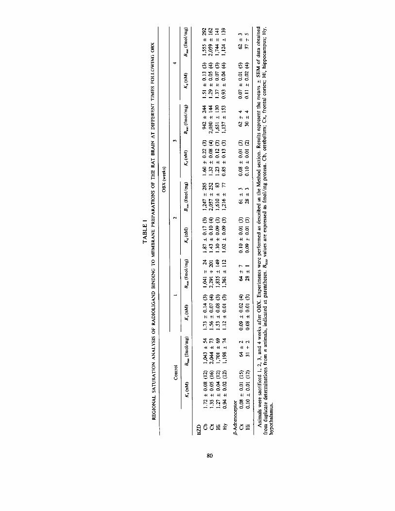
Analyses for the binding parameters of the four ligands and structures investigated were carried out using tissues from OBX rats and their corresponding sham-operated controls. In all cases, there were no significant differences (ANOVA) in Kd or Bma x values between control groups from each time point; thus, these values were pooled. The maximal binding capacities (Bm~) of GABAA receptors increased in the frontal cortex by 1 week (38°70) following OBX and reached statistical significance at 2 weeks (49%). This augmentation persisted throughout the study (54 and 60070 after 3 and 4 weeks, respec- tively) and was not accompanied by any change in Kd values (Fig. IA). Bm~, values were also increased significantly in the hippocampus after 1 week (53070) but were not significantly different thereafter (Fig. 1A). No changes in GABAA binding parameters were seen in the hypothalamus or cerebellum (Fig. IA). Kd values in the cortex, hippocampus, hypothalamus, and cerebellum were 50.0 + 4.0, 48.0 + 5.1, 43.3 + 3.3, and 66.4 _+ 9.0 nM, respectively.
Despite the observed variations in GABA A binding densi- ties in the frontal cortex, these increases were not accompa- nied by any parallel changes in total BDZ receptor binding densities (Table 1). Indeed, no significant changes in [3H]flun- itrazepam binding parameters were observed in any of the structures investigated throughout the study (Table 1). More- over, no variations in BDZ, or BDZ: subtype populations were seen (data not shown).
In contrast, decreases in GABAa receptor binding densities with no change in Kd values were observed in the frontal cortex (Fig. IB). Bm~ values were significantly decreased by 38 and 41070 1 and 2 weeks after OBX, respectively. However, even though binding densities were still found to be 27 and 23070 lower than sham-operated control levels at 3 and 4 weeks, respectively, these decreases were no longer statistically signif-
OLFACTORY BULBECTOMY AND GABA RECEPTORS 79
A 5000
"• 4000
"5 E 3000
X
2000" E m
1000.
Cb Cx Hi Hy
B 600
500
400,
300'
E 200 m
100
Cb Cx Hi Hy
FIG. 1. Time course of the effects of bilateral olfactory bulbectomy (OBX) on Bm~ values (fmol/mg protein) of: (A) specific high-affinity GABAA reptors and (B) specific high-affinity GABAa receptors. Ex- periments were performed as described m the Method section. Results represent the means + SEM of data obtained from duplicate determi- nations from n animals, indicated at the foot of each column. In all cases, the first and subsequent columns represent sham-operated animals followed by OBX at 1, 2, 3, and 4 weeks, respectively. Kd values were not significantly changed at any time in any of the struc- tures investigated. Cb, cerebellum; Cx, frontal cortex; Hi, hippocam- pus; Hy, hypothalamus. */7 < 0.05.
icant (Fig. IB). Bm~ values in the cerebellum increased by 96070 after 1 week and returned to normal levels by 3 weeks (Fig. 1B). GABA B binding parameters in the hippocampus and hy- pothalamus were not altered at any time following OBX (Fig. 1B). g d values in the cortex, hippocampus, hypothalamus, and cerebellum were 47.5 _+ 3.9, 52.2:1: 5.4, 53.7 + 5.9, and 43.4 + 5.8 nM, respectively.
Binding parameters for [3H]CGP-12177 binding to /~- adrenoceptors in the frontal cortex or hippocampus were not modified throughout the time period investigated following OBX (Table 1).
DISCUSSION
The results of the present study show that, over the time period investigated, persistent significant increases in GABAA receptor binding densities, with no change in Kd, are observed in the frontal cortex following OBX. Conversely, during the first 2 weeks following surgery cortical GABAB binding densi- ties are significantly decreased. However, these decreases in Bm~ values show a gradual tendency to normalize and are no longer statistically significant 3 and 4 weeks after OBX.
The present results confirm and extend the previous reports on GABA B binding of Lloyd et al. (13,16,17). Indeed, the results of our study show good agreement in the variations of GABAB binding at the time points (14 and 29 days) chosen by the previous authors (13,16,17). In the present study, Bm~ values for GABAB receptors in the rat frontal cortex are sig- nificantly decreased by 40070 compared to sham-operated con- trols both 1 and 2 weeks after OBX, comparable to the 40- 5007o decrease observed after 2 weeks by Lloyd (16,17). Varia- tions in cerebellar GABAa receptor densities are similar after 2 weeks, but our study shows a marked, transient increase after 1 week that tends to normalize. Moreover, confirming their previous observations (16,17), no changes were observed in the hippocampus in the present study. In addition, 4 weeks after OBX we still observed a 23% reduction in GABAB bind- ing in the cortex whereas the previous authors (13) reported a 27070 decrease using a single concentration of radioligand. The fact that the decrease in GABA a binding we observed 4 weeks after OBX was not statistically significant while theirs was (13) after 29 days may be explained by the large population of animals used in their study. Importantly, our results suggest that, over the time period studied, GABA a receptor densities tend to normalize. These observations are of critical impor- tance to the GABA hypothesis of antidepressant drug action as proposed by Lloyd (18,24). The study of Joly et al. (13) suggests that upregulation of cortical GABAa receptor density following prolonged treatment with desipramine is associated with a normalization of the behavioral deficit in a stepdown passive avoidance paradigm. However, cortical GABA a bind- ing levels in the desipramine-nonresponding group were also normalized in their study (13). Further, the trend toward a normalization of GABAB receptor binding densities with time observed in the present study is consistent with that seen in the various studies performed by these authors (13, 16,17). Thus, as behavioral deficits persist (10,13) while GABAB binding densities tend to normalize, it would appear that GABAa receptor density normalization is not the sole requisite to reverse the deficit in stepdown passive avoidance behavior.
As for the other regions studied, a transient increase in GABAA Bm~ values was observed in the hippocampus 1 week after bulb ablation. In the hypothalamus and cerebellum, no changes in GABA A binding parameters were observed. In ad- dition, no changes in BDZ binding parameters were noted in any of the structures studied (Table 1). This observation is consistent with the results of Hirsch (7), who found no change in [3H]diazepam binding 16 weeks after OBX. However, one may have expected an increase in [3Hlflunitrazepam binding in the frontal cortex parallel to that of GABAA receptors. There are a number of possible explanations as to why this was not the case. In a recent study, Weizman et al. (33) pointed out that measurements of variations in BDZ binding parameters can yield differing results between in vitro and in vivo conditions. These authors suggest that in vivo binding may be a more precise reflection of receptor changes (33). Second, the present binding studies were carried out on mem- brane preparations. This may mask discrete regional changes that could perhaps be detected using autoradiographic proce- dures; such experiments are presently being carried out in our laboratory. Finally, it is generally accepted that high-affinity GABAA receptors are not linked to BDZ binding sites (30), and in the present study only high-affinity [aH]GABAA bind- ing was measured. Thus, this would suggest that bulbectomy results in an upregulation of cortical high-affinity GABAA receptor binding sites that are not associated with BDZ recep-
TA
BL
E
1
RE
GIO
NA
L S
AT
UR
AT
ION
AN
AL
YSI
S O
F R
AD
IOL
IGA
ND
B
IND
ING
TO
ME
MB
RA
NE
PR
EPA
RA
TIO
NS
OF
TH
E R
AT
BR
AIN
AT
DIF
FER
EN
T
TIM
ES
FOL
LO
WIN
G
OB
X
OB
X (
wee
ks)
Con
trol
1
2 3
4
K d
(n
M)
B~
(fm
ol/m
g)
Kd
(nM
) B
~ (
fmol
/mg)
K
d (n
M)
B~
(fm
ol/m
g)
K d (
nM)
B~
(fm
ol/m
g)
Kd
(nM
) B
~ (
fmol
/mg)
Oo
BZ
D
Cb
1.72
+
0.
08
(12)
1,
043
_ 54
1.
73
+
0.14
(3
) 1,
041
+
24
1.87
±
0.
17
(3)
1,24
7 ±
28
5 1.
60
±
0.22
(3
) 94
2 ±
24
4 1.
51 ±
0.
13
(3)
1,55
5 ±
29
2 C
x 1.
35
±
0.05
(1
6)
2,04
4 ±
73
1.
56
±
0.07
(4
) 2,
291
±
201
1.43
±
0.
10
(4)
2,05
7 ±
25
2 1.
32
±
0.08
(4
) 2,
030
+
144
1.29
±
0.
05
(4)
2,05
9 ±
16
2 H
i 1.
27
±
0.04
(1
2)
1,70
1 +
69
1.
53
±
0.08
(3
) 1,
835
+
149
1.30
+
0.
09
(3)
1,61
0 ±
83
1.
23
±
0.12
(3
) 1,
651
±
130
1.37
±
0.
07
(3)
1,74
4 +
14
1 H
y 0.
94
±
0.02
(1
2)
1,19
8 +
74
1.
12
±
0.01
(3
) 1,
361
±
112
1.02
+
0.
09
(3)
1,21
6 ±
77
0.
85
±
0.13
(3
) 1,
137
±
153
0.93
±
0.
04
(4)
1,12
4 +
13
9
/3-A
dren
ocep
tor
Cx
0.08
±
0.
01
(15)
64
±
2
0.09
±
0.
02
(4)
64
±
7 0.
10
:t: 0
.01
(3)
61
±
3 0.
08
±
0.01
(3
) 62
±
4
0.07
+
0.
01
(5)
62
±
3 H
i 0.
10
+
0.01
(1
2)
31
±
2 0.
08
±
0.01
(3
) 28
±
1
0.09
±
0.
01
(3)
28
±
3 0.
10
_+ 0
.01
(2)
30
±
4 0.
11
±
0.02
(4
) 37
±
5
An
imal
s w
ere
sacr
ific
ed
1, 2
, 3,
an
d 4
wee
ks a
fter
OB
X.
Ex
per
imen
ts w
ere
per
form
ed a
s de
scri
bed
m t
he M
eth
od
sec
tion
. R
esul
ts r
epre
sent
the
mea
ns
+
SE
M o
f da
ta o
bta
ined
fr
om
dup
lica
te d
eter
min
atio
ns
fro
m n
an
imal
s, i
ndic
ated
m p
aren
thes
es.
Bm
~x v
alue
s ar
e ex
pres
sed
as f
mo
l/m
g p
rote
in.
Cb,
cer
ebel
lum
; C
x,
fro
nta
l co
rtex
; H
i, h
ipp
oca
mp
us;
Hy,
h
yp
oth
alam
us.
OLFACTORY BULBECTOMY AND GABA RECEPTORS 81
tors. Moreover, this effect is unlikely to be a consequence of postoperative stress. Physical and psychological stress will result in rapid cerebral and plasmatic elevations of neuroactive steroids that are highly potent allosteric modulators of GABA A receptors (23,26). However, various stressors have been shown to decrease in vivo [3H]Ro 15-1788 binding in the cortex but also to increase mRNAs for cq- and 3,2-subunits of GABA A receptors and low-affinity GABA^ binding densities (3,14,25,32). Thus, the fact that stress primarily affects GABAA/BDZ receptors lends further support to the notion that the upregulation of high-affinity GABAA receptors in the frontal cortex observed in the present study is a consequence of OBX and not stress effects.
Jesberger and Richardson (11), using [3H]dihydroalpreno- lol, reported an increase in/~-adrenoceptor binding in the hip- pocampus of OBX rats 7 weeks after surgery. In a more recent study, this increase in binding was further characterized using ['25I]iodocyanopindolol and shown to be the result of a 30% increase in affinity with no change in Bm~ values (29). This increased affinity was observed both in the hippocampus and amygdala, but not in the cerebral cortex, and was proposed to result from an increased coupling of the receptors with G proteins (29). In the present study, we confirmed a lack of significant variations in the binding parameters of [3H]CGP-12177 to /~-adrenoceptors in the cortex. However, there was no change in the affinity of this ligand in the hippo- campus either. Thus, our results do not corroborate these previous observations (29). This discrepancy may be due to methodological differences, that is, the radioligand used
rather than the time points investigated. Although [3H]CGP- 12177 displays high affinity for/3-adrenoceptors (0.10 nM in our hands), [~25Iliodocyanopindolol has 5- to 10-fold greater affinity (22,29) but is less selective and demonstrates high af- finity toward 5-hydroxytryptaminete (5-HTIB) receptors (22). In light of the small change in affinity observed by the previ- ous authors (29), it is possible that the use of [3H]CGP-12177 in the present study may have precluded the detection of such a change.
In conclusion, following OBX persistent significant in- creases in high-affinity GABAA receptor binding site densities were observed in the frontal cortex throughout the time period studied. Conversely, in the same region GABAB receptor bind- ing site densities were significantly decreased 1 and 2 weeks after surgery and more moderately so after 3 and 4 weeks. These changes in GABA receptor densities are likely to result in or from alterations of GABAergic neurotransmission. The fact that cortical GABA a receptor densities tend to normalize with time while increased GABAA binding densities persist would suggest that the upregulation of GABA A receptors, rather than the downregulation of GABAa receptors, might underlie the behavioral deficits elicited by OBX.
ACKNOWLEDGEMENTS
This work was supported by the Medical Research Council of Canada (Grant MT 11025). T.D. is a Scholar of the Fonds de la Recherche en Sant6 du Qu6bec. The authors thank H. A. Cameron for secretarial assistance.
REFERENCES
1. B6navid~s, J.; Cornu, P.; Dennis, T.; Dubois, A.; Hauw, J. J.; MacKenzie, E. T.; Sazdovitch, V; Scatton, B. Imaging of human brain lesions with an ~o 3 site radioligand. Ann. Neurol. 24:708- 712; 1988.
2. B6navid~s, J.; Fage, D.; Carter, C. J.; Scatton, B. Peripheral type benzodiazepine binding sites are a more sensitive index of excitotoxic lesions of the rat striatum than the neuronal marker enzymes choline acetyltransferase or glutamate decarboxylase. Brain Res. 421:167-172; 1987.
3. Biggio, G.; Concas, A.; Mele, S.; Corda, M. G. Changes in GABAergic transmission induced by stress, anxiogenic and anxio- lyric/~-carbolines. Brain Res. Bull. 19:301-308; 1990.
4. Butler, J.; Tannian, M.; Leonard, B. E. The chronic effects of desipramine and sertraline on platelet and synaptosomal 5-HT uptake in olfactory bulbectomised rats. Prog. Neuropsychophar- macol. Biol. Psychiatry 12:585-594; 1988.
5. Healy, D.; Carney, P. A.; O'Halleran, A.; Leonard, B. E. Pe- ripheral adrenoceptors and serotonin receptors in depression: Changes associated with response to treatment with trazodone or amitriptyline. J. Affect. Dlsord. 9:285-296; 1985.
6. Hill, D. R.; Bowery, N. G.; Hudson, A. L. Inhibition of GABAa receptor binding by guanyl nucleotides. J. Neurochem. 42:652- 657; 1984.
7. Hirsch, J. D. Opiate and muscarinic hgand binding in five limbic areas after bilateral olfactory bulbectomy. Brain Res. 198:271- 283; 1980.
8. Hong, K. W.; Lee, W. S.; Rhim, B. Y. Role of central cq- adrenoceptors on the development of muricidal behavior in olfac- tory bulbectomized rats: Effect of ct2-adrenoceptor antagonists. Physiol. Behav. 39:535-539; 1987.
9. Jancsar, S. M.; Leonard, B. E. Changes in neurotransmitter me- tabolism following olfactory bulbectomy in the rat. Prog. Neu- ropsychopharmacoi. Biol. Psychiatry 8:263-269; 1984.
10. Jesberger, J. A.; Richardson, J. S. Effects of antidepressant
drugs on the behavior of olfactory bulbectomized and sham- operated rats. Behav. Neurosci. 100:256-274; 1986.
11. Jesberger, J. A.; Richardson, J. S. Differential effects of antide- pressant drugs on [3H]dihydroalprenolol and [3H]imipramine li- gand recognition sites in olfactory bulbectomized and sham- lesioned rats. Gen. Pharmacol. 17:293-307; 1986.
12. Jesberger, J. A.; Richardson, J. S. Brain output dysregulation induced by olfactory bulbectomy: An approximation in the rat of major depressive disorder in humans? Int. J. Neurosci. 38:241- 265; 1988.
13. Joly, D.; Lloyd, K. G.; Pichat, P.; Sanger, D. J. Correlation between the behavioural effect of desipramine and GABAB recep- tor regulation in the olfactory bulbectomized rat. Br. J. Pharma- col. 90:125P; 1987.
14. Kang, I.; Thompson, M. L.; Heller, J.; Miller, L. G. Persistent elevation in GABA A receptor subunit mRNAs following social stress. Brain Res. Bull. 26:809-812; 1991.
15. Leonard, B. E.; Tuite, M. Anatomical, physiological and behav- ioral aspects of olfactory bulbectomy in the rat. Int. Rev. Neuro- biol. 22:251-286; 1981.
16. Lloyd, K. G.; Pichat, P. Decrease in GABAa binding in the fron- tal cortex of olfactory bulbectomized rats. Br. J. Pharmacol. 87: 36P; 1985.
17. Lloyd, K. G.; Pichat, P. GABA synapses, depression and antide- pressant drugs. In: Dahl, S. G.; Gram, L. F.; Paul, S. M.; Potter, W. Z., eds. Clinical pharmacology in psychiatry. Berlin: Spring- er-Verlag; 1987:113-126.
18. Lloyd, K. G.; Thuret, F.; Pilc, A. Upregulation of 3,-aminobutyric acid (GABA)B binding sites in rat frontal cortex: A common action of repeated administration of different classes of antidepressants and electroshock. J. Pharmacol. Exp. Ther. 235:191-199; 1985.
19. Lo, M. M. S.; Nlehoff, D. L.; Kuhar, M. J.; Snyder, S. H. Differential localization of type I and type II benzodiazepine binding sites in substantia nigra. Nature 306:57-60; 1983.
82 DENNIS , B E A U C H E M I N A N D L A V O I E
20. Martin, P.; Pichat, P.; Massol, J.; Soubrie, P.; Lloyd, K. G.; Puech, A. J. Decreased GABA B receptors in helpless rats: Rever- sai by tricychc antidepressants. Neuropsychobiology 22:220-224; 1989.
21. McLaughlin, N. J.; Collins, G. G. S. Binding characteristics of the selective cq-adrenoceptor antagonist [3H]idazoxan to rat ol- factory cortex membranes. Eur. J. Pharmacol. 121:91-96; 1986.
22. Morin, D.; Zini, R.; Urien, S.; Sapena, R.; Tillement, J. P. Labelling of rat brain /~-adrenoceptors: [3H]CGP-12177 or [~25I]iodocyanopindolol? J. Receptor Res. 12:369-387; 1992.
23. Paul, S. M.; Purdy, R. H. Neuroactive steroids. FASEB J. 6: 2311-2322; 1992.
24. Pilc, A.; Lloyd, K. G. Chronic antidepressants and GABA B re- ceptors: A GABA hypothesis of antidepressant drug action. Life Sci. 35:2149-2154; 1984.
25. Primus, R. J.; Gailager, D. W. GABA^ receptor subunit mRNA levels are differentially influenced by chronic FG 7142 and diaze- pam exposure. Eur. J. Pharmacol. Mol. Pharmacol. Sect. 226: 21-28; 1992.
26. Purdy, R. H.; Morrow, A. L.; Moore, P. H., Jr.; Paul, S. M. Stress-reduced elevations of 3,-aminobutyric acid type A receptor- active steroids in the rat brain. Proc. Natl. Acad. Sct. USA 88: 4553-4557; 1991.
27. Rtva, M. A.; Creese, I. Comparison of two putatively selective radioligands for labeling central nervous system ~-adrenergic re- ceptors: Inadequacy of [3H]dihydroalprenolol. Mol. Pharmacol. 36:201-210; 1989.
28. Suranyi-Cadotte, B. E.; Dam, T. V.; Quirion, R. Antidepressant- anxiolytic interaction: Decreased density of benzodiazepine recep- tors in rat brain following chronic administration of antidepres- sants. Eur. J. Pharmacol. 106:673-675; 1984.
29. Tiong, A. H. K.; Richardson, J. S. Differential effects of olfac- tory bulbectomy on/3-adrenoceptors in rat amygdaia, hippocam- pus and cerebral cortex. Brain Res. 531:269-275; 1990.
30. Unnerstall, J. R.; Kuhar, M. J.; Niehoff, D. L.; Palacios, J. M. Benzodiazepine receptors are coupled to a subpopulation of 3,-aminobutyric acid (GABA) receptors: Evidence from a quanti- tative autoradiographic study. J. Pharmacol. Exp. Ther. 218: 797-804; 1981.
31. Van Riezen, H.; Leonard, B. E. Effects of psychotropic drugs on the behavior and neurochemistry of olfactory bulbectomized rats. Pharmacol. Ther. 47:21-34; 1990.
32. Weizman, A.; Weizman, R.; Kook, K. A.; Vocci, F.; Deutsch, S. I.; Paul, S. M. Adrenalectomy prevents the stress-induced de- crease m m vtvo [3H]RoI5-1788 binding to GABAA-benzodiaze- pine receptors in the mouse. Brain Res. 519:347-350; 1990.
33. Weizman, R.; Weizman, A.; Kook, K. A.; Vocci, F.; Deutsch, S. I.; Paul, S. M. Repeated swim stress alters brain benzodiazepme receptors measured in vivo. J. Pharmacol. Exp. Ther. 249:701- 707; 1989.
34. Willner, P. Animal models of depression: An overview. Pharma- col. Thee 45:425-455; 1990.