Differentiated Adipose-Derived Stem Cell Cocultures forBone Regeneration in Polymer Scaffolds In Vivo
Amita R. Shah, MD, PhD,* Agustin Cornejo, MD,* Teja Guda, PhD,† David E. Sahar, MD,*Stacy M. Stephenson, MD,* Shiliang Chang, MD,* Naveen K. Krishnegowda, MD,*
Ramaswamy Sharma, PhD,‡ and Howard T. Wang, MD*
Abstract: Critical-sized bone defects can lead to significant mor-bidity, and interventions are limited by the availability and donor-site morbidity of bone grafts. Polymer scaffolds seeded with cellshave been explored to replace bone grafts. Adipose-derived stemcells have shown great promise for vascularization and osteogenesisof these constructs, and cocultures of differentiated stem cells arebeing explored to augment vessel and bone formation. Adipose-derived stem cells were differentiated into endothelial cells andosteoblasts, and in vitro studies showed increased proliferation ofcocultured cells compared with undifferentiated adipose-derivedstem cells and monocultures of endothelial cells and osteoblasts.The cells were seeded into polylactic acid gas-plasma–treated scaffoldsas cocultures and monocultures and then implanted into critical-sized rat calvarial defects. The cocultures were in a 1:1 osteoblastto endothelial cell ratio. The increase in proliferation seen by thecocultured cells in vitro did not translate to increased vasculariza-tion and osteogenesis in vivo. In vivo, there were trends of increasedvascularization in the endothelial cell group and increased osteo-genesis in the osteoblast and endothelial monoculture groups, butno increase was seen in the coculture group compared with the undif-ferentiated adipose-derived stem cells.
Endothelial cells enhance vascularization and osteoblast and en-dothelial cell monocultures enhance bone formation in the polymerscaffold. Predifferentiation of adipose-derived stem cells is promis-ing for improving vascularization and osteogenesis in polymer scaffoldsbut requires future evaluation of coculture ratios to fully character-ize this response.
Traumatic injuries can lead to significant morbidity if the injuryinvolves bone defects that exceed the body's ability to repair it-
self to its preinjury state.1 Bone autografts are the criterion standardfor implant materials for critical-sized defects but are limited byavailability of materials and donor-site morbidity.2–4 Strategies forovercoming these shortcomings have involved synthetic or allogenicscaffolding with implantation of cells for bone regeneration.5,6
Adipose-derived stem cells (ASCs) have been investigatedas a source of cells for implantation in scaffolds for bone regener-ation.7,8 They serve as a source of autologous stem cells that canbe in adequate supply and are easily accessible compared with othersources of cells such as bone marrow or vessels. These cells are ca-pable of differentiating into adipocytes, chondrocytes, osteoblasts(OBs), fibroblasts, and endothelial cells (ECs) 9–11 and have beensuccessfully differentiated into ECs and OBs by our group.5
Because diffusion in a scaffold is limited to 100 to 150 μm,12
it is important to not only regenerate bone but also allow for the de-velopment of a vascularized network to allow for the delivery ofnutrients, growth factors, and oxygen to the newly developing bone.Seeding of undifferentiated ASCs or a single type of cell has beenlimited in forming a robust vessel network or adequate mineraliza-tion in large defects. Therefore, cocultures are being examined asa possible solution to this problem.
Coculture is the culture of cells together such as ECs withOBs. During the natural regeneration of bone, there are multiplesynergistic interactions between these 2 types of cells.13–16 Cocul-ture studies have shown that ECs accelerate bone formation via an-giogenesis and production of osteogenic factors, whereas OBsproduce angiogenic factors such as vascular endothelial growthfactor and matrix components.16–18 These cocultures can be easilyseeded within a polymeric scaffold.
Polylactic acid polymer scaffolds have the advantage of be-ing degradable, easily moldable, and adequate in supply but are lim-ited by the hydrophobicity of the surface.19 Gas plasma treatment isa method of sterilization and surface treatment for polymers that im-prove attachment and proliferation of cells by attaching oxygenradicals to the surface and making the surface more hydrophilic.20,21
It has also been shown to enhance angiogenesis in subcutaneousmouse models.22
The objective of this study was to implant gas-plasma–treated poly (D,L-lactide) polymer (PLA) scaffolds, seeded with ei-ther cocultures or monocultures of ASCs predifferentiated into ECsor OBs, into a critical-sized calvarial defect and assess bone regen-eration and vessel formation. It is hypothesized that there will be a
From the *Division of Plastic and Reconstructive Surgery, University ofTexas Health Science Center at San Antonio, †Department of Biomedi-cal Engineering, University of Texas at San Antonio, and ‡Cellular andStructural Biology, University of Texas Health Science Center at SanAntonio, San Antonio, Texas.
Received November 3, 2013.Accepted for publication January 7, 2014.Address correspondence and reprint requests to Howard T. Wang, MD,
Division of Plastic and Reconstructive Surgery, The University of TexasHealth Science Center at San Antonio, 7703 Floyd Curl Dr MSC 7844,San Antonio, Texas 78229; E-mail: [email protected]
Supported by the National Endowment for Plastic Surgery and PlasticSurgery Foundation.
greater amount of bone volume and vasculogenesis in the defectsimplanted with scaffolds containing cocultured cells.
MATERIALS AND METHODS
Adipose-Derived Stem Cell Isolation and CultureThe study protocol was approved by the University of Texas
Health Science Center at San Antonio animal use committee. Isola-tion and differentiation of the ASCs were performed as done inprevious studies by our group.5 The ASC differentiation was basedon protocols established by Zuk et al for endothelial differentia-tion.9 Confirmation ASC differentiation to ECs was previouslydone by our group,5,8 which included immunohistochemistry withCD31, von Willebrand Factor (VWF) western blot, angiogenicassays, and histologic assay. The same protocols were used for thisstudy. Osteogenic differentiation of ASCs has also been confirmedin our previous studies with immunohistochemistry withosteocalcin as well as osteopontin, and the ability for calcium de-position was demonstrated with von Kossa staining.8
Under general anesthesia using isoflurane, adipose tissue ex-cised from the perinephric and peritesticular fat pad of 2-month-oldLewis rats was placed in sterile pH-balanced Hank's balanced saltsolution containing 1% bovine serum albumin (BSA) for furtherprocessing. The adipose tissue was finely minced using sharp dis-section and centrifuged for 5 minutes at 500 � g. The free-floatingfat was removed, and the remaining tissue/cell suspension wasplaced into 25 mL of 1% BSA Hank's balanced salt solutioncontaining collagenase (200 U/mL) and allowed to agitate at 37°Cfor 1 hour. The digest was filtered through 100-μm nylon meshtwice and then centrifuged for 5 minutes at 500 � g. The superna-tant was removed and the pellet was resuspended in MesenPro me-dia (Invitrogen, Carlsbad, CA) to a concentration of 4 � 104 cellsper milliliter and plated overnight and incubated at 37°C with 5%CO2. Cells were used for differentiation after the third passage.
Osteogenic InductionAfter the third passage, the ASCs were cultured in an osteo-
genic differentiation medium consisting of Dulbecco's ModifiedEagle Medium, 10% fetal bovine serum, 1% penicillin/streptomycin,0.1-μM dexamethasone, 10-μM β-glycerol phosphate, and 50-μMascorbic acid. The osteogenic medium was changed every thirdday for 21 days.
Endothelial InductionAfter the third passage, the ASCs were cultured in a vascu-
logenic medium consisting of Dulbecco's Modified Eagle Medium,10% fetal bovine serum, 1% penicillin/streptomycin, and 50-ng/mLrecombinant rat vascular endothelial growth factor-C (Promocell,Heidelberg, Germany). The media was changed every other day for8 days before implantation in the bone allografts. The vascular endo-thelial growth factor-C was added fresh to stock media before eachmedia change.
ImmunocytochemistryAfter the cells were cultured within the scaffolds for 21 days,
the cells were fixed with 4% (w/v) paraformaldehyde, washed withphosphate-buffered saline (PBS; pH, 7.4), and permeabilized withPBS containing 0.2% Tween-20 for 30 minutes. The cells were thenwashed thrice with PBS and blocked with PBS containing 1% BSAand 10% goat serum for 1 hour at room temperature (RT) on ashaker. Primary antibody to osteocalcin was then added in blockingsolution (sc-23790; Santa Cruz Biotech, Santa Cruz, CA), and the
cells were kept at 4°C overnight on the shaker. After washing thenext day, secondary antibody conjugated to Alexa 488 fluorochrome(A11055, Invitrogen, Carlsbad, CA) was added for 1 hour at RT.The cells were washed again as well as blocked for 30 minutes atRT, and primary antibody to VWF (sc-14014; Santa Cruz Biotech)was added to cells at 4°C overnight. The cells were washed again,and the secondary antibody conjugated to Alexa 568 (A11036or A11004; Invitrogen) was added for 1 hour at RT, along withHoechst 33258 (Invitrogen). The cells were then washed withPBS and rinsed in water, and the cover slips were transferred to a slidewith an antifade (VectaShield; Vector Laboratories, Burlingame,CA),sealed with nail polish, and visualized in an Olympus FV1000 confocalmicroscope (Olympus, Center Valley, PA). Images were ac-quired sequentially and analyzed using FV10-ASW version 2.1at 40-fold oil immersion magnification.
Preparation of PLA ScaffoldsThe PLA scaffolds were fabricated using a vibrating particle
salt-leaching method, which yields an open cell construct withinterconnected pores, porosity of 90%, and pores 250 to 600 μm in di-ameter. Poly (D,L-lactic) polymer (DURECTCorp, Birmingham, AL)was dissolved in acetone, and the polymer solution was added tosodium chloride and vibrated under continuous flow conditions.Eight-millimeter–diameter disks of 1.2-mm thickness were punchedout of the polymer mixture after drying, and the sodium chloridewas leached out in sterile deionized water. After 2 days in water,the scaffolds were lyophilized. Before seeding, gas-plasma treat-ment was performed in a pure oxygen environment in a glowdischarge system (PDC-32G, Plasma Cleaner/Sterilizer; HarrickScientific, Inc, Pleasantville, NY) for 3 minutes at 100 W as a sur-face treatment and for sterilization.
Seeding PLA ScaffoldsBefore seeding, the scaffolds were degassed in culture media
under vacuum and placed in a 24-well ultralow attachment tissueculture plate (Corning Life Sciences, Lowell, MA). The cells wereresuspended to a concentration of 100,000 cells per 20 μL beforebeing added dropwise to the scaffold surface. The seeding effi-ciency of this method is approximately 80% based on our studies.The cells were allowed to incubate for 30 minutes at 37ºC 5%CO2 before adding 500 μL of the culture media to each well.
Proliferation AssayProliferation of ASC, ECs, OBs, and cocultured ECs and
OBs in the scaffolds was measured on days 1, 3, 7, 14, and 21 usingthe AlamarBlue (Invitrogen, Carlsbad, CA) assay. At each timepoint, the scaffolds were incubated for 1.5 hours at 37°C with 5%CO2 in 500 μL of a 10% AlamarBlue solution in a new 24-wellplate. After the incubation, the scaffolds were removed from thewell plates, rinsed with PBS, transferred to a new plate with newmedia, and placed back in the incubator for further culture. Fluo-rescence was measured using a fluorescence plate reader at excita-tion at 540 nm and emission at 590 nm (n = 3).
Preparation of In Vivo Implant SamplesThe PLA scaffolds were degassed and seeded in the same man-
ner as that for the in vitro samples with a total of 100,000 cells perscaffold. The scaffolds were divided into 4 groups: (1) undifferenti-ated ASC (n = 8), (2) adipose tissue–derived OBs (Osteo, n = 5),(3) adipose tissue–derived ECs (Endo, n = 7), and (4) adipose tissue–derived EC and OB coculture in a 1:1 ratio (Osteo-Endo, n = 8).
The Journal of Craniofacial Surgery • Volume 25, Number 4, July 2014 Bone Regeneration Stem Cell Cocultures
In Vivo AssayTwo-month-old Lewis rats were anesthetized with isoflurane,
and the scalps were shaved and prepared. The rat calvarias wereexposed using a midline scalp incision, and the skin and periosteumreflected laterally. An 8-mm defect was created using an 8-mmtrephine burr, leaving the dura mater intact. The critical-sizedcalvarial defects were treated with the previously seeded PLAscaffolds. The implants were secured in place with silk suture,and the scalp was closed with staples. At 8 weeks postoperatively,the rats were euthanized and their calvaria were harvested for radio-graphic and histologic examination.
Microcomputed Tomographic AnalysisThe samples were scanned using microcomputed tomogra-
phy (μCT) SkyScan 1076 (Skyscan, Aartselaar, Belgium) at 100-kVsource voltage and 100-μA source current with no filter used andat a spatial resolution of 8.77 μm. The reconstructions were per-formed using NRecon software (Skyscan, Aartselaar, Belgium)and resulted in grayscale images that spanned a density range from0.96 to 2.57 g/cm3 corresponding to grayscale values of 0 to 255.DataViewer (Skyscan, Aartselaar, Belgium) was used to reslicethe μCT images along coronal and sagittal axes, which were usedto reorient the μCT slices to be perpendicular to the cranial-caudal axisof the calvaria, and μCT thresholding was performed to includeonly ossified tissues. The thresholds were selected as the histogramminima between the peaks representing the formalin in which thesample was scanned and the bone volume of the sample. The geo-metric mean of the threshold for all 100 samples in the study wasused to determine the upper and lower threshold for binarizationas 32 and 250. The region of interest (ROI) was chosen by creatinga volume that spanned 8 mm around the defect generated in the cal-varia and repaired with allograft to include the entire defect gener-ated. Additional analyses were performed by choosing concentricROI of 7 mm in diameter and the remainder of the native calvaria.All ROIs comprised the full thickness of the calvaria. The total vol-ume of bone ingrowth in this three-dimensional volume was com-puted using CTAn software (Skyscan, Aartselaar, Belgium), andthe mean bone density of the ossified tissues within these three-dimensional volumes was compared.
Histologic Evaluation of VascularizationAfter μCT, the specimens were fixed in 10% neutral
phosphate-buffered solution formalin, demineralized in EDTA so-lution, dehydrated in a series of alcohol washes, and embedded inparaffin. The specimens were sectioned in the coronal plane inthe center of the bone scaffolds at a thickness of 6 μm and stainedwith hematoxylin and eosin for visualization through conventionalqualitative bright-field microscopy analysis. For the quantificationof blood vessels, 3 sections per animal stained with a rat-specificanti-CD34 antibody (R&D Systems AF4117/Goat pAb) and counter-stained with hematoxylin were analyzed. Microscopic pictures wererandomly taken at 10-fold magnification including 3 central and3 peripheral areas per section for a total of 6 pictures. To determinethe microvascular density (mean number of capillaries per squaremillimeter), the number of structures with lumen surrounded byCD34-positive cells was counted manually. Histomorphometricanalysis of the tissue area on the pictures was performed using Im-age J (National Institutes of Health, Bethesda, MD). Image collec-tion and quantification were performed by 1 author blinded to thetreatment groups.
Statistical AnalysisThe Kruskal-Wallis 1-way analysis of variance on ranks was
used for statistical analysis using Statistical Analysis Software (SAS
Institute, Cary, NC) and was performed for all acquired μCT andmicrovessel quantification data. All data are presented as themean ± standard deviation. In all statistical evaluations, P < 0.05was considered as statistically significant.
RESULTS
Endothelial DifferentiationSuccessful differentiation of ASCs into ECs for this experi-
ment was confirmed using immunocytochemistry analysis. Therewas an increase in VWF expression of EC-differentiated ASC com-pared with nondifferentiated ASCs after 8 days of differentiation inendothelial differentiation media (Fig. 1).
Osteogenic DifferentiationOsteocalcin was prominently expressed on ASCs differenti-
ated in osteogenic media for 21 weeks as shown by prominentexpression of osteocalcin by the cells using immunofluorescence(Fig. 1).
Proliferation in PLA ScaffoldsThere was increased proliferation of cells at all time points in
scaffolds seeded with cocultured ECs and OBs (Osteo-Endo) com-pared with undifferentiated ASCs and monocultured EC (Endo)and OBs (Osteo). At day 21, there was a significantly higheramount of cells in the Osteo-Endo group compared with that inthe other groups. Undifferentiated ADSCs had the lowest amountof proliferation at all time points. The amount of fluorescence atday 21 for the Osteo-Endo group was 11.112 ± 0.083 comparedwith 6.172 ± 0.080 (ASC), 7.081 ± 0.046 (Endo), and 9.093 ± 0.118(Osteo) (Fig. 2).
Osteogenesis in PLA ScaffoldsQuantification of Bone Volume
Quantification of bone volume was performed using μCTanalysis. The bone volume was measured in the 8-mm–diameter de-fect and also the 7-mm–diameter defect, which excludes the outer0.5 mm of the defect to account for migration of native OBs andbone formation due to those cells. For the 8- and 7-mm ROIs, therewas no statistically significant difference in bone volume, but therewas an increased trend of increased bone volume in the Osteogroup (Fig. 3).
Quantification of Bone Mineral DensityBone mineral density was measured in gram hydroxyapatite
per milliliter in the 8-mm defect 8 weeks after the implantation.The observed mineral density ranged from 1.132 to 1.309 mg HA/
FIGURE 1. Immunofluorescence images of scaffolds seeded in vitro for 21 days.Blue indicates Hoechst = nucleus; green, OCN; red, VWF (40� oil-immersionmagnification). OCN, osteocalcin.
Shah et al The Journal of Craniofacial Surgery • Volume 25, Number 4, July 2014
mLwith the Endo and Osteo groups, showing increased trend of bonedensity compared with the ASC and Osteo-Endo groups. There wasno statistically significant difference between the groups usingKruskal-Wallis 1-way analysis of variance (P = 0.151) (Fig. 4).
Vasculogenesis in PLA ScaffoldsMicrovessel density was measured as vessel area/tissue area
(%) in the 8-mm defect 8 weeks after the implantation. The medianobserved microvessel density ranged from 0.932% for the Endogroup, 0.817% for the ASC group, 0.744% for the Osteo group,and 0.899% for the Osteo-Endo group, with the Endo group show-ing a slightly increased trend of increased microvessel density com-pared with the ASC, Osteo, and Osteo-Endo groups. There was nostatistically significant difference between the groups (P = 0.619).The Kruskal-Wallis 1-way analysis of variance on ranks was usedfor statistical analysis (Fig. 5).
DISCUSSIONThe PLA scaffolds were engineered to have porosity, inter-
connectivity, and surface morphology favorable to EC attachmentand bone formation.23,24 In addition, the gas-plasma treatment ofthe PLA scaffold has been shown to increase vascularization.22
For these reasons, PLA scaffolds were used for this study and itwas hypothesized that the use of PLA scaffolds with cocultured dif-ferentiated ASCs would result in improved vasculogenesis and os-teogenesis within a critical-sized rat calvarial defect.
Although increased proliferation of cocultured cells was seenin vitro, this did not translate in vivo. There were no statistically sig-nificant differences seen between the 4 groups for vascularization or
osteogenesis, but there were trends of increased vascularization inthe Endo group and increased osteogenesis in the Osteo and Endogroups but none in the coculture group.
In previous studies from our group using allografts and PLAscaffolds in the same rat calvarial defect model using monocultureddifferentiated and undifferentiated ASCs, it was shown that therewas an increase in vascularization when ECs were used in boththe allograft and PLA implants, but bone formation was differentbetween the 2 materials.5,8 The PLA scaffolds displayed increasedbone formation with OB seeding, and allografts had increasedbone formation with endothelial seeding. The differences seen inthese studies are thought to be directly caused by the materialdifferences.
Endothelial cell and OB cocultures have been shown to havea synergistic effect between the 2 cell types, but as with other cellcultures, the results are dependent on the environment in which theyare cultured.25 In this study, the cocultured group had increased pro-liferation in vitro compared with the monocultured and undifferen-tiated culture groups, which did not translate into increasedangiogenesis or osteogenesis in vivo. The limitation of the in vitroproliferation study is that it reflects the total number of cells anddoes not give a distribution of ECs to OBs present once the cul-ture starts. Factors such as cell density, distribution, and timingof seeding within the scaffold contribute to formation of boneand vessels.25,26
Cocultured ECs and OBs were used in the allograft study,and it was believed that the lack of adequate porosity of the allo-graft and the use of the 1:1 coculture ratio were responsible forthe limitations of the vasculogenic and osteogenic response. For
FIGURE 3. Bone volume quantification measured in cubic millimeters withinthe 8-mm scaffold 8 weeks after the implantation. The results are presented asmedian as well as 25% and 75% quartiles with outliers indicated by black dots.
FIGURE 5. Microvessel density measured in cubic millimeters within the 8-mmscaffold 8 weeks after the implantation. The results are presented as median aswell as 25% and 75% quartiles with outliers indicated by black dots.
FIGURE 2. Cell proliferation: Cocultured ECs and OBs had significantlyincreased proliferation over 21 days compared with monocultured cells.The asterisk indicates P < 0.05).
FIGURE 4. Bone mineral density measured in cubic millimeters within the8-mm scaffold 8 weeks after the implantation. The results are presentedas median as well as 25% and 75% quartiles with outliers indicated byblack dots.
The Journal of Craniofacial Surgery • Volume 25, Number 4, July 2014 Bone Regeneration Stem Cell Cocultures
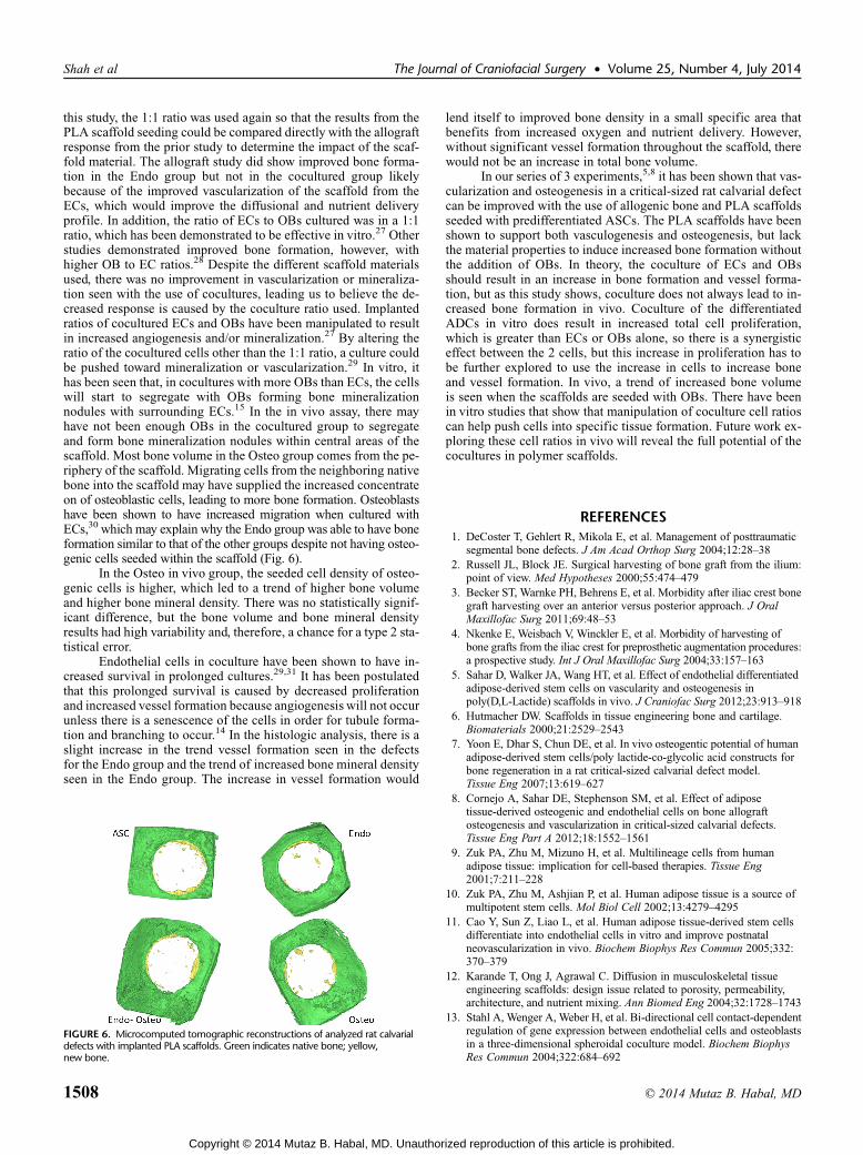
this study, the 1:1 ratio was used again so that the results from thePLA scaffold seeding could be compared directly with the allograftresponse from the prior study to determine the impact of the scaf-fold material. The allograft study did show improved bone forma-tion in the Endo group but not in the cocultured group likelybecause of the improved vascularization of the scaffold from theECs, which would improve the diffusional and nutrient deliveryprofile. In addition, the ratio of ECs to OBs cultured was in a 1:1ratio, which has been demonstrated to be effective in vitro.27 Otherstudies demonstrated improved bone formation, however, withhigher OB to EC ratios.28 Despite the different scaffold materialsused, there was no improvement in vascularization or mineraliza-tion seen with the use of cocultures, leading us to believe the de-creased response is caused by the coculture ratio used. Implantedratios of cocultured ECs and OBs have been manipulated to resultin increased angiogenesis and/or mineralization.27 By altering theratio of the cocultured cells other than the 1:1 ratio, a culture couldbe pushed toward mineralization or vascularization.29 In vitro, ithas been seen that, in cocultures with more OBs than ECs, the cellswill start to segregate with OBs forming bone mineralizationnodules with surrounding ECs.15 In the in vivo assay, there mayhave not been enough OBs in the cocultured group to segregateand form bone mineralization nodules within central areas of thescaffold. Most bone volume in the Osteo group comes from the pe-riphery of the scaffold. Migrating cells from the neighboring nativebone into the scaffold may have supplied the increased concentrateon of osteoblastic cells, leading to more bone formation. Osteoblastshave been shown to have increased migration when cultured withECs,30 which may explain why the Endo group was able to have boneformation similar to that of the other groups despite not having osteo-genic cells seeded within the scaffold (Fig. 6).
In the Osteo in vivo group, the seeded cell density of osteo-genic cells is higher, which led to a trend of higher bone volumeand higher bone mineral density. There was no statistically signif-icant difference, but the bone volume and bone mineral densityresults had high variability and, therefore, a chance for a type 2 sta-tistical error.
Endothelial cells in coculture have been shown to have in-creased survival in prolonged cultures.29,31 It has been postulatedthat this prolonged survival is caused by decreased proliferationand increased vessel formation because angiogenesis will not occurunless there is a senescence of the cells in order for tubule forma-tion and branching to occur.14 In the histologic analysis, there is aslight increase in the trend vessel formation seen in the defectsfor the Endo group and the trend of increased bone mineral densityseen in the Endo group. The increase in vessel formation would
lend itself to improved bone density in a small specific area thatbenefits from increased oxygen and nutrient delivery. However,without significant vessel formation throughout the scaffold, therewould not be an increase in total bone volume.
In our series of 3 experiments,5,8 it has been shown that vas-cularization and osteogenesis in a critical-sized rat calvarial defectcan be improved with the use of allogenic bone and PLA scaffoldsseeded with predifferentiated ASCs. The PLA scaffolds have beenshown to support both vasculogenesis and osteogenesis, but lackthe material properties to induce increased bone formation withoutthe addition of OBs. In theory, the coculture of ECs and OBsshould result in an increase in bone formation and vessel forma-tion, but as this study shows, coculture does not always lead to in-creased bone formation in vivo. Coculture of the differentiatedADCs in vitro does result in increased total cell proliferation,which is greater than ECs or OBs alone, so there is a synergisticeffect between the 2 cells, but this increase in proliferation has tobe further explored to use the increase in cells to increase boneand vessel formation. In vivo, a trend of increased bone volumeis seen when the scaffolds are seeded with OBs. There have beenin vitro studies that show that manipulation of coculture cell ratioscan help push cells into specific tissue formation. Future work ex-ploring these cell ratios in vivo will reveal the full potential of thecocultures in polymer scaffolds.
REFERENCES1. DeCoster T, Gehlert R, Mikola E, et al. Management of posttraumatic
segmental bone defects. J Am Acad Orthop Surg 2004;12:28–382. Russell JL, Block JE. Surgical harvesting of bone graft from the ilium:
point of view. Med Hypotheses 2000;55:474–4793. Becker ST, Warnke PH, Behrens E, et al. Morbidity after iliac crest bone
graft harvesting over an anterior versus posterior approach. J OralMaxillofac Surg 2011;69:48–53
4. Nkenke E, Weisbach V, Winckler E, et al. Morbidity of harvesting ofbone grafts from the iliac crest for preprosthetic augmentation procedures:a prospective study. Int J Oral Maxillofac Surg 2004;33:157–163
5. Sahar D, Walker JA, Wang HT, et al. Effect of endothelial differentiatedadipose-derived stem cells on vascularity and osteogenesis inpoly(D,L-Lactide) scaffolds in vivo. J Craniofac Surg 2012;23:913–918
6. Hutmacher DW. Scaffolds in tissue engineering bone and cartilage.Biomaterials 2000;21:2529–2543
7. Yoon E, Dhar S, Chun DE, et al. In vivo osteogentic potential of humanadipose-derived stem cells/poly lactide-co-glycolic acid constructs forbone regeneration in a rat critical-sized calvarial defect model.Tissue Eng 2007;13:619–627
8. Cornejo A, Sahar DE, Stephenson SM, et al. Effect of adiposetissue-derived osteogenic and endothelial cells on bone allograftosteogenesis and vascularization in critical-sized calvarial defects.Tissue Eng Part A 2012;18:1552–1561
9. Zuk PA, Zhu M, Mizuno H, et al. Multilineage cells from humanadipose tissue: implication for cell-based therapies. Tissue Eng2001;7:211–228
10. Zuk PA, Zhu M, Ashjian P, et al. Human adipose tissue is a source ofmultipotent stem cells. Mol Biol Cell 2002;13:4279–4295
11. Cao Y, Sun Z, Liao L, et al. Human adipose tissue-derived stem cellsdifferentiate into endothelial cells in vitro and improve postnatalneovascularization in vivo. Biochem Biophys Res Commun 2005;332:370–379
12. Karande T, Ong J, Agrawal C. Diffusion in musculoskeletal tissueengineering scaffolds: design issue related to porosity, permeability,architecture, and nutrient mixing. Ann Biomed Eng 2004;32:1728–1743
13. Stahl A, Wenger A, Weber H, et al. Bi-directional cell contact-dependentregulation of gene expression between endothelial cells and osteoblastsin a three-dimensional spheroidal coculture model. Biochem BiophysRes Commun 2004;322:684–692
FIGURE 6. Microcomputed tomographic reconstructions of analyzed rat calvarialdefects with implanted PLA scaffolds. Green indicates native bone; yellow,new bone.
Shah et al The Journal of Craniofacial Surgery • Volume 25, Number 4, July 2014
14. Villars F, Guillotin B, Amedee T, et al. Effect of HUVEC onhuman osteoprogenitor cell differentiation nees heterotypicgap junction communication. Am J Physiol Cell Physiol 2002;282:775–785
15. Wenger A, Stahl A, Weber H, et al. Modulation of in vitro angiogenesisin a three-dimensional spheroidal coculture model for bone tissueengineering. Tissue Eng 2004;10:1536–1547
16. Clarkin CE, Emery RJ, Pitsillides AA, et al. Evaluation ofVEGF-mediated signaling in primary human cells reveals a paracrineaction for VEGF in osteoblast-mediated crosstalk to endothelial cells.J Cell Physiol 2008;214:537–544
17. Wang DS, Miura M, Demura H, et al. Anabolic effects of1,25-dihydroxyvitamin D3 on osteoblasts are enhanced by vascularendothelial growth factor produced by osteoblasts and by growthfactors produced by endothelial cells. Endocrinology 1997;138:2953–2962
18. Villars F, Bordenave L, Bareille R, et al. Effect of human endothelialcells on human bone marrow stromal cell phenotype: role of VEGF?J Cell Biochem 2000;79:672–685
19. Agrawal C, Ray R. Biodegradable polymeric scaffolds for musculoskeletaltissue engineering. J Biomed Mater Res 2001;55:141–150
20. Shah A, Shah S, Mani G, et al. Endothelial cell behaviour ongas-plasma-treated PLA surfaces: the roles of surface chemistry androughness. J Tissue Eng Regen Med 2010;5:301–312
21. Chim H, Ong JL, Schantz J-T, et al. Efficacy of glow discharge gas plasmatreatment as a surface modification process for three-dimensional poly(D,L-lactide) scaffolds. 2003
22. Polan JL, Morse B, Wetherold S, et al. VEGF analysis induced byendothelialized gas-plasma treated d,l-PLA scaffolds. CardiovascRadiat Med 2002;3:176–182
23. Cheung H-Y, Lau K-T, Lu T-P, et al. A critical review on polymer-basedbio-engineered materials for scaffold development. Compos Part B-Eng2007;38:291–300
24. Shah A. Endothelial cell and osteoblast co-cultures on biphasiccomposite scaffolds for bone tissue engineering [Ph.D. dissertation].San Antonio: Biomedical Engineering, The University of Texas atSan Antonio, 2010
25. Sorrell JM. A self-assembled fibroblast-endothelial cell co-culturesystem that supports in vitro vasculogenesis by both human umbilicalvein endothelial cells and human dermal microvascular endothelial cells.Cells Tissues Organs 2007;186:157–168
26. Chen RR, Silva EA, Yuen WW, et al. Spatio-temporal VEGF and PDGFdelivery patterns blood vessel formation and maturation. Pharm Res2007;24:258–264
27. Ma J, van den Beucken J, Yang F, et al. Coculture of osteoblasts andendothelial cells: optimization of culture medium and cell ratio.Tissue Eng Part C 2011;17:349–357
28. Koob S, Torio-Padron N, Stark GB, et al. Bone formation andneovascularization mediated by mesenchymal stem cells andendothelial cells in critical-sized calvarial defects. Tissue EngPart A 2011;17:311–321
29. Unger RE, Sartoris A, Peters K, et al. Tissue-like self-assembly incocultures of endothelial cells and osteoblasts and the formation ofmicrocapillary-like structures on three-dimensional porous biomaterials.Biomaterials 2007;28:3965–3976
30. Shah A, Shah S, Oh S, et al. Migration of co-cultured endothelial cellsand osteoblasts in composite hydroxyapatite/polylactic acid scaffolds.Ann Biomed Eng 2011;39:2501–2509
31. Koike N, Fukumura D, Gralla O, et al. Tissue engineering: creation oflong-lasting blood vessels. Nature 2004;428:138–139
The Journal of Craniofacial Surgery • Volume 25, Number 4, July 2014 Bone Regeneration Stem Cell Cocultures