29
1 The Digestive System
| Date post: | 04-Aug-2015 |
| Category: |
Education |
| Upload: | not-yet-working-i-m-still-studing |
| View: | 11 times |
| Download: | 0 times |
1
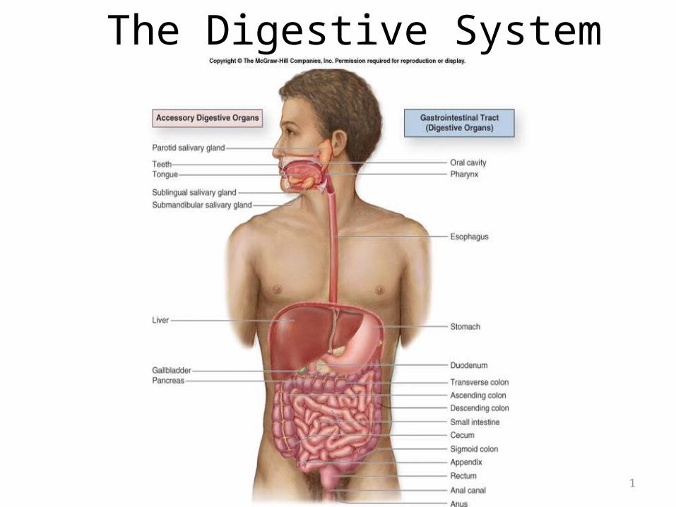
The Digestive System
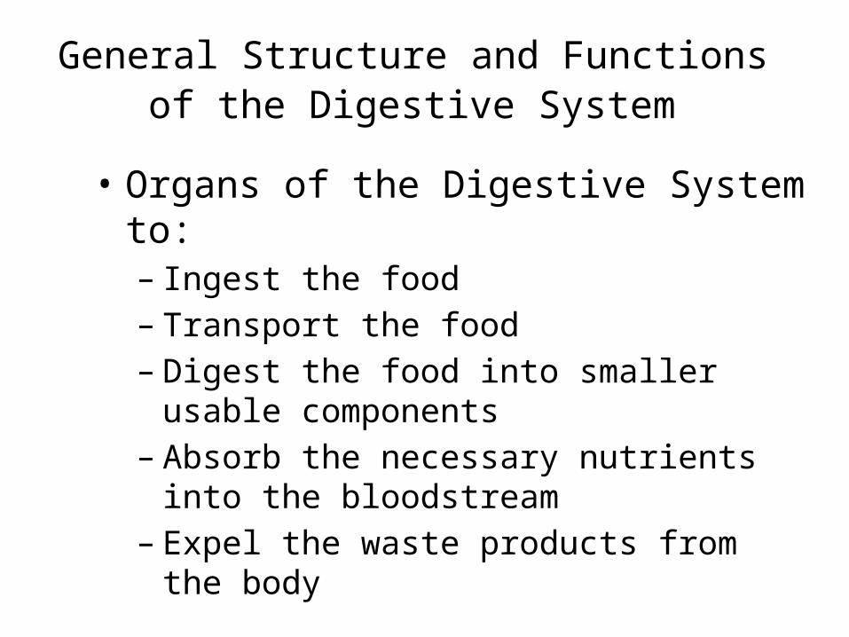
General Structure and Functions of the Digestive System
• Organs of the Digestive System to:– Ingest the food– Transport the food– Digest the food into smaller usable components– Absorb the necessary nutrients into the
bloodstream– Expel the waste products from the body
26-3
General Structure and Functions of the Digestive System
• Composed of two separate categories of organs: – digestive organs – accessory digestive organs
• Digestive organs collectively make up the:– gastrointestinal (GI) tract– Also called:
• the digestive tract• alimentary canal
4
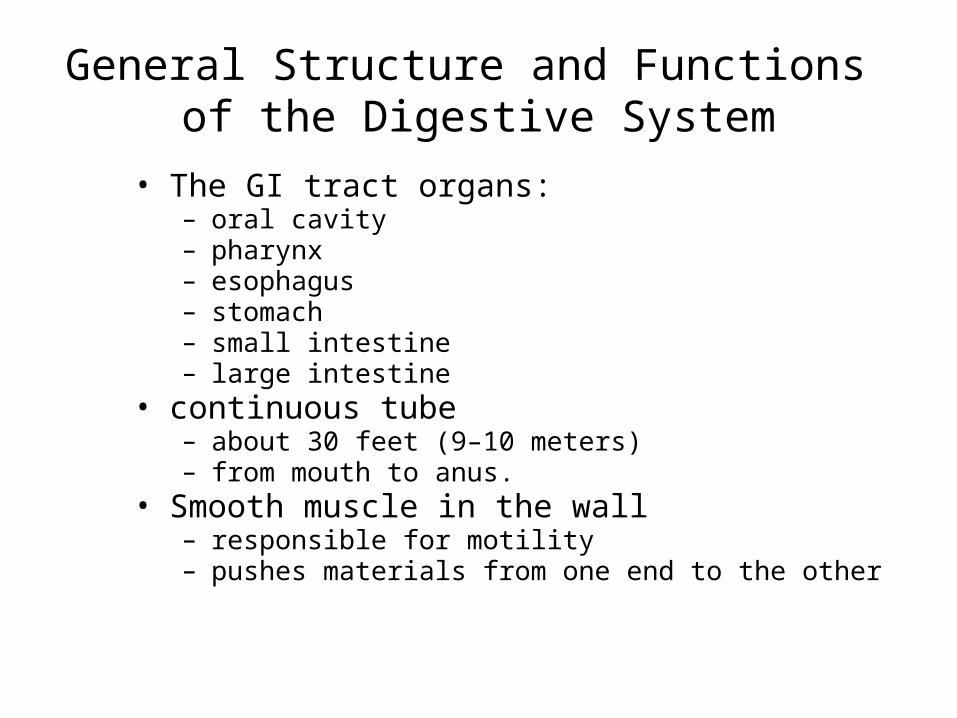
General Structure and Functions of the Digestive System
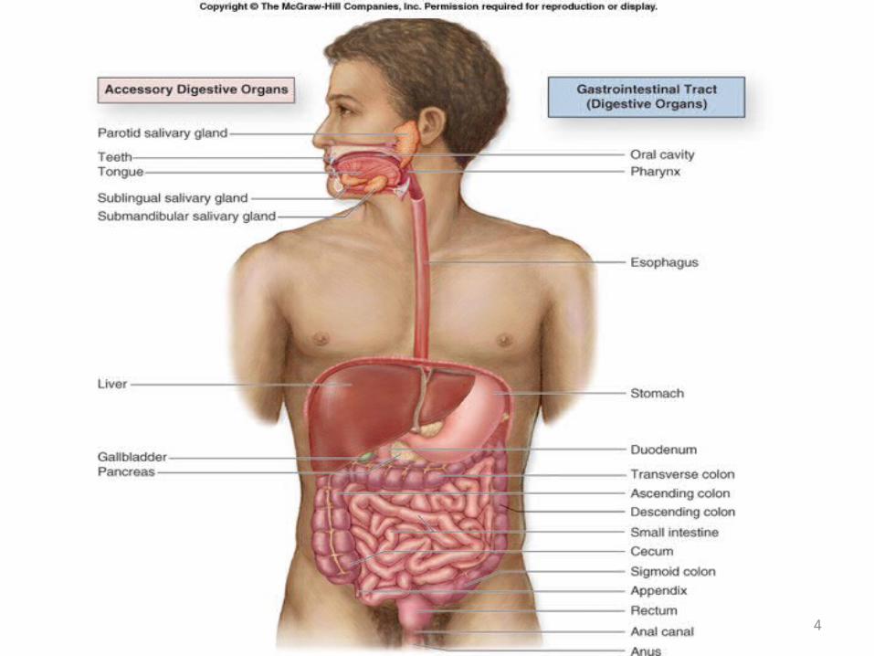
• The GI tract organs: – oral cavity– pharynx– esophagus – stomach– small intestine – large intestine
• continuous tube– about 30 feet (9–10 meters)– from mouth to anus.
• Smooth muscle in the wall– responsible for motility– pushes materials from one end to the other
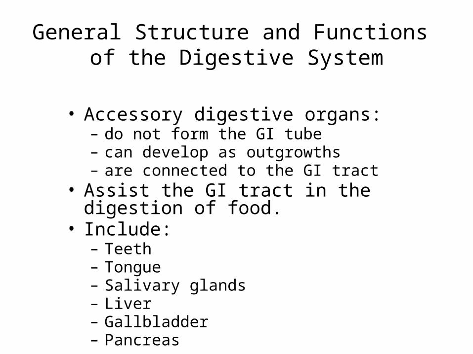
General Structure and Functions of the Digestive System
• Accessory digestive organs: – do not form the GI tube – can develop as outgrowths– are connected to the GI tract
• Assist the GI tract in the digestion of food.• Include:
– Teeth– Tongue– Salivary glands– Liver– Gallbladder– Pancreas
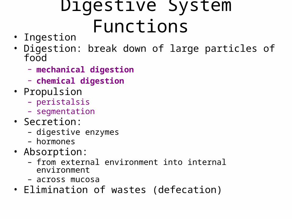
Digestive System Functions • Ingestion• Digestion: break down of large particles of food
– mechanical digestion – chemical digestion
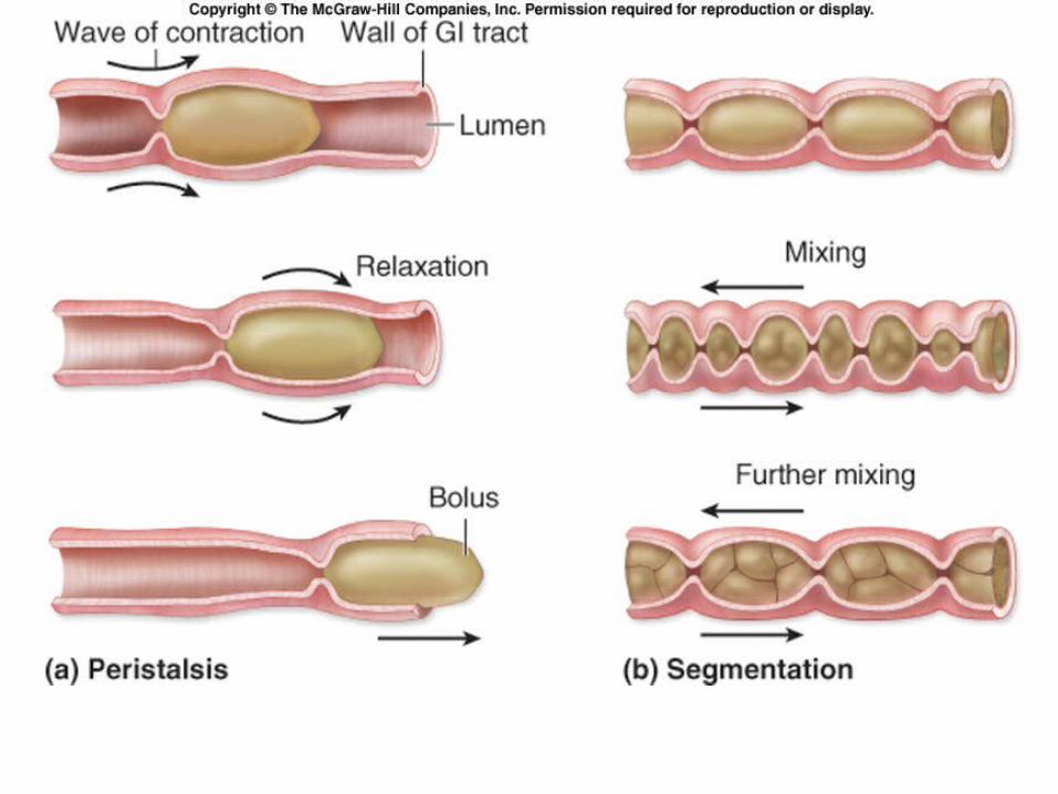
• Propulsion – peristalsis– segmentation
• Secretion: – digestive enzymes– hormones
• Absorption:– from external environment into internal environment– across mucosa
• Elimination of wastes (defecation)
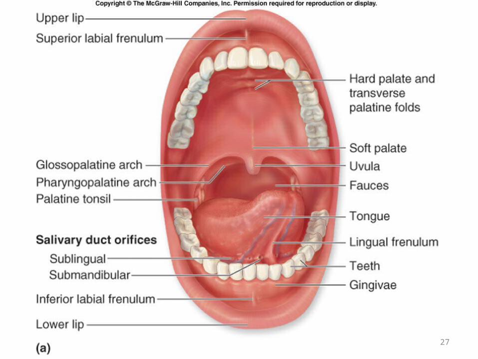
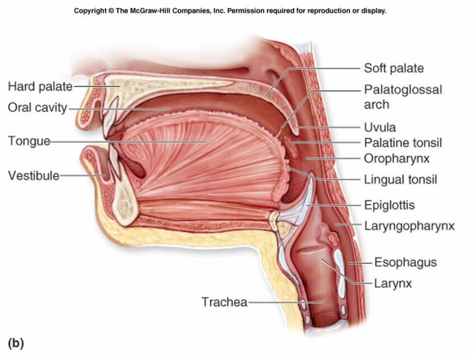
BASIC ORAL ANATOMY
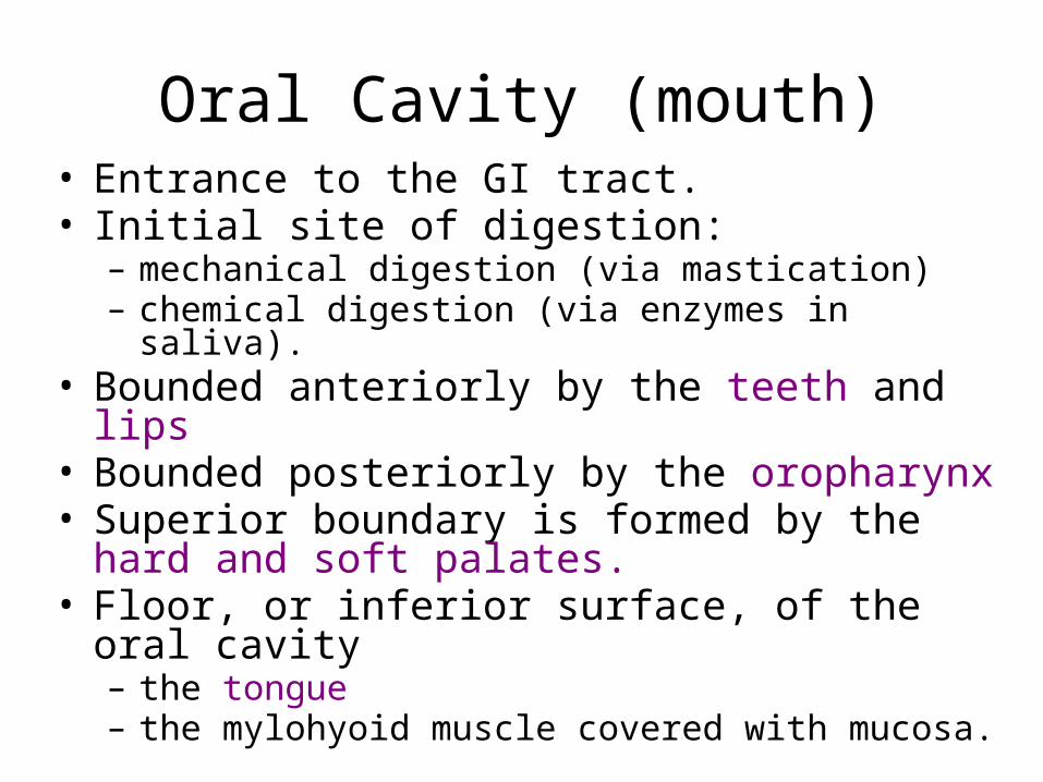
Oral Cavity (mouth)• Entrance to the GI tract.• Initial site of digestion:
– mechanical digestion (via mastication)– chemical digestion (via enzymes in saliva).
• Bounded anteriorly by the teeth and lips• Bounded posteriorly by the oropharynx• Superior boundary is formed by the hard and soft
palates. • Floor, or inferior surface, of the oral cavity
– the tongue– the mylohyoid muscle covered with mucosa.
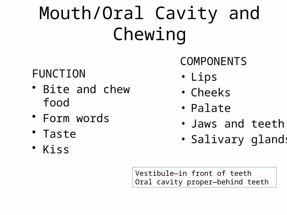
Mouth/Oral Cavity and Chewing
COMPONENTS• Lips• Cheeks• Palate• Jaws and teeth• Salivary glands
FUNCTION• Bite and chew food• Form words• Taste• Kiss
Vestibule—in front of teethOral cavity proper—behind teeth
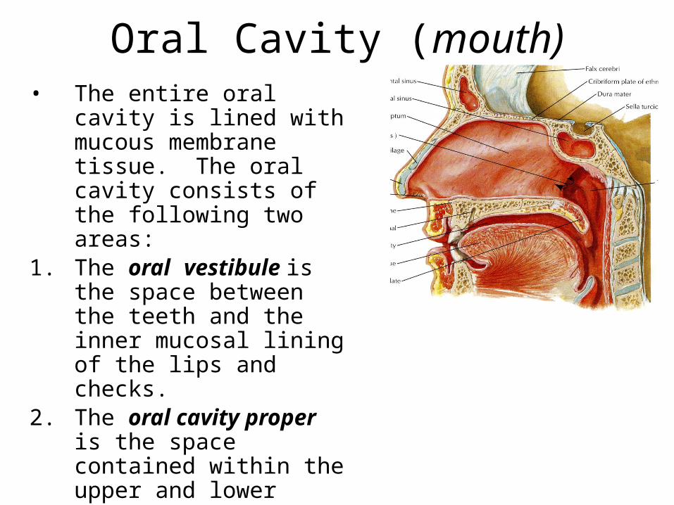
Oral Cavity (mouth)• The entire oral cavity is lined
with mucous membrane tissue. The oral cavity consists of the following two areas:
1. The oral vestibule is the space between the teeth and the inner mucosal lining of the lips and checks.
2. The oral cavity proper is the space contained within the upper and lower dental arches.
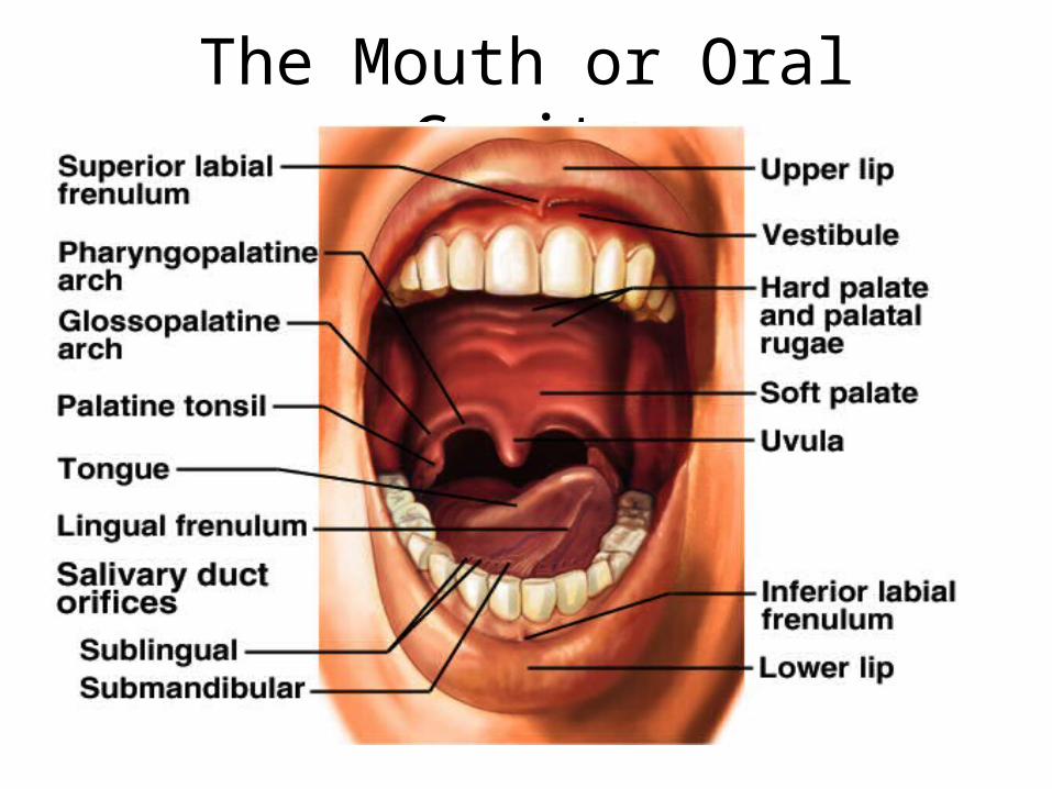
The Mouth or Oral Cavity
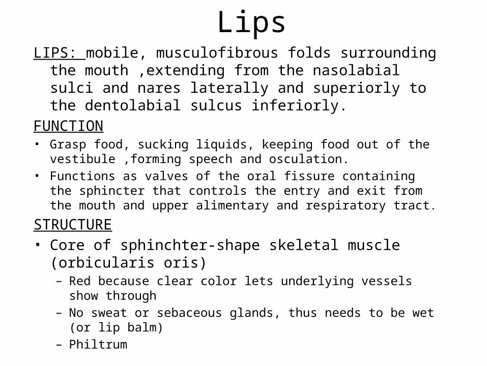
LipsLIPS: mobile, musculofibrous folds surrounding the
mouth ,extending from the nasolabial sulci and nares laterally and superiorly to the dentolabial sulcus inferiorly.
FUNCTION• Grasp food, sucking liquids, keeping food out of the vestibule ,forming
speech and osculation.• Functions as valves of the oral fissure containing the sphincter that
controls the entry and exit from the mouth and upper alimentary and respiratory tract.
STRUCTURE• Core of sphinchter-shape skeletal muscle (orbicularis oris)
– Red because clear color lets underlying vessels show through– No sweat or sebaceous glands, thus needs to be wet (or lip balm)– Philtrum
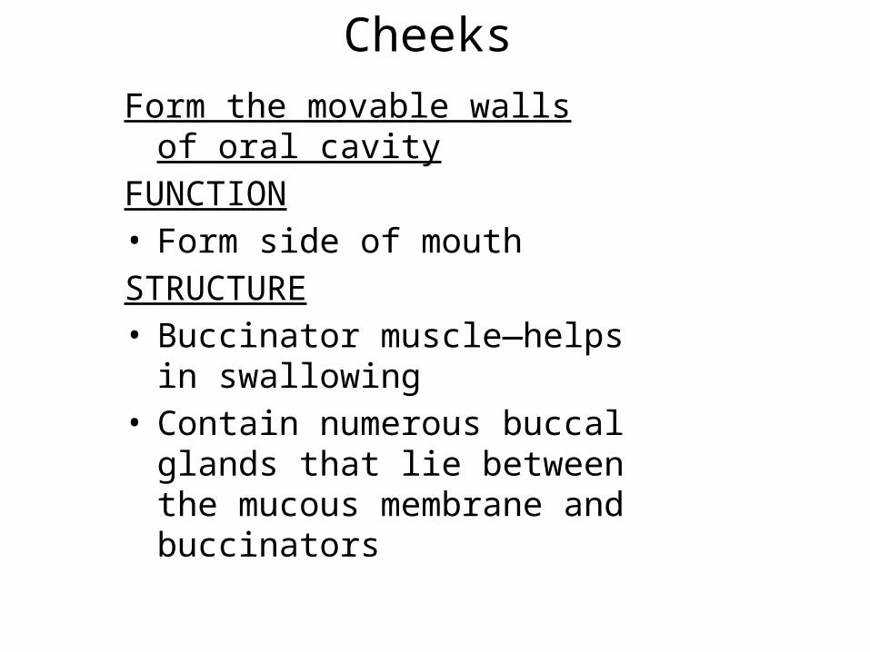
CheeksForm the movable walls of oral
cavityFUNCTION• Form side of mouthSTRUCTURE• Buccinator muscle—helps in
swallowing • Contain numerous buccal glands
that lie between the mucous membrane and buccinators
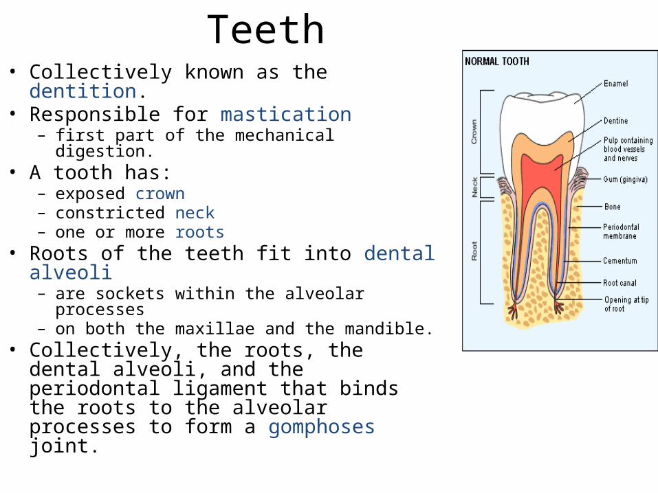
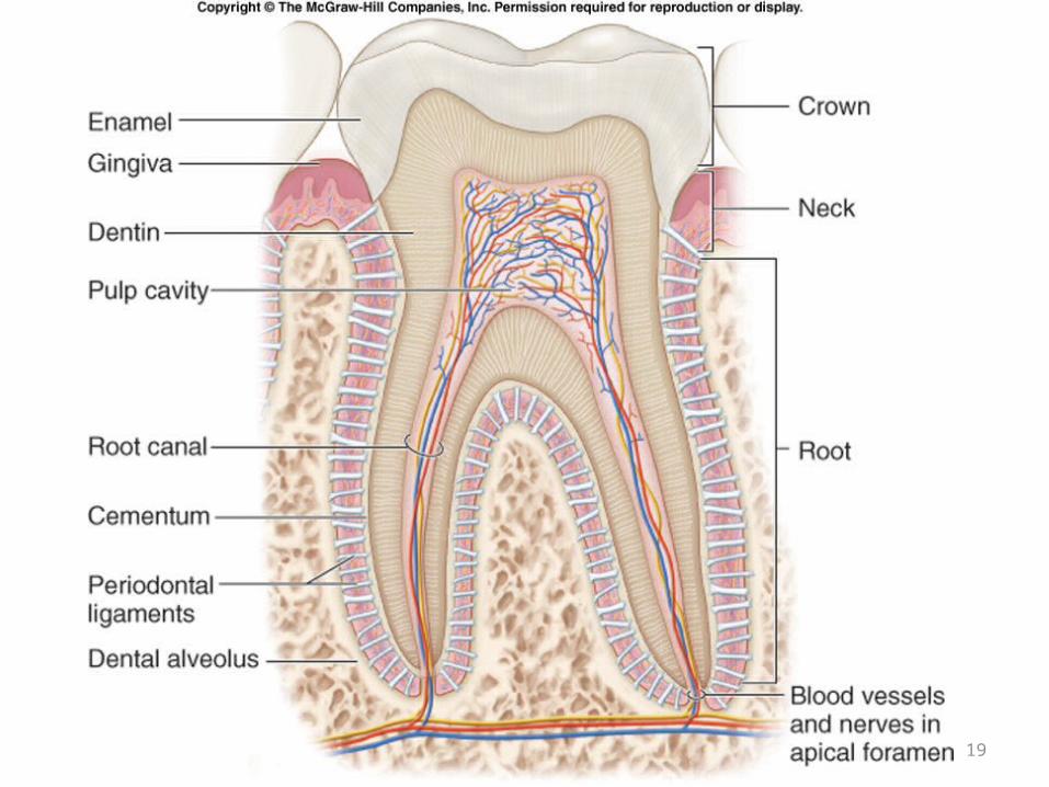
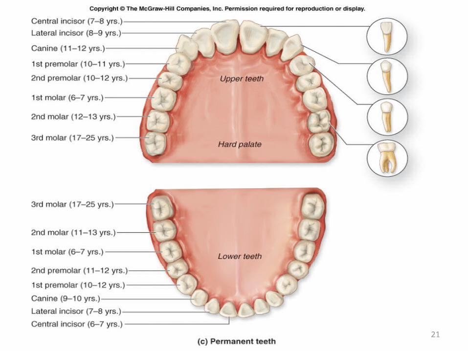
Teeth • Collectively known as the dentition. • Responsible for mastication
– first part of the mechanical digestion. • A tooth has:
– exposed crown– constricted neck– one or more roots
• Roots of the teeth fit into dental alveoli– are sockets within the alveolar processes– on both the maxillae and the mandible.
• Collectively, the roots, the dental alveoli, and the periodontal ligament that binds the roots to the alveolar processes to form a gomphoses joint.
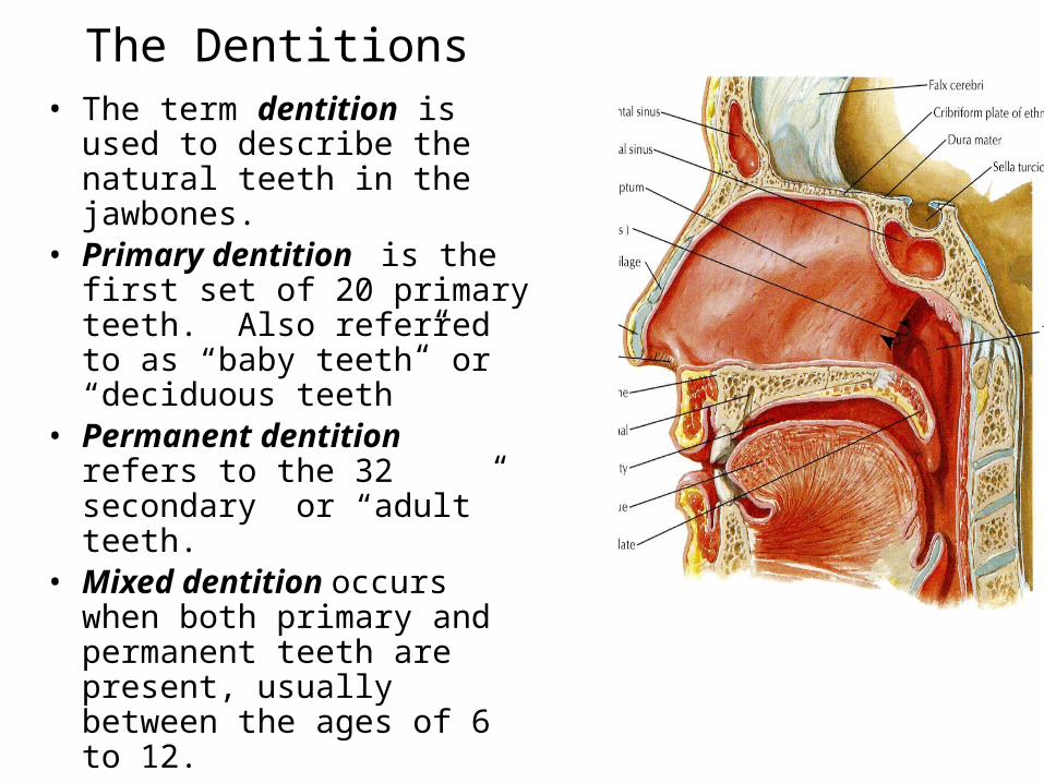
The Dentitions• The term dentition is used to
describe the natural teeth in the jawbones.
• Primary dentition is the first set of 20 primary teeth. Also referred to as “baby teeth” or “deciduous teeth”
• Permanent dentition refers to the 32 secondary or “adult” teeth.
• Mixed dentition occurs when both primary and permanent teeth are present, usually between the ages of 6 to 12.
19
20
21
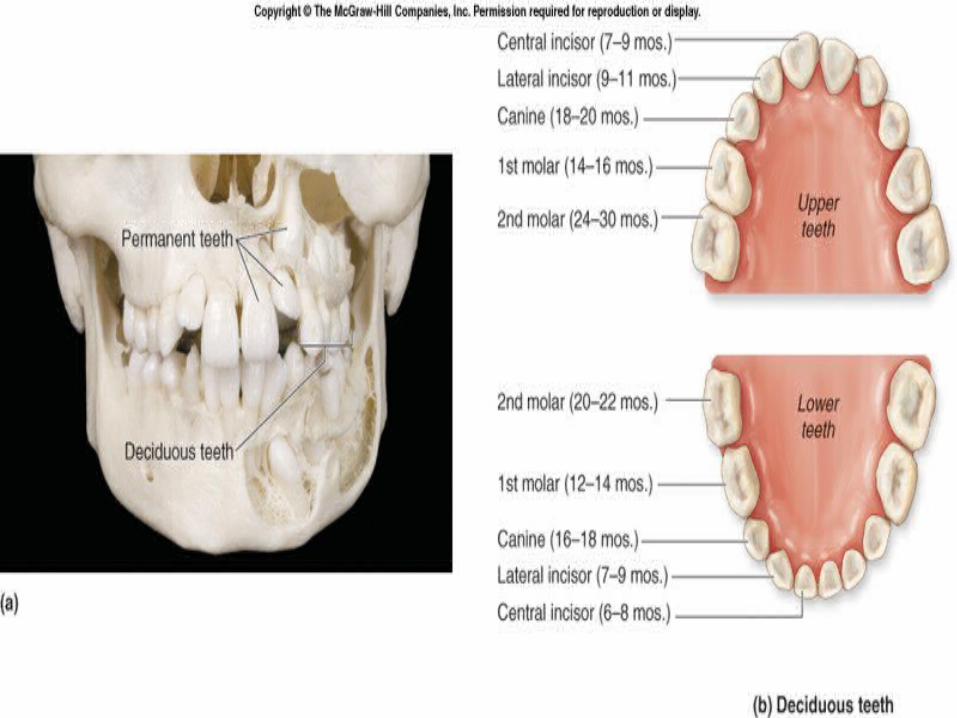
Teeth• Two sets of teeth • 20 deciduous teeth, also called “milk teeth,” erupt between 6
months and 30 months after birth. • These teeth are eventually lost and replaced by 32 permanent
teeth. • The more anteriorly placed permanent teeth tend to appear first,
followed by the posteriorly placed teeth. • The last teeth to erupt are the third molars, often called “wisdom
teeth,” in the late teens or early 20’s. • Often the jaw lacks space to accommodate these final molars, and
they may either emerge only partially or grow at an angle and become impacted.
• Impacted teeth cannot erupt properly because of the angle of their growth.
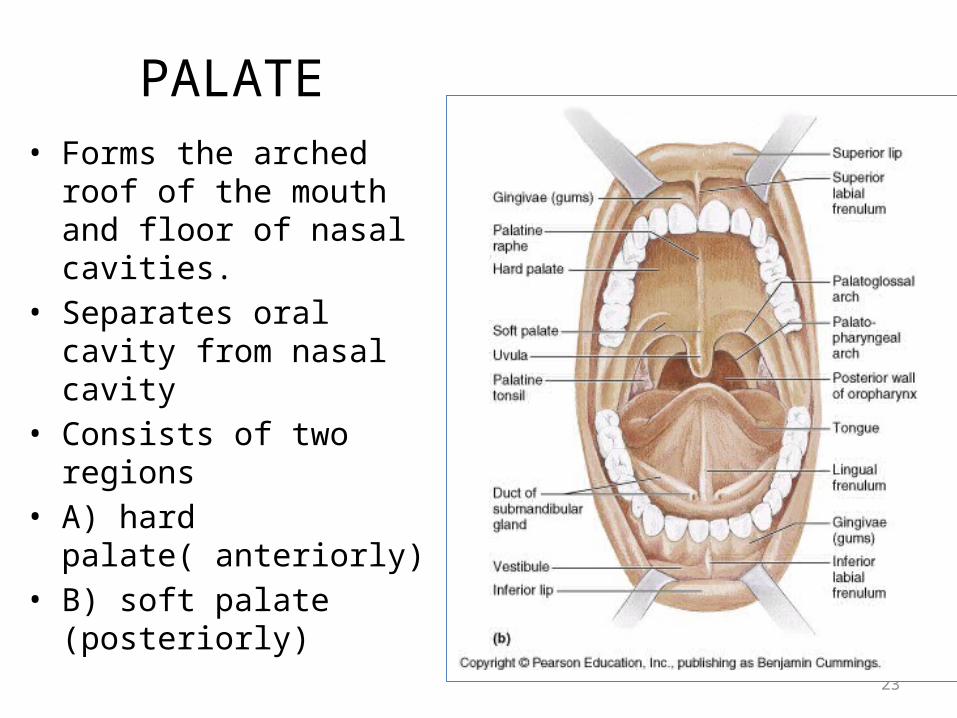
PALATE • Forms the arched roof of
the mouth and floor of nasal cavities.
• Separates oral cavity from nasal cavity
• Consists of two regions • A) hard palate( anteriorly)• B) soft palate (posteriorly)
23
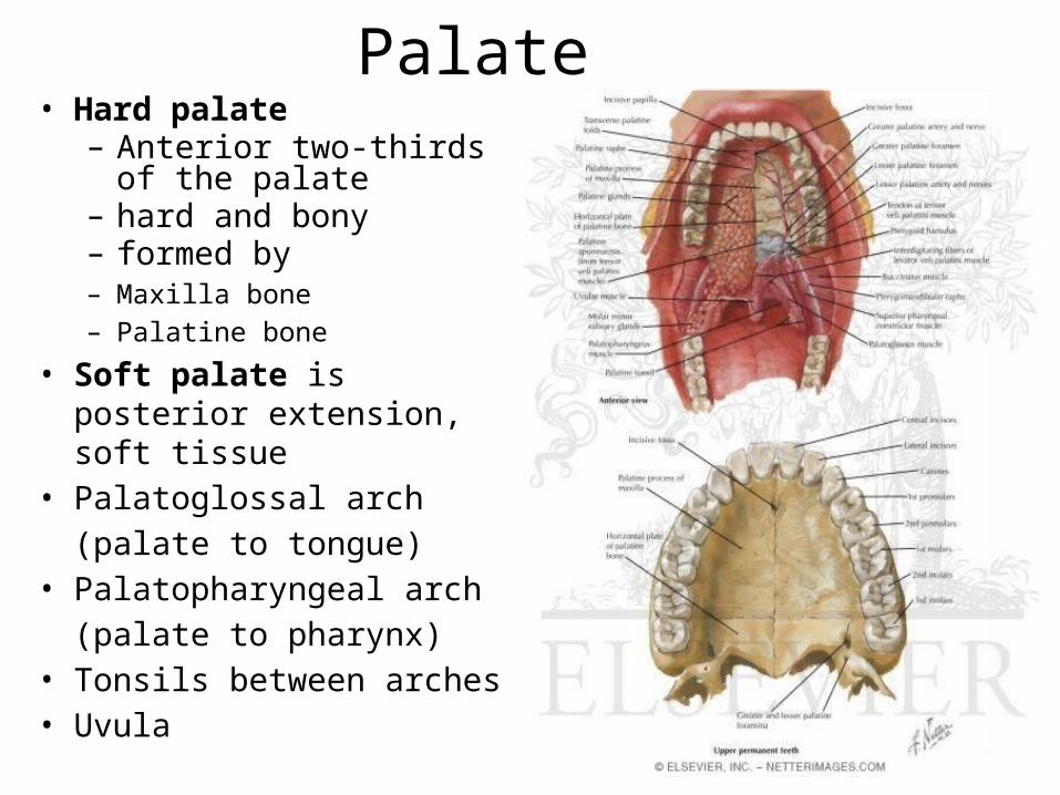
Palate• Hard palate
– Anterior two-thirds of the palate
– hard and bony – formed by – Maxilla bone– Palatine bone
• Soft palate is posterior extension, soft tissue
• Palatoglossal arch(palate to tongue)
• Palatopharyngeal arch(palate to pharynx)
• Tonsils between arches• Uvula

25

Palate• Soft palate
– Movable Posterior one-third– soft and muscular, no bony
skeleton– primarily composed of skeletal
muscle.– Extending inferiorly from the
posterior part of the soft palate is the uvula.
• When swallowing, the soft palate and the uvula elevate to close off the opening of the nasopharynx.
27
26-28
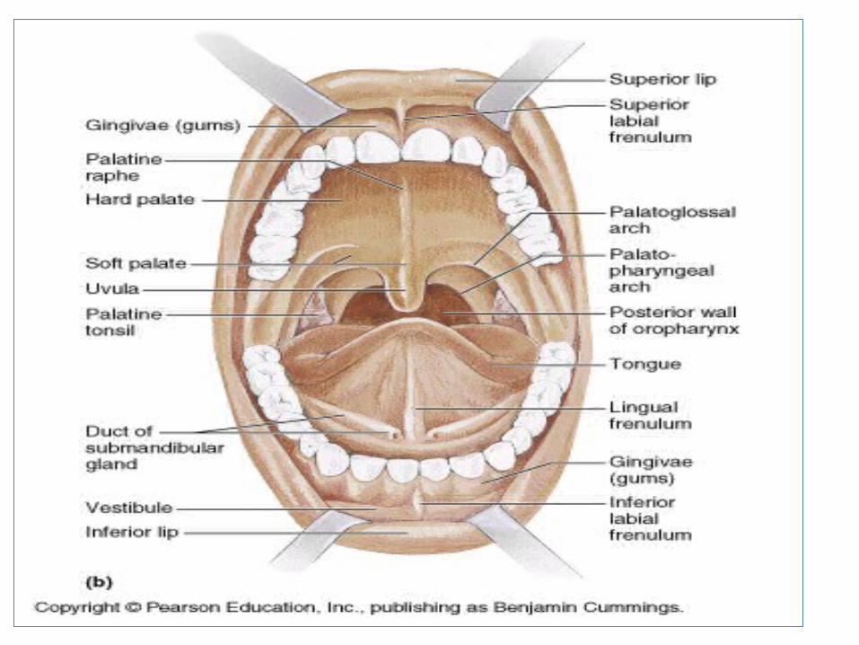
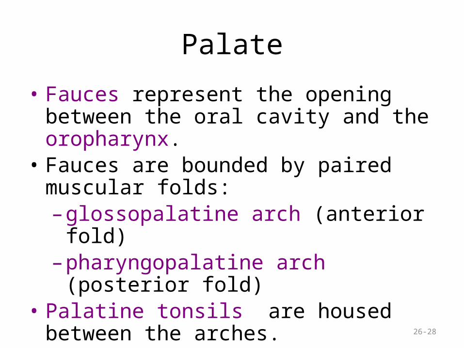
Palate
• Fauces represent the opening between the oral cavity and the oropharynx.
• Fauces are bounded by paired muscular folds:– glossopalatine arch (anterior fold) – pharyngopalatine arch (posterior fold)
• Palatine tonsils are housed between the arches.
29


