HAL Id: tel-02156725 https://tel.archives-ouvertes.fr/tel-02156725 Submitted on 14 Jun 2019 HAL is a multi-disciplinary open access archive for the deposit and dissemination of sci- entific research documents, whether they are pub- lished or not. The documents may come from teaching and research institutions in France or abroad, or from public or private research centers. L’archive ouverte pluridisciplinaire HAL, est destinée au dépôt et à la diffusion de documents scientifiques de niveau recherche, publiés ou non, émanant des établissements d’enseignement et de recherche français ou étrangers, des laboratoires publics ou privés. Discovery of active secondary metabolites from Paenibacillus odorifer, a lichen-associated bacterium Thi Bach Le Nguyen To cite this version: Thi Bach Le Nguyen. Discovery of active secondary metabolites from Paenibacillus odorifer, a lichen- associated bacterium. Pharmacology. Université Rennes 1, 2018. English. NNT : 2018REN1S098. tel-02156725
Transcript
HAL Id: tel-02156725https://tel.archives-ouvertes.fr/tel-02156725
Submitted on 14 Jun 2019
HAL is a multi-disciplinary open accessarchive for the deposit and dissemination of sci-entific research documents, whether they are pub-lished or not. The documents may come fromteaching and research institutions in France orabroad, or from public or private research centers.
L’archive ouverte pluridisciplinaire HAL, estdestinée au dépôt et à la diffusion de documentsscientifiques de niveau recherche, publiés ou non,émanant des établissements d’enseignement et derecherche français ou étrangers, des laboratoirespublics ou privés.
Discovery of active secondary metabolites fromPaenibacillus odorifer, a lichen-associated bacterium
Thi Bach Le Nguyen
To cite this version:Thi Bach Le Nguyen. Discovery of active secondary metabolites from Paenibacillus odorifer, a lichen-associated bacterium. Pharmacology. Université Rennes 1, 2018. English. �NNT : 2018REN1S098�.�tel-02156725�
PART 1: STATE OF THE ART ON LICHEN-ASSOCIATED BACTERIA ........................... 6 1.1. INTRODUCTION ON LICHENS .......................................................................................... 6
1.1.1. The lichens ........................................................................................................................ 6 1.1.1.1 Description .................................................................................................................. 6 1.1.1.2 Morphology................................................................................................................. 8 1.1.1.3 Metabolites produced by lichen thallus .................................................................... 10
1.2. BACTERIA ASSOCIATED WITH LICHENS .................................................................... 14 1.2.1. State of art about bacterial communities on lichens ....................................................... 14 1.2.2. The roles of bacteria ....................................................................................................... 26
1.3. METABOLITES FROM THE LICHEN-ASSOCIATED BACTERIA ............................... 27 1.4. FOCUS ON RHIZOCARPON GEOGRAPHICUM ............................................................ 34
1.4.1. Description of Rhizocarpon genus ................................................................................. 34 1.4.2. Morphology of R. geographicum ................................................................................... 36 1.4.3. Studies on R. geographicum ........................................................................................... 38 1.4.4. Bacteria communities of R. geographicum .................................................................... 42
1.5. NATURAL CYTOTOXIC COMPOUNDS FROM BACTERIAL CULTURE .................. 43 1.6. THE OBJECTIVES OF THE WORK .................................................................................. 60
PART II. RESULTS ...................................................................................................................... 63
CHAPTER 1: ISOLATION OF BACTERIAL STRAINS FROM R. GEOGRAPHICUM..... 65
CHAPTER 2: THE SELECTION OF PAENIBACILLUS ODORIFER AS A PROMISING SOURCE OF INTERESTING METABOLITES ....................................................................... 82
TABLE OF CONTENTS
Page ii
CHAPTER 3: OPTIMIZATION OF THE CULTURE OF PAENIBACILLUS ODORIFER
........................................................................................................................................................ 102 3.1. THE FIRST OPTIMIZATION OF PROCESS ................................................................... 102
3.1.1. The selected parameters................................................................................................ 102 3.1.2. Results .......................................................................................................................... 105 3.1.3. The application to bioreactor fermentation .................................................................. 106
3.2. THE SECOND OPTIMIZATION PROCESS .................................................................... 107 3.2.1. The selected parameters................................................................................................ 107 3.2.2. Results .......................................................................................................................... 108 3.2.3. The application of these results for cytotoxic compounds discovery ........................... 113
CHAPTER 4: ISOLATION OF METABOLITES FROM PAENIBACILLUS ODORIFER ... 1 4.1 THE PROCESS FOR THE PRODUCTION OF CRUDE EXTRACTS FROM FERMENTATION ......................................................................................................................... 1 4.2. ISOLATION OF A POLYSACCHARIDE UNIT .................................................................. 4
4.2.1. General presentation of bacterial polysaccharides: structure and properties ................... 4 4.2.2. Production of one polysaccharide fraction from P. odorifer ............................................ 5
4.3. ISOLATION OF TERT-BUTYL PHENOLIC COMPOUNDS ........................................... 20 4.3.1. State of art about tert-butyl compounds ......................................................................... 20 4.3.2. Production of the tert-butyl phenol compounds ............................................................. 22
4.4. THE ISOLATION PROCESS OF A NOVEL ALKALOID ................................................ 37 4.4.1. General presentation of alkaloids ................................................................................... 37 4.4.2 Isolation of alkaloid ......................................................................................................... 39
4.5. DESCRIPTION OF THE OTHER ISOLATED METABOLITES ...................................... 50 4.5.1. Structural elucidation of compounds .............................................................................. 53
CHAPTER 5: CONCLUSIONS AND PERSPECTIVES .......................................................... 64
6.2.1. The process for the production of crude extracts from fermentation ............................. 71 6.2.2. Analytical methods used for isolation steps ................................................................... 71
ANNEXE 1: SUPPORTING INFORMATION FOR THE ARTICLE OF TERT-BUTYLPHENOL COMPOUNDS .............................................................................................. 125
ANNEXE 2: SUPPORTING INFORMATION FOR THE ARTICLE OF ALKALOID COMPOUND................................................................................................................................ 137
ANNEXE 3: NMR SPECTRA OF THE METABOLITES FROM P. ODORIFER .............. 143
ABREVIATIONS
Page v
ABREVIATIONS
ACN Acetonitrile
CHCl3 Chloroform
CoA Coenzyme A
COSY Correlation Spectroscopy
CFU Colony- forming unit
Da Dalton
DAD Diode Array Detector
DCM DiChloroMethane
DMSO DiMethylSulfOxide
OD Optical Density
DPPH DiPhenylPicrylHydrazine
EDTA EthyleDiamine Tetra Acetic acid
ESI ElectroSpray Ionization
EtOAc Ethyl acetate
EtOH Ethanol
FISH Fluorescent In Situ Hybridization
FTIR Fourier Transform Infra-Red
GC Gas Chromatography
HMBC Heteronuclear Multiple Bond Correlation
HPLC High Performance Liquid Chromatography
HRESIMS High Resolution ElectroSpray Ionization
HRMS High Resolution Mass Spectrometry
ABREVIATIONS
Page vi
HSQC Heteronuclear Single Quantum Coherence spectroscopy
IC50 Inhibitory Concentration of 50%
IR Infra-Red
MeOH Methanol
MS Mass Spectrometry
NBT NitroBlueTetrazolium
NI Negative Ionization
NMR Nuclear Magnetic Resonance
NOESY Nuclear Overhauser Effect Spectroscopy
ORF Open Reading Frame
P Para-phenylene diamine
PCB Poly Chlorinated Biphenyl
PCR Polymerase Chain Reaction
PDA Photo Diode Array
PI Positive Ionization
ppm part per million
Rf Retention front
rpm rotation per minute
TLC Thin Layer Chromatography
tR Retention time
UV/Vis UltraViolet/Visible
LIST OF FIGURES
Page vii
LIST OF FIGURES
Figure 1.1 Exchange nutrients between lichen symbiotic partners (R. geographicum as the model) (adapted from Grube et al., 2015). ......................................................................................................7
Figure 1.2 Types of the lichens ...........................................................................................................9
Figure 1.3 Putative pathways of the major groups of lichen metabolites (adapted from Elix J.A. and Stocker-Wörgötter 2008) ...........................................................................................................11
Figure 1.4 Structure of typical lichen products derived from the Acetyl-polymalonyl pathway .....12
Figure 1.5 Structure of typical lichen products derived from the Shikimic acid pathway ...............13
Figure 1.6 Structure of typical lichen products derived from the Mevalonic acid pathway .............14
Figure 1.7 Roles of bacteria in lichens (adapted from Grube et al., 2015) .......................................26
Figure 1.8 Cross section of R. geographicum ...................................................................................35
Figure 1.9: Map of the sites of Rhizocarpon geographicum in France ............................................37
Figure 1.10 Morphology of Rhizocarpon geographicum .................................................................37
Figure 1.11 Stuctures of some compounds from R. geographicum ..................................................38
Figure 2.3.1 The curves of bacterial growth at 15oC and at 25°C with different culture conditions105
Figure 2.3.4 The OD and number of CFU/mL for each experiment at different culture conditions110
Figure 2.3.5 Chemical profiles obtained by HPLC-DAD of crude extracts from resin (R1: extract from resin of experiment number 1, similar for R2, R3, R4, R5, R7, R8, R9) ...............................111
Figure 2.4.1 Tert-butylphenols isolated from nature (from Dembitsky et al., 2006) .......................21
Figure 2.4.2 Tert-Butylphenols produced by various bacteria .........................................................22
Figure 2.4.3 Some structural features found in alkaloids .................................................................37
Figure 2.4.4 Structures of some drugs as natural alkaloids ..............................................................38
Figure 2.4.5 Some cytotoxic alkaloids recently isolated from bacteria ............................................39
Figure 2.4.6 Structures of no cytotoxic or well-known compounds isolated during this work ........53
Figure 2.4.7 Key correlations for the structural assignment of 1 ......................................................54
Figure 2.4.8 Key correlations for the structural assignment of 2 ......................................................55
Figure 2.4.9 Key HMBC correlations in 3 ........................................................................................56
Figure 2.4.10 Key HMBC correlations in 4 and 5 ............................................................................57
Figure 2.4.11 Key COSY (black line) and HMBC (arrows H to C) correlations for compound 7 ..58
Figure 2.4.12 Key COSY (black lines) and HMBC (arrows H to C) correlations for compound 8 .59
Figure 2.4.13 Key HMBC correlations for compound 9 ..................................................................60
Figure 2.4.14 Key HMBC correlations for compound 11 ................................................................60
Figure 2.6.1 Gradient of elution for the separation of extracts by flash chromatography using a 40g SiOH Chromabond column...............................................................................................................72
Figure 2.6.2 Gradient of elution in flash chromatography using a reverse phase C18 Reveleris (Grace) column .................................................................................................................................72
Figure 2.6.3 Gradient of solvent in HPLC analysis using Prevail C18 column .................................73
Figure 2.6.4 Gradients of elution used in semi-preparative HPLC (using Prevail C18 column).......74
Figure 2.6.5 The process of viable plate counts................................................................................78
LIST OF TABLES
Page ix
LIST OF TABLES
Table 1.1 Some lichen subtances which give colour reactions with chemical reagents (Huneck and Yoshimura, 1996) ...............................................................................................................................9
Table 1.2 Summary of bacterial communities associated with lichens ............................................19
Table 1.3 Summary of metabolites from lichen-associated bacteria ................................................29
Table 1.4 Classification of Rhizocarpon subgenus (Innes, 1985) ....................................................35
Table 1.5 Summary of studies on Rhizocarpon geographicum ........................................................41
Table 1.6 Summary of some cytotoxic compounds produced by bacteria .......................................44
Table 2.2.1 Summary about chemical studies on Sphingomonas genus...........................................82
Table 2.2.2 Summary about chemical studies from Burkholderia genus (Betaproteobacteria) ......83
Table 2.2.3 Summary about chemical studies on Paenibacillus genus (Bacilli) ..............................85
Table 2.2.4 Summary about chemical studies on Lysinibacillus genus (Bacilli) .............................87
Table 2.2.5 Chemical studies on Bacillus genus (Bacilli) ................................................................87
Table 2.3.1 Parameters for the first optimization ...........................................................................102
Table 2.3.2 The parameters for second optimization ......................................................................108
Table 2.3.3 The mass of crude extracts (mg) from experiments obtained during the second optimization step .............................................................................................................................110
Table 2.3.4 The results of the second optimization (in Gym Streptomyces medium supplemented with CaCO3 at 25oC, pH = 7) ..........................................................................................................112
Table 2.4.1 Comparison of 1H NMR (500MHz, CD3OD) and 13C NMR (75MHz, CD3OD) spectroscopic data of compounds 1 and 2.........................................................................................55
Table 2.4.2 Comparison of NMR data between compound 7 and literature ....................................58
Table 2.4.3 Comparison of NMR data between compound 11 and reference ..................................61
Table 2.6.1 Ingredient of media used to isolate bacterial strains from R. geographicum ................70
Table 2.6.2 Parameters for GC-MS process .....................................................................................75
INTRODUCTION
INTRODUCTION
Page 1
INTRODUCTION
Since the discovery of penicillin at the beginning of the twentieth century, natural products
have become candidates for the development of new pharmaceutical agents. Over 50% of
anticancer drugs approved by United States Foods and Drugs Administration since 1960 derived
from natural products (Lammer et al., 2017). A large percentage of natural products have been
isolated from a variety of microorganisms. Over 7600 compounds have been isolated from bacteria
and almost from Streptomyces genus (Keohn et al., 2005). Thus finding metabolites from other
bacterial lineages represent new interests for chemists. Among that, lichens are admitted as a rich
source of new bacterial lineages and novel bacterial compounds (Suzuki et al., 2015). Therefore,
microorganism communities associated with lichens became interesting subjects with a great
potential for the production of active natural compounds.
In this thesis, we focus our work on the isolation of bacterial lineages from the lichen Rhizocarpon
geogaphicum, one of the most popular crustose lichen dwelling on the rock. From the strains
isolated, a bacterial species was selected for further work to produce active compounds. Therefore,
this thesis is divided into two parts.
In part I, a state of the art about lichens detailing the morphology of lichens, metabolites from
lichens, bacteria associated with lichens, metabolites from lichen-associated bacteria is introduced.
Besides, a general view about Rhizocarpon geographicum is described.
Part II reports the achievements of this work and is divided into 6 chapters.
Chapter 1 deals with the isolation of bacterial lineages from the lichen Rhizocarpon
geographicum.
Chapter 2 gives details about various active metabolites which have been already isolated from
strains closer to the isolates. The reasons for the selection of Paenibacillus odorifer for the
production of metabolites of interest will be given.
Chapter 3 describes the optimization process to find the best conditions to produce active
compounds from the culture of P. odorifer.
INTRODUCTION
Page 2
Chapter 4 reports all the metabolites isolated from P. odorifer. In this chapter, the results will be
displayed either as an article or as a common part of the thesis.
Chapter 5 corresponds to conclusions and perspectives.
Chapter 6 details materials and methods used for this work.
The annexes provide the spectroscopic data of the metabolites isolated.
All strategies were summarized in Scheme 1.
INTRODUCTION
Page 3
Scheme 1: General strategies used for the study described in the thesis
Part I: State of the art about lichens and associated bacteria
Lichen (Rhizocarpon geographicum) Part II- Chapter 1: Isolation of
microbial strains from R. geographicum
Part II- Chapter 2: The selection of Paenibacillus odorifer as a promising source of interesting metabolites
Part II- Chapter 3: Optimization of the culture of P. odorifer
OO OH
OH
OHPart II- Chapter 4: Metabolites from P. odorifer
Part II- Chapter 5: Conclusions and Perspectives
Part II- Chapter 6: Materials and Methods
O
N
HO
NH2
INTRODUCTION
Page 4
References
Lammers, A., Wang, R., Cetnar, J., Prasad, V., 2017. Time from US Food and Drug Administration approval to
publication of data for cancer drugs: a comparison of first and subsequent approvals. Blood Cancer Journal 7,
637. https://doi.org/10.1038/s41408-017-0008-9
Suzuki, M.T., Parrot, D., Berg, G., Grube, M., Tomasi, S., 2015. Lichens as natural sources of biotechnologically
Part I : State of the art on lichen-associated bacteria
Page 30
NH
NCl
MeO
R
F R = HG R = Cl
NHMe
O OHO
Angucyline derivative Butenolide derivative
ClOH O
O
OH
OH
O
ClCl
O
O
OH
OH
Angucyline: - cytotoxic activity against human cervical carcinoma Hela (IC50 values of 36 µM) and human malignant pleural mesothelioma ACC-MESO-1 cell lines (IC50 of 52 µM) - Antibacterial against Micrococcus luteus, at 25 µg.
Streptomyces
sp.
Unidentified
lichen
Motohashi
et al., 2010
Coumabiocins A – F
O O O
NH
O
O
R
MeOO
NH2
OOH
OH
O
A R = OHB R = H
Coumabiocins A-E :
significant inhibition
activity against
Streptomyces 85E at
concentration of 10 µg
Coumabiocin F : inactivity
Streptomyces
caeruleus
Cladonia
gracilis
Cheenpracha
et al., 2010
Part I : State of the art on lichen-associated bacteria
Page 31
O O O
NH
O
MeOO
NH2
OOH
OH
O
C R = HD R = OH
O
R
O O O
NH
O
MeOO
NH2
OOH
OH
O
EOHOH
HO O
NH
O
OH
O
OHOH
F
Novobiocin (G) and isonovobiocin (H)
O O O
NH
O
MeOOR2
OR1OH
O
OH
G R1 = H; R2 = CONH2H R1 = CONH2; R2 = H
Novobiocin : potent
inhibition activity
Isonovobiocin : weaker
activity
Unciaphenol
HNO
O OH
OH
HO
O OH
in vitro anti-HIV activity
against drug-resistant
isolates of the virus.
Streptomyces
uncialis
Cladonia
uncialis
Williams et
al., 2015
Cyaneodimycin Cyaneomycin Cyaneodimycin : cytotoxic Streptomyces Lichina Parrot et al.,
Part I : State of the art on lichen-associated bacteria
Page 32
O
O
O
OOH
OO
O
O
O
OOH
HO
Diketopiperazines : Cyclo-(L-Phe-L-Pro); Cyclo-
(L-Leu-L-Pro)
(3-hydroxyacetyl)indole
N-methyldactinomycin
Usnic acid
O
O
HO
OHO
O
O
against human keratinocyte
HaCaT (IC50 of 47±11 µM),
murine melanoma B16
(IC50 of 27±4 µM) and
leukemic (Jurkat) cell lines
(IC50 = 18.5±0.5 μM).
N-
methyldactinomycin
cytotoxic activities (IC50 ≈ 0.05 μM) against various normal or cancer cell lines after 48 h of incubation
Section Alpicola and section Superficiale can then be further subdivided by the size of the spores,
the hymenial dimensions and colour of the apihymenium. In the sections with pluriseptate spores,
Rhizocarpon and Viridiatrum apihymenium colour and the reaction of the medulla to iodine
(turning blue) are common distinguishing characteristics (Benedict, 1988). A cross-section of a
Rhizocarpon thallus can be found in Figure 1.8 and demonstrates the characteristic of a stratified
thallus concluding three parts: upper cortex, alga layer and medulla part.
Part I : State of the art on lichen-associated bacteria
Page 35
Figure 1.8: Scheme of the cross section of R. geographicum
http://www.imlichenit.com/imlichenit/about.html)
Table 1.4 Classification of Rhizocarpon subgenus (Innes, 1985)
Genus Section Species
Rhizocarpon
Superficiale
R. dispersum Runem. R. ejjiguratum (Anzi) Th.Fr. R. norvegicum Ras. R. parvum Runem. R. pusillum Runem. R. superjiciale (Schaer.) Vain
Alpicola R. alpicola (Hepp.) Rahb. R. eupetraeoides (Nyl.) 810mb. R. inarense (Vain.) Vain.
Viridiatrum
R. atrovirellum (Nyl.) Zahlbr. R. kakurgon Poelt R. lusitanicum (Nyl.) Arnold R. oportense (Vain.) Ras. R. subtile Runem. R. tetras porum Runem. R. viridiatrum (Wulf.) Koerb
Rhizocarpon
R. atroglavescens Lynge R. carpaticum Runem. R. ferax H. Magn. R.furax Poelt et V. R. geographicum (L.) DC. R. intermediellum Ras. R. lecanorinum Anders R. macros porum Ras. R. pulverulentum (Schaer.) Ras. R. rapax V. Wirth et Poelt R. riparium Ras. R. saanense Ras. R. sphaerosporum Ras. R. sublucidum
Parrot, D., Legrave, N., Intertaglia, L., Rouaud, I., Legembre, P., Grube, M., Suzuki, M.T., Tomasi, S., 2016b.
Cyaneodimycin, a Bioactive Compound Isolated from the Culture of Streptomyces cyaneofuscatus Associated
with Lichina confinis. Eur. J. Org. Chem. 2016, 3977–3982. https://doi.org/10.1002/ejoc.201600252
Parrot, D., Peresse, T., Hitti, E., Carrie, D., Grube, M., Tomasi, S., 2015. Qualitative and Spatial Metabolite Profiling of Lichens by a LC–MS Approach Combined With Optimised Extraction. Phytochemical Analysis 26, 23–33. https://doi.org/10.1002/pca.2532
Printzen, C., Fernández-Mendoza, F., Muggia, L., Berg, G., Grube, M., 2012. Alphaproteobacterial communities in
geographically distant populations of the lichen Cetraria aculeata. FEMS Microbiology Ecology 82, 316–325.
https://doi.org/10.1111/j.1574-6941.2012.01358.x
Proctor, M.C.F., 1983. Sizes and Growth-Rates of Thalli of the Lichen Rhizocarpon Geographicum on the Moraines of
the Glacier De Valsorey, Valais, Switzerland. The Lichenologist 15, 249–261.
https://doi.org/10.1017/S0024282983000389
Qin, L.-L., Zhou, B., Ding, W., Ma, Z., 2018. Bioactive metabolites from marine-derived Streptomyces sp. A68 and its
Chapter 1 : Isolation of bacterial strains from R. geographicum
Page 73
Figure 3: The pure strains isolated from Rhizocarpon geographicum. (a:GC-GYM-YT; b: PW-GYM-WS; c: PC-GYM-TO; d: PW-GYM-LY and PW-GYM-CY; e: PY-GYM +10%NaCl-PY; f: PW-MA-YF and PW-MA-LG; g: PW-MA-LB; h: PW-MA-OF; i: GC-MA-OP; j: GC-Thio-DG; k: GC-Bac-LW) Table 3 : The most common colony types in samples after 14 days of incubation Samples Dominant
colonies Other colony types
GC A I,J,K PC C PW D,F B,E,G,H
GC: thin layer at the center position on the surface of R. geographicum collected at the farthest distance from the sea PC: thin layer at the center position on the surface of R. geographicum collected at the medium distance PW: whole of Rhizocarpon geographicum collected at the medium distance
A clear difference in the dominance of the colony morphology was observed in agar plates
obtained from Rhizocarpon geographicum samples collected at the different locations on the
thallus. Most colony types were scarce, with one or few representatives per plate, the samples
being strongly dominated by one or few colony types (See Table 3). The crustose lichen-associated
strains appear to be characterized by a strong presence of bacteria forming colony type D, F, A, C.
However, the individual samples appear to harbor populations distinct from another type with GC
a b d f c e
g h i j k
Chapter 1 : Isolation of bacterial strains from R. geographicum
Page 74
samples harboring populations strongly dominated by colony type A, whereas colony type C
dominated on PC samples and the PW samples are co-dominated by bacteria forming colony types
D and F.
Microbial identification
The isolates were identified by partial 16S rRNA gene sequence analysis (see Table 4). All
sequences matched with entries in GenBank with similarity ranging from 85% to 100%. The
collection was found to contain members of three classes such as Alphaproteobacteria,
Betaproteobacteria and Bacilli. The taxonomic diversity of selected strains is shown in Figure 4.
Chapter 1 : Isolation of bacterial strains from R. geographicum
Page 75
Table 4: Identify of cultured strains as revealed by partial 16S rRNA gene sequencing
Chapter 2: The selection of P. odorifer as a promising source of interesting metabolites
Page 84
Burkholderia vietnamiensis
Endotoxin Immunostimulatory activity on human myelomonocytic U937 cells.
Ieranò et al., 2009
Burkholderia thailandensis
Bactobolin
Bactobolins A, B: antibiotics against MRSA and V. parahemolyticus with MICs < 1 µg/mL
Seyedsayamdost et al., 2010
Spiruchostatins
Antitumor towards LOX IMVI melanoma cells using murine hollow fiber assay
Klausmeyer et al., 2011
B. dolosa Endotoxin Lorenzo et al., 2013
Burkholderia gladioli pv. agaricicola
Volatile organic compounds (major compound as 1-methyl-4-(1-methylethenyl)-cyclohexene)
Antifungal activity against Botrytis cinerea, Aspergillus flavus, Aspergillus niger, Penicillium digitatum, Penicillium expansum, Sclerotinia sclerotiorum and Phytophthora cactorum using agar diffusion test
Phytotoxicity against pine callus at EC50 of 171.98 μg ml−1
Dang et al., 2011
The Paenibacillus genus possess the ability to produce some metabolites with industrial
applications such as xylanase in paper industry (Zheng et al., 2012; Yeasmin et al., 2010),
Chapter 2: The selection of P. odorifer as a promising source of interesting metabolites
Page 85
acetoin in cosmetics and foods (Zhang et al., 2012). Especially, the genus produced
fusaracidin - a compound having remarkable antibiotic activity against Micrococcus luteus
(Kim et al., 2014) or abling to control Phytophthora blight infection (a disease blighted
leaves) caused by Phytophthora capsic (Lee et al., 2012). This genus thus is promising as a
source of interesting compounds. The data of some species production belonging to this genus
was reported in Table 2.2.3.
Table 2.2.3 Summary about chemical studies on Paenibacillus genus (Bacilli)
Producing strains Compounds Nature of bioactivities
References
P. campinasensis Xylanase Industrial application: pulp bleaching pretreatment.
Zheng et al., 2012
P. polymyxa Acetoin
OH
O
Cosmetic, food Zhang et al., 2012
Xylanase enzyme Paper industry Yeasmin et al., 2010
Lipopeptide Anti-phytopathogen Xanthomonas campestris using agar diffusion test
Mageshwaran et al., 2011
Fusaracidin
Antibiotic : control Phytophthora blight infection caused by Phytophthora capsic at concentration 0.1 ppm
Lee et al., 2012
Antimicrobial activity against Micrococcus luteus using agar diffusion test
Kim et al., 2014
-1,4-mannanase Production of mannooligosaccharide
Hori et al., 2014
Paenibacillus sp. Fibrinolytic enzyme Vijayaraghavan et
Chapter 2: The selection of P. odorifer as a promising source of interesting metabolites
Page 86
al, 2014
Mutanase (α-1,3-glucan) An oral hygiene product
Shimotsuura et al., 2008
Chitinase Meena et al., 2013
KB 425796-A (nikkomycin)
KB 425796-B
Antifungal activity against Aspergillus fumigatus (MIC of 0.5 and 2.5 µg.mL-1, respectively.)
Kai et al., 2013b
-1,3-glucanase PgIA Fungal disease biocontrol
Cheng et al., 2013
KB 425796-C macrocyclic lipopeptidolactone
Anifungal activity activities against T. asahii (MEC of 1.56 mg.ml-1, MIC of 3.13 mg.ml-1) and Aspergillus fumigatus (MEC of 3.13 mg.ml-1, MIC > 50 mg.ml-1).
Kai et al., 2013a
P. terrae Xylanase
(endo-β-1,4-xylanase KRICT PX-3)
Song et al., 2014
P. woosongensis Alkaline keratinolytic protease In the laundry industry Paul et al., 2014
P.alvei Cyclic lipopeptide Antimicrobial activity against E. coli, Salmonella, and Staphylococcus aureus using agar diffusion test
Knolhoff et al., 2015
P. elgii Lipopeptide Antibiotic Ding et al., 2011
To date, to our knowledge, the genus Lysinibacillus was not already found as symbiotic
partner of lichens. Herein, it was firstly reported from R. geographicum. The studies
describing the production of its metabolites were also limited. Some researches (summarized
in Table 2.2.4) highlighted that some bioactive compounds were isolated from this genus as
spirobacillenes, soraphinol A and kurasoin B (Park et al., 2012), biosulfactants which possess
Chapter 2: The selection of P. odorifer as a promising source of interesting metabolites
Page 87
potent activity as anticancer agent with cytotoxic effect on human embryonic kidney
cancerous cell (HEK-293) with LC50 75 μg.ml−1 (Pradhan et al., 2014).
Table 2.2.4 Summary about chemical studies on Lysinibacillus genus (Bacilli)
Producing strains Compounds Nature of bioactivities References
Lysinibacillus fusiformis Spirobacillenes A and B:
Soraphinol A
HNOH
O
HO Kurasoin B
HN
O
HO
Activity against the production of nitric oxide (NO) and reactive oxygen species
Park et al., 2012
Protease and esterase Prabha et al., 2014
Biosurfactants Cytotoxicity effect on human embryonic kidney cancerous cells (HEK-293) with LC50 75 μg ml−1
Pradhan et al., 2014
To date, the Bacillus genus has already attracted many interests for its metabolites. It
produced many significant bioactive compounds (see Table 2.2.5). Diverse metabolites
extracted from its culture were isolated e.g. enzymes as β-mannanase (Zang et al., 2015),
lipoamide (Berrue et al., 2009), a series of bioactive compounds as surfactin, macrolactin
(Nastro et al., 2013; Mondol et al., 2011a, 2011b) and diketopiperazines (Kumar et al., 2013).
Table 2.2.5 Chemical studies on Bacillus genus (Bacilli)
Producing strains
Compounds Nature of bioactivities References
Chapter 2: The selection of P. odorifer as a promising source of interesting metabolites
Page 88
Bacillus pumilus β-mannanase Monno-oligosaccharides production
Zang et al., 2015
Lipoamide A
Lipopeptides 1-4
Amicoumacins A, B
O
NH
O
H2N
O
OOHOH
OH
H2N
No active
Antibacterial activity against S. aureus, P. vulgaris, E. faecalis (MIC from 6.5-25 µg/mL)
Antibacterial activity against S. aureus, P. vulgaris, E. faecalis, S. aureus (MIC from 6.5-50 µg/mL)
Berrue et al., 2009
Bacillus amyloliquefaciens
Surfactin
Fengycin
Iturin A
Macrolactin
Difficidin
Bacillaene
Nastro et al., 2013
Difficidin
Oxidifficidin
Bacillaene
Chen et al., 2006
Macrolactin S
Macrolactin V
Macrolactin S : antibacterial activity against E.coli and S.aureus (MIC of 0.3 and 0.1µg.mL-
1, respectively)
Macrolactin V : antibacterial activity against Bacillus subtilis, E.coli, S.aureus (MIC of 0.1µg.mL-1)
Gao et al., 2010
Chapter 2: The selection of P. odorifer as a promising source of interesting metabolites
Page 89
Bacillopeptin B1 and B Antifungal activity Ma et al., 2014
Bacillus lichenformis
Glycolipids : ieodoglucomide C and ieodoglycolipid
Antibiotic properties against Staphylococcus aureus, Bacillus subtilis, Bacillus cereus, Salmonella typhi, Escherichia coli and Pseudomonas aeruginosa with MICs ranging from 0.01 to 0.05 μM
Tareq et al., 2015
Ieodoglucomides A and B
O
OHHOHO
O
O
O
HN
OH
O
H8
O OHO
A : R = CH3B : R = H
Antimicrobial activity
B: Anti-lung cancer (GI50 25.18 µg.mL1), stomach cancer (GI50 17.78 µg.mL1)
Tareq et al., 2012
Bacteriocin BL8 Antimicrobial activity Smitha and Bhat, 2013
Bacillus subtilis Tetraprenyl-β-curcumene
Tetraprenyl-α-curcumene
C35-terpenol
Takigawa et al., 2010
Chitinase emzyme Antifungal activity
Anti-insecticid pests
Senol et al., 2014
Chapter 2: The selection of P. odorifer as a promising source of interesting metabolites
Page 90
Bacilosarcins A
Bacilosarcins B
Bacilosarcin A showed 82% inhibition at 50 mM against growth of barnyard millet
Azumi et al., 2008
7-O-malonyl macrolactins A
R = CO-CH2-COOH
Antibacterial activity Romero-Tabarez et al., 2006
1-deoxynojirimycin
HN
HO OH
OHHO
Increasing of GLUT4 and glucose uptake into adipocytes at 0.5 μM
Lee et al., 2013
Sporulene
H
H
H
Terpenoid
Kontnik et al., 2008
Bacillus claucii β-geranylfarnesene
β-hexaprene
Sato et al., 2013
Chapter 2: The selection of P. odorifer as a promising source of interesting metabolites
Antimycobacterial activity : cyclo-(L-Pro-L-Met): MIC values of 4 μg/ml against M. tuberculosis H37Rv
Kumar and Mohandas, 2014
Bogorol A
Antibacterial activity: against
MRSA (MIC 2 µg/mL) and VRE (10 µg/mL), E. coli (35 µg/mL)
Barsby et al., 2001
Loloatin B Antibacterial activity: against Staphylococcus aureus, Enterococcus sp, Streptococcus pneumoniae with MICs of 1-2 µg/mL
Gerard et al., 1996
Bacillamide
S
N
ONH
HN
O
Antialgal activity against Cochlodinium polykrikoides with LC50 of 3.2 µg/ml.
Jeong et al., 2003
Macrolactin A Macrolactin Q
Macrolactin W
Macrolactin W: antibacterial activity: against Bacillus subtilis, Staphylococcus aureus, Escherichia coli and Pseudomonas aeruginosa at MIC of 64 μg/mL
Mondol et al., 2011a
Macrolactins 1 Macrolactins 2
Macrolactins 3
Antibacterial activity: MIC of 0.16 μM against Bacillus subtilis and Escherichia coli ; MICs against Saccharomyces cerevisiae 0.16, 0.02, and 0.16 μM, respectively.
Mondol et al., 2011b
Chapter 2: The selection of P. odorifer as a promising source of interesting metabolites
Page 92
Lichenase Maktouf et al., 2015
Semiquinone glucoside Antioxidant Mishra et al., 2014
3,5-dihydroxy-4-isopropyl stilbene
HO
HO
Antioxidant at concentration 100 µg/ml
Anticancer against cervical cancer cell line (HeLa), growth inhibition at IC50 of 25 µg/ml
Kumar et al., 2013
3,4’,5-trihydroxystilbene
OHHO
OH
3,5-dihydroxy-4-isopropyl stilbene
Antiphytopathogen: P. expansum (MIC of 4 µg/mL) Fusarium oxysporum (MIC of 2 µg/mL)
Antifungal : P. expansum (MIC of 8 µg/mL), Rhizotocnia solani (MIC of 8 µg/mL)
Kumar et al., 2012
Macrolactin F (1) and 7-O-succinyl macrolactin F (2), and A (3)
Antibacterial activity against B. subtilis and S. aureus (inhibition zones of 8-28 mm at 50-100 µg/disk
Jaruchoktaweechai et al., 2000
Bacillus marinus Marihysin A
Antifungal activity against Alternaria solani, Fusarium oxysporum, Verticillium alboatrum, F.graminearum, Sclerotium sp., Penicillium sp., Rhizoctonia solani, and Colletotrichum sp. with MIC values of 100 – 200 mg/ml
Liu et al., 2010
Macrolactins T and U Macrolactin B: antifungal activity against Alternaria solani with MIC values of
Xue et al., 2008
Chapter 2: The selection of P. odorifer as a promising source of interesting metabolites
Page 93
Macrolactins A,B,D,O and S
7.5 and 20.1 µg/mL Pyricularia oryzae and antibacterial Staphylococcus aureus (MIC of 4.5 µg/mL)
Bacillus hunanensis
Hunanamycin A
Antibacterial activity against Salmonella enterica (MIC of 12.4 μM)
Hu et al., 2013
In summary, via a screening of the references dealing with bacterial compounds of some
genera isolated from R. geographicum, Paenibacillus genus exhibited a great promising
potential in production of bioactive metabolites. So far, this genus has almost produced
macromolecules as enzyme, polysaccharide, macrolactins… (See Table 2.2.3) that presented
significant antifungal and antibacterial activities. Therefore, this genus was selected for
further studies to isolate other interesting metabolites.
2.2. Chemical studies on Paenibacillus odorifer
To our knowledge, there are no reports about the production of metabolites from P.
odorifer belonging to Paenibacillus genus to date. We have decided to work on the
unexpoited P. odorifer isolated from R. geographicum for further bioactive metabolites study.
We have focused our research on the discovery of new and potent cytotoxic compounds as
bacteria have often been highlighted as a promising source of active metabolites (Newman
and Cragg, 2016).
Chapter 2: The selection of P. odorifer as a promising source of interesting metabolites
Page 94
Chapter 2: The selection of P. odorifer as a promising source of interesting metabolites
Page 95
References
Azumi, M., Ogawa, K., Fujita, T., Takeshita, M., Yoshida, R., Furumai, T., Igarashi, Y., 2008. Bacilosarcins A and B, novel bioactive isocoumarins with unusual heterocyclic cores from the marine-derived bacterium Bacillus subtilis. Tetrahedron 64, 6420–6425. https://doi.org/10.1016/j.tet.2008.04.076
Barsby, T., Kelly, M.T., Gagné, S.M., Andersen, R.J., 2001. Bogorol A Produced in Culture by a Marine Bacillus sp. Reveals a Novel Template for Cationic Peptide Antibiotics. Org. Lett. 3, 437–440. https://doi.org/10.1021/ol006942q
Berrue, F., Ibrahim, A., Boland, P., Kerr, R.G., 2009. Newly isolated marine Bacillus pumilus (SP21): A source of novel lipoamides and other antimicrobial agents. Pure and applied chemistry 81, 1027–1031.
Chen, J.W., Koh, C.-L., Sam, C.-K., Yin, W.-F., Chan, K.-G., 2013. Short Chain N-acyl Homoserine Lactone Production by Soil Isolate Burkholderia sp. Strain A9. Sensors 13, 13217–13227. https://doi.org/10.3390/s131013217
Chen, X.-H., Vater, J., Piel, J., Franke, P., Scholz, R., Schneider, K., Koumoutsi, A., Hitzeroth, G., Grammel, N., Strittmatter, A.W., Gottschalk, G., Süssmuth, R.D., Borriss, R., 2006. Structural and Functional Characterization of Three Polyketide Synthase Gene Clusters in Bacillus amyloliquefaciens FZB 42. J Bacteriol 188, 4024–4036. https://doi.org/10.1128/JB.00052-06
Cheng, R., Chen, J., Yu, X., Wang, Y., Wang, S., Zhang, J., 2013. Recombinant production and characterization of full-length and truncated β-1,3-glucanase PglA from Paenibacillus sp. S09. BMC Biotechnology 13, 105. https://doi.org/10.1186/1472-6750-13-105
Dang, Q.L., Son, S.W., Cheon, H.-M., Choi, G.J., Choi, Y.H., Jang, K.S., Lim, C.H., Kim, J.-C., 2011. Pyochelin isolated from Burkholderia arboris KRICT1 carried by pine wood nematodes exhibits phytotoxicity in pine callus. Nematology 13, 521–528. https://doi.org/10.1163/138855410X528271
Ding, R., Wu, X.-C., Qian, C.-D., Teng, Y., Li, O., Zhan, Z.-J., Zhao, Y.-H., 2011. Isolation and identification of lipopeptide antibiotics from Paenibacillus elgii B69 with inhibitory activity against methicillin-resistant Staphylococcus aureus. J Microbiol. 49, 942–949. https://doi.org/10.1007/s12275-011-1153-7
El-Banna, Winkelmann, 1998. Pyrrolnitrin from Burkholderia cepacia: antibiotic activity against fungi and novel activities against streptomycetes. Journal of Applied Microbiology 85, 69–78. https://doi.org/10.1046/j.1365-2672.1998.00473.x
Elshafie, H.S., Camele, I., Racioppi, R., Scrano, L., Iacobellis, N.S., Bufo, S.A., 2012. In Vitro Antifungal Activity of Burkholderia gladioli pv. agaricicola against Some Phytopathogenic Fungi. Int J Mol Sci 13, 16291–16302. https://doi.org/10.3390/ijms131216291
Gao, C.-H., Tian, X.-P., Qi, S.-H., Luo, X.-M., Wang, P., Zhang, S., 2010. Antibacterial and antilarval compounds from marine gorgonian-associated bacterium Bacillus amyloliquefaciens SCSIO 00856. The Journal of Antibiotics 63, 191–193. https://doi.org/10.1038/ja.2010.7
Gerard, J., Haden, P., Kelly, M.T., Andersen, R.J., 1996. Loloatin B, A cyclic decapeptide antibiotic produced in culture by a tropical marine bacterium. Tetrahedron Letters 37, 7201–7204. https://doi.org/10.1016/0040-4039(96)01624-3
Comment [ST1]: Write in ref Zahringer D-talan
Chapter 2: The selection of P. odorifer as a promising source of interesting metabolites
Page 96
Goh, S.Y., Tan, W.-S., Khan, S.A., Chew, H.P., Kasim, N.H.A., Yin, W.-F., Chan, K.-G., 2014. Unusual Multiple Production of N-Acylhomoserine Lactones a by Burkholderia sp. Strain C10B Isolated from Dentine Caries. Sensors (Basel) 14, 8940–8949. https://doi.org/10.3390/s140508940
Hori, K., Kawabata, Y., Nakazawa, Y., Nishizawa, M., Toeda, K., 2014. A Novel β-1,4-mannanase Isolated from Paenibacillus polymyxa KT551. Food Science and Technology Research 20, 1261–1265. https://doi.org/10.3136/fstr.20.1261
Hu, Y., Wang, K., MacMillan, J.B., 2013. Hunanamycin A, an Antibiotic from a Marine-Derived Bacillus hunanensis. Org. Lett. 15, 390–393. https://doi.org/10.1021/ol303376c
Hwang, J., Chilton, W.S., Benson, D.M., 2002. Pyrrolnitrin production by Burkholderia cepacia and biocontrol of Rhizoctonia stem rot of poinsettia. Biological Control 25, 56–63. https://doi.org/10.1016/S1049-9644(02)00044-0
Ieranò, T., Silipo, A., Sturiale, L., Garozzo, D., Bryant, C., Lanzetta, R., Parrilli, M., Aldridge, C., Gould, F.K., Corris, P.A., Khan, C.M.A., Soyza, A.D., Molinaro, A., 2009. First structural characterization of Burkholderia vietnamiensis lipooligosaccharide from cystic fibrosis-associated lung transplantation strains. Glycobiology 19, 1214–1223. https://doi.org/10.1093/glycob/cwp112
Jaruchoktaweechai, C., Suwanborirux, K., Tanasupawatt, S., Kittakoop, P., Menasveta, P., 2000. New Macrolactins from a Marine Bacillus sp. Sc026. J. Nat. Prod. 63, 984–986. https://doi.org/10.1021/np990605c
Jeong, S.-Y., Ishida, K., Ito, Y., Okada, S., Murakami, M., 2003. Bacillamide, a novel algicide from the marine bacterium, Bacillus sp. SY-1, against the harmful dinoflagellate, Cochlodinium polykrikoides. Tetrahedron Letters 44, 8005–8007. https://doi.org/10.1016/j.tetlet.2003.08.115
Kai, H., Yamashita, M., Nakamura, I., Yoshikawa, K., Nitta, K., Watanabe, M., Inamura, N., Fujie, A., 2013a. Synergistic antifungal activity of KB425796-C in combination with micafungin against Aspergillus fumigatus and its efficacy in murine infection models. J Antibiot 66, 479–484. https://doi.org/10.1038/ja.2013.57
Kai, H., Yamashita, M., Takase, S., Hashimoto, M., Muramatsu, H., Nakamura, I., Yoshikawa, K., Kanasaki, R., Ezaki, M., Nitta, K., Watanabe, M., Inamura, N., Fujie, A., 2013b. Identification of ten KB425796-A congeners from Paenibacillus sp. 530603 using an antifungal assay against Aspergillus fumigatus in combination with micafungin. J Antibiot 66, 473–478. https://doi.org/10.1038/ja.2013.64
Karapetyan, G., Kaczynski, Z., Iacobellis, N.S., Evidente, A., Holst, O., 2006. The structure of the O-specific polysaccharide of the lipopolysaccharide from Burkholderia gladioli pv. agaricicola. Carbohydrate Research 341, 930–934. https://doi.org/10.1016/j.carres.2006.02.010
Kawahara, K., Seydel, U., Matsuura, M., Danbara, H., Rietschel, E.T., Za¨hringer, U., 1991. Chemical structure of glycosphingolipids isolated fromSphingomonas paucimobilis. FEBS Letters 292, 107–110. https://doi.org/10.1016/0014-5793(91)80845-T
Keum, Y.S., Lee, Y.J., Lee, Y.H., Kim, J.H., 2009. Effects of nutrients on quorum signals and secondary metabolite productions of Burkholderia sp. O33. J. Microbiol. Biotechnol. 19, 1142–1149.
Kim, H.-R., Park, S.-Y., Kim, S.-B., Jeong, H., Choi, S.-K., Park, S.-H., 2014. Inactivation of the phosphoglucomutase gene pgm in Paenibacillus polymyxa leads to overproduction of fusaricidin. J Ind Microbiol Biotechnol 41, 1405–1414. https://doi.org/10.1007/s10295-014-1470-z
Chapter 2: The selection of P. odorifer as a promising source of interesting metabolites
Kmunícek, J., Hynková, K., Jedlicka, T., Nagata, Y., Negri, A., Gago, F., Wade, R.C., Damborský, J., 2005. Quantitative Analysis of Substrate Specificity of Haloalkane Dehalogenase LinB from Sphingomonas paucimobilis UT26†. Biochemistry 44, 3390–3401. https://doi.org/10.1021/bi047912o
Knolhoff, A.M., Zheng, J., McFarland, M.A., Luo, Y., Callahan, J.H., Brown, E.W., Croley, T.R., 2015. Identification and Structural Characterization of Naturally-Occurring Broad-Spectrum Cyclic Antibiotics Isolated from Paenibacillus. J. Am. Soc. Mass Spectrom. 26, 1768–1779. https://doi.org/10.1007/s13361-015-1190-2
Kontnik, R., Bosak, T., Butcher, R.A., Brocks, J.J., Losick, R., Clardy, J., Pearson, A., 2008. Sporulenes, Heptaprenyl Metabolites from Bacillus subtilis Spores. Org Lett 10, 3551–3554. https://doi.org/10.1021/ol801314k
Kubota, M., Takimoto, H., Kaneko, M., Inoue, J., Kumazawa, Y., 2009. Potentiation of murine innate immunity by α-galacturonosyl-type glycosphingolipids isolated from Sphingomonas yanoikuyae and S. terrae. Immunopharmacology and Immunotoxicology 31, 363–369. https://doi.org/10.1080/08923970802438409
Kumar, S. N., Siji, J. V., Rajasekharan, K. N., Nambisan, B., Mohandas, C., 2012. Bioactive stilbenes from a Bacillus sp. N strain associated with a novel rhabditid entomopathogenic nematode. Letters in Applied Microbiology 54, 410–417. https://doi.org/10.1111/j.1472-765X.2012.03223.x
Kumar, S.N., Mohandas, C., 2014. Antimycobacterial activity of cyclic dipeptides isolated from Bacillus sp. N strain associated with entomopathogenic nematode. Pharmaceutical Biology 52, 91–96. https://doi.org/10.3109/13880209.2013.815635
Kumar, S.N., Nambisan, B., Kumar, B.S.D., Vasudevan, N.G., Mohandas, C., Cheriyan, V.T., Anto, R.J., 2013. Antioxidant and anticancer activity of 3,5-dihydroxy-4-isopropylstilbene produced by Bacillus sp. N strain isolated from entomopathogenic nematode. Arch. Pharm. Res. 1–11. https://doi.org/10.1007/s12272-013-0207-2
Lee, S.H., Cho, Y.E., Park, S.-H., Balaraju, K., Park, J.W., Lee, S.W., Park, K., 2012. An antibiotic fusaricidin: a cyclic depsipeptide from Paenibacillus polymyxa E681 induces systemic resistance against Phytophthora blight of red-pepper. Phytoparasitica 41, 49–58. https://doi.org/10.1007/s12600-012-0263-z
Lee, S.-M., Do, H.J., Shin, M.-J., Seong, S.-I., Hwang, K.Y., Lee, J.Y., Kwon, O., Jin, T., Chung, J.H., 2013. 1-Deoxynojirimycin isolated from a Bacillus subtilis stimulates adiponectin and GLUT4 expressions in 3T3-L1 adipocytes. J. Microbiol. Biotechnol. 23, 637–643.
Liu, R.-F., Zhang, D.-J., Li, Y.-G., Tao, L.-M., Tian, L., 2010. A New Antifungal Cyclic Lipopeptide from Bacillus marinus B-9987. HCA 93, 2419–2425. https://doi.org/10.1002/hlca.201000094
Lorenzo, F.D., Sturiale, L., Palmigiano, A., Fazio, L.L.-, Paciello, I., Coutinho, C.P., Sá-Correia, I., Bernardini, M., Lanzetta, R., Garozzo, D., Silipo, A., Molinaro, A., 2013. Chemistry and Biology of the Potent Endotoxin from a Burkholderia dolosa Clinical Isolate from a Cystic Fibrosis Patient. ChemBioChem 14, 1105–1115. https://doi.org/10.1002/cbic.201300062
Ma, Z., Hu, J., Wang, X., Wang, S., 2014. NMR spectroscopic and MS/MS spectrometric characterization of a new lipopeptide antibiotic bacillopeptin B1 produced by a marine sediment-derived Bacillus amyloliquefaciens SH-B74. J Antibiot 67, 175–178. https://doi.org/10.1038/ja.2013.89
Chapter 2: The selection of P. odorifer as a promising source of interesting metabolites
Page 98
Mageshwaran, V., Walia, S., Annapurna, K., 2011. Isolation and partial characterization of antibacterial lipopeptide produced by Paenibacillus polymyxa HKA-15 against phytopathogen Xanthomonas campestris pv. phaseoli M-5. World J Microbiol Biotechnol 28, 909–917. https://doi.org/10.1007/s11274-011-0888-y
Maktouf, S., Moulis, C., Miled, N., Ellouz Chaabouni, S., Remaud-Simeon, M., 2015. A highly thermostable lichenase from Bacillus sp. UEB-S: Biochemical and molecular characterization. Journal of Molecular Catalysis B: Enzymatic 115, 8–12. https://doi.org/10.1016/j.molcatb.2015.01.016
Meena, S., Gothwal, R.K., Saxena, J., Mohan, M.K., Ghosh, P., 2013. Chitinase production by a newly isolated thermotolerant Paenibacillus sp. BISR-047. Ann Microbiol 64, 787–797. https://doi.org/10.1007/s13213-013-0715-9
Mondol, M.A., Kim, J.H., Lee, H.-S., Lee, Y.-J., Shin, H.J., 2011a. Macrolactin W, a new antibacterial macrolide from a marine Bacillus sp. Bioorganic & Medicinal Chemistry Letters 21, 3832–3835. https://doi.org/10.1016/j.bmcl.2010.12.050
Mondol, M.A.M., Tareq, F.S., Kim, J.H., Lee, M. ah, Lee, H.-S., Lee, Y.-J., Lee, J.S., Shin, H.J., 2011b. Cyclic Ether-Containing Macrolactins, Antimicrobial 24-Membered Isomeric Macrolactones from a Marine Bacillus sp. J. Nat. Prod. 74, 2582–2587. https://doi.org/10.1021/np200487k
Nastro, R.A., Arguelles-Arias, A., Ongena, M., Costanzo, A.D., Trifuoggi, M., Guida, M., Fickers, P., 2013. Antimicrobial Activity of Bacillus amyloliquefaciens ANT1 Toward Pathogenic Bacteria and Mold: Effects on Biofilm Formation. Probiotics & Antimicro. Prot. 5, 252–258. https://doi.org/10.1007/s12602-013-9143-1
Newman, D.J., Cragg, G.M., 2016. Natural Products as Sources of New Drugs from 1981 to 2014. J. Nat. Prod. 79, 629–661. https://doi.org/10.1021/acs.jnatprod.5b01055
Oguma, T., Kitao, S., Kobayashi, M., 2014. Purification and Characterization of Cycloisomaltooligosaccharide Glucanotransferase and Cloning of cit from Bacillus circulans U-155. Journal of Applied Glycoscience advpub. https://doi.org/10.5458/jag.jag.JAG-2013_017
Pan, W., Perrotta, J.A., Stipanovic, A.J., Nomura, C.T., Nakas, J.P., 2011. Production of polyhydroxyalkanoates by Burkholderia cepacia ATCC 17759 using a detoxified sugar maple hemicellulosic hydrolysate. J Ind Microbiol Biotechnol 39, 459–469. https://doi.org/10.1007/s10295-011-1040-6
Park, H.B., Kim, Y.-J., Lee, J.K., Lee, K.R., Kwon, H.C., 2012. Spirobacillenes A and B, Unusual Spiro-cyclopentenones from Lysinibacillus fusiformis KMC003. Org. Lett. 14, 5002–5005. https://doi.org/10.1021/ol302115z.
Park, C.H., Kim, K.M., Elvebakk, A., Kim, O.-S., Jeong, G., Hong, S.G., 2015. Algal and Fungal Diversity in
Antarctic Lichens. Journal of Eukaryotic Microbiology 62, 196–205. https://doi.org/10.1111/jeu.12159
Paul, T., Das, A., Mandal, A., Halder, S.K., Jana, A., Maity, C., DasMohapatra, P.K., Pati, B.R., Mondal, K.C., 2014. An efficient cloth cleaning properties of a crude keratinase combined with detergent: towards
Chapter 2: The selection of P. odorifer as a promising source of interesting metabolites
Page 99
industrial viewpoint. Journal of Cleaner Production 66, 672–684. https://doi.org/10.1016/j.jclepro.2013.10.054
Prabha, M.S., Divakar, K., Priya, J.D.A., Selvam, G.P., Balasubramanian, N., Gautam, P., 2014. Statistical analysis of production of protease and esterase by a newly isolated Lysinibacillus fusiformis AU01: purification and application of protease in sub-culturing cell lines. Ann Microbiol 65, 33–46. https://doi.org/10.1007/s13213-014-0833-z
Pradhan, A.K., Pradhan, N., Mohapatra, P., Kundu, C.N., Panda, P.K., Mishra, B.K., 2014. Cytotoxic Effect of Microbial Biosurfactants Against Human Embryonic Kidney Cancerous Cell: HEK-293 and Their Possible Role in Apoptosis. Appl Biochem Biotechnol 174, 1850–1858. https://doi.org/10.1007/s12010-014-1168-8.
Romero-Tabarez, M., Jansen, R., Sylla, M., Lünsdorf, H., Häußler, S., Santosa, D.A., Timmis, K.N., Molinari,
G., 2006. 7-O-Malonyl Macrolactin A, a New Macrolactin Antibiotic from Bacillus subtilis Active
against Methicillin-Resistant Staphylococcus aureus, Vancomycin-Resistant Enterococci, and a Small-
Colony Variant of Burkholderia cepacia. Antimicrob Agents Chemother 50, 1701–1709.
Senol, M., Nadaroglu, H., Dikbas, N., Kotan, R., 2014. Purification of Chitinase enzymes from Bacillus subtilis bacteria TV-125, investigation of kinetic properties and antifungal activity against Fusarium culmorum. Ann Clin Microbiol Antimicrob 13, 35. https://doi.org/10.1186/s12941-014-0035-3
Seyedsayamdost, M.R., Chandler, J.R., Blodgett, J.A.V., Lima, P.S., Duerkop, B.A., Oinuma, K.-I., Greenberg, E.P., Clardy, J., 2010. Quorum-Sensing-Regulated Bactobolin Production by Burkholderia thailandensis E264. Org Lett 12, 716–719. https://doi.org/10.1021/ol902751x
Shimomura, H., Matsuura, M., Saito, S., Hirai, Y., Isshiki, Y., Kawahara, K., 2003. Unusual Interaction of a Lipopolysaccharide Isolated from Burkholderia cepacia with Polymyxin B. Infect Immun 71, 5225–5230. https://doi.org/10.1128/IAI.71.9.5225-5230.2003
Shimotsuura, I., Kigawa, H., Ohdera, M., Kuramitsu, H.K., Nakashima, S., 2008. Biochemical and Molecular Characterization of a Novel Type of Mutanase from Paenibacillus sp. Strain RM1: Identification of Its Mutan-Binding Domain, Essential for Degradation of Streptococcus mutans Biofilms. Appl Environ Microbiol 74, 2759–2765. https://doi.org/10.1128/AEM.02332-07
Smitha, S., Bhat, S. g., 2013. Thermostable Bacteriocin BL8 from Bacillus licheniformis isolated from marine sediment. J Appl Microbiol 114, 688–694. https://doi.org/10.1111/jam.12097
Song, H.Y., Lim, H.K., Kim, D.R., Lee, K.I., Hwang, I.T., 2014. A new bi-modular endo-β-1,4-xylanase KRICT PX-3 from whole genome sequence of Paenibacillus terrae HPL-003. Enzyme and Microbial Technology 54, 1–7. https://doi.org/10.1016/j.enzmictec.2013.09.002
Takigawa, H., Sugiyama, M., Shibuya, Y., 2010. C35-Terpenes from Bacillus subtilis KSM 6-10. J. Nat. Prod. 73, 204–207. https://doi.org/10.1021/np900705q
Chapter 2: The selection of P. odorifer as a promising source of interesting metabolites
Page 100
Tareq, F.S., Kim, J.H., Lee, M.A., Lee, H.-S., Lee, Y.-J., Lee, J.S., Shin, H.J., 2012. Ieodoglucomides A and B from a Marine-Derived Bacterium Bacillus licheniformis. Org. Lett. 14, 1464–1467. https://doi.org/10.1021/ol300202z
Tareq, F.S., Lee, H.-S., Lee, Y.-J., Lee, J.S., Shin, H.J., 2015. Ieodoglucomide C and Ieodoglycolipid, New Glycolipids from a Marine-Derived Bacterium Bacillus licheniformis 09IDYM23. Lipids 50, 513–519. https://doi.org/10.1007/s11745-015-4014-z
Tran, D.-T., Chen, C.-L., Chang, J.-S., 2012. Immobilization of Burkholderia sp. lipase on a ferric silica nanocomposite for biodiesel production. Journal of Biotechnology 158, 112–119. https://doi.org/10.1016/j.jbiotec.2012.01.018
Vijayaraghavan, P., Prakash Vincent, S.G., Vijayaraghavan, P., Prakash Vincent, S.G., 2014. Medium Optimization for the Production of Fibrinolytic Enzyme by Paenibacillus sp. IND8 Using Response Surface Methodology, Medium Optimization for the Production of Fibrinolytic Enzyme by Paenibacillus sp. IND8 Using Response Surface Methodology. The Scientific World Journal, The Scientific World Journal 2014, 2014, e276942. https://doi.org/10.1155/2014/276942, 10.1155/2014/276942
Wang, J.-H., Quan, C.-S., Qi, X.-H., Li, X., Fan, S.-D., 2010. Determination of diketopiperazines of Burkholderia cepacia CF-66 by gas chromatography–mass spectrometry. Anal Bioanal Chem 396, 1773–1779. https://doi.org/10.1007/s00216-009-3379-3
Xue, C., Tian, L., Xu, M., Deng, Z., Lin, W., 2008. A New 24-membered Lactone and a New Polyene δ-Lactone from the Marine Bacterium Bacillus marinus. The Journal of Antibiotics 61, 668–674. https://doi.org/10.1038/ja.2008.94
Yeasmin, S., Kim, C.H., Park, H.J., Sheikh, M.I., Lee, J.Y., Kim, J.W., Back, K.K., Kim, S.H., 2010. Cell Surface Display of Cellulase Activity–Free Xylanase Enzyme on Saccharomyces Cerevisiae EBY100. Appl Biochem Biotechnol 164, 294–304. https://doi.org/10.1007/s12010-010-9135-5
Zähringer, U., Rettenmaier, H., Moll, H., Senchenkova, S.N., Knirel, Y.A., 1997. Structure of a new 6-deoxy-α-D-talan from Burkholderia (Pseudomonas) plantarii strain DSM 6535, which is different from the O-chain of the lipopolysaccharide. Carbohydrate Research 300, 143–151. https://doi.org/10.1016/S0008-6215(96)00304-7
Zang, H., Xie, S., Wu, H., Wang, W., Shao, X., Wu, L., Rajer, F.U., Gao, X., 2015. A novel thermostable GH5_7 β-mannanase from Bacillus pumilus GBSW19 and its application in manno-oligosaccharides (MOS) production. Enzyme and Microbial Technology 78, 1–9. https://doi.org/10.1016/j.enzmictec.2015.06.007
Zhang, L., Chen, S., Xie, H., Tian, Y., Hu, K., 2012. Efficient acetoin production by optimization of medium components and oxygen supply control using a newly isolated Paenibacillus polymyxa CS107. J. Chem. Technol. Biotechnol. 87, 1551–1557. https://doi.org/10.1002/jctb.3791
Zheng, H., Liu, Y., Liu, X., Wang, J., Han, Y., Lu, F., 2012. Isolation, purification, and characterization of a thermostable xylanase from a novel strain, Paenibacillus campinasensis G1-1. J. Microbiol. Biotechnol. 22, 930–938.
Page 101
CHAPTER 3: OPTIMIZATION OF THE CULTURE OF
PAENIBACILLUS ODORIFER
Chapter 3: Optimization of the culture of P. odorifer
Page 102
CHAPTER 3: OPTIMIZATION OF THE CULTURE OF PAENIBACILLUS
ODORIFER
When P. odorifer is cultivated in liquid media, the selection of culture parameters in this
process is crucial because they could dramatically affect the production of bioactive
compounds. We have decided to perform the optimization of the culture in order to obtain
sufficient amount of active extracts for further purification steps.
In this chapter, we will describe two optimal processes used for the culture of this strain.
The first optimization was based on the selection of culture parameters as pH, temperature,
and CaCO3 supplementation in medium to obtain the best bacterial growth. The second
process was then applied using the results of the first one and was set up using new
parameters corresponding to stirring rate, inoculum ratio, quantity and biological activities of
the crude extracts obtained.
3.1. THE FIRST OPTIMIZATION OF PROCESS
3.1.1. The selected parameters
P. odorifer was isolated from Gym Streptomyces agar medium at 15o and 25oC. The
DSMZ (German collection of microorganisms and cell culture) suggested that CaCO3 can be
removed if Gym Streptomyces medium was liquid. The supplementation with or without
CaCO3 in liquid medium was selected as factors for optimal process. Beside, the pH values of
medium were also chosen to measure its eventual impact on bacterial growth. Indeed, the
parameters selected for the first optimization were pH of medium (4, 7, 8, 9 and 10),
temperature (15°C and 25°C) and medium supplementation with or without CaCO3.
The experimental plans were carried out at small scale (25 mL of medium). The cultures
were performed in Gym Streptomyces medium with parameters shown in Table 2.3.1.
c) The analysis of chemical profile of each extract by HPLC
The analysis by HPLC-DAD was performed using the same concentration of 1 mg/mL for
each sample, with a volume injection of 20 µL, using a reverse phase (Prevail C18 column), and a
gradient of CH3CN and H2O as mobile phase (See Chapter 6, part 6.2; 6.2.2.4).
Chapter 3: Optimization of the culture of P. odorifer
Page 111
Figure 2.3.5 Chemical profiles obtained by HPLC-DAD of crude extracts from resin (R1: extract from resin of experiment number 1, similar for R2, R3, R4, R5, R7, R8, and R9)
Figure 2.3.6 Chemical profiles obtained by HPLC--DAD of crude extracts from supernatant (S1: extract from supernatant of experiment number 1)
The data reported in Figures 2.3.5 and 2.3.6 highlighted a similar chemical profiling of
extracts of all the experiments depending on the nature of these extracts (resin or supernatant).
Moreover, the resin extracts seem richer in term of metabolites than the supernatants which
contained more polar metabolites (compounds with Tr between around 13 and 24 min).
R1
R3 R4 R5
R7 R8 R9
R2
R6
S1 S2 S3 S4 S5 S6 S7 S8 S9
Chapter 3: Optimization of the culture of P. odorifer
Page 112
d) The evaluation of biological activities
The cytotoxic effect on all the crude extracts were evaluated using a MTT assay against
HaCaT human keratinocyte and B16 murine melanoma cell lines with doxorubicine as positive
control (See Chapter 6 - part 6.2 - 6.2.7). The results showed that the extracts possessing the
highest cytotoxicity (IC50 of 26 ± 3 and 17 ± 7) against the two cell lines were the resin extract 7,
which was harvested from culture at 25oC, 120 rpm of stirring with 1% inoculum in medium
supplemented with CaCO3 at pH 7, followed by resin extract 5. However the supernatant extract
of experiment 7 (in the same conditions of culture) exhibited low values of cytotoxicity (IC50 of
93±11 and 150±10 for HaCaT and B16, respectively). The difference can be explained that the
vast majority of cytotoxic metabolites in the broth number 7 were present on extract resin in
comparison to the supernatant extract.
Table 2.3.4 The results of the second optimization (in Gym Streptomyces medium supplemented with CaCO3 at 25oC, pH = 7)
Experimental conditions Results
Crude extract from resin Crude extract from supernatant
Experiment Stirring (rpm)
Inoculum (%)
Amount (mg) (in 4.0 L medium) (mg/L yield)
IC50 (µg/mL) Amount (mg) (in 4.0 L medium) mg/mL yield)
From the comparison of the NMR spectroscopic data, minor differences in the chemical shifts
of protons and carbons were highlighted for Compound 1 and standard Santonox. Thus, the
identification of the exact structures could not be based on these data, with the exception of NOE
correlations. For Santonox, the NOE correlations were clearly displayed between two series of
protons at δH 7.00 (aromatic proton, H-3) and δH 1.28 (tert-butyl protons), and at δH 6.54 (aromatic
proton, H-6) and δH 2.27 (methyl protons). For Compound 1, the NOE experiments only highlighted
the correlations between δH 7.00 (aromatic proton, H-3) and δH 1.28 (tert-butyl protons). The absence
of the NOE correlation between H-6 (δH 6.56) and protons of the tert-butyl group (δH 1.28) indicated
that H-6 in Compound 1 was not close to the tert-butyl group. NOE predictions obtained through
molecular dynamics simulations confirmed the NOE experimental data (see Supplementary
Materials, Figure S20 and Table S1). As a result, the data from NOE correlations finally highlighted
that Compound 1 was an isomer of Santonox. Moreover, to our best knowledge, it is the first report
of the isolation of Compound 1 from a bacterial culture.
Molecules 2018, 23, x
28 of 294
Table 1. One-dimensional (1D) and two-dimensional (2D) NMR data for Compound 1 and standard Santonox in CDCl3 (300 MHz for 1H-NMR, and 75 MHz for 13C-NMR).
Compound 1 Standard Santonox
Position δC,
ppm
δH, (ppm)
mult. (J in Hz)
HMBC
(H → C) COSY NOESY Position δC, ppm
δH, (ppm) mult.
(J in Hz)
HMBC
(H → C) COSY NOESY
1/1′ 153.4 - - - 1/1′ 153.2 - - - -
2/2′ 134.6 - - - 2/2′ 134.5 - - - -
3/3′ 130.8 7.00, s 1, 4, 5, 8 7, 9/10/11 9/10/11 3/3′ 130.6 7.00, s 1, 4, 5, 6,
7, 8 7, 9/10/11 9/10/11
4/4′ 125.5 - - - 4/4′ 137.7 - - - -
5/5′ 137.8 - - - 5/5′ 125.4 - - - -
6/6′ 118.8 6.56, s 1, 2, 4, 7 7 6/6′ 118.7 6.54, s 1, 4, 5, 7,
8 7 7
7/7′ 19.8 2.28, s 4, 5, 6 3, 6 7/7′ 19.7 2.27, s 4, 5, 6 3, 6 6
8/8′ 34.4 - - 8/8′ 34.3 - - -
9, 10, 11
/9′,10, 11′ 29.7 1.28, s
9/10/11,
2, 8 3 3
9, 10,
11/9′,10, 11′ 29.5 1.28, s
9/10/11,
2, 8 3 3
OH 4.72, br - OH
Chapter 4: Isolation of metabolites from P. odorifer
Page 29
Compound 2 was isolated as a white solid and had a C35H62O3 molecular formula determined by the [M +
Na]+ peak at m/z 553.4592 from its (+)HRESIMS data. The analysis of 1H-NMR and Jmod, along with HSQC data
(Table 2), indicated the presence of two aromatic protons, 19 methylene groups (one of them oxygenated), seven
methyl groups (six of them as singlets), four aromatic quaternary carbons, and one carbonyl carbon. The
presence of two aromatic protons at δH 6.99 (2H, s, H-3′/H-5′) pointed to the existence of a 1,2,4,6-
tetrasubstituted phenyl moiety (Figure 4). Four spin systems could be revealed via analysis of COSY
correlations, corresponding to the C-1 to C-2, C-1′’ to C-2′’, C-3′’ to C-17′’, and C-17′’ to C-18′’ fragments. The
HMBC correlations from an exchangeable proton (δH 5.07, 1H, bs) to C-1′ demonstrated that C-1′ might be
substituted by a hydroxyl group, and it was confirmed by the 13C shift of C-1′ at δC 152.1. Compound 2 also
presented two tert-butyl groups containing six methyl groups at δC/H 30.3/1.43 (18H, s, H-8′, H-9′, H-10′, H-12′,
H-13′, H-14′) connected with two quaternary carbons at δC 34.3 (C-7′/C-11′) as two of the substituted groups on
the aromatic ring. In addition, the HMBC data provided correlations from the protons of tert-butyl groups to C-
7′/C-11′, C-2′/C-6′ (δC 135.8), C-3′/C-5′ (δC 124.8), and C-4′ (δC 131.1); and from H-3′/H-5′ to C-1′ (δC 152.1), C-3′/C-
5′ (δC 124.8), and C-1 (δC 31.0). Moreover, the presence of two coupled methylene groups at δH 2.85 (2H, dd, J =
9.1, 6.9 Hz, H-1) and δH 2.60 (2H, dd, J = 9.1, 6.9 Hz, H-2) was also observed. The HMBC correlations from H-1 to
C-4′, C-3′/C-5′, C-2 (δC 36.5), and C-3 (δC 173.4); and from H-2 to C-4′, C-1, and C-3 provided more evidence that
this group made a linkage between a phenyl nucleus and a carbonyl carbon (Figure 4). Furthermore, the
oxygenated methylene at δH 4.07 (2H, t, J = 6.8Hz, H-1′’), δC 64.6 (C-1′’) was linked to carbonyl carbon C-3 and to
other several methylene groups as indicated by the HMBC data. Thus, the structure of Compound 2 was
established as octadecyl 1-(2′,6′-di-tert-butyl-1′-hydroxyphenyl)propanoate, introduced in Figure 4. This
structure was already reported in the literature [3]; however, its full NMR data are not yet published, and this is
the first report of its isolation from a culture of P. odorifer.
O
O14
12 3
7'8 ' 11' 12'
1 ''2 '' 3 ''
OH1'
2 '
3 '5 '
HMBC COSY
Figure 4. Key correlations for the structural assignment of 2.
Table 2. 1D and 2D NMR data for Compound 2 in CDCl3 (300 MHz for 1H-NMR, and 75 MHz for 13C-NMR).
Compound 2
Position δC Type δH, mult. (J in Hz) COSY HMBC (H → C)
1′ 152.1 C - -
2′ 135.8 C - -
3′ 124.8 CH 6.99, s 1′, 5′, 12′
4′ 131.1 C -
5′ 124.8 CH 6.99, s 1′, 3′, 1, 8′
6′ 135.8 C -
1 31.0 CH2 2.85, dd (9.1, 6.9) 2 3′/5′, 4′, 2, 3
2 36.5 CH2 2.60, dd (9.1, 6.9) 1 5′, 1, 3
3 173.4 C -
1′’ 64.6 CH2 4.07, t (6.8) 2″ 3, 2″, 3″
2′’ 29.7 CH2 1.56–1.61, m 1″, 3″ 1″, 3″
3′’ 29.7 CH2 1.56–1.61,m 2″ 2′’
4″–17″ 22.7–32.0 CH2 1.24, m 18″ 5″–17″, 2″, 3″, 18″
18′’ 14.1 CH3 0.88, t (6.7) 17″ 17′’
Chapter 4: Isolation of metabolites from P. odorifer
Page 30
7′/11′c 34.3 C -
8′,9‘,10′/12′,13‘,14′ a 30.3 CH3 1.43, s 8′,9‘,10′/12′,13‘,14′, 7′/11′, 2′, 6′, 3′/5′
OH 5.07, bs 1′, 2′, 6′
a Carbons 7′/11′ and 8′,9’,10′/12′,13’,14′ form a single peak each.
2.3. Supplementation Assays
The structure of Compound 1 was determined to be closely related to that of butylated hydroxyanisole
(BHA) which is approved as an antioxidant ingredient added to polymers, foods, and food-related products
[14–16]. To respond to this issue, a supplementation of the culture of P. odorifer was carried out with standard
BHA, put either in a culture flask (a kind of plastic vessel) or in an Erlenmeyer (a kind of glass vessel).
Furthermore, the controls were based on the medium incubated in a culture flask and the culture of P. odorifer in
an Erlenmeyer flask. The results from the LC–MS data are shown in Figure 5, and they highlighted that both
extracts from the cultures supplemented with BHA in the culture flask and Erlenmeyer flask provided [M − H]−
ions at m/z 357 with a retention time of 35.7 min, which is characteristic of Compound 1. However, Compound 1
could neither be found in the extract from the medium incubated in the culture flask, nor from the culture of P.
odorifer in the Erlenmeyer flask.
Additionally, the analysis of Fraction 1′, which was a mixture of non-separable BHA and Compound 1,
partially purified from the culture supplemented with BHA in the Erlenmeyer flask, showed similar NOE
correlations to Compound 1 between δH 6.99 and δH 1.28 (tert-butyl protons; see Supplementary Materials,
Figure S13). Accordingly, we concluded that Compound 1 was converted by P. odorifer from BHA, which was
detected in the medium incubated in the culture flask. Therefore, the biosynthetic pathway of Compound 1 is
proposed in Figure 6, following the mechanism suggested by Fontecave [17,18] with some modifications. After
an oxidative step of BHA, the formed phenoxy radical could react with cysteine as a sulfur donor to produce
Compound 1 after further reactions. This reaction could be supported by an iron–sulfur cluster protein that was
already reported in the genome of P. odorifer (gene symbol PODO_RS22860), described in the NCBI bank.
(f)
(b)
Compound 1
(a)
(c)
(d)
(e)
Chapter 4: Isolation of metabolites from P. odorifer
Page 31
Figure 5. HPLC chromatograms of Compound 1 (a), of the extracts from P. odorifer culture supplemented with
butylated hydroxyanisole (BHA) in the culture flask (b), or in the glass Erlenmeyer flask (c), of medium in the
culture flask (d), and of the Paenibacillus odorifer (P. odorifer) culture in the Erlenmeyer flask (e). Electrospray
ionisation (ESI)-MS (−) spectra of extracts from the P odorifer culture supplemented with BHA in the culture flask
(f), or in the glass Erlenmeyer flask (g).
OH
OMeBHA
O
OMe
O
OMe
SS
Fe3+
CysO
OMe
SFe3+
O
OMe
Cys-S
OH
OMe
SO
OMeOH
S
OH
H
OH
OMe
SFe3+
OH
OMe
SOH
OMe1
Figure 6. Putative biosynthetic pathway for Compound 1 from BHA supported by an iron–sulfur cluster protein
with cysteine as a sulfur donor.
Compound 2, as with Compound 1, was isolated from the culture process using a culture flask in the pre-
culture stage. In order to discover the origin of Compound 2, butylated hydroxytoluene (BHT), with its close
structure to that of Compound 2, was used as a supplemented material during the culture of P. odorifer, put
either in a culture flask or in an Erlenmeyer flask. The blank controls were the medium (without bacteria) in the
culture flask and the culture of P. odorifer in the Erlenmeyer flask. The HPLC-MS data introduced in Figure 7
exhibited that Compound 2, with a retention time at 38 min, was associated with an ion at m/z 296, which
occurred in extracts from media supplemented with standard BHT in both the culture flask and the Erlenmeyer
flask. This ion was detected in the MS spectrum of Compound 2 due to the hydrolysis of its ester group in LC–
MS process. However, Compound 2 was not found in the medium put in the culture flask, but was found in the
culture broth of P. odorifer in the Erlenmeyer flask. On the other hand, this compound was already reported in
the literature [3] from Oakwood. Therefore, we propose that Compound 2 came from the bioconversion of BHT,
or as a natural metabolite from the culture of P. odorifer. Furthermore, our P. odorifer strain could be considered
as a new example of tert-butylphenol-utilizing bacterium.
(g)
O
O
OH
14(a)
Chapter 4: Isolation of metabolites from P. odorifer
Page 32
Figure 7. ESI-MS (+) spectra of Compound 2 (a), of the extracts from the culture supplemented with BHT in either
the culture flask (b) or the Erlenmeyer flask (c), and of the culture of P. odorifer in the Erlenmeyer flask (d).
2.4. Cytotoxic Activity
The biological activities of Compounds 1 and 2 were tested using a 3-(4,5-dimethylthiazol-2-yl)-2,5-
diphenyltetrazolium bromide (MTT) assay on HaCaT (human keratinocytes) and B16 (murine melanoma) cell
lines (Table 3) [19]. Although neither compound showed activity significantly greater than the positive control
(doxorubicin) against the two cell lines, Compound 1 exhibited a significant half maximal inhibitory
concentration (IC50) on B16 (4.75 µM) and HaCaT (8.38 µM), while Compound 2 was less active. Additionally,
DNA damage assays, using γH2AX as a biomarker, were performed with Compound 1 on U2OS cells (Table 4).
These cells are frequently used since they are sensitive to DNA damage. Although the compound was highly
cytotoxic at 1 µM (cell death > 90%), no significant induction in γH2AX foci was observed at 1 µM or 0.1 µM
within the nuclei, suggesting that no significant DNA damage was triggered compared to untreated cells. These
results suggest that the cytotoxicity of Compound 1 was not driven by DNA damage.
Table 3. Cytotoxic assay of Compounds 1 and 2.
Compound IC50 (µM)
HaCaT B16
1 8.38 4.75
2 >377.4 169.8 ± 1
Doxorubicin 0.096 ± 0.009 0.034 ± 0.001
(b)
(c)
(d)
Chapter 4: Isolation of metabolites from P. odorifer
Page 33
Table 4. DNA damage assay of Compound 1.
Concentration (µM)
γH2AX Foci/Nuclei
0 12.9 ± 0.4
0.1 12.8 ± 0.2
1 3.6 ± 0.3
3. Materials and Methods
3.1. General Experimental Procedures
One-dimensional (1D) and two-dimensional (2D) NMR spectroscopic data were recorded in MeOH-d4 and
CDCl3 on a Bruker DMX 300 spectrometer (300 MHz (1H) and 75 MHz (13C), Bruker BioSpin, Billerica, MA,
USA). NMR spectroscopic data were processed using the MestRenoVa version 10.0 software (Mestrelab
Research, S.L., Santiago de Compostela, Spain). HRMS measurements for exact mass determination were
performed with a Q-Extractive Focus at the Centre Regional de Mesure Physique de l’Ouest (CRMPO), Rennes,
France. Analytical HPLC and semi-preparative HPLC were performed on a 5-µm Prevail C18 column (250 mm ×
4.6 mm for the former, and 250 mm × 10 mm for the later), GRACE, Columbia, MD, USA.
3.2. Collection and Phylogenetic Analysis of PC-GYM-TO Strain
The PC-GYM-TO strain was isolated from the crustose lichen, Rhizocarpon geographicum, collected in
Brittany, France in February 2015. The strain was identified at Banyuls/mer Platform (L. Intertaglia) as
Paenibacillus odorifer based on 16S ribosomal RNA (rRNA) gene sequence analysis (GenBank accession number
AJ223990). A comparative BLAST similarities search of the 16S rRNA gene sequence gave a 98.46% similarity to
that of P. odorifer (Gene bank entry PODO_RS03805). After culture in GYM Streptomyces medium (containing 4 g
of glucose (Sigma-Aldrich, St Louis, MO, USA), 4 g of yeast extract (Sigma-Aldrich, St Louis, MO, USA), 10 g of
malt extract (Sigma-Aldrich, St Louis, MO, USA), 2 g of CaCO3 (Merck KGaA, St Frankfurter, Darmstadt,
Germany), and 12 g of agar (Sigma-Aldrich, St Louis, MO, USA) in 1 L), the bacterium was stored in a mixture
of 47.5% (v/v) glycerol, 47.5% (v/v) H2O, and 5% (v/v) DMSO at −80 °C with a reference of PC-GYM-TO
(CORINT collection).
3.3. Cultivation and Extraction
P. odorifer (strain PC-GYM-TO) was cultured on GYM Streptomyces medium agar (2 g of glucose, 4 g of
yeast extract, 4 g of malt extract, 2 g of CaCO3, and 12 g of agar in 1 L at pH 7). The inoculum was prepared by
transferring one loop full of culture (PC-GYM-TO) from agar medium to a 250-mL culture flask, containing 50
mL of liquid GYM Streptomyces medium (2 g of glucose, 4 g of yeast extract, 4 g of malt extract, and 2 g of
CaCO3 in 1 L at pH 7). The bacterium culture was grown at 25 °C on a rotary shaker incubator at 120 rpm for
seven days. After seven days for pre-culture, 42 mL of bacterium culture was transferred into 14 Erlenmeyer
flasks (500 mL), each containing 300 mL of liquid GYM Streptomyces medium. The fermentation culture was
then incubated at 25 °C with 120-rpm shaking for seven days. After seven days of culture, the fermentation
broth was collected and centrifuged at 3500 rpm, at 4 °C for 15 min. After removal of the pellet, sterilized XAD-
7-HP resin (40 g/L) was added to the supernatant to absorb the organic products from the culture, and the resin
was then shaken at 220 rpm for 4 h. The resin was filtered and de-adsorbed by a mixture of solvent
acetone/MeOH (50/50, v/v). This mixture of solvent was removed under reduced pressure; the resulting
aqueous layer was extracted with ethyl acetate (EtOAc; 3 × 300 mL). The EtOAc/solute extract was dried under
vacuum to yield 439.5 mg of organic extract from 4.0 L of the culture.
Chapter 4: Isolation of metabolites from P. odorifer
Page 34
Supplementation assays: BHA was supplemented with a quantity of 0.1 mg per 25 mL of liquid GYM
Streptomyces medium at day zero of the culture to check the origin of isolated compounds.
3.4. Purification and Isolation
The organic extract (439.5 mg) from strain PC-GYM-TO, after biological assays, was subjected to flash
chromatography with a 50-g SiOH Chromabond® Flash column, using a sequential mixture of solvent with
increasing polarity from cyclohexane to dichloromethane, EtOAc, and MeOH for 4 h to furnish 14 fractions.
Guided by HPLC analysis, the first fraction containing Compound 2 (FA, 39.6 mg) and the second fraction
containing Compound 1 (FB, 46.6 mg) were purified with semi-preparative HPLC (using a Prevail® C18 column
with a gradient of 0% to 100% CH3OH in H2O for 60 min, and a flow rate of 2.5 mL/min) and preparative TLC to
afford Compound 1 (5.0 mg) and Compound 2 (5.9 mg), with yields of 1.25 mg/L and 1.5 mg/L, respectively.
LC–MS was applied using a Prevail® C18 column, with a gradient of 0% to 100% CH3CN in H2O for 60 min,
a flow rate of 0.8 mL/min, a sample concentration of 1 mg/mL, and an MS full range from 100–1200.
5,5′-Thiobis(2-tert-butyl-4-methylphenol) (Compound 1): white amorphous powder, retention time = 35.7
min; Rf = 0.45 (chloroform 100%). 1H NMR (300 MHz, CDCl3) and 13C NMR (75 MHz, CDCl3) are described in
Chapter 4: Isolation of metabolites from P. odorifer
Page 39
N
1-methyl-1,4-dihydroquinoline(Uzair et al., 2006)
N
OO
ClNH2
H2N
O
NHO
Ammosamide D(Pan et al., 2012)
NN
OO
OHOHOH
OH
Hunanamycin D(Hu et al., 2013)
NN
O
O
H3CO
H2N
H2N
CO2H
OH
OCH3OCH3
Streptonigrin(Wang et al., 2001)
O
O OH
HN OHO OH
Uncialamycin(Davies et al., 2005)
NH
N
N
R1
MeO
MeOO
R2
HO
A R1 = Cl; R2 = HB R1 = R2 = ClC R1 = R2 = H
NH
N
HN
Cl
MeO
O
R
D R = HE R = Cl
OOH
Me
NH
NCl
MeO
R
F R = HG R = Cl
NHMe
O OHO
Cladoniamides (Williams et al., 2008)
HNO
O OH
OH
HO
O OH
Unciaphenol(Williams et al., 2015)
OH
Figure 2.4.5 Some cytotoxic alkaloids recently isolated from bacteria
4.4.2 Isolation of alkaloid
In our study, a new cytotoxic alkaloid was also isolated. Its structure was a combination of
dihydronaphthalene moiety fused to a pyrrolooxazine unit. Moreover, it exhibited a cytotoxicity
against B16 murine melanoma and HaCaT human keratinocyte cell lines with micromolar IC50 values.
This property promised it as a potent agent for further pharmaceutical researches. The isolation,
structural elucidation and bioactive evaluation of this alkaloid were displayed as an article. The
supporting information of the publication will be reported in ANNEXE 2.
Chapter 4: Isolation of metabolites from P. odorifer
Page 40
References
Achan, J., Talisuna, A.O., Erhart, A., Yeka, A., Tibenderana, J.K., Baliraine, F.N., Rosenthal, P.J., D’Alessandro, U., 2011. Quinine, an old anti-malarial drug in a modern world: role in the treatment of malaria. Malar J 10, 144. https://doi.org/10.1186/1475-2875-10-144
Ain, Q.-U.-, Khan, H., Mubarak, M.S., Pervaiz, A., 2016. Plant Alkaloids as Antiplatelet Agent: Drugs of the Future in the Light of Recent Developments. Front Pharmacol 7. https://doi.org/10.3389/fphar.2016.00292
Asif, M., 2014. Chemical Characteristics, Synthetic Methods, and Biological Potential of Quinazoline and Quinazolinone Derivatives [WWW Document]. International Journal of Medicinal Chemistry. https://doi.org/10.1155/2014/395637
Braun, G.G., Schywalsky, M., Wölfel, L., Müller, H., Danner, U., Albert, R., 1993. [A comparison of the two anticholinergic agents atropine and glycopyrrolate during antagonism of a muscle relaxation with pyridostigmine]. Anaesthesiol Reanim 18, 120–122, 125–127.
Davies, J., Wang, H., Taylor, T., Warabi, K., Huang, X.-H., Andersen, R.J., 2005. Uncialamycin, A New Enediyne Antibiotic. Org. Lett. 7, 5233–5236. https://doi.org/10.1021/ol052081f
Dembitsky Valery M., 2005. Astonishing diversity of natural surfactants: 6. Biologically active marine and terrestrial alkaloid glycosides. Lipids 40, 1081. https://doi.org/10.1007/s11745-005-1473-2
Fodstad, O., Breistøl, K., Pettit, G.R., Shoemaker, R.H., Boyd, M.R., 1996. Comparative antitumor activities of halichondrins and vinblastine against human tumor xenografts. J. Exp. Ther. Oncol. 1, 119–125.
Honig, S., Murray, K.A., 1984. An appraisal of codeine as an analgesic: single-dose analysis. J Clin Pharmacol 24, 96–102.
Hu, Y., Wang, K., MacMillan, J.B., 2013. Hunanamycin A, an Antibiotic from a Marine-Derived Bacillus hunanensis. Org. Lett. 15, 390–393. https://doi.org/10.1021/ol303376c
Pan, E., Jamison, M., Yousufuddin, M., MacMillan, J.B., 2012. Ammosamide D, an Oxidatively Ring Opened Ammosamide Analog from a Marine-Derived Streptomyces variabilis. Org. Lett. 14, 2390–2393. https://doi.org/10.1021/ol300806e
Santos F. A., Rao V. S. N., 2011. A Study of the Anti‐ pyretic Effect of Quinine, an Alkaloid Effective Against Cerebral Malaria, on Fever Induced by Bacterial Endotoxin and Yeast in Rats. Journal of Pharmacy and Pharmacology 50, 225–229. https://doi.org/10.1111/j.2042-7158.1998.tb06180.x
Uzair, B., Ahmed, N., Ahmad, V.U., Kousar, F., 2006. A new antibacterial compound produced by an indigenous marine bacteria--fermentation, isolation, and biological activity. Nat. Prod. Res. 20, 1326–1331. https://doi.org/10.1080/14786410601102017
Wang, H., Yeo, S.L., Xu, J., Xu, X., He, H., Ronca, F., Ting, A.E., Wang, Y., Yu, V.C., Sim, M.M., 2002. Isolation of Streptonigrin and Its Novel Derivative from Micromonospora as Inducing Agents of p53-Dependent Cell Apoptosis. J. Nat. Prod. 65, 721–724. https://doi.org/10.1021/np0104572
Williams, D.E., Bottriell, H., Davies, J., Tietjen, I., Brockman, M.A., Andersen, R.J., 2015. Unciaphenol, an Oxygenated Analogue of the Bergman Cyclization Product of Uncialamycin Exhibits Anti-HIV Activity. Org. Lett. 17, 5304–5307. https://doi.org/10.1021/acs.orglett.5b02664
Chapter 4: Isolation of metabolites from P. odorifer
Page 41
Williams, D.E., Davies, J., Patrick, B.O., Bottriell, H., Tarling, T., Roberge, M., Andersen, R.J., 2008. Cladoniamides A−G, Tryptophan-Derived Alkaloids Produced in Culture by Streptomyces uncialis. Org. Lett. 10, 3501–3504. https://doi.org/10.1021/ol801274c
Yu, J., Ma, Y., Drisko, J., Chen, Q., 2013. Antitumor Activities of Rauwolfia vomitoria Extract and Potentiation of Carboplatin Effects Against Ovarian Cancer. Curr Ther Res Clin Exp 75, 8–14. https://doi.org/10.1016/j.curtheres.2013.04.001
Chapter 4: Isolation of metabolites from P. odorifer
Page 42
Article
Paeniloxazine, a new alkaloid isolated from Paenibacillus
odorifer a lichen-associated bacterium
Le Thi Bach Nguyen, Isabelle Rouaud, Solenn Ferron, Sophie Tomasi*
Univ Rennes, CNRS, ISCR – UMR 6226, F-35000 Rennes, France ; [email protected] (L.N);
solid, Rf = 0.45 (in CHCl3/EtOAc = 2/1)); HR-MS-ESI at m/z 365.1261 of [M+Na]+ ion, calcul. C22H18N2O2 molecular
formula; FT-IR max (cm-1): 3400, 2921, 1705, 1610, 1415; UV-vis (MeOH,) max (log ): 224 (3.27), 281(2.47); 1H-
NMR (CDCl3) and 13C-NMR (CDCl3) see Table 1. [α]D20 was not measurable at the operating wavelength of the
polarimeter (589 nm) at c, 0.01 in EtOH.
Chapter 4: Isolation of metabolites from P. odorifer
Page 48
3.5. Cytotoxic evaluation
The cytotoxic effects were evaluated on crude extract and pure compounds (with mother concentration for
each sample as 40 mg/mL) against HaCaT human keratinocytes and B16 murine melanoma cell line described in
literature [11]. HaCaT (2000 cells/well) and B16 (1800 cells/well) were cultivated in RMPI 1640 medium
supplemented with 5% of fetal calf serum (FCS) and antibiotic in atmosphere of 5% CO2 at 37oC. After 24h
culture, the samples were added at different concentrations (1, 10, 50, 100 and 200 µg/mL) and each 96-well
plate was continuously incubated at the same temperature and atmosphere as above. After 48 culture, cell
growth and viability were then measured at 540 nm using a MTT (3-[4,5-dimethylthiazol-2-yl]-2,5-
diphenyltetrazolium bromide) assay. Doxorubicin was used as a positive control. Each experiment was
repeated three times.
4. Conclusions
In summary, a new tetracyclic alkaloid skeleton was firstly isolated from P. odorifer associated with the
lichen Rhizocarpon geographicum. Its structure consists of dihydronaphthalene unit that is a well-known group of
natural products, fused to a rare pyrrolooxazine moiety. This compound exhibited a weak cytotoxicity against
HaCaT and B16 cells. Further experiments will be done to determine its activity on a larger panel of cancer cell
lines and its eventual mechanism of action.
Supplementary Materials: Supplementary Materials are available online
Author Contributions: S.T. and L.N. conceived of and designed the experiments; L.N. performed the
experiments; L.N. analyzed the data; I.R. realized the biological assays; S.F. helps for UV measurement; L.N.
and S.T. wrote the paper.
Acknowledgments: We sincerely thank Dr. D. Delmail for his help for the isolation of the strain P. odorifer from
R. geographicum, to A. Sauvager for technical assistance in chiral HPLC analysis, to Dr O. Delalande for
discussion about NOEs calculation, to Dr P. Jehan from CRMPO for the measurement of HRMS, to Pr. P. Uriac
and Dr. A. Bondon for critical discussion in NMR spectra, and to Vietnamese Government for PhD grant of
Nguyen T. B. Le.
Conflicts of Interest: The authors declare no conflict of interest.
References
1. Grube, M.; Cernava, T.; Soh, J.; Fuchs, S.; Aschenbrenner, I.; Lassek, C.; Wegner, U.; Becher, D.; Riedel, K.; Sensen, C.W.; Berg, G. Exploring Functional Contexts of Symbiotic Sustain within Lichen-Associated Bacteria by Comparative Omics. ISME J 2015, 9 (2), 412–424.
2. Suzuki, M.T.; Parrot, D.; Berg, G.; Grube, M.; Tomasi, S. Lichens as Natural Sources of Biotechnologically Relevant Bacteria. Appl Microbiol Biotechnol 2015, 100 (2), 583–595.
3. Davies, J.; Wang, H.; Taylor, T.; Warabi, K.; Huang, X.-H.; Andersen, R.J. Uncialamycin, A New Enediyne Antibiotic. Org. Lett. 2005, 7 (23), 5233–5236.
4. Nicolaou, K.C.; Chen, J.S.; Zhang, H.; Montero, A. Asymmetric Synthesis and Biological Properties of Uncialamycin and 26-Epi-Uncialamycin. Angewandte Chemie International Edition 47 (1), 185–189.
5. Estévez, V.; Villacampa, M.; Menéndez, J.C. Recent Advances in the Synthesis of Pyrroles by Multicomponent Reactions. Chem Soc Rev 2014, 43 (13), 4633–4657.
Chapter 4: Isolation of metabolites from P. odorifer
Page 49
6. Bailly, C. Anticancer Properties of Lamellarins. Mar Drugs 2015, 13 (3), 1105–1123.
7. Reddy, M.V.R.; Rao, M.R.; Rhodes, D.; Hansen, M.S.T.; Rubins, K.; Bushman, F.D.; Venkateswarlu, Y.; Faulkner, D.J. Lamellarin α 20-Sulfate, an Inhibitor of HIV-1 Integrase Active against HIV-1 Virus in Cell Culture. J. Med. Chem. 1999, 42 (11), 1901–1907.
8. Kashman, Y.; Koren-Goldshlager, G.; Gravalos, M.D.G.; Schleyer, M. Halitulin, a New Cytotoxic Alkaloid from the Marine Sponge Haliclona Tulearensis. Tetrahedron Letters 1999, 40 (5), 997–1000.
9. Yun, K.; Leutou, A.S.; Rho, J.-R.; Son, B.W. Formoxazine, a New Pyrrolooxazine, and Two Amines from the Marine–Mudflat-Derived Fungus Paecilomyces Formosus. Bulletin of the Korean Chemical Society 37 (1), 103–104.
10. Dewick, P.M. The Acetate pathway. Medicinal Natural Products; John Wiley & Sons, Ltd, 2009; pp 507, .
11. Millot, M.; Tomasi, S.; Studzinska, E.; Rouaud, I.; Boustie, J. Cytotoxic Constituents of the Lichen Diploicia Canescens. J. Nat. Prod. 2009, 72 (12), 2177–2180.
Sample Availability: Samples of the compounds 1 is available from the authors.
of the Creative Commons Attribution (CC BY) license (http://creativecommons.org/licenses/by/4.0/).
Chapter 4: Isolation of metabolites from P. odorifer
Page 50
4.5. DESCRIPTION OF THE OTHER ISOLATED METABOLITES
Along with cytotoxic compounds reported above, other known and no cytotoxic metabolites
were also isolated from the different cultures carried out during this work including two diol
compounds (1, 2) (found from sawdust of Oak wood reported in Rukachaisirikul et al., 2012), an
aliphatic ester (3), two furfural derivatives (4, 5) a butyrolactone (6), an acrylate derivative
cyaneodimycin (7) (isolated from Streptomyces cyaneofuscatus - Parrot et al., 2016), a cyclopentene
(8), a salicylate ester (9) (Green et al., 2008), a diphenol (10) and an indole compound (11) (found
from Nocardia ignorata Noël et al 2017) (Figure 2.4.6). The physical and chemical properties of these
compounds will be introduced in Chapter 6 – 6.3 and their NMR spectra will be provided in ANNEXE
3. Among them, the two diols and (1, 2) and cyaneodimycin (7) were found in both of the resin
extracts from the culture performed using bioreactor or Erlenmeyer flasks. The other compounds were
isolated from extracts obtained by culture in either bioreactor or Erlenmeyer with too small yield. The
Schemes 2.4.1 to 2.4.3 described the process of isolation of these compounds. The isolation process is
currently in progress from the Erlenmeyer 40L culture.
Scheme 2.4.1: Description of the isolated process on resin extract from culture in bioreactor with
the yield of compounds in mg/L.
P. odorifer culture in bioreactor (4.5 L)
EtOAc extract from resin (353.7 mg)
Fraction 14
21.8 mg
Fraction 20
116.4 mg
Flash chromatography using C18 column
Compound 1 2.0 mg
(0.45 mg/L)
Compound 2 1.9 mg
(0.42 mg/L)
Compound 7 0.6 mg
(0.13 mg/L)
Polysaccharide (PO-QL1) Semi-preparative HPLC using Prevail C18 column
Chapter 4: Isolation of metabolites from P. odorifer
Page 51
P. odorifer culture in Erlenmeyer (4.0 L)
Resin extract (439.5 mg)
Fraction 1 39.6 mg
Fraction 2 46.6 mg
Fraction 3 24.7 mg
Flash chromatography using silica gel column
Fraction 4 39.8 mg
Fraction 5 23.3 mg
Fraction 7 13.7 mg
Subfraction 1 18.6 mg
Column chromatography using silica gel 60A, elution
of cyclohexan/DCM and DCM/EtOAc
Subfraction 2 12.6 mg
Compound 3 3.6 mg
(0.9 mg/L)
Semi-preparative
HPLC
R7-FA-2*
5.9 mg (1.5 mg/L)
Semi-preparative
HPLC
R7-FB-1* 5 mg
1.25 mg/L)
Semi-preparative
HPLC
Compound 4 2.93 mg
(0.73 mg/L)
Semi-preparative
HPLC
Compound 5 2.3 mg
(0.57 mg/L)
Semi-preparative
HPLC
Subfraction 18.6 mg
Compound 6 1.5 mg
(0.38 mg/L)
Semi-preparative
TLCa
Fraction12 8.9 mg
Compound 7 0.9 mg
(0.26 mg/L)
Semi-preparative
HPLC
Compound 8 1.9 mg
(0.48 mg/L)
Semi-preparative
TLCa
Compound 9 0.9 mg
(0.23 mg/L)
Semi-preparative
HPLC
Scheme 2.4.2: Description of the isolation process on resin extract from culture in Erlenmeyer (4.0 L) with the yield of compounds. * showed tert-butyl compound reported in Chapter 4 – part 4.3. a preparative TLC with elution solvent as n-hexane/chloroform/ethyl acetate/methanol 2/2/2/0.5 (v/v/v/v).
Chapter 4: Isolation of metabolites from P. odorifer
Page 52
Scheme 2.4.3: Description of the isolation process on resin extract from culture in Erlenmeyer
(40.0 L) with the production yield of compounds (mg/L). * An alkaloid reported in Chapter 4 – part
4.4, ** tert-butylphenols found via HPLC-MS analysis, reported in Chapter 4 – part 4.3
P. odorifer culture in Erlenmeyer (40.0 L)
Resin extract (3.9 g)
Fraction 8 192.0 mg
Fraction 7 62.0 mg
Flash chromatography using silica gel column
An alkaloid* 18 mg
0.45 mg/L
Semi-preparative HPLC using Prevail C18 column
Compound 11 1.19 mg
0.03 mg/L
Compound 10 16.5 mg
0.41 mg/L
Compound 1 39.8 mg 1.9 mg/L
Semi-preparative HPLC using Prevail C18 column
Compound 2 4.3 mg
0.1 mg/L
Fraction 6 130.mg
R7-FB-1**
Comment [ST3]: In scheme the yield is indicated in % to change
Chapter 4: Isolation of metabolites from P. odorifer
7a - 137.8 - - - 139.1 8-CHO 9.90, s 190.0 C-3, C-4 - 9.89, s 187.2
Conclusions
The compounds isolated from the culture of P. odorifer showed a variety of skeleton from
primary metabolite (polysaccharide) to secondary metabolites (alkaloid, tert-butylphenol compounds,
diol compounds, furfural derivatives, ester salicylate, indole carbaldehyde...). Although
polysaccharides were reported in many cultures of bacteria, it was firstly found in P. odorifer species.
Excepted the alkaloid, most compounds isolated from P. odorifer were either rare as natural products
(e.g. tert-butylphenol compounds) or were synthetic compounds (furfural derivative, ester salicylate).
A novel compound but no cytotoxic has been also described. Besides, indole carbaldehyde, a well-
known compound that was found from many bacteria, was isolated from this bacterial species.
However, no diketopiperazines which act as chemical signals (Brelles-Mariño and Bedmar, 2001)
were found from P. odorifer.
Chapter 4: Isolation of metabolites from P. odorifer
Page 62
References
McKay, M.J., Carroll, A.R., Quinn, R.J., Hooper, J.N.A., 2002. 1,2-Bis(1H-indol-3-yl)ethane-1,2-dione, an Indole Alkaloid from the Marine Sponge Smenospongia sp. J. Nat. Prod. 65, 595–597. https://doi.org/10.1021/np010347v
Noël, A., Ferron, S., Rouaud, I., Gouault, N., Hurvois, J.-P., Tomasi, S., 2017. Isolation and Structure Identification of Novel Brominated Diketopiperazines from Nocardia ignorata—A Lichen-Associated Actinobacterium. Molecules 22, 371. https://doi.org/10.3390/molecules22030371
Parrot, D., Legrave, N., Intertaglia, L., Rouaud, I., Legembre, P., Grube, M., Suzuki, M.T., Tomasi, S., 2016. Cyaneodimycin, a Bioactive Compound Isolated from the Culture of Streptomyces cyaneofuscatus Associated with Lichina confinis. Eur. J. Org. Chem. 2016, 3977–3982. https://doi.org/10.1002/ejoc.201600252
Rigal, L., Gaset, A., 1983. Direct preparation of 5-hydroxymethyl-2-furancarboxaldehyde from polyholosides: a chemical valorisation of the Jerusalem artichoke (Helianthus tuberosus L.). Biomass 3, 151–163. https://doi.org/10.1016/0144-4565(83)90003-3
Rukachaisirikul, V., Rodglin, A., Sukpondma, Y., Phongpaichit, S., Buatong, J., Sakayaroj, J., 2012. Phthalide and Isocoumarin Derivatives Produced by an Acremonium sp. Isolated from a Mangrove Rhizophora apiculata. J. Nat. Prod. 75, 853–858. https://doi.org/10.1021/np200885e
Silverstein M. R., Webster X. F., Kiemle J. D., 2005, spectrometric identification of organic compounds, 550, John Wiley & Son, INC, USA, p. 172.
Shankar, T., Muthusubramanian, S., Gandhidasan, R., 2009. A chromanone alkaloid from Derris ovalifolia stem. Nat. Prod. Res. 23, 1652–1656. https://doi.org/10.1080/14786410903306126
Wang, P., Kong, F., Wei, J., Wang, Y., Wang, W., Hong, K., Zhu, W., 2014. Alkaloids from the Mangrove-Derived Actinomycete Jishengella endophytica 161111. Mar Drugs 12, 477–490. https://doi.org/10.3390/md12010477
Page 63
CHAPTER 5: CONCLUSIONS AND PERSPECTIVES
Chapter 5: Conclusions and Perspectives
Page 64
CHAPTER 5: CONCLUSIONS AND PERSPECTIVES
Lichens are complex organisms which can grow on many different supports. They can be
found on trees, soil, rock surfaces and even inside rocks as well as on glass ceramics, metal
objects... Lichens are also perfect self-supply mini-ecosystems growing very slowly and formed
by the association of three partners such as fungi (mycobiont), algae or/and cyanobacteria
(photobiont) and bacterial communities. Each partner plays distinct roles and participates together
to permit to this organism to survive under drastic conditions of light, temperature and water
supply, from the poles to the tropics, from the intertidal zones to peaks of the mountains (Brodo et
al., 2001).
Among partners of the lichen, the third partner corresponding to the bacterial communities
became a new interesting source for chemists to study due to their ability to produce many active
compounds. In our efforts, using a culture-based strategy, in discovery and study these
communities found on Rhizocarpon geographicum, one of the crustose lichens living on the rock
surface, 13 pure strains including 10 of bacteria, one cyanobacterium and one fungus (Chapter 1)
have been collected. The ten bacteria strains belonged to the two phyla of Firmicutes and
Proteobacteria. Indeed, unlike to the other lichens, Proteobacteria was not the predominant class of
bacteria among all the symbiotic microorganisms of R. geographicum. Instead, Firmicutes phylum
took place in this dominance among these bacterial communities. Moreover, in this phylum, the
genus which was present with the highest percentage (33%) was Paenibacillus genus. Throughout
the studies of literature already published on the strains of this genus, Paenibacillus odorifer was
selected to isolate its metabolites due to its potential production of active compounds (Chapter 2).
The next step of this work was to determine the optimal parameters of culture of P. odorifer
in liquid medium. The first optimization (Chapter 3 – 3.1) carrying out with small scale (25 mL)
resulted that P. odorifer grew the best in Gym Streptomyces medium supplemented with CaCO3 at
pH =7 and 25oC. These parameters chosen from the first optimization step were applied for the
culture by bioreactor and yielded polysaccharide (Chapter 4 – 4.2) and two diol compounds
(Chapter 4 – 4.5).
The polysaccharide, firstly identified by IR and NMR data, was formed by three
sugar units of glucuronic acid, fructose and fucose with an average molecular ratio of 4/2/1,
respectively. The results were determined from the comparison of retention time observed in
Chapter 5: Conclusions and Perspectives
Page 65
HPLC chromatograms between sugar units derived from the hydrolysis of polysaccharide and
those of standard monosaccharides. Then, a series of reactions involving methylation, hydrolysis,
sodium borohydride reduction, acetylation by acetic anhydride and final GC-MS analysis of
alditol acetates led to a hypothesis of the structure of this polysaccharide fraction as →2)- β-D-
Further, the culture with a large volume (40 L in Erlenmeyer) using the best conditions
selected following the results of the second optimization was carried out to find bioactive
metabolites with higher amounts. A new alkaloid was found from a cytotoxic fraction (fraction 8).
Its structure consists of a dihydronaphthalene moiety, which is a well-known structural group in
natural products, fused to a rare pyrrolooxazine unit (Chapter 4 – 4.4). This new compound was
the first example of this skeleton produced by any living organisms (and especially P. odorifer).
This compound also presented week cytotoxicity with IC50 values of 76.0 µM and 78.9 µM on
B16 and HaCaT cell lines, respectively. Moreover, two well- known compounds were also found
from the fraction 8 such as 4,4'-(propane-2,2-diyl)diphenol (10) and 1H-indole-3-carbaldehyde
(11) which were no active.
By comparing the report of all compounds isolated from each culture of P. odorifer using
different means as bioreator and Erlenmeyer, it seems that the polysaccharide was only found
from the culture using bioreactor. This can be explained by the fact that for culture in bioreator,
the inoculum ratio was not controlled and the stirring was fixed at 150 rpm during the culture.
These factors are different to the optimal parameters selected from optimization stages whereas
these factors seem to play an important role for the production of active metabolites (Chapter 3 –
3.2.2). Moreover, the difference in the aeration method used could be also one of the reasons for
this difference in production. Therefore, the realization of a culture in the bioreactor following the
best conditions found during the second optimization could afford an accurate conclusion for the
production of this polysaccharide fraction by P. odorifer. Endeavors will be done to control in a
better way all the parameters of the culture in bioreactor (aeration rate, stirring, inoculum ratio
Chapter 5: Conclusions and Perspectives
Page 67
etc…).
In the same way, for the other metabolites, the two tert-butylphenols were found from the
culture in Erlenmeyer but were absent in culture using bioreactor. The origin of this kind of
compounds must be studied and especially their putative biosynthetic pathway. We envisaged
realizing the entire genome sequencing in order to emphasize some interesting metabolic
pathways.
Unlike, the diol compounds 1 and 2 were reported from the culture in both using bioreactor
and Erlenmeyer flask. Thus, we could conclude that they are one of the major compounds
produced by P. odorifer. Interestingly, when the volume of culture increased the mass of these
compounds also increased (see in Scheme 2.3.1 and 2.4.2). In the same manner, to increase the
mass of cytotoxic compounds, it is better to cultivate this strain in a large volume of medium.
Besides, two furfural derivatives (4, 5) were also found in the cultures using Erlenmeyer
flask. The medium used for the culture of P. odorifer contained malt extract which is a mixture of
cereal seeds. It is important to note that furfural was reported to be formed from the treatment of
cereals at high temperature (Mesias et al., 2017). Correspondingly, we could suggest that the
furfural derivatives (4, 5) were formed by the bioaccumulation of furfural which was present in
medium after sterilization step and following by biotransformation by P. odorifer. Therefore, the
bioaccumulation and biotransformation seem to be a characteristic behavior of P. odorifer. As
discussed above this particular behavior must be studied in further experiments. We also suggest
submitting this interesting strain in the biotransformation studies of lichen compounds.
Finally, P. odorifer is an interesting producer as this strain could produce original
compounds such as a new alkaloid, some diol compounds in a large amount, some furfural
derivatives, and some tert-butylphenols which could be derived from the biotransformation of
particular precursors. Interestingly, while a significant amount of diketopiperazines have already
been isolated from the culture of various lichen-associated bacteria, none of these compounds,
classically found from cultures of microorganisms, were found herein. These observations
highlight that this bacterium could be an interesting strain for biotransformation or producer of
novel active compounds.
Chapter 5: Conclusions and Perspectives
Page 68
The isolation of other potential active metabolites is in progress from other active fractions
obtained from our culture in large volume. Other evaluation of activities such as antibiotic or
antifungal properties will be undertaken to valorize these compounds particularly the diol
compounds obtained in large amount. We also envisage studying the other extracts as supernatant
of from pellets which could contain potentially active intracellular compounds.
Moreover, the co-culture between P. odorifer and the fungus which has already been
isolated from R. geographicum should be done to give some hypothesis about the ability of this
bacterium to produce defensive weapons in the presence of this fungus. These further experiments
will give us some argues to highlight the competition between various microorganisms which can
be appeared inside this complex micro-ecosystem which could be the lichen.
References
Brodo I.M., Sharnoff S.D., Sharnoff S. (2001). Lichens of North Ameica. Yale University Press, New Haven CT. 828pp.
Mesías, M., Morales, F.J., 2017. Effect of Different Flours on the Formation of Hydroxymethylfurfural, Furfural, and Dicarbonyl Compounds in Heated Glucose/Flour Systems. Foods 6. https://doi.org/10.3390/foods6020014
Rukachaisirikul, V., Rodglin, A., Sukpondma, Y., Phongpaichit, S., Buatong, J., Sakayaroj, J., 2012. Phthalide and Isocoumarin Derivatives Produced by an Acremonium sp. Isolated from a Mangrove Rhizophora apiculata. J. Nat. Prod. 75, 853–858. https://doi.org/10.1021/np200885e.
S1. 1H-NMR spectrum of compound 1 in CDCl3 (300 MHz)
S
OH OH
12
345
6
7
8
9
10
1'
11
1
ANNEXE 1
Page 127
S2. Jmod spectrum of compound 1 in CDCl3 (75 MHz)
S3. 2D-NMR HSQCedit spectrum of compound 1 in CDCl3 (300 MHz)
S
OH OH
12
345
6
7
8
9
10
1'
11
1
ANNEXE 1
Page 128
S4. 2D-NMR HMBC spectrum of compound 1 in CDCl3 (300 MHz)
S5. 2D-NMR COSY spectrum of compound 1 in CDCl3 (300 MHz)
S6. 2D-NMR NOESY spectrum of compound 1 in CDCl3 (300 MHz)
S
OH OH
12
345
6
7
8
9
10
1'
11
1
ANNEXE 1
Page 129
S7. 1H- NMR spectrum of compound Santonox in CDCl3 (300 MHz)
S8. Jmod-spectrum of compound Santonox in CDCl3 (75 MHz)
S
HO OH
12
35
6
7
8
9 10
1'
Santonox
11
S
HO OH
12
35
6
7
8
9 10
1'
Santonox
11
ANNEXE 1
Page 130
S9. 2D-NMR HSQC spectrum of Santonox in CDCl3 (300 MHz)
S10. 2D-NMR HMBC spectrum of Santonox in CDCl3 (300 MHz)
S
HO OH
12
35
6
7
8
9 10
1'
Santonox
11
ANNEXE 1
Page 131
S11. 2D-NMR COSY spectrum of Santonox in CDCl3 (300 MHz)
S12. 2D-NMR NOESY spectrum of Santonox in CDCl3 (300 MHz)
S
HO OH
12
35
6
7
8
9 10
1'
Santonox
11
ANNEXE 1
Page 132
S13: NOESY spectrum of compound 1’ (mixture of BHA and compound 1) from the extract of the culture
supplemented with BHA in Erlenmeyer flask
S14: 1H – NMR spectra of compound 1’ (mixture of compound 1 and BHA) (a); compound 1 (b) and BHA (c)
H-6
Compound 1
Compound 1
H-3
Compound 1
H-9/10/11
(a)
(b)
(c)
OH
OMe
OH
OMe
BHA
S
OH OH
12
345
6
7
8
9
10
1'
11
1
ANNEXE 1
Page 133
S15. 1H-NMR spectrum of compound 2 in CDCl3 (300 MHz)
S16. Jmod-NMR spectra of compound 2 in CDCl3 (75 MHz)
O
O
OH
14
2
ANNEXE 1
Page 134
S17. 2D-NMR HSQCedit spectrum of compound 2 in CDCl3 (300 MHz)
S18. 2D-NMR HMBC spectrum of compound 2 in CDCl3 (300 MHz)
O
O
OH
14
2
ANNEXE 1
Page 135
S19. 2D-NMR COSY of compound 2 in CDCl3 (300 MHz)
O
O
OH
14
2
ANNEXE 1
Page 136
Table 1: Predicted inter-protons distances on the base of molecular models corresponding to the major conformers of compound 1 and santonox extracted from the molecular dynamics simulation (see S20 ).
Santonox Santonox Isoform
H3-tBut 3,58 5,66
H6-tBut 5,83 3,87
S20: Major conformers for the compound 1 (0.34) and santonox (0.46) extracted from the molecular dynamics simulations performed in chloroform.
ANNEXE 2
Page 137
ANNEXE 2: Supporting information for the article of alkaloid compound
Paeniloxazine, a new alkaloid isolated from Paenibacillus odorifer a lichen-associated
bacterium.
Le Thi Bach Nguyen, Isabelle Rouaud, Solenn Ferron, Sophie Tomasi*,
Univ Rennes, CNRS, ISCR – UMR 6226, F-35000 Rennes, France
Résumé : Les bactéries qui sont des sources prolifiques d'antibiotiques et des fournisseurs importants d’agents pharmaceutiques peuvent produire une grande variété de métabolites. Ainsi, la découverte de métabolites issus bactéries est un nouveau challenge pour les chimistes. Parmi ces sources, les lichens sont admis comme niches intéressantes de nouvelles bactéries et de nouveaux composés bactériens. Par conséquent, les communautés de micro-organismes associées aux lichens sont devenues des sources prometteuses pour la production de composés naturels actifs.
Dans cette thèse, nous avons concentré notre travail sur l'isolement des bactéries de Rhizocarpon geogaphicum, l'un des lichens crustacés les plus populaires vivant sur la roche. Parmi les souches isolées, Paenibacillus odorifer a été sélectionnée pour poursuivre les travaux visant à produire des composés actifs.
Après des étapes d’optimisation de culture, l’étude des extraits issus des cultures de P. odorifer soit par le bioréacteur soit en Erlenmeyer a permis l’isolement des métabolites : un polysaccharide antioxydant, deux dérivés tert-butylphénoliques cytotoxiques issus de la bioaccumulation et de la biotransformation de précurseurs, d'un nouvel alcaloïde cytotoxique, de deux diols, de deux dérivés de type furfural et quelques autres composés connus. Des hypothèses de biosynthèse ont pu être proposés pour certains composés.
La diversité des métabolites isolés de P. odorifer indique que cette espèce possède un grand potentiel de production des composés actifs et est une bactérie utilisatrice de substrats tert-butyl phénoliques.
Title: : Discovery of active secondary metabolites from Paenibacillus odorifer, a lichen-associated
Abstract: Bacteria which are prolific sources of antibiotics and important suppliers to the pharmaceutical agents can produce a wide variety of metabolites. Thus finding metabolites from the bacterial lineages represented new interests for chemists. Among that, lichens are admitted as a rich source of new bacterial lineages and novel bacterial compounds. Therefore, microorganism communities associated with lichens became significant subjects as great potential for the production of active natural compounds.
In this thesis, we focus our work on the isolation of bacterial lineages from the lichen Rhizocarpon
geogaphicum, one of the most popular crustose lichens dwelling on the rock. Among the strains isolated, Paenibacillus odorifer was selected for further work to produce active compounds.
After the culture optimization steps, the study of extracts from the P. odorifer cultures either in the bioreactor or in Erlenmeyer flask led to the production of metabolites: an antioxidant polysaccharide, two cytotoxic tert-butylphenol derivatives which came from the bioaccumulation and biotransformation of precursors, a novel and cytotoxic alkaloid compound, two diol compounds, two furfural derivatives and some other known compounds. Putative biosynthetic pathways have been proposed for some compounds.
The diversity of metabolites isolated from P. odorifer highlighted that this species possessed a great potential of the production active compounds and were a new case of tert-butyl phenol utilizing bacterium.
1
LE RÉSUMÉ DE LA THÈSE par Thi Bach Le NGUYEN
Discovery of active secondary metabolites from Paenibacillus odorifer, a lichen-
associated bacterium
Les lichens sont des organismes complexes qui peuvent se développer sur de nombreux
supports différents. On les trouve sur les arbres, les sols, les surfaces rocheuses et même à
l'intérieur des roches, ainsi que sur la vitrocéramique, les objets métalliques… Les lichens
sont également des mini-écosystèmes auto-approvisionnements parfaits qui se développent
très lentement et sont formés par l'association de trois partenaires tels que champignons
(mycobionte), algues et / ou cyanobactéries (photobionte) et communautés bactériennes.
Chaque partenaire joue un rôle distinct et participe ensemble pour permettre à cet organisme
de survivre dans des conditions extrêmes de lumière, de température et d’eau, des pôles aux
tropiques, des zones intertidales aux sommets des montagnes. (Brodo et al., 2001).
Parmi les partenaires du lichen, le troisième partenaire correspondant aux communautés
bactériennes est devenu une nouvelle source intéressante à étudier pour les chimistes en raison
de leur capacité à produire de nombreux composés actifs. Dans nos efforts, en utilisant une
stratégie basée sur la culture, pour découvrir et étudier ces communautés trouvées sur
Rhizocarpon Geographicum, l'un des lichens crustacés vivant à la surface de la roche, 13
souches pures comprenant 10 bactéries, une cyanobactérie et un champignon ont été collectés.
Les dix souches de bactéries appartenaient aux deux phylums de Firmicutes et
Proteobacteria. En effet, contrairement aux autres lichens, les protéobactéries ne constituaient
pas la classe prédominante de bactéries parmi tous les microorganismes symbiotiques de R.
geographycum. Au lieu de cela, le phylum Firmicutes a eu lieu dans cette domination parmi
ces communautés bactériennes. De plus, dans ce phylum, le genre Paenibacillus présentait le
pourcentage le plus élevé (33%). Tout au long des études de la littérature déjà publiées sur les
souches de ce genre, Paenibacillus odorifer a été sélectionné pour isoler ses métabolites en
raison de la production potentielle de composés actifs.
La prochaine étape de ce travail consistait à déterminer les paramètres optimaux de la
culture de P. odorifer en milieu liquide. La première optimisation effectuée à petite échelle
(25 ml) a permis à P. odorifer de bénéficier de la meilleure croissance dans le milieu Gym
Streptomyces supplémenté en CaCO3 à pH 7 et à 25°C. Ces paramètres choisis dans la
2
première étape d’optimisation ont été appliqués à la culture par bioréacteur et ont donné le
polysaccharide et deux composés diols.
Le polysaccharide, identifié premièrement par les données IR et RMN, était
formé de trois unités de sucre d'acide glucuronique, de fructose et de fucose avec un rapport
moléculaire moyen de 4/2/1, respectivement. Les résultats ont été déterminés à partir de la
comparaison du temps de rétention observé dans les chromatogrammes HPLC entre les unités
de sucre dérivées de l'hydrolyse de polysaccharide et celles de monosaccharides classiques.
Ensuite, une série de réactions impliquant la méthylation, l'hydrolyse, la réduction du
borohydrure de sodium, l'acétylation par l'anhydride acétique et l'analyse finale par GC-MS
des acétates d'alditol ont conduit à une hypothèse de la structure de cette fraction de
(méthoxyméthyl) cyclopent-3-énécarboxylate d'éthyle (8); le 2-hydroxybenzoate d'hexyle (9).
De plus, la culture avec un volume important (40 L dans l’Erlenmeyer flask) en utilisant
les meilleures conditions choisies à la suite des résultats de la seconde optimisation a été
réalisée pour trouver des métabolites bioactifs en plus grande quantité. Un nouvel alcaloïde a
été trouvé dans une fraction cytotoxique (fraction 8). Sa structure consiste à une fraction
dihydronaphtalène, qui est un groupe structurel bien connu dans les produits naturels,
fusionnée à une unité rare de pyrrolooxazine. Ce nouveau composé était le premier exemple
de ce squelette produit par des organismes vivants (notamment P. odorifer). Ce composé
présentait également une cytotoxicité hebdomadaire avec des valeurs de CI50 de 76,0 µM et
78,9 µM sur les lignées cellulaires B16 et HaCaT, respectivement. De plus, deux composés
bien connus ont également été trouvés dans la fraction 8, tels que le 4,4 '- (propane-2,2-diyl)
diphénol (10) et le 1H-indole-3-carbaldéhyde (11) qui n'étaient pas actifs.
4
Par comparer le compte-rendu de tous les composés isolés de chaque culture de P.
odorifer en utilisant différents moyens comme bioréacteur et Erlenmeyer flasks, il semble que
le polysaccharide n'ait été trouvé que dans la culture utilisant le bioréacteur. Ceci s'explique
que, pour la culture en bioréacteur, le rapport d'inoculum n'était pas contrôlé et l'agitation était
fixée à 150 tr / min pendant la culture. Ces facteurs diffèrent des paramètres optimaux choisis
parmi les étapes d'optimisation, alors qu'ils semblent jouer un rôle important dans la
production de métabolites actifs. De plus, la différence de méthode d'aération utilisée pourrait
également être l'une des raisons de cette différence de production. Par conséquent, la
réalisation d'une culture dans le bioréacteur en respectant les meilleures conditions trouvées
lors de la deuxième optimisation pourrait permettre de conclure avec précision pour la
production de cette fraction de polysaccharide par P. odorifer. Ces efforts seront faits pour
mieux contrôler tous les paramètres de la culture dans le bioréacteur (taux d'aération,
brassage, rapport d'inoculum, etc.).
De la même manière, pour les autres métabolites, les deux tert-butylphénols ont été
trouvés dans la culture à Erlenmeyer flasks mais étaient absents dans la culture au bioréacteur.
L’origine de ce type de composés doit être étudiée particulièrement par leur voie de
biosynthèse supposée. Nous avons envisagé de réaliser le séquençage complet du génome afin
de mettre en évidence certaines voies métaboliques intéressantes.
Contrairement aux composés 1 et 2 du diol, ils ont été rapportés de la culture à la fois
par utiliser un bioréacteur et une fiole Erlenmeyer flask. Nous pourrions donc conclure qu'ils
constituent l'un des principaux composés produits par P. odorifer. Fait intéressant, lorsque le
volume de culture a augmenté, la masse de ces composés a également augmenté. De la même
manière, pour augmenter la masse de composés cytotoxiques, il est préférable de cultiver cette
souche dans un grand volume de milieu.
En outre, deux dérivés du furfural (4, 5) ont également été trouvés dans les cultures en
utilisant un erlenmeyer. Le milieu utilisé pour la culture de P. odorifer contenait de l'extrait de
malt, qui est un mélange de graines de céréales. Il est important de noter que le furfural aurait
été formé par le traitement des céréales à haute température (Mesias et al., 2017). De manière
correspondante, nous pourrions suggérer que les dérivés de furfural (4, 5) ont été formés par
la bioaccumulation de furfural présent dans le milieu après l'étape de stérilisation et ensuite
par biotransformation par P. odorifer. Par conséquent, la bioaccumulation et la
5
biotransformation semblent être un comportement caractéristique de P. odorifer. Comme
discuté ci-dessus, ce comportement particulier doit être étudié dans des expériences
ultérieures. Nous suggérons également de soumettre cette souche intéressante dans les études
de biotransformation de composés du lichen.
Enfin, P. odorifer est un producteur intéressant car cette souche pourrait produire des
composés originaux tels qu'un nouvel alcaloïde, certains composés de diol en grande quantité,
certains dérivés du furfural et certains tert-butylphénols qui pourraient être dérivés de la
biotransformation de précurseurs particuliers. Fait intéressant, bien qu'une quantité importante
de dicétopipérazines ait déjà été isolée de la culture de diverses bactéries associées au lichen,
aucun de ces composés, classiquement trouvé dans des cultures de microorganismes, n'a été
trouvé ici. Ces observations montrent que cette bactérie pourrait constituer une souche
intéressante pour la biotransformation ou produire de nouveaux composés actifs.
L'isolement d'autres métabolites actifs potentiels est en cour traiter sur les autres
fractions actives obtenues de notre grand volume de culture. D'autres évaluations d'activités
telles que les propriétés antibiotiques ou antifongiques seront entreprises pour valoriser ces
composés, particulièrement une grande quantité de ces composées diols a été obtenue. Nous
envisageons également d’étudier les autres extraits en tant que surnageant de pellets pouvant
contenir des composés intracellulaires potentiellement actifs.
De plus, la co-culture entre P. odorifer et le champignon qui a déjà été isolé de R.
geographicum devrait être réalisée afin de donner une hypothèse sur la capacité de cette
bactérie à produire des armes défensives sur la présence de ce champignon. Ces expériences
ultérieures nous donneront quelques arguments pour mettre en évidence la compétition parmi
les divers microorganismes pouvant apparaître dans ce micro-écosystème complexe qui
pourrait être le lichen.
References
Brodo I.M., Sharnoff S.D., Sharnoff S. (2001). Lichens of North Ameica. Yale University Press, New Haven CT. 828pp.
Mesías, M., Morales, F.J., 2017. Effect of Different Flours on the Formation of Hydroxymethylfurfural, Furfural, and Dicarbonyl Compounds in Heated Glucose/Flour Systems. Foods 6. https://doi.org/10.3390/foods6020014
Rukachaisirikul, V., Rodglin, A., Sukpondma, Y., Phongpaichit, S., Buatong, J., Sakayaroj, J., 2012. Phthalide and Isocoumarin Derivatives Produced by an Acremonium sp. Isolated from a Mangrove Rhizophora apiculata. J. Nat. Prod. 75, 853–858. https://doi.org/10.1021/np200885e.


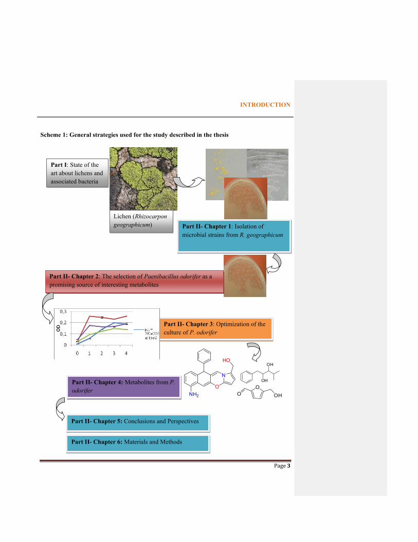


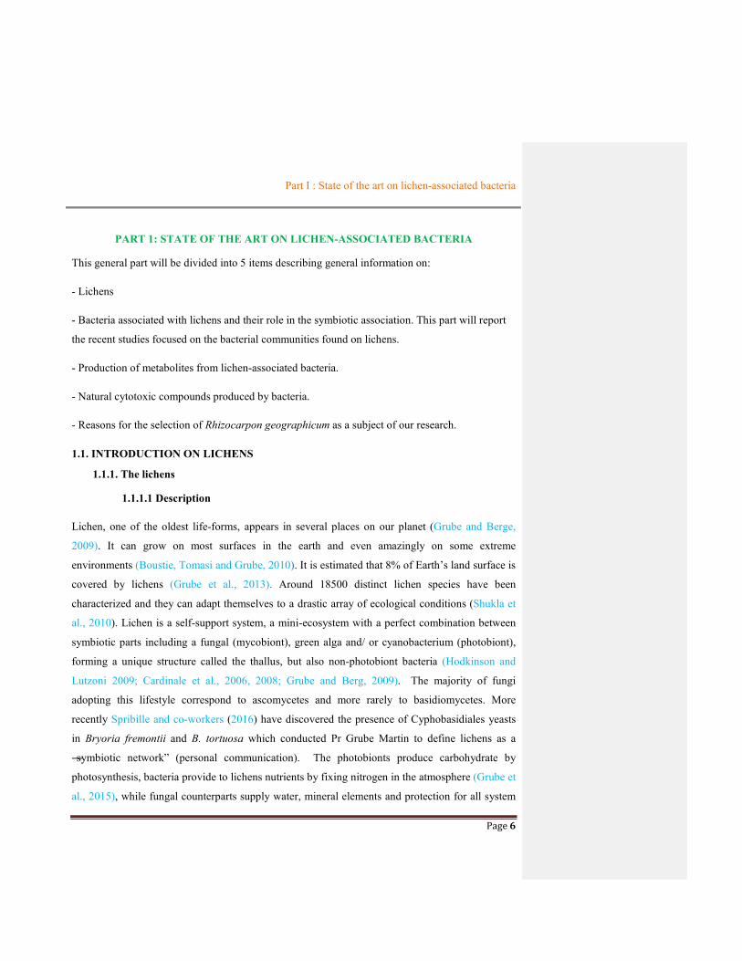

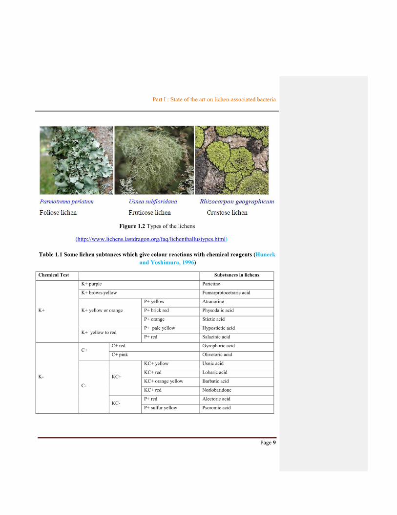

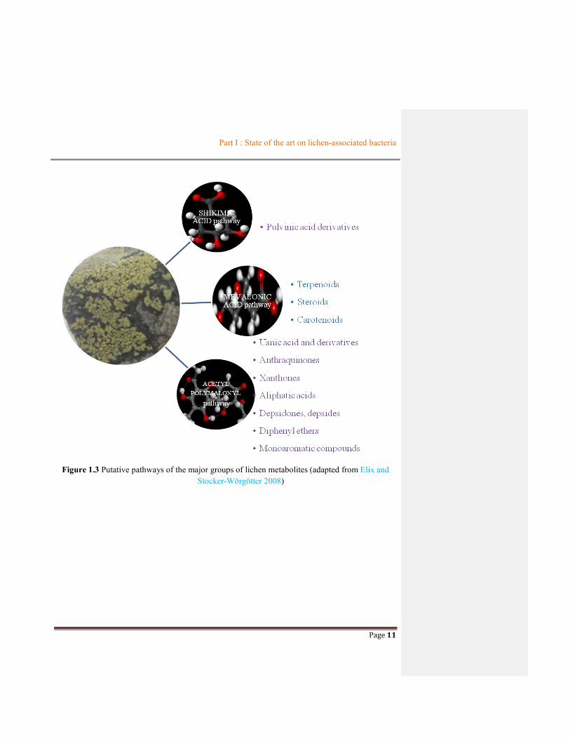


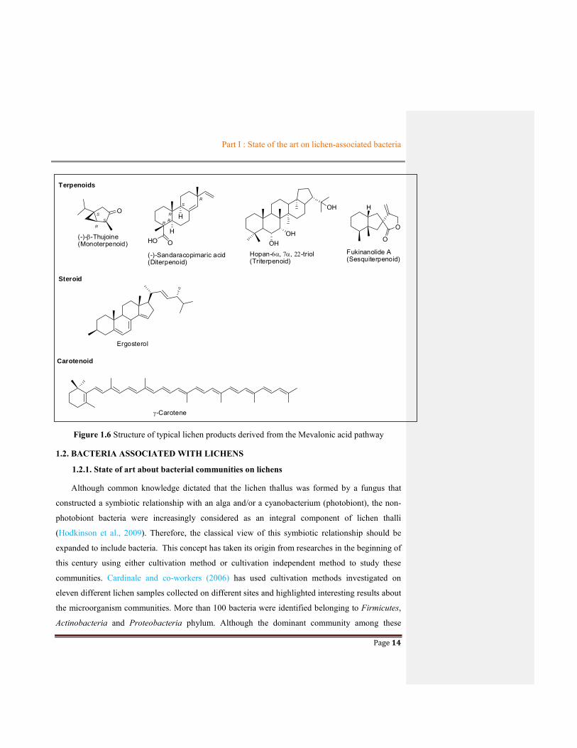


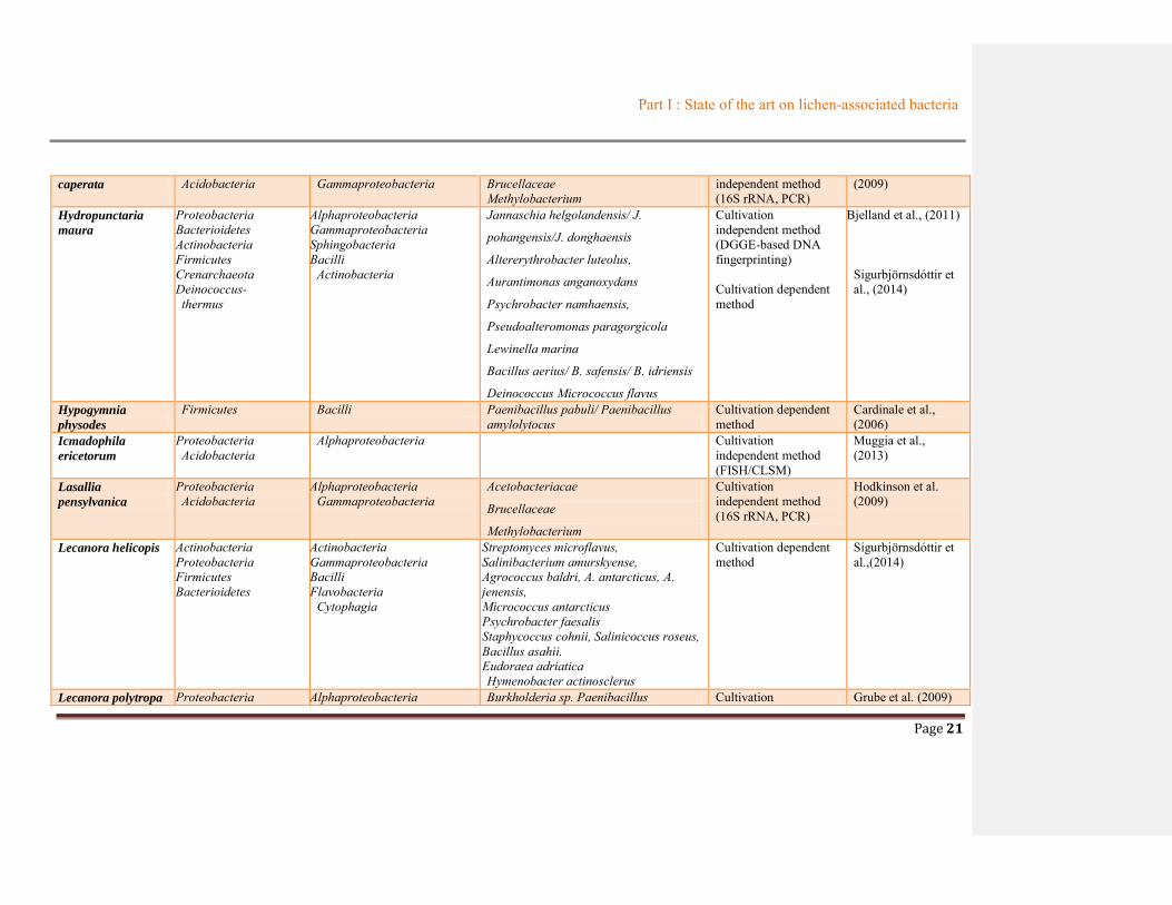
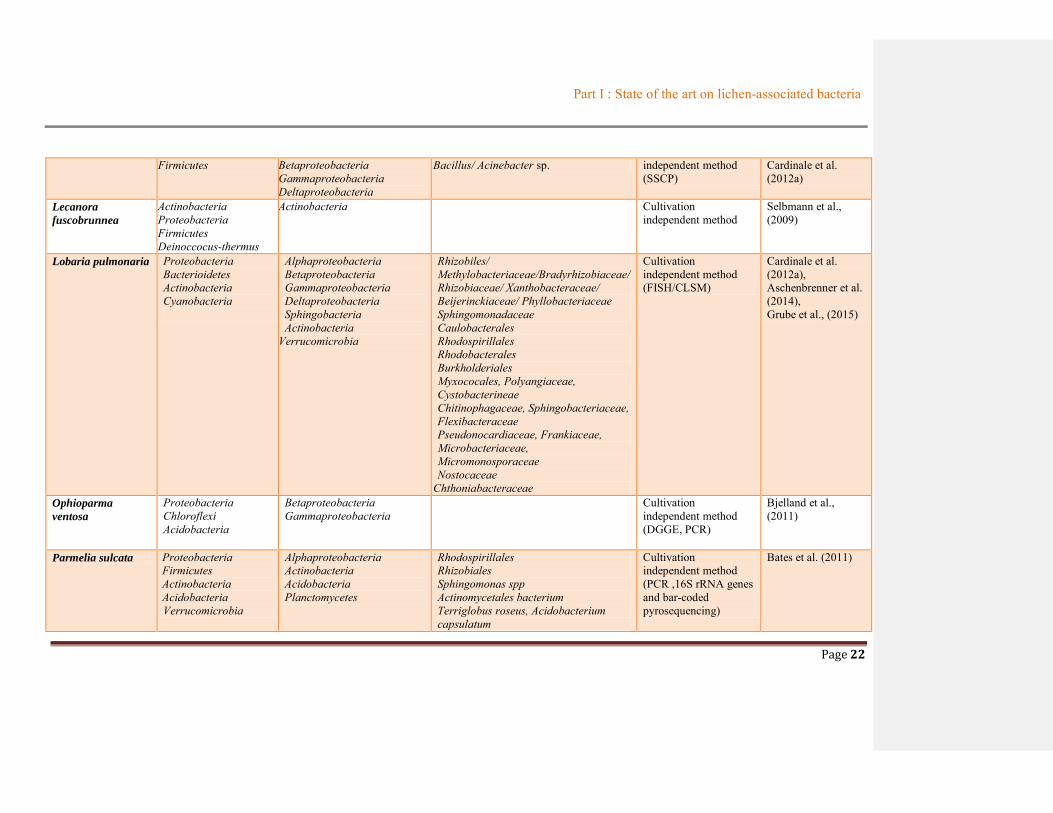
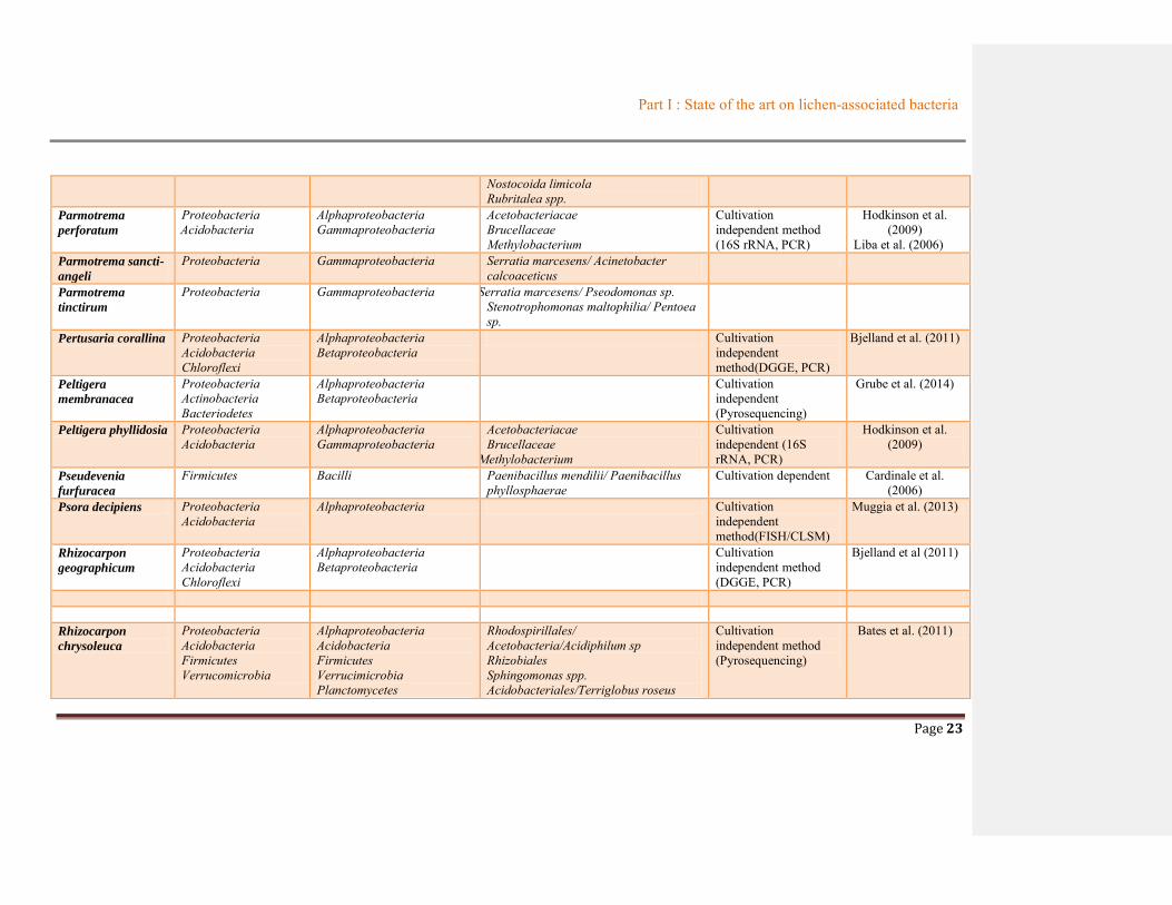
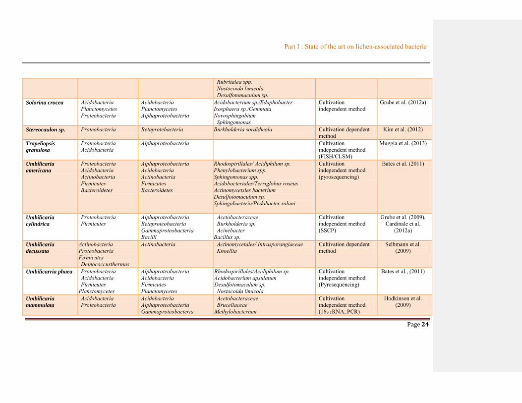

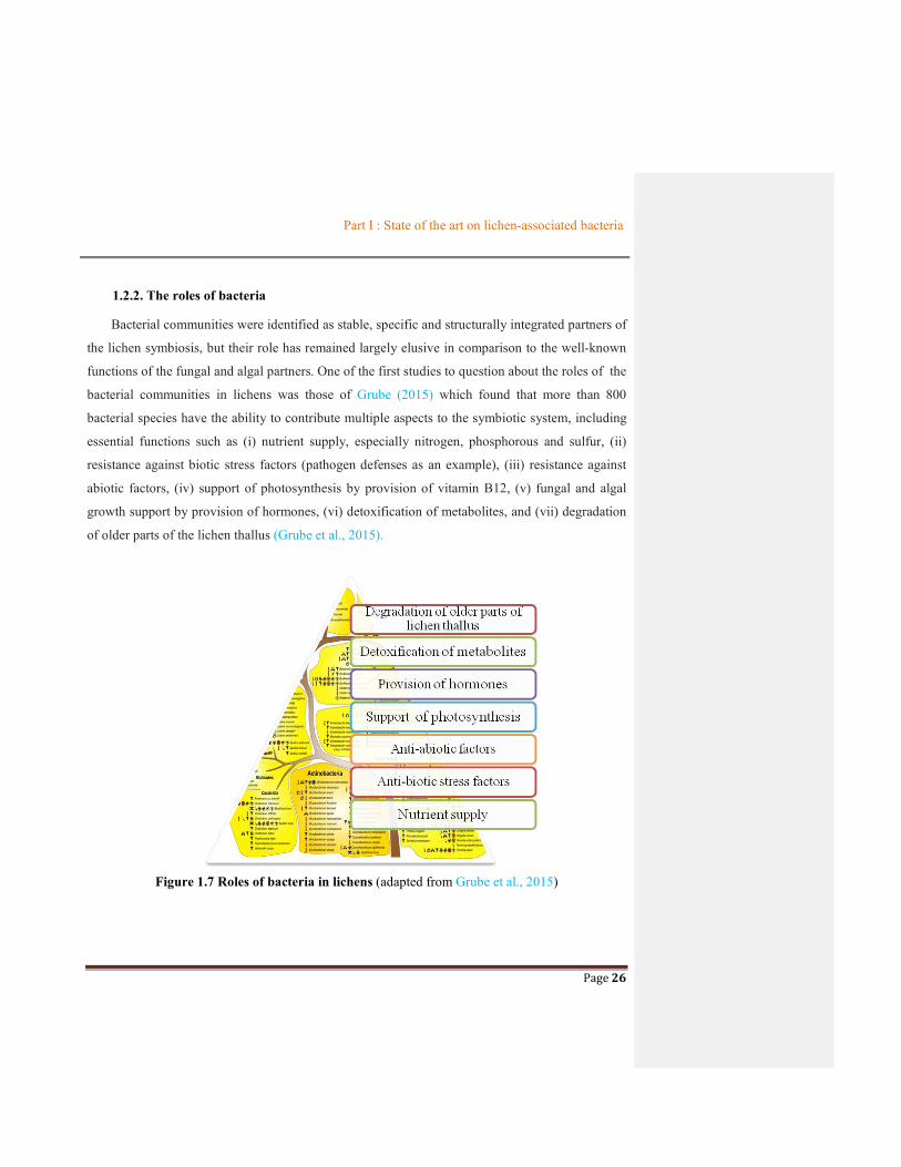


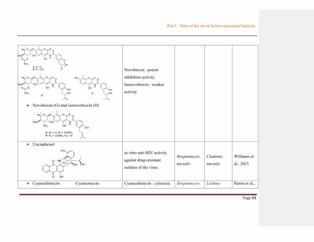
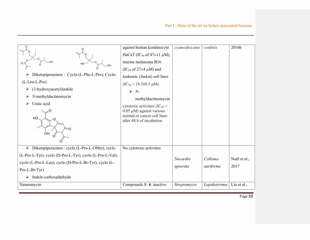
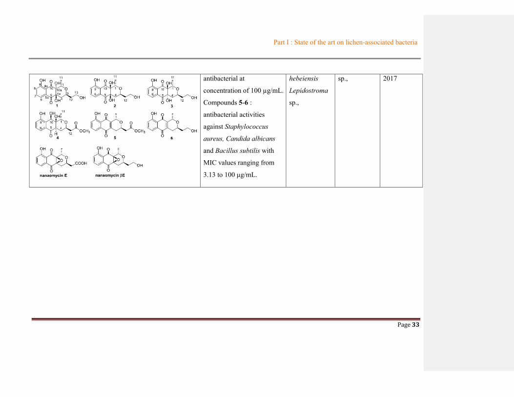
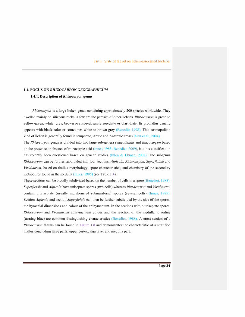


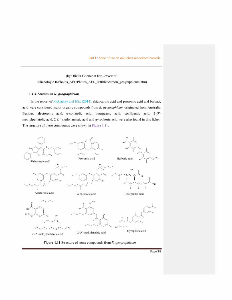


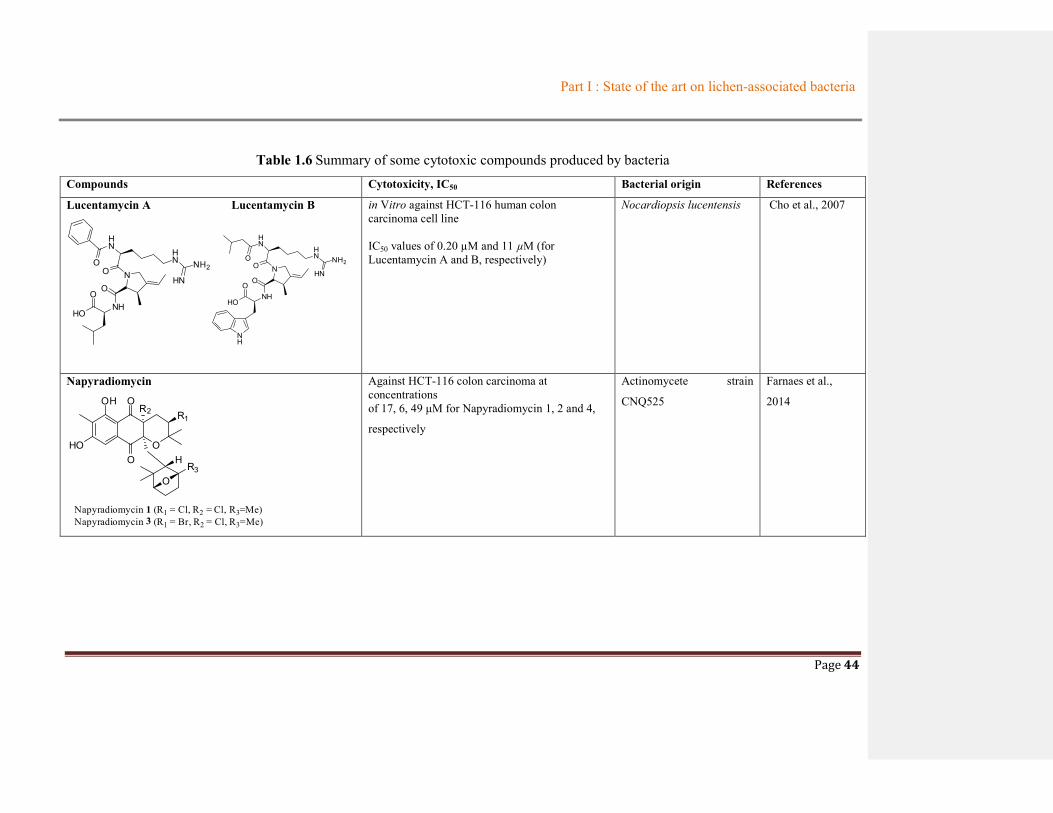


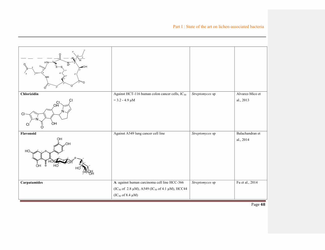


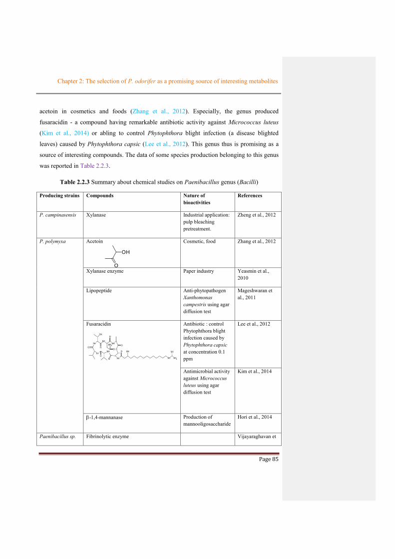

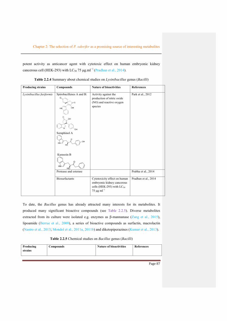


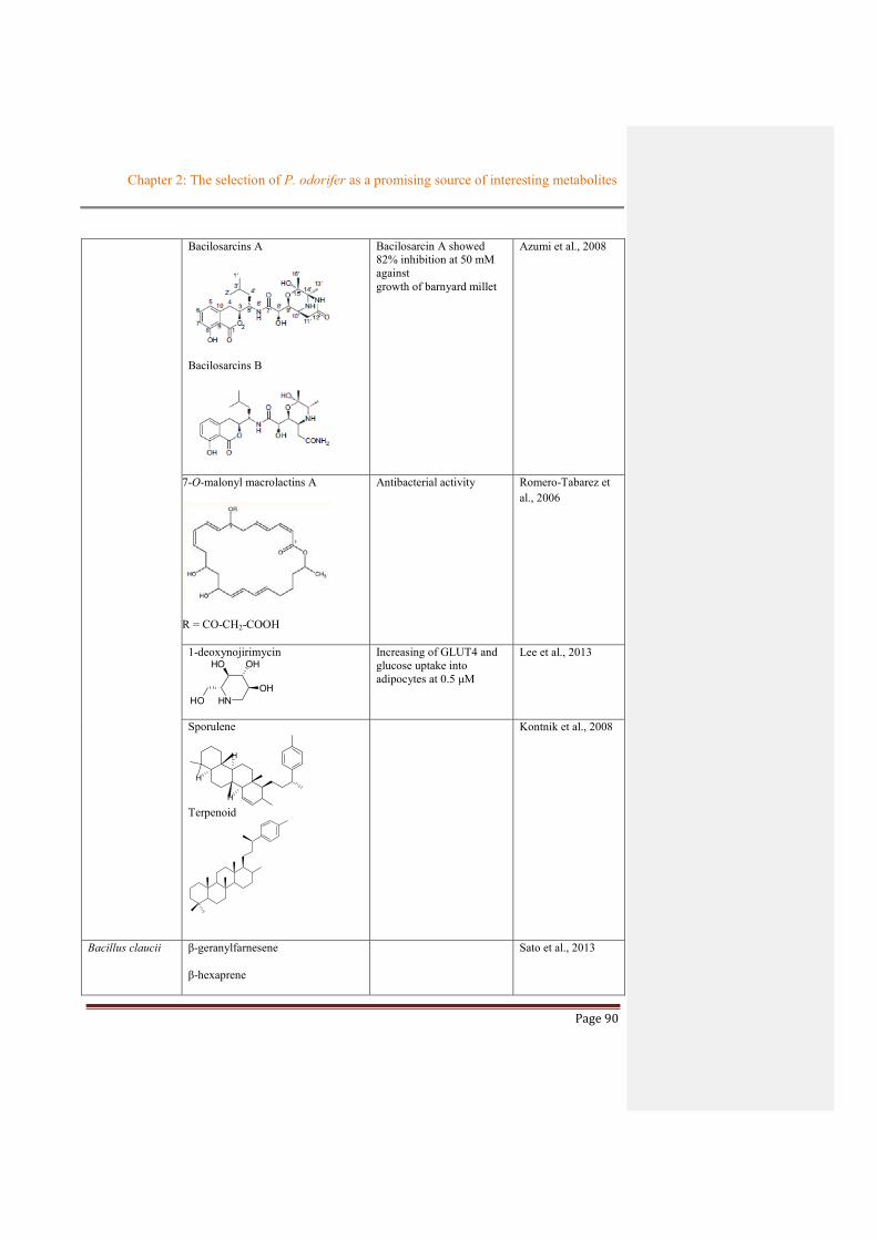


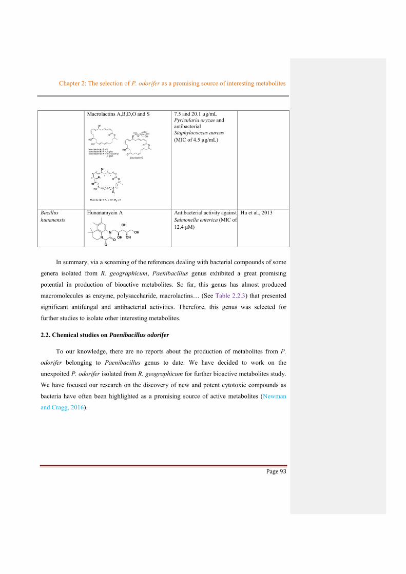


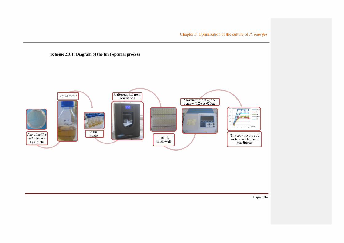

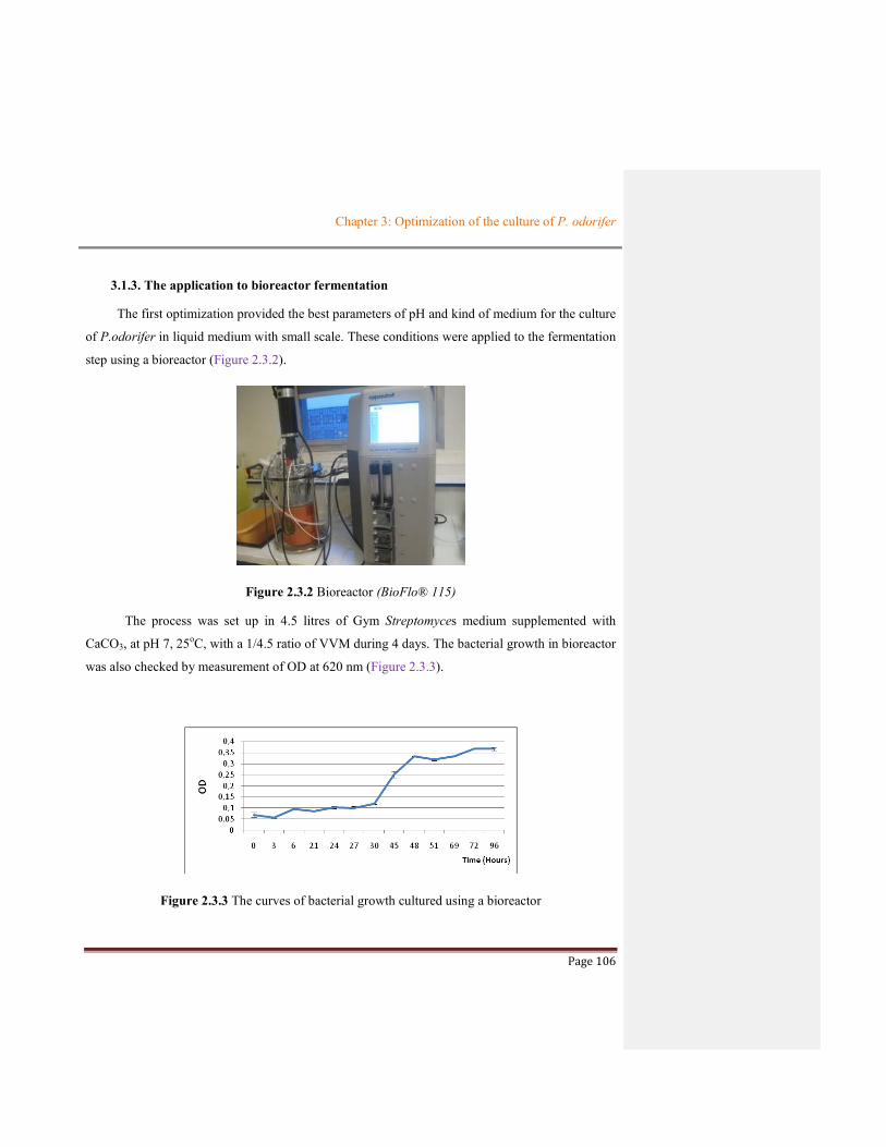


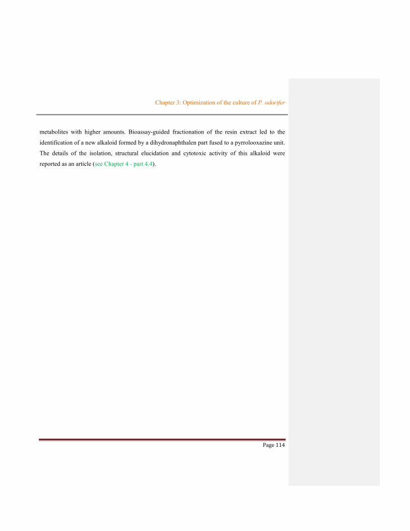


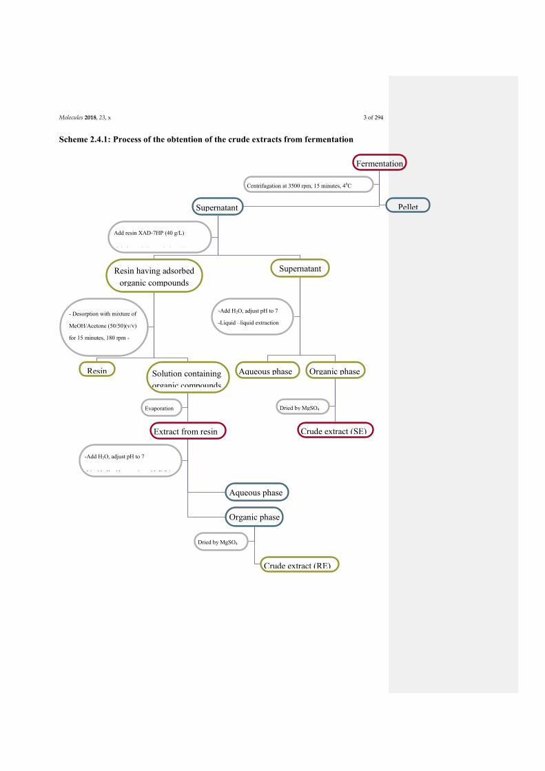


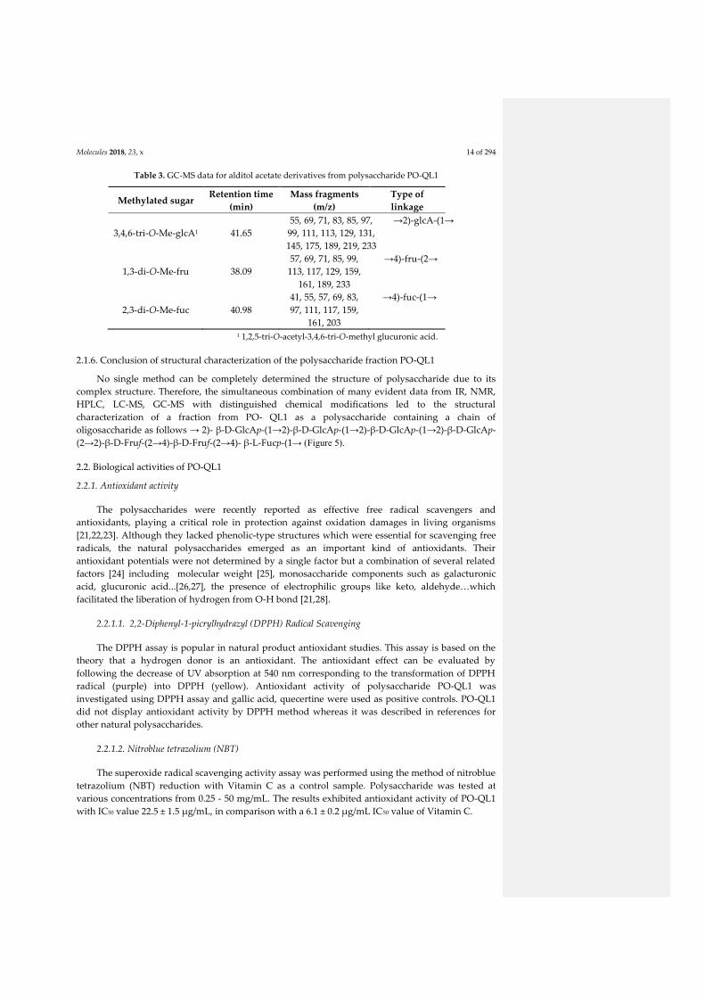


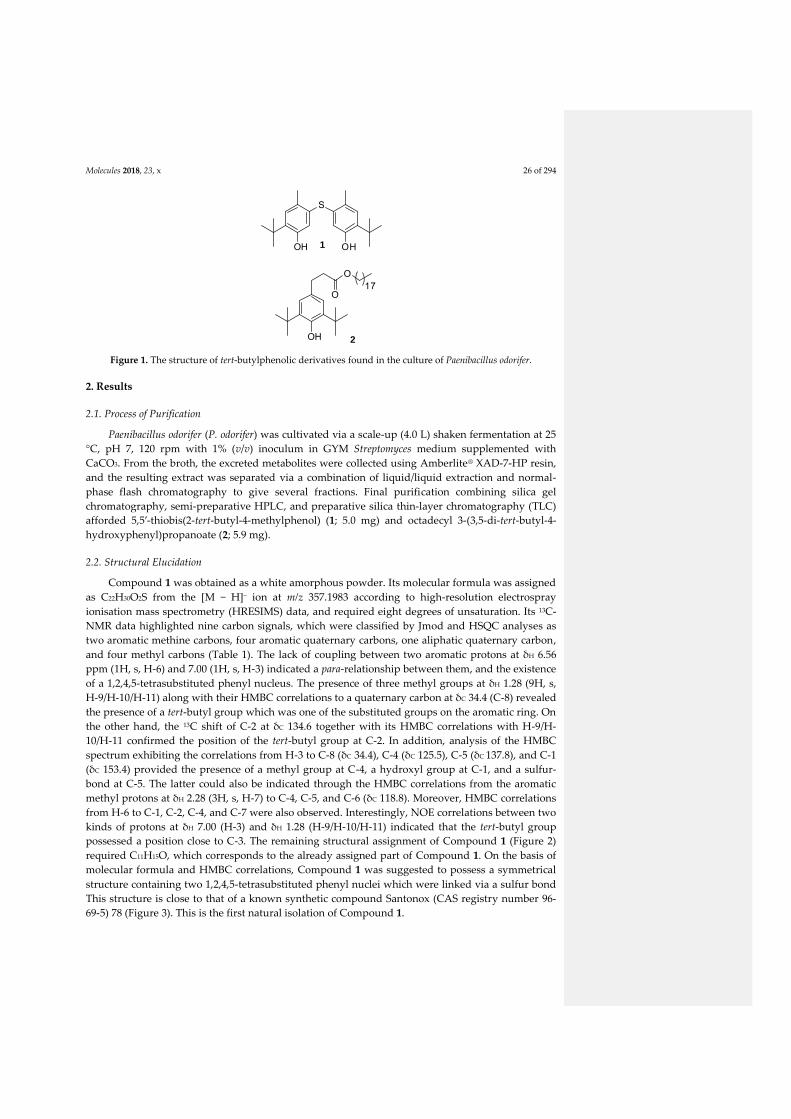

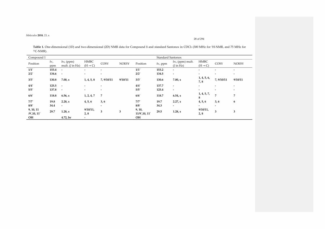


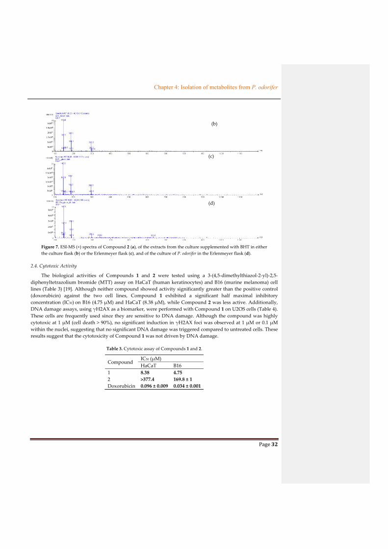

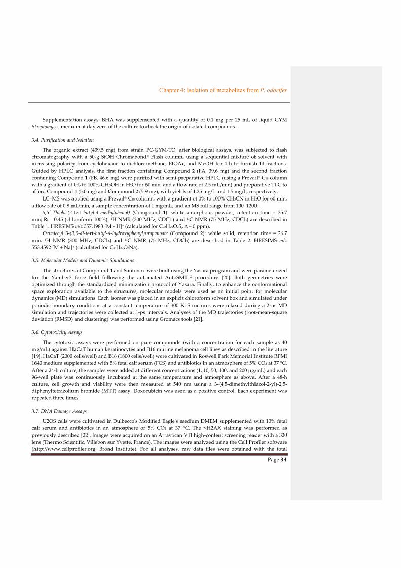

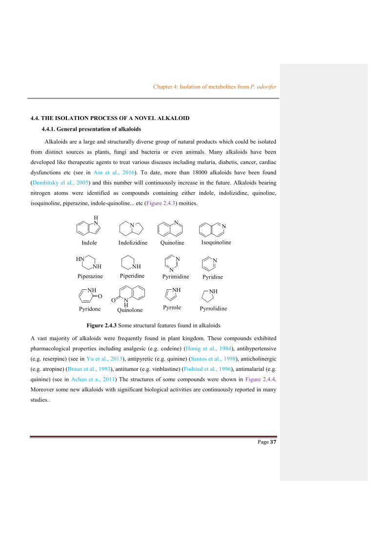
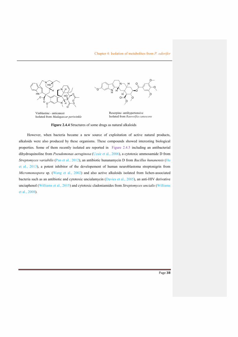


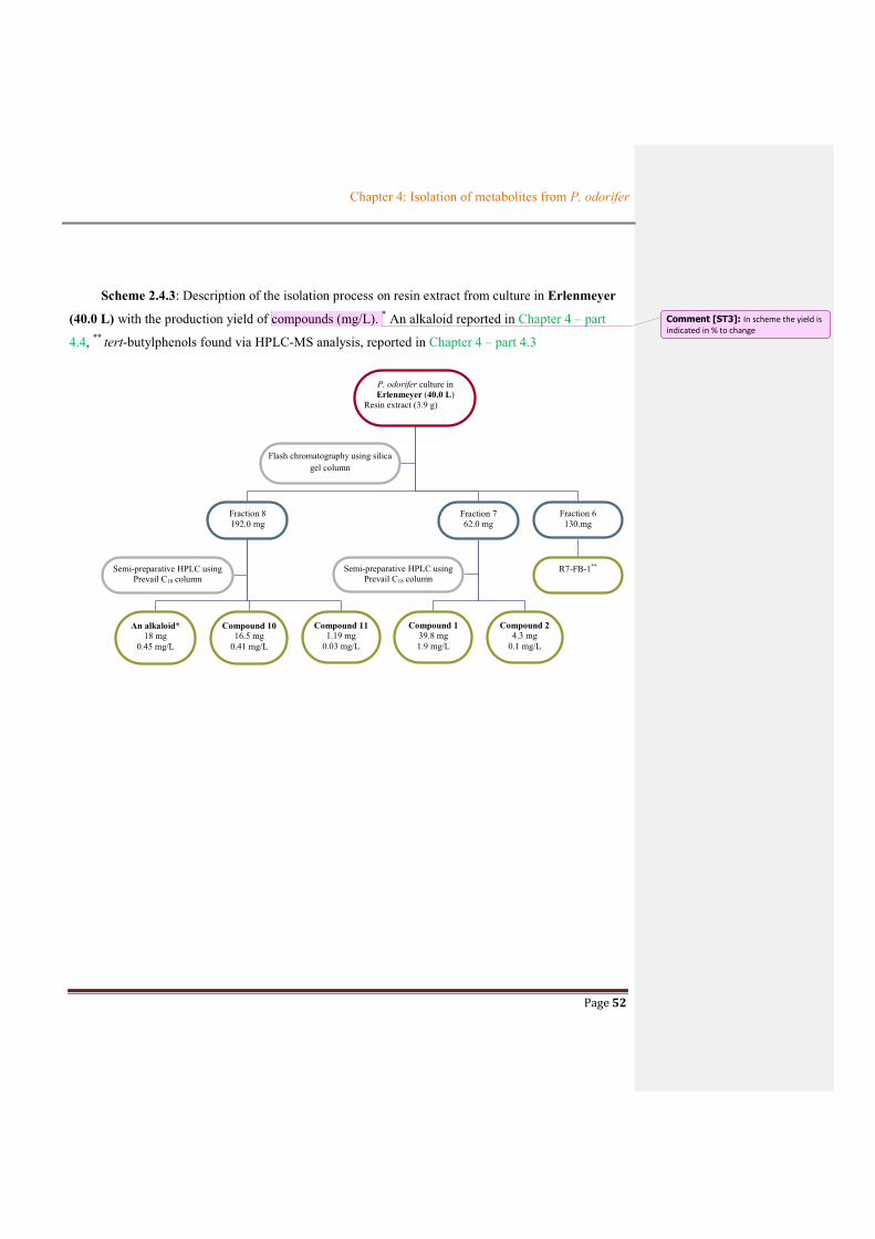

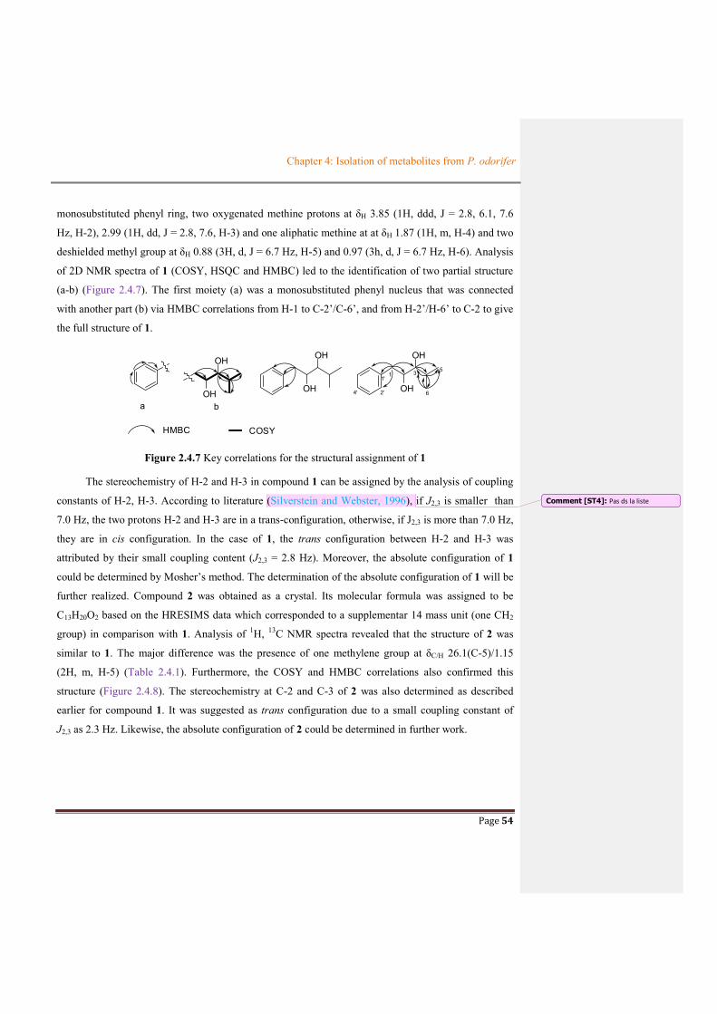


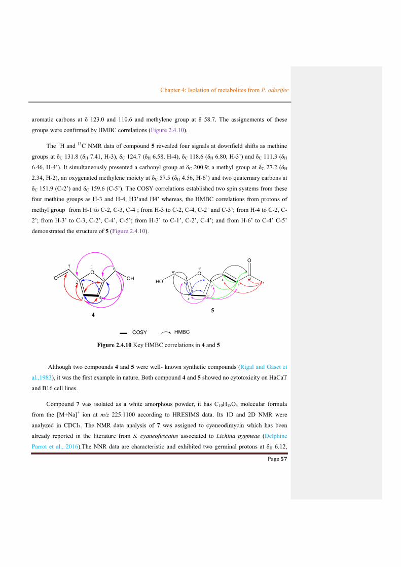
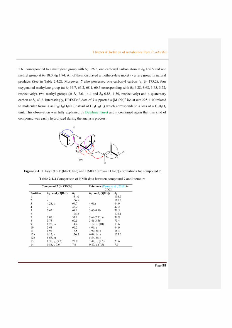


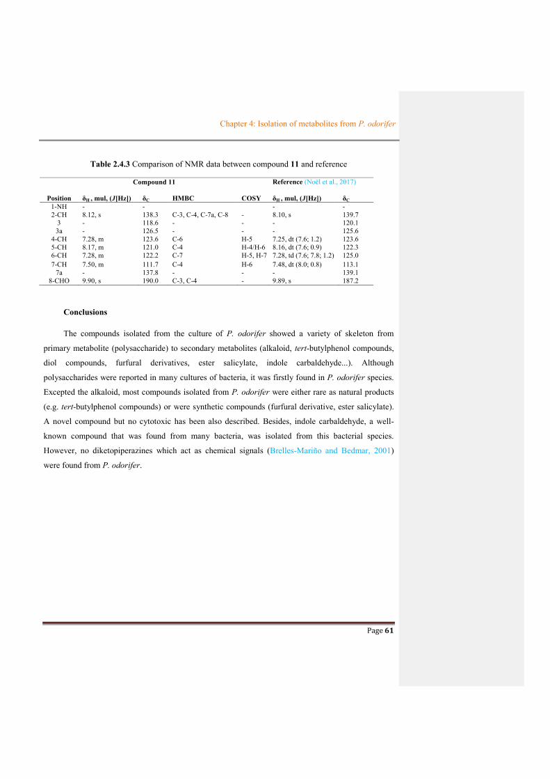


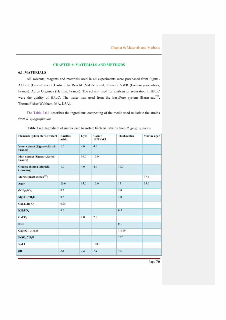


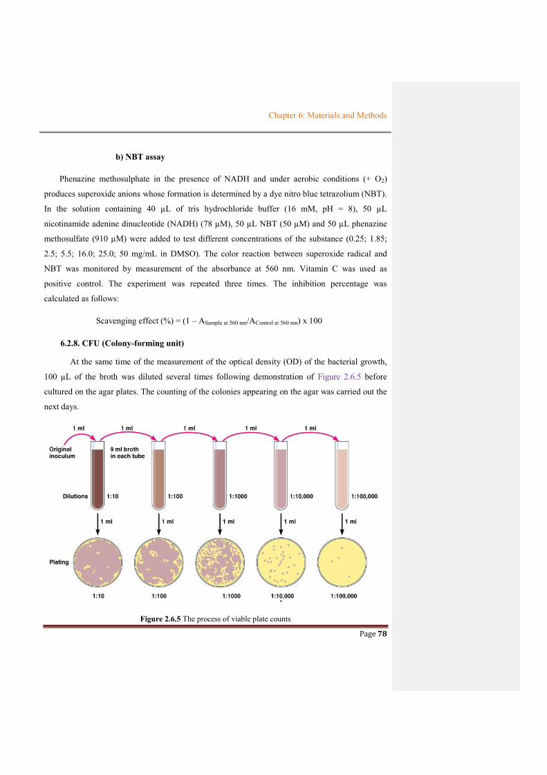


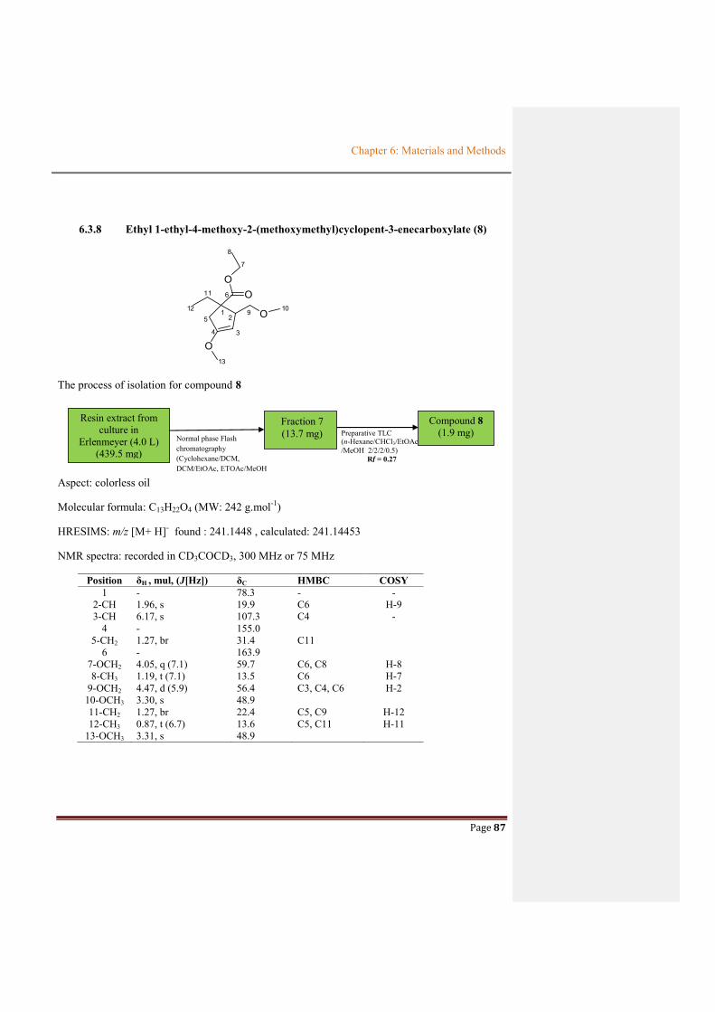


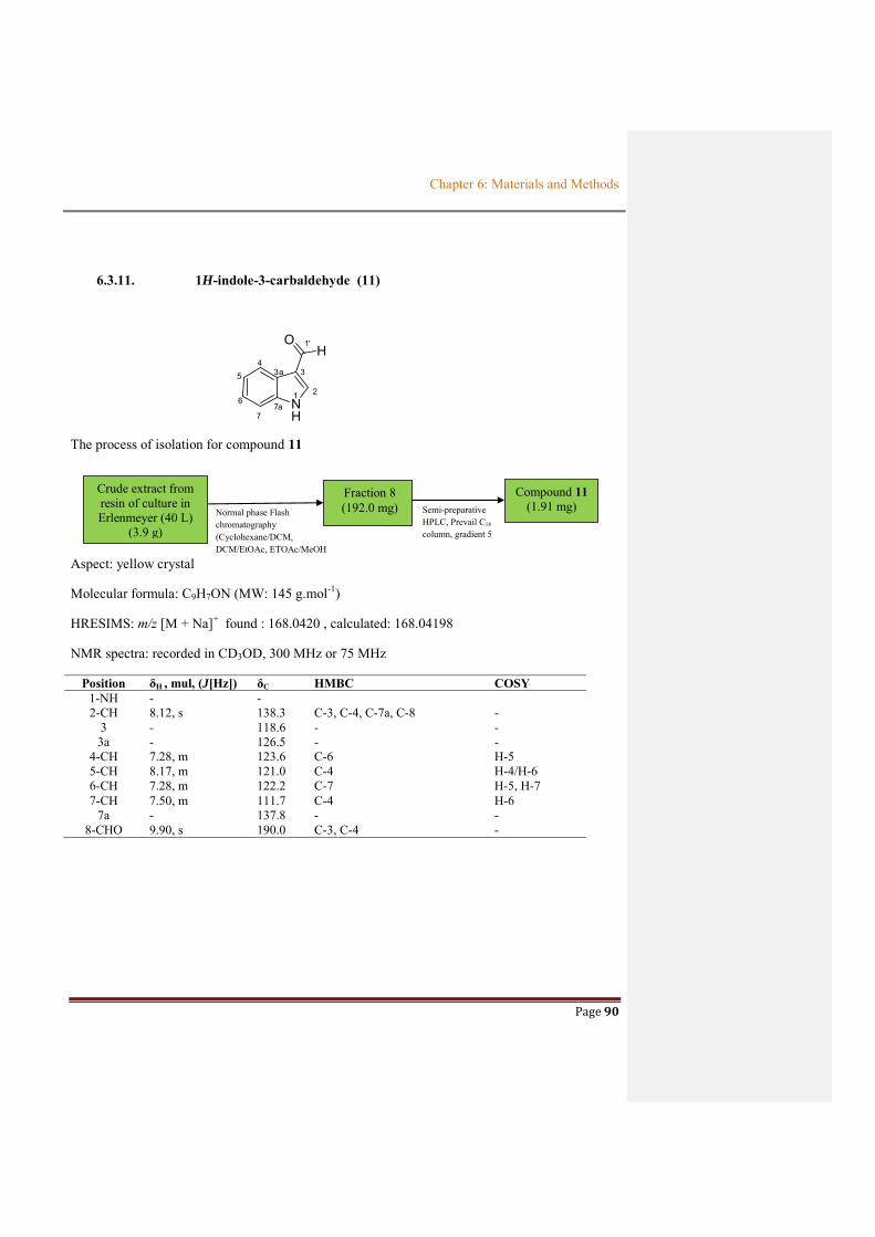


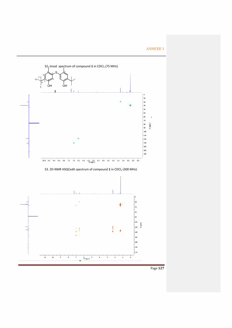
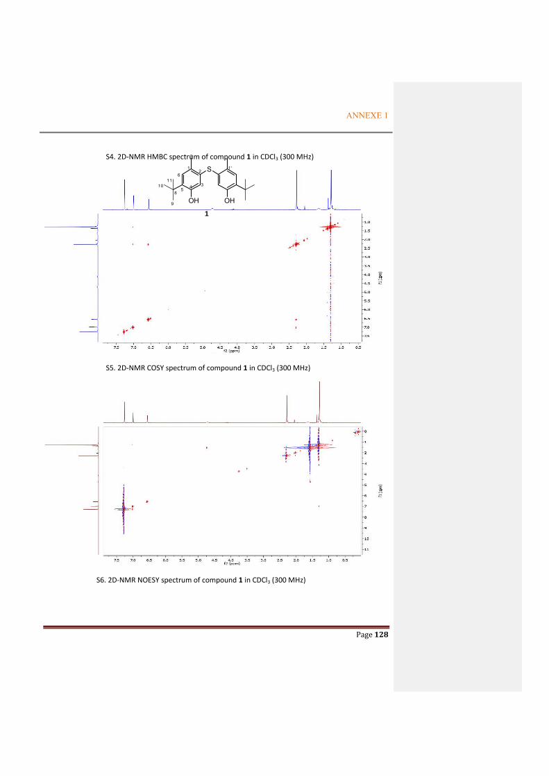

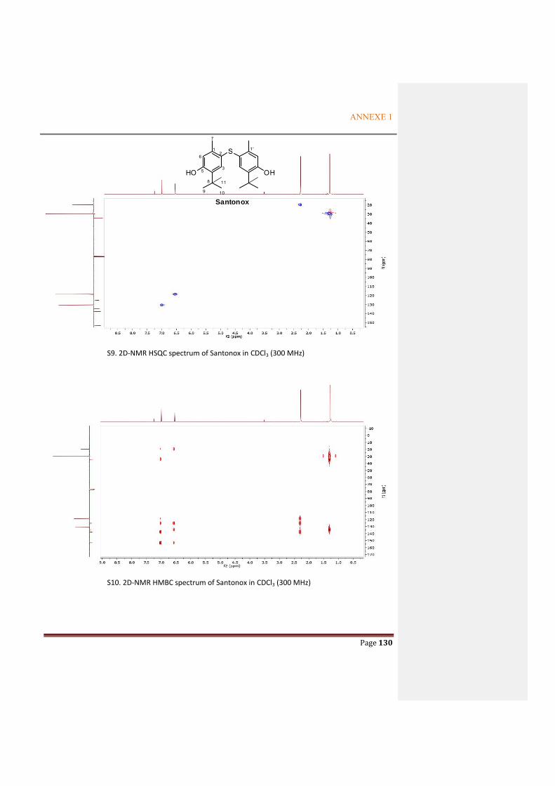


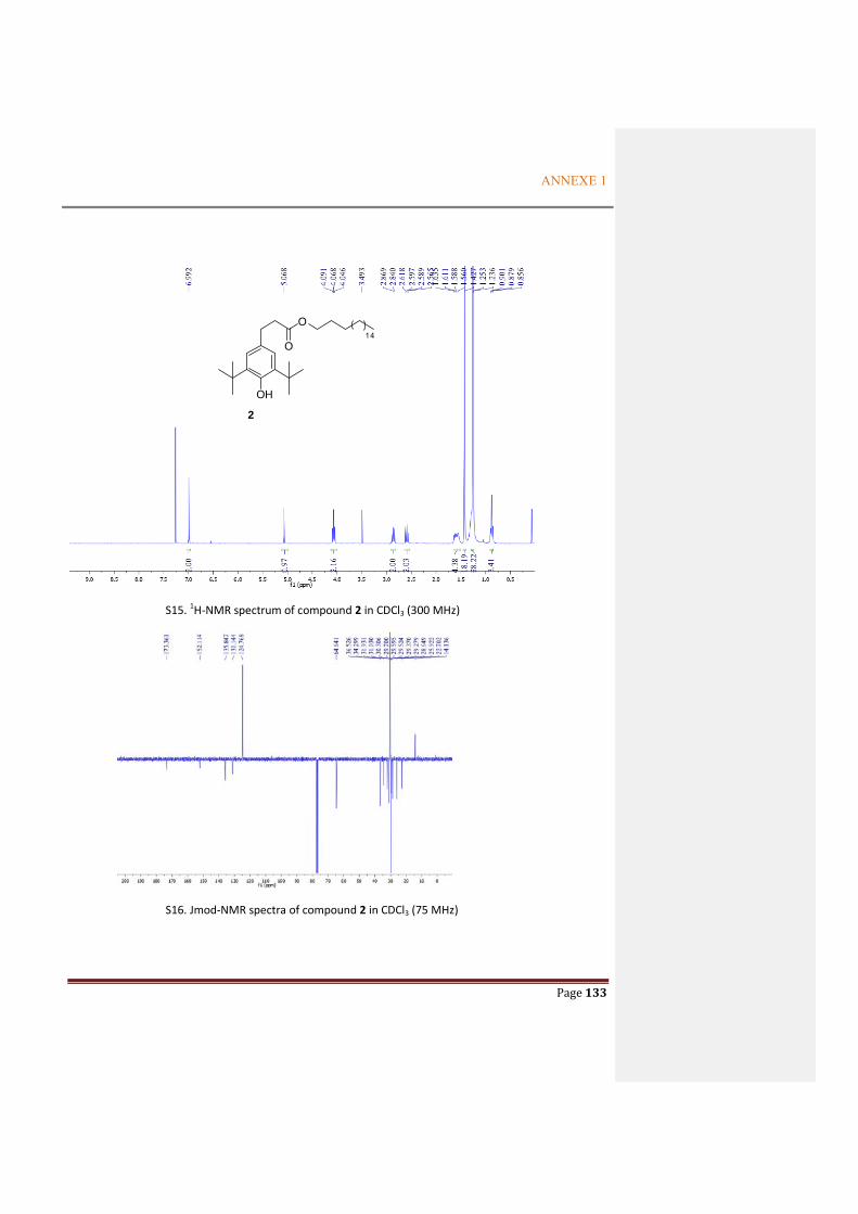
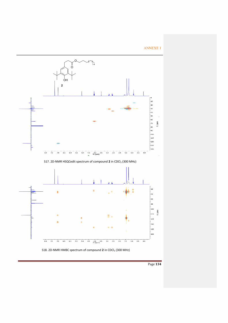


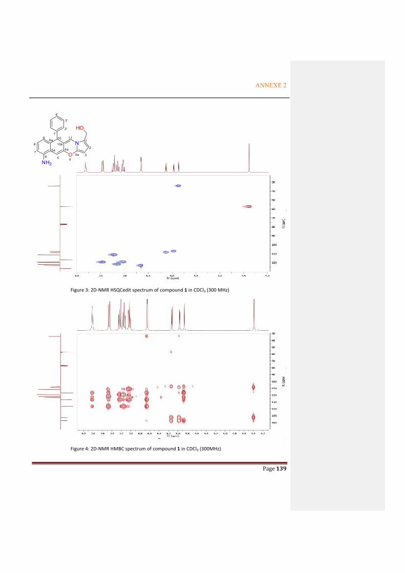


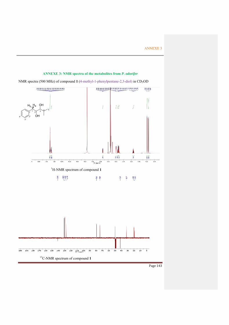
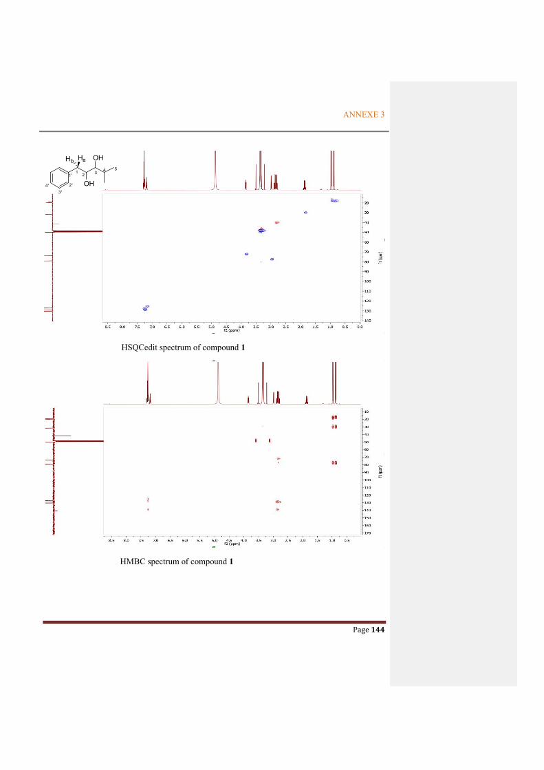
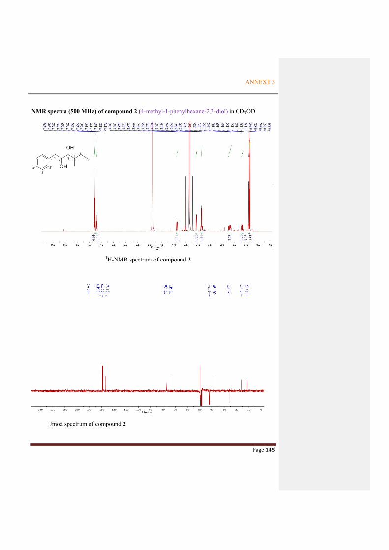
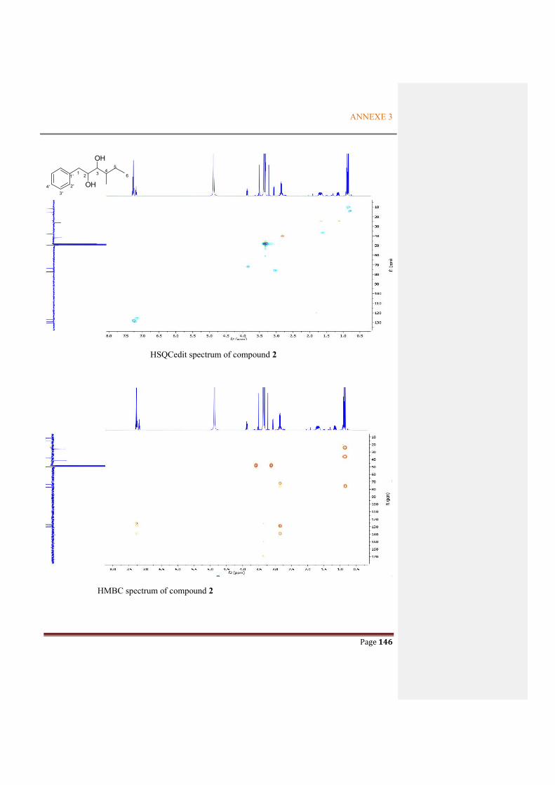

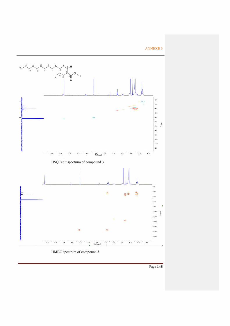
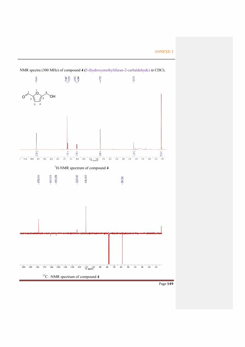
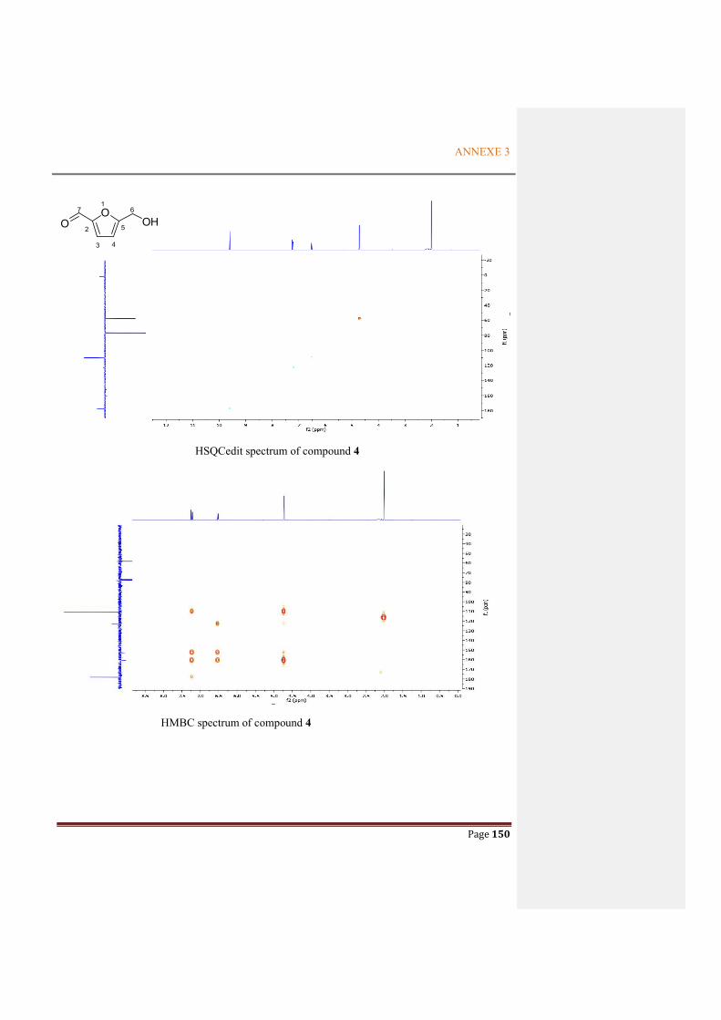

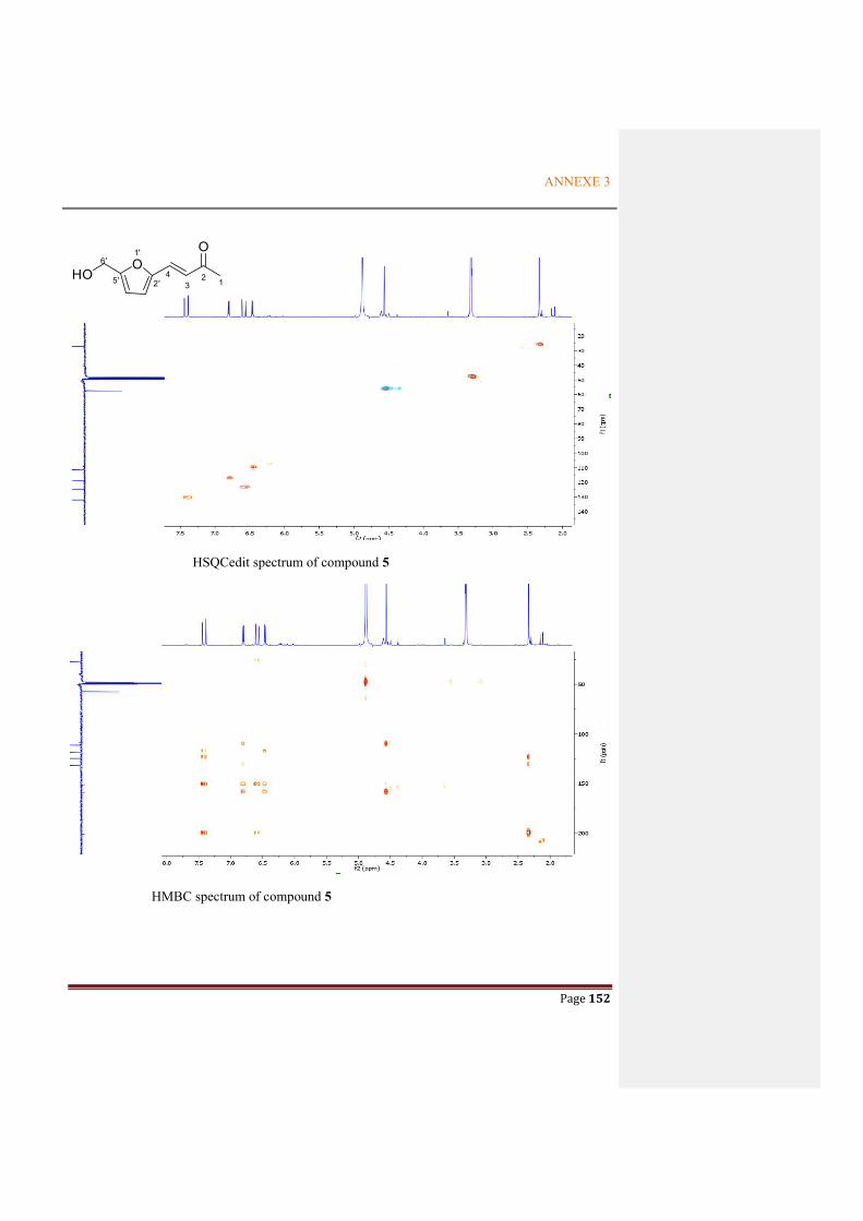
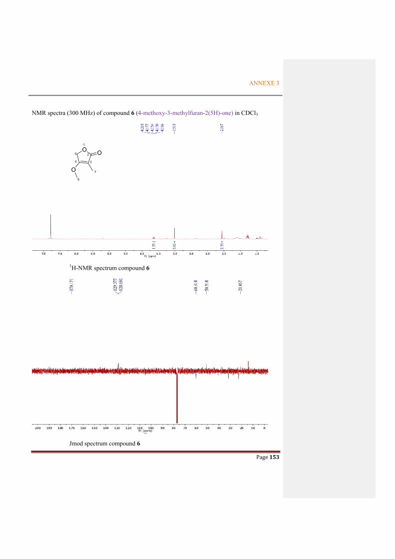


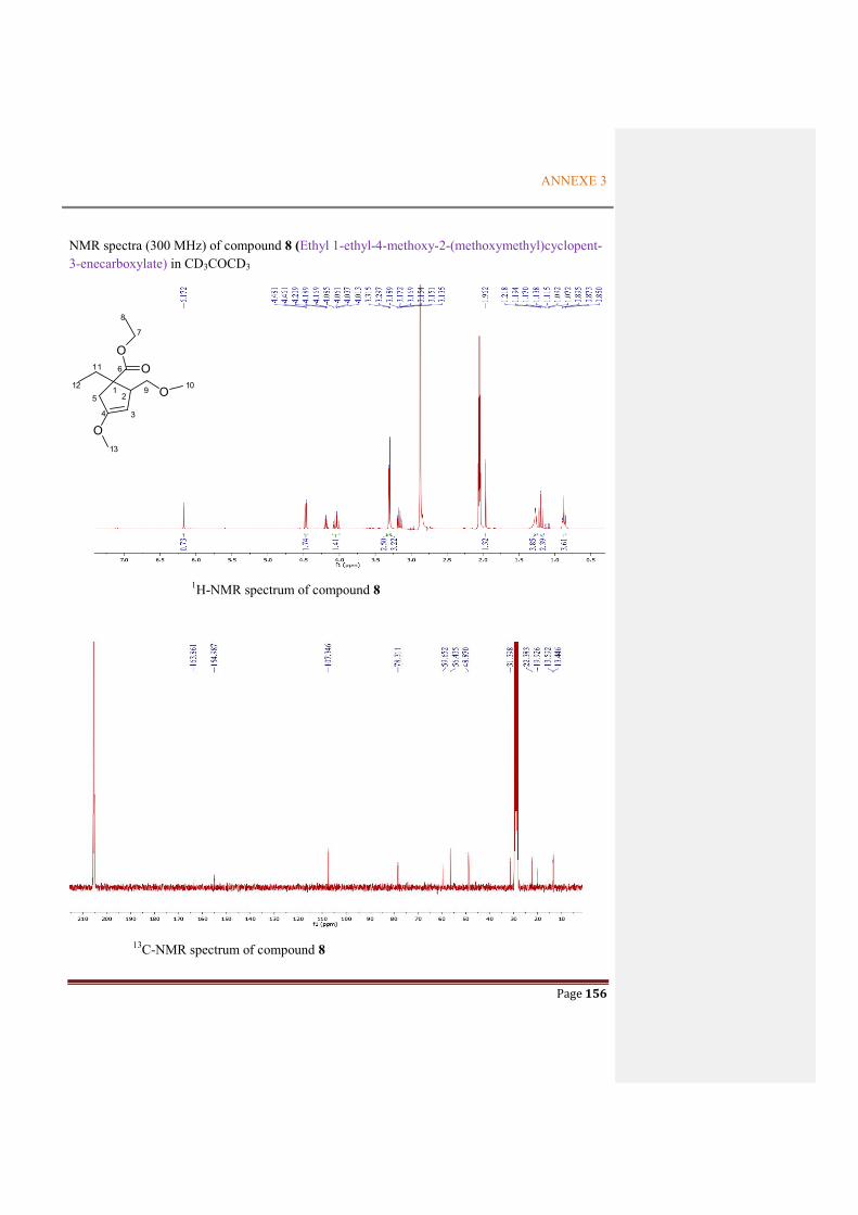
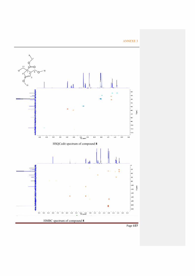

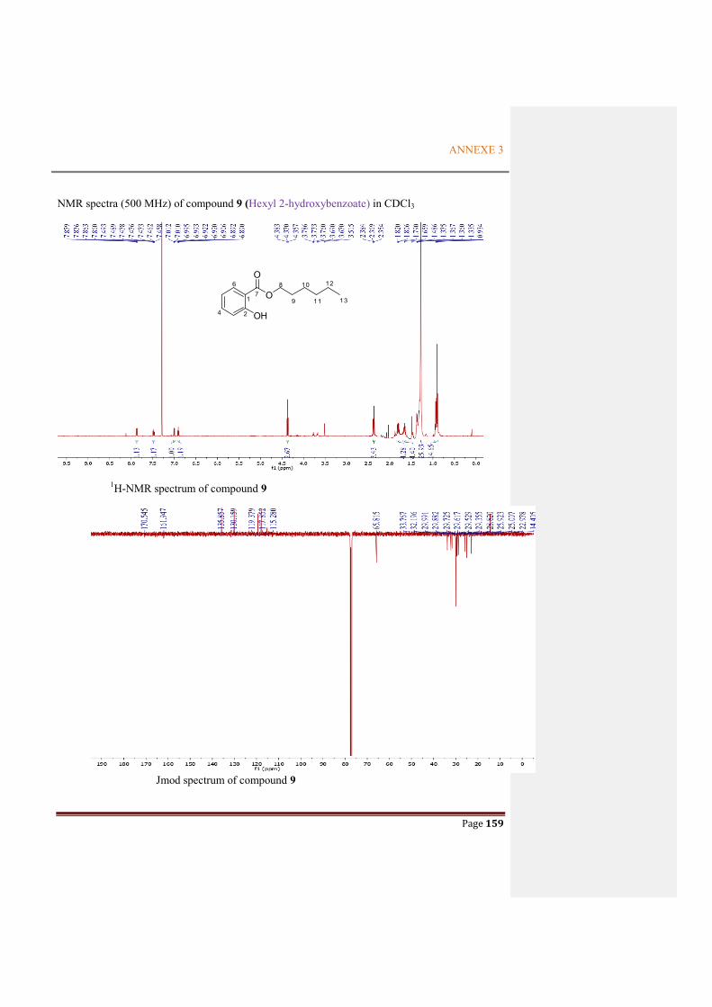


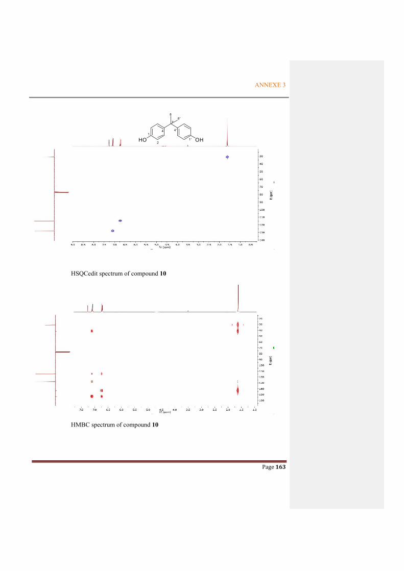


![Dereplication Guided Discovery of Secondary Metabolites of ... · library of microbial metabolites [27] as well as the AntiBase 2012 natural products database [28] were used for identification.](https://static.documents.pub/doc/80x56/5fab84b707a45c0328219b2d/dereplication-guided-discovery-of-secondary-metabolites-of-library-of-microbial.jpg)


